Note: Descriptions are shown in the official language in which they were submitted.
i
CA 02322452 2000-09-O1
WO 99144523 PCTIUS99104942
PMR DEVICE AND METHOD
Related A~ulications
The present application is related to U.S. Provisional Patent Application
Serial No. 60/064,210, filed on November 4, 1997, and entitled
TRANSMYOCARDIAL REVASCULARIZATION GROWTH FACTOR
MEDIUMS AND METHOD, U.S. Patent Application Serial No. 08/812,425,
filed on March 6, 1997, entitled TRANSMYOCARDIAL
REVASCULARIZATION CATHETER AND METHOD, U.S. Patent
Application Serial No. 08/810,830, filed March 6, 1997, entitled
RADIOFREQUENCY TRANSMYOCARDIAL REVASCULARIZATION
APPARATUS AND METHOD, and U.S. Patent Application Serial No.
filed on March 5, 1998, and entitled EXPANDABLE PMR
DEVICE AND METHOD herein incorporated by reference.
Field of the Invention
The present invention relates generally to medical devices for forming
hales in heart chamber interior walls in percutaneous myocardial
revascularization (PMR) procedures. More specifically, the present invention
relates to intravascular PMR devices having generally annular tips.
Backeround of the Invention
A number of techniques are available for treating cardiovascular disease
such as cardiovascular by-pass surgery, coronary angiaplasty, laser
angioplasty
1
CA 02322452 2000-09-O1
WO 99/44523 PCT/US99/04942
and atherectomy. These techniques are generally applied to by-pass or open
lesions in coronary vessels to restore and increase blood flow to the heart
muscle.
In some patients, the number of lesions are so great, or the location so
remote in
the patient vasculature that restoring blood flow to the heart muscle is
difficult.
S Percutaneous myocardial revascularization (PMR) has been developed as an
alternative to these techniques which are directed at by-passing or removing
lesions.
Heart muscle may be classified as healthy, hibernating and "dead". Dead
tissue is not dead but is scarred, not contracting, and no longer capable of
contracting even if it were supplied adequately with blood. Hibernating tissue
is
not contracting muscle tissue but is capable of contracting, should it be
adequately
re-supplied with blood. PMR is performed by boring channels directly into the
myocardium of the heart.
PMR was inspired in part by observations that reptilian hearts muscle is
supplied primarily by blood perfusing directly from within heart chambers to
the
heart muscle. This contrasts with the human heart, which is supplied by
coronary
vessels receiving blood from the aorta. Positive results have been
demonstrated
in some human patients receiving PMR treatments. These results are believed to
be caused in part by blood flowing from within a heart chamber through patent
channels formed by PMR to the myocardial tissue. Suitable PMR holes have
been burned by laser, cut by mechanical means, and burned by radio frequency
current devices. Increased blood flow to the myocardium is also believed to be
caused in part by the healing response to wound formation. Specifically, the
2
CA 02322452 2000-09-O1
WO 99144523 PCTNS99/04942
formation of new blood vessels is believed to occur in response to the newly
created wound.
~umm~v of the Invention
The present invention pertains to a device and method for performing
percutaneous myocardial revascularization (PMR). The device of the present
invention can be used to form crater wounds in the myocardium of the patient's
heart. A crater wound can be viewed as a wound having a width greater than its
depth, whereas a channel wound is one having a depth greater than its width. A
hole in the myocardium is a volumetric removal of tissue. The device can also
be
used to form channel wounds, but the configuration of the device's electrodes)
makes the device particularly suitable for creating crater wounds.
In the preferred form of the method in accordance with the present
invention, a crater wound is made through the endocardium and into the
myocardium. The wound, and thus the healing response, including angiogenisis
and subsequent perfusion of tissue is enhanced by collateral damage to the
myocardium. The collateral damage is preferably induced by directing
pressurized saline, contrast media, drug or a combination into the crater site
. . ~ dough. ~e . endocardium and into the myocardium. This causes . he:
vessels,
capillaries and sinuses to rupture. By creating the collateral damage, the
number
of wounds which need to be made during the PMR procedure can be substantially
reduced as the size of each wound is increased in view of the collateral
damage.
Additionally, and arguably as significant as the reduction in the number of
wounds which must be formed during the procedure, is the reduction of the
3
CA 02322452 2000-09-O1
WO 99/44523 PCTNS99/04942
likelihood of a myocardial perforation. This reduction is possible because the
holes can be limited in depth to just through the endocardium. Once the
endocardium is perforated, pressure from infused fluid can rupture the
myocardial
vessels without further ablation or removal of tissue.
In a preferred embodiment, a catheter in accordance with the present
invention includes an elongate shaft having a proximal end and a distal end,
and a
conductor extending therethrough. An electrode is disposed at the distal end
of
the shaft and connected to the conductor. The electrode has a generally
annular
transverse cross-sectional shape. The annular shape defines an opening within
the
electrode. An insulator surrounds the elongate shaft.
A stop is disposed in the opening a predetermined distance proximally of
the distal end of the electrode. The shaft preferably defines a lumen in fluid
communication with the opening through the electrode. In one embodiment, a
needle can be disposed within the opening and be in fluid communication with
the
lumen to deliver contrast media, growth factors or drugs to the wound.
In another embodiment, the annular shape of the electrode is generally
circular. The annular shape can be continuous or in an alternate embodiment,
~s~ntinuous and formed from a plurality of discrete electrodes. positioned in
an
array. The electrode can also include a serrated edge that produces a
plurality of
electrode contact points.
A method for performing PMR in accordance with the present invention
includes providing a catheter having an elongate shaft including a proximal
end
and a distal end. A generally annular shaped electrode is disposed at the
distal
4
CA 02322452 2000-09-O1
WO 99/44523 PCT/US99/04942
end of the shaft. The electrode is advanced to proximate the endocardial
surface
of the myocardium of the patient's heart. The electrode is energized and
advanced
into the myocardium to form an annular shaped crater wound. Depth is
controlled
by a mechanical stop.
Brief Description of the Drawing
Figure 1 is a cross-sectional, perspective view of an annular shaped crater
wound in a patient's myocardium formed by a device in accordance with the
present invention;
Figure 2 is a perspective, cross-sectional view of a catheter in accordance
with the present invention;
Figure 3 is a cross-sectional view of the catheter of Figure 2 in use;
Figure 4 is a perspective, cross-sectional view of an alternate embodiment
of the catheter in accordance with the present invention;
Figure 5 is a cross-sectional view of the catheter of Figure 4 in use;
Figure 6 is a perspective view of the distal end of yet another alternate
embodiment of a catheter in accordance with the present invention;
Figure-7 is a perspective view of yet another alternate embodiment of the
catheter in accordance with the present invention;
Figure 8 is a perspective view of yet another alternate embodiment of the
catheter in accordance with the present invention;
Figure 9 is a perspective view of yet another alternate embodiment of the
catheter in accordance with the present invention;
5
CA 02322452 2000-09-O1
WO 99/44523 PCT/US99/04942
Figure 10 is a cross-sectional view of the catheter of Figure 8;
Figure 11 is a cross-sectional view of the catheter of Figure 8;
Figure 12 is a cross-sectional view of the catheter of Figure 8;
Figure I3 is a top view of a crater formed in the endocardium;
Figure 14 is a cross-sectional view of the crater of Figure 12;
Figure 15 is a front view of a catheter electrode in accordance with the
present invention;
Figure 16 is a back view of the electrode of Figure 14;
Figure 17 is a side view of the electrode of Figure 14;
Figure 18 is a front view of yet another embodiment of an electrode in
accordance with the present invention; and
Figure 19 is a back view of the electrode of Figure 17.
Detailed Description of the Invention
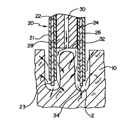
Referring now the drawings wherein like reference numerals refer to like
elements throughout the several views, Figure 1 is a perspective, partial
cross-
sectional view of a heart wall 10 having an annular hole 12 formed in the
myocardium by a catheter made in accordance with the present invention. Figure
2 is a perspective, partial cross-sectional view of a catheter 20 in
accordance with
the present invention. Catheter 20 includes a shaft 21 having a proximal end
and
a distal end. Shaft 21 preferably includes an elongate hypotube sandwiched
between an inner insulator 24 and an outer insulator 26. Hypotube 22 can be
formed from stainless steel or Nitinol or other conductive material. It can be
6
CA 02322452 2000-09-O1
WO 99/44523 PCTIUS99/04942
desirable to use a Nitinol hypotube as the highly flexible material can act as
a
shock absorber while catheter 20 is pressured against the beating heart during
the
PMR procedure. Insulators 24 and 26 may be formed from, for example,
polyethylene, polyimide or PTFE. Those skilled in the art would appreciate
that
other biocompatible materials can be used to form these elements. The distal
end
of hypotube 22 is preferably left uninsulated to form an annularly-shaped
electrode 23.
A stop 28 is preferably disposed within shaft 21. Stop 28 preferably
defines a lumen 30 extending therethrough. Stop 28 includes a distal end 32
spaced a predetermined distance from a distal end 34 of electrode 23. This
predetermined distance can be used to control the depth of holes 12 formed in
the
myocardium of a patient's heart. Those skilled in the art will recognize the
non-
conductive, biocompatible materials available to form stop 28, for example
PEPI.
In view of the discussion below regarding the use of catheter 20, those
i 5 skilled in the art of catheter construction would recognize the various
possibilities
for manifolds to be disposed at the proximal end of catheter 20, and that a
suitable
radio frequency (RF) generator can be conductively connected to hypotube 22 to
deliver RF energy to electrode 23. , . ,
Figure 3 is a cross-sectional view of catheter 20 in use. In Figure 3,
electrode 23 has been energized with RF energy and advanced into heart wall 10
to form hole 12. As shown by the arrows, contrast medium, growth factor or
other drugs are being infused through lumen 30 into hole 12, and then into
myocardium 10. It can be noted that in Figure 3 that distal end 32 of stop 28
is
7
CA 02322452 2000-09-O1
WO 99/44523 PCT/US99/04942
spaced a predetermined distance from distal end 34 of electrode 23 such that
the
depth of hole 12 is approximately equal to its width. The predetermined
distance
can be varied such that shallower holes or craters are formed, or
alternatively the
distance can be increased to form channels.
Figure 4 is a perspective, partial cross-sectional view of catheter 20
modified to include a hypotube 36 extending distally from lumen 30. The distal
end of hypotube 36 includes a sharpened end 38, and a lumen defined
therethrough in fluid communication with lumen 30. Hypotube 36 can also act as
a bi-polar ground
Figure 5 is a cross-sectional view of catheter 20 including hypotube 36.
This view is similar to that of Figure 3, except that rather than infusion
fluid into
hole 12, as shown by the arrows, fluid is directed into the myocardium.
Figure 6 is an alternate embodiment of a catheter 120 in accordance with
the present invention. Many elements of catheter 120 are similar to that of
catheter 20 as shown in Figure 2. Rather than shaft 121 including a hypotube
22,
shaft 121 includes a plurality of elongate conductive members 122 embedded in
a
tubular insulator 124. A distal portion of members 122 is preferably left
,.. , . uninsulated to form a-generally annularly shaped array of electrodes
123. A stop
128 is disposed within tubular member 124. Stop 128 defines a lumen 130
extending therethrough. Stop 128 includes distal end 132 spaced a
predetermined
distance proximally of distal ends 134 at electrodes I23 to control the depth
of the
holes created by catheter 123. It can be appreciated by those skilled in the
art that
catheter 120 can be used in substantially the same manner to perform PMR as
8
CA 02322452 2000-09-O1
WO 99144523 PCTNS99/04942
catheter 20 shown in Figure 3. A plurality of electrodes, having a surface
area
less than a continuous annular electrode requires less energy to arc or
ablate. A
plurality of electrodes will also tend to grab tissue, stabilizing the
electrode on a
moving heart wall.
Figure 7 is a perspective view of a modified embodiment of catheter 20 of
Figure 2. In particular, the distal end of hypotube 22 has been serrated to
form a
serrated electrode 40. Serrating electrode 40 changes the surface of the
electrode
contacting the tissue and thus reduces the power needed to arc. Serrated
electrode
40 will also grab tissue, securing electrode 40 to a moving heart wall during
crater
formation.
Figure 8 is a view of yet another embodiment of catheter 20 in accordance
with the present invention. To catheter 20 has been added a second grounded or
return electrode 31 to form a bi-polar RF PMR catheter. It can be appreciated
that
this electrode can also be added to catheter 120 of Figure 6 and catheter 20
of
Figure 7 to make each of these embodiments bi-polar as well.
Figure 9 is a perspective view of yet another embodiment of a catheter 210
in accordance with the present invention disposed within a guide catheter 212.
Catheter 210 includes ,an elongate shaft 214. Elongate shaft 214 is ,
preferably
formed from an elongate tubular, and conductive member such as a stainless
steel
or Nitinol hypotube. Shaft 214 defines an infusion lumen therethrough. The
wall
of the lumen and the exterior shaft 214 are preferably insulated, by a layer
of, for
example, polyethylene. An electrode 216 is connected to shaft 214 by solder or
another conductive connection.
9
CA 02322452 2000-09-O1
WO 99/44523 PCTIUS99/04942
Electrode 216 can be formed from a wire or ribbon shaped member which
extends distally from shaft 214 to a generally linearly and transversely
extending
distal end 218. All but distal end 218 of electrode 216 can be insulated with,
for
example, PTFE to focus RF energy at end 218. Electrode 216 can be partially or
completely surrounded by a hood 220 extending from shaft 214. Hood 220
preferably defines an infusion lumen in fluid communication with the infusion
lumen of shaft 2I4. All or a portion of electrode 216 can be disposed in the
infusion lumen. Hood 220 includes a distal end 222. Distal end 218 could be
plated with gold or other radiopaque material to act as a marker.
Figure 10 is a cross-sectional view of hood 220 showing electrode 218
extending distally beyond distal end 222. By contrast, in Figure 11, electrode
216
is entirely disposed proximally of end 222. In Figure 12, distal end 218 of
electrode 216 is disposed flush with end 222 of hood 220. The relative
positioning of hood 220 and electrode 216 can have an effect on the depth of
craters formed by catheter 210, as explained in more detail below.
Figure 13 is a view directly into a crater 223 formed by a typical electrode
218 viewed from a perspective perpendicular to a surface 224 of endocardium
226. Crater 223 extends-into myocardium 228 of~a patient's heart. Figure 14 is
a
cross-sectional view of crater 223 of Figure 13.
The depth D of crater 223 is a function of the power delivered to electrode
216 and the relative position of the electrode 216 to distal end 222 of hood
220.
The more power delivered to electrode 216, the greater the depth of crater
223.
With respect to the position of electrode 216 relative to hood 220, the
position of
CA 02322452 2000-09-O1
WO 99144523 PCTNS99I04942
electrode distal end 218 relative hood distal end 222 of Figure 10 creates the
deepest crater. The positioning shown in Figure 11 would create the
shallowest,
whereas the positioning of Figure 12 would create a crater of intermediate
depth.
The width W of crater 223 is a function of the transverse extent of distal
end 218 of electrode 216, and the power delivered to the electrode. The
greater
the transverse extent of distal end 218, the greater the width of crater 223.
The
more power that is delivered to electrode 216, the wider will be crater 223.
In use, catheter 210 is preferably advanced percutaneous to the
endocardium of a patient's heart. This route will normally be by way of the
femoral artery and the aorta to the left ventricle. Distal end 222 is brought
into
contact with the endocardium, preferably, such that the perimeter of distal
end
222 is entirely in contact with the endocardium. Electrode 216 disposed in one
of
the positions shown in Figures 10-12, is energized to form a crater. A fluid
under
pressure is then forced into the crater by way of the infusion lumen through
shaft
214 and hood 220. This fluid can be saline, contrast media, a drug or any
combination of these. By forcing fluid under pressure into the myocardium, the
vessels, capillaries, and sinuses will be collaterally damaged within an area
230
about crater 223. This will increase the healing response. by: - angiogenisis
associated with the crater. The likelihood of perforating the myocardium is
reduced as the depth of the crater need only be sufficient to penetrate the
endocardium.
The following are exemplary technical specifications for catheter 210 as
configured in Figure 12:
11
CA 02322452 2000-09-O1
WO 99/44523 PCTIUS99/04942
A. Output power vs. impedance specifications-channel or crater
making PMR device;
1. Output power vs. impedance is preferably flat across a wide range
of impedance values for desired therapeutic power level.
2. Exemplary power requirements: a) output power approximately
30-40 watts into 100 to 10,000 ohms; b) output voltage
approximately 1,200 to 2,000 V P-P into approximately 100 to
10,000 ohms; c) output current approximately 100 to 300 ma P-P
into about 100 to 10,000 ohms voltage is preferably large enough
to sustain cutting effect for a given electrode while delivery current
as low as possible.
B. The RF wave form is preferably 500 KHz or higher unmodulated
continuous sine wave.
C. The delivery type can be mono-polar delivery with small area
dispersive electrode for lower power applications.
D. RF delivery control.
1. Preferably fixed power to provide cutting effect.
2. Delivery controlled by application timer preferably fixed at
about 0.6 to 1.0 seconds.
It can be appreciated, that angiogenisis is also stimulated by the thermal
injury creating the crater, and fluid pressure entering the myocardium from
the
left ventricle through the endocardium by way of the crater. Hemorrhaging of
the
12
CA 02322452 2000-09-O1
WO 99/44523 PC'T/US99104942
subendocardial vasculature may also occur in response to adjacent tissue
ruptures
or ablation.
Figure 15 is a front view of an elongate electrode 300 having an angled
distal end 302. Disposed on the front of electrode 300 is an asymmetrical
radiopaque marker 304. Marker 304 could be formed from, for example, gold or
platinum. As electrode 300 is rotated 180° around its longitudinal
axis, electrode
300 will appear as shown in Figure 16. Figure I6 is a fluoroscopic back side
view
of electrode 300 wherein marker 304 appears in mirror image to its position
Figure 1 S.
Figure I7 is a side view of electrode 300 rotated 90° round about
its
longitudinal axis relative to its position in Figure 15. It can be appreciated
that by
providing an asymmetrical marker band, the relative rotational position of the
catheter or electrode in a patient can be determined by fluoroscopy.
Figures 18 and 19 are views of the front and back, respectively of
electrode 300 including an alternate marker 306 configured as an F. It can be
appreciated that various asymmetrical marker configurations can be used in
accordance with the present invention.
It is -noted several times above that contrast media can be infused into the
holes, craters, wounds, or channels formed during a PMR procedure. Normal
contrast media formulations will tend to dissipate rapidly into the patient's
blood
stream as the patient's heart continues to beat. In order to retain the
contrast
media within the crater for an extended period of time, a mixture of 498
LoctiteT""
adhesive can be radiopaque loaded with platinum or other biocompatible
13
CA 02322452 2000-09-O1
WO 99/44523 PCT/US99/04942
radiopaque material to a weight percentage sufficient to be visible under
fluoroscopy.
In use, the catheters of the present invention can be advanced
percutaneously to a chamber of a patient's heart, for example, the left
ventricle.
The percutaneous route for advancement will generally be by way of the femoral
artery and the aorta. The electrode is then brought into close proximity with
the
chamber wall. The electrode is energized and repeatedly plunged into the
myocardium to form a plurality of holes.
Numerous advantages of the invention covered by this document have
been set forth in the foregoing description. It will be understood, however,
that
this disclosure is, in many respects, only illustrative. Changes may be made
in
details, particularly in matters of shape, size, and arrangement of parts
without
exceeding the scope of the invention. The inventions's scope is, of course,
defined in the language in which the appended claims are expressed.
14