Note: Descriptions are shown in the official language in which they were submitted.
CA 02322675 2000-10-10
Electrode carrying surgical drape and method
TECHNICAL FIELD
The present invention relates to surgical drapes, and more particularly to an
electrode carrying surgical drape which reliably maintains the sterility of an
established sterile field, and further relates to a method of sealingly
securing and
reinforcing a traversing wire lead through sterile and non-sterile fields
without
compromising established sterility.
BACKGROUND OF INVENTION
The establishment and maintenance of sterile fields during surgical procedures
is
of the utmost importance, with swift and full recovery otherwise at risk. The
sterility of a surgical procedure is only as good as its weakest link.
A great number of surgical procedures require sterile liquids to be maintained
and used to lower or raise body cavity temperatures. Numerous methods and
apparatuses for heating and cooling sterile surgical liquids and collecting
sterile
surgical slush are known in the art.
Methods of providing sterile surgical slush typically involve the scraping of
congealed sterile liquid from a sterile liquid basin, more particularly from a
basin conform ing surgical drape lining such a basin. As scraping methods
generally jeopardize the integrity of the sterile field vis-a-vis the
potential
damage to the surgical drape, improved methods have focused upon indirectly
breaking up the congealed liquid adhered to the drape (i.e., lifting or
otherwise
agitating the drape to dislodge congealed liquid). Although the risk of
surgical
drape leaks has been greatly reduced via indirect dislodging techniques, no
means were provided to otherwise prevent damage to the surgical drape, as for
instance by heating or cooling a "dry" basin, and thereby insure the integrity
of
the sterile field.
Techniques for preventing damage to surgical drapes and to heating and cooling
mechanisms used in conjunction with apparatus for containing and thermally
treating sterile liquid all require the sensing of environmental conditions
external
-1- ~~__ .
CA 02322675 2000-10-10
to the sterile field (e.g., temperature, conductivity, etc.). Heretofore
surgical
drapes have been outfitted with a variety of sensors, all having a drape plug
connector (i.e., a conventional plug having socket receiving pins) positioned
at a
terminal end opposite the sensing element. The drape plug connectors are
integral to the drapes, being attached by insertion through a grommet filled
hole
in the surgical drape, or by using conventional snap fasteners in combination
with holders carried by the drape.
Drape plug connectors are noted to be cumbersome in the surgical room,
expensive to manufacture, and subject to breaches about the grommet (i.e.,
more
generally the interface of the plug with the drape), thereby compromising the
established sterile field. Furthermore, such connectors lack the versatile
required
in the variety of applications confronting surgical teams.
Accordingly, it is therefore advantageous and desirable to provide a surgical
drape having an unadorned wire lead traversing through sterile and non-sterile
fields without comprising sterility, and an inexpensive method of producing
same.
It is likewise advantageous and desirable to provide a surgical drape having a
laminate structure about an electrode site that permits passage of a wire lead
through sterile and non-sterile fields without comprising established
sterility.
It is further beneficial and desirable to provide a surgical drape having a
laminate structure about an electrode site from which portions of a drape
traversing electrode extend such that connections can be made through sterile
and non-sterile fields without comprising established
sterility.
Similarly, it is desirous to provide a surgical drape having a reinforcingly
secured wire lead traversing through sterile and non-sterile fields at an
electrode
site without comprising established sterility.
SUMMARY OF THE INVENTION
The present invention is directed to an electrode carrying surgical drape and
method, specifically providing a drape traversing lead wire which passes
therethrough and effectively "links" sterile and non-sterile fields without
compromising established sterility. The electrode carrying surgical drape of
the
-2-
CA 02322675 2000-10-10
present invention includes a polymeric film having opposing surfaces and an
electrode receiving aperture. An electrode is disposed in and through the
electrode receiving aperture. Electrode receiving aperture patches sealingly
affix
portions of the electrode to each of the opposing surfaces of the polymeric
film
in the vicinity of the electrode receiving aperture so as to thereby form a
reinforced laminate structure capable of maintaining the sterility of an
established sterile field.
More specific features and advantages will become apparent with reference to
the DETAILED DESCRIPTION OF THE INVENTION, appended claims, and the
accompanying drawing
figures.
BRIEF DESCRIPTION OF THE DRAWINGS
FIG. 1 is an exploded top view of the electrode carrying surgical drape of the
subject invention.
FIG. 2 is an exploded sectional view of the electrode site of the drape taken
through the length the electrode.
FIG. 3 is a plan view particularly illustrating the electrode site.
DETAILED DESCRIPTION OF THE INVENTION
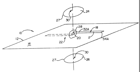
With reference to the drawings, the electrode carrying surgical drape 10
generally includes a polymeric film 12 having opposing surfaces 14 & 16, an
electrode 18 received in an electrode receiving aperture 20 to thereby define
an
electrode site 22 for the surgical drape 10, and a pair of reinforcing patches
24 &
26 affixed to each of the opposing sides 14 & 16 of the polymeric film 12,
each
positioned to sealingly overlay the electrode receiving aperture 20. An
adhesive
sealant 28 occupies any spaces or voids between the electrode 18 and the
electrode receiving aperture 20, and generally coats the polymeric film 12 in
the
vicinity of the electrode site 22 so as to sealingly engage and adhere at
least the
central portion 30 of the reinforcing patches 24 & 26 to the opposing sides 14
&
16 of the polymeric film 12.
The surgical drape 10 is generally deployed so as to establish, delimit and
-3-
CA 02322675 2000-10-10
maintain sterile and non-sterile fields, and is generally formed from a
polymeric
surgical drape film 12, preferably a polyurethane film having a thickness of
about 0.005 inches. Alternate drape synthetics suitable to establish and
maintain
a sterile field are likewise contemplated, those being well known to those of
skill
in the art.
An integrally placed electrode 18 traverses the film 12 to thereby permit the
passage of electricity, in the form of current or voltage, between the sterile
and
non-sterile fields without jeopardizing or compromising the sterility
established
by the surgical drape. Preferably the electrode 18 is a thin, dual lead, film
backed
conductor. In a broad sense, the electrode 18 is, in effect, a conduit linking
the
"environments" existing adjacent each of the opposing surfaces 14 & 16 of the
polymeric film 12 when the drape 10 is deployed for use. In addition to, or
beyond the passage of electricity through the established fields by the
conduit,
information or conditions (i.e., "data" more generally) may be passed
therethrough as the application warrants, when for instance fiber optic
sensors
are used for a variety of detection purposes. It is to be understood that the
term
"electrode" used herein is not limited to electrical conductance but to
conductance in its broadest sense (i.e., an electrode as a connector or
linkage).
The electrode 18 is received in an aperture or passage 20 centrally positioned
in
the electrode site 22 so to pass through the drape film 12 (i.e.,
substantially
intersect the plane of the drape film 12). The aperture 20 is preferable a
slit (i.e.,
a cut with no removal of material from the drape film) dimensioned to accept
the
electrode 18 therethrough. Openings through the drape film may also include
holes or perforations, with methods of making such openings well know to those
of skill in the art of such methods.
Referring now specifically to FIGS. 1& 2, electrode receiving aperture patches
24 & 26 form a sealed, reinforced "sandwich" (i.e., a laminate structure)
which
includes several layers or partial layers, namely, a first adhesively coated
polymeric patch 24, a segment 32A of a first portion 17 of the electrode 18,
the
polymeric film 12, a medical grade adhesive 28, a segment 32B of a second
portion 19 of the electrode 18, and a second adhesively coated polymeric patch
26. This arrangement provides a strong yet simple and supremely efficient seal
of the polymeric film 12 about the drape intersecting electrode 18 in the
vicinity
of the electrode receiving aperture or passage 20. As particularly illustrated
in
-4-
CA 02322675 2000-10-10
FIGS. 2 & 3, non-sandwiched segments 34A & 34B of the first 17 and second
19 portions of the electrode 18 extend beyond the boundary 27 of the
reinforcing and sealing patches 24 & 26 so as to be suitably connected or
otherwise linked to peripherals, thereby completing a transmission path
between
the fields.
The electrode receiving aperture patches are preferably formed from a
polyethylene film or sheeting. In addition to performing a sealing function,
the
patches perform a reinforcement function, and as such are generally but not
necessarily more rigid than the surgical drape film, and otherwise posses at
least
equivalent tear resistance and strength when compared to the surgical drape
film.
The patches 24 & 26 are preferably centered about the electrode receiving
aperture 20 on both sides of the polymeric surgical drape film 12, and
preferably
carry a coating of pressure sensitive adhesive on one of their surfaces so as
to
easily apply and affix the patch to the electrode site. Preferably the patches
are
circular, however other geometries are suitable. The patch is preferably
dimensioned to be about 5 to 10 times the maximum dimension of the electrode
aperture, however, dimensions outside this range may be more appropriate based
upon factors such as aperture dimension, surgical drape material and
thickness,
and the physical qualities of the electrode, to name but a few parameters.
The thickness of the patches is variable, being dependent in part upon the
nature
of the film and patch material. Generally, sufficient sealing and
reinforcement is
readily achieved in the vicinity of the electrode site with patches having a
thickness not greater than that of the surgical drape film itself.
In the method of forming an electrode carrying surgical drape, the poly55meric
surgical drape film is first slit or otherwise cut to receive an electrode.
Slitting is
advantageous as no material is removed from the polymeric film, thereby
providing a form fit for the electrode placed therethrough. The electrode is
next
placed in the slit so as to form first and second electrode portions. One of
the
electrode portions (i.e., the first electrode portion) is partially covered by
a
single sided adhesive backed reinforcement patch positioned to be centered
about the slit and adhered thereto. Approximately 0.1 milliliter of medical
grade
surgical adhesive is placed in the slit on the unpatched side of the polymeric
surgical drape (i.e., in the "open" slit), and is further used to coat the
segment of
the second portion of the electrode to be sandwiched by the opposing patches.
A
-5-
- - ----- _ , ~
CA 02322675 2000-10-10
second adhesive backed reinforcement patch is centered over the silicone
adhesive
filled slit and affixed to the surgical drape film so as to form a laminate
structure.
It will be understood that this disclosure, in many respects, is only
illustrative.
Changes may be made in details, particularly in matters of shape, size,
material,
and arrangement of parts without exceeding the scope of the invention.
Accordingly, the scope of the invention is as defined in the language of the
appended claims.
15
25
35
-6-