Note: Descriptions are shown in the official language in which they were submitted.
CA 02322713 2000-10-31
1 , r
Case 20487
Background of the Invention
In order for metastasis of cancer to occur, several hurdles must be overcome,
such as
degradation of the extracellular matrix and basal membrane, intra- and
extravasation of
vessels of the blood and of the lymphatic system, escape by the attack of the
immune
system, and homing and colonization of distant organs (Pardee, A.B., Advances
in Cancer
Res. 65 (1994) 213-227; Ponta, H., et al., Biochem. Biophys. Acta 1198 (1994)
1-10). A
further level of complexity is achieved in that different types of cancers
make use of
different molecular mechanisms for metastasis and exhibit different tropism of
metastasis.
Metastasizing and non-metastasizing human melanoma cell lines have been
important
tools in identifying differentially expressed genes and for investigation of
their role in
metastasis (Weterman, M.A.J., et al., Cancer Res. 52 (1992) 1291-1296;
Weterman, M.A.J.,
et al., Int. J. Cancer 53 (1993) 278-284; Van Groningen, J.M., et al., Cancer
Res. 55 (1995)
6237-6243; Weterman, M.A.J., et al., Int. J. Cancer 60 (1995) 73-81; van
Muijen, G.N.P., et
al., Int. J. Cancer 48 (1991) 85-91; van Muijen, G.N.P., et al., Clin. Exp.
Metastasis 9 (1991)
259-272).
Cell adhesion molecules play an important role in the invasion, dissemination,
extravasation and lodging of tumor cells. The interaction of disseminated
tumor cells with
endothelium and tissue stroma is supposed to be one of the critical steps in
tumor
progression and metastasis formation (Ebnet, K., et al., Annu. Rev. Immunol.
14 (1996)
155-177; Varner, J.A., and Cheresh, D.A., Curr. Opin. Cell Biol. 8 (1996) 724-
730; Albelda,
S.M., Lab. Invest. 68 (1993) 4-17).
CTp 11 is a polypeptide homologous to the polypeptide sequences described in
EMBL
Database A1962751, AA412605, and AA412270 as well as in SEQ ID NO:18 and
SEQ ID NO:75 of WO 99/46374.
Summary of the Invention
In accordance with the present invention, it was surprisingly found that a
protein, termed
CTp 11 (cancer/testis-associated protein of 11 kD), is upregulated in
metastatic cancer cells
as compared to their non-metastatic counterparts. CTp11 may be involved in
promotion of
several steps of the metastatic cascade. CTp 11 is a specific marker of
metastatic cancer cells,
due to the fact that it can be presented in an MHC Class I complex on
cytotoxic T cells but
CA 02322713 2000-10-31
-2-
is not presented naturally because the only non-tumor cells (testis cells) in
which CTp 11 is
found do not present antigens in an MHC Class I context. The CTp 11 gene codes
for a
polypeptide of SEQ ID NO:2.
The present invention provides a process for detecting the presence or absence
of at least
one specific nucleic acid or mixture of nucleic acids, or distinguishing
between two
different sequences in said sample, wherein the sample is suspected of
containing said
sequence or sequences, which process comprises the following steps in order:
(a) incubating said sample under stringent hybridization conditions with a
nucleic acid
probe which is selected from the group consisting of:
(i) a nucleic acid sequence of SEQ ID NOS: 1 and 3 to 6;
(ii) a nucleic acid sequence which is exactly complementary to a nucleic acid
sequence of (i);
(iii) a nucleic acid sequence which hybridizes under stringent conditions with
the
sequence of (i); and
(iv) a nucleic acid sequence which hybridizes under stringent conditions with
the
sequence of (ii); and
(b) determining whether said hybridization has occurred.
Moreover, the present invention provides a process for determining whether or
not a
cancer cell-containing test sample has potential for tumor progression or
metastasis,
wherein the test sample and a cancer cell-containing sample which is free from
metastasis
and wherein both samples are obtained from the same individual or different
individuals of
the same species, which process comprises the following steps:
(a) incubating each respective sample under stringent hybridization conditions
with a
nucleic acid probe which is selected from the group consisting of:
(i) a nucleic acid sequence of SEQ ID NOS: 1 and 3 to 6;
(ii) a nucleic acid sequence which is exactly complementary to a nucleic acid
sequence of (i);
(iii) a nucleic acid sequence which hybridizes under stringent conditions with
the
sequence of (i); and
(iv) a nucleic acid sequence which hybridizes under stringent conditions with
the
sequence of (ii); and
(b) determining the approximate amount of hybridization of each respective
sample with
said probe, and
CA 02322713 2000-10-31
-3-
(c) comparing the approximate amount of hybridization of the test sample to an
approximate amount of hybridization of the sample which is free from
metastasis, to
identify whether or not the test sample contains a greater amount of the
specific
nucleic acid or mixture of nucleic acids than does the sample which is free
from
metastasis.
Detailed Descriution of the Invention
The present invention provides methods of use and the use of CTp11 gene
(cancer/testis-
associated protein of 11 kD) for diagnostics, especially in the field of
cancer diagnosis. In
particular, the invention involves the identification of said gene CTp 11 in
malignant tumor
cells having a metastatic and/or progression potential.
Differential Display Technique applied to non-metastatic melanoma cell line 1
F6 and its
metastatic subcell line 1F6m resulted in identification of CTpll which was at
least 40 fold
up-regulated in the metastatic cell line (Fig. 1).
According to the invention, the nucleic acid molecule (CTp 11) has upregulated
expression
in metastatic tumor cells and is capable of inducing tumor progression and/or
metastasis,
especially in malignant melanoma and mammary carcinoma cells. The nucleic acid
(CTp11) has the sequence SEQ ID NO:I or it is a nucleic acid which, because of
the
degeneracy of the genetic code, differs from SEQ ID NO:1, but which encodes
the amino
acid sequence encoded by the nucleic acid of SEQ ID NO:1.
The isolated CTpll polypeptide can occur in natural allelic variations which
differ from
individual to individual. Such variations of the amino acids are usually amino
acid
substitutions. However, they may also be deletions, insertions or additions of
amino acids
to the total sequence. The CTp 11 protein according to the invention -
depending, both in
respect of the extent and type, on the cell and cell type in which it is
expressed - can be in
glycosylated or non-glycosylated form. Polypeptides with metastatic activity
can be
identified by transfection of CTp 11 -negative non-metastasizing tumor cells
with expression
vectors for CTpll, establishment of stable transfectants and evaluation of in
vitro
invasiveness in Matrigel invasion assays and their metastatic capacity after
xenografting
into nude mice.
"Polypeptide with CTp11 activity or CTp11 " means also proteins with minor
amino acid
variations but with substantially the same CTpll activity. Substantially the
same means
CA 02322713 2000-10-31
-4-
that the activities are of the same biological properties and the polypeptides
show (at least
90%, preferably more than 95%) homology, or preferably identity, in amino acid
sequence.
Homology can be examined by using the BLAST algorithm described by Altschul,
S.F., et
al., Nucleic Acids Res. 25 (1997) 3389-3402.
The term "nucleic acid molecule or nucleic acid" denotes a polynucleotide
molecule which
can be, for example, a DNA, RNA, or derivatized active DNA or RNA. DNA and/or
RNA
molecules are preferred, however.
The term "hybridize under stringent conditions" means that two nucleic acid
fragments are
capable of hybridization to one another under standard hybridization
conditions described
in Sambrook et al., Molecular Cloning: A Laboratory Manual (1989) Cold Spring
Harbor
Laboratory Press, New York, USA. More specifically, "stringent conditions" as
used herein
refer to hybridization at 65 C in a hybridization buffer consisting of 250
mmol/1 sodium
phosphate buffer pH 7.2, 7% (w/v) SDS, 1% (w/v) BSA, 1 mmol/l EDTA and 0.1
mg/ml
single-stranded salmon sperm DNA. A final wash is performed at 65 C in 125
mmol/1
sodium phosphate buffer pH 7.2, 1 mmol/1 EDTA and 1% (w/v) SDS.
The phrase "nucleic acid or polypeptide" as used throughout this application
refers to a
nucleic acid or polypeptide having a CTp11 activity which is substantially
free of cellular
material or culture medium when produced by recombinant DNA techniques, or
substantially free of chemical precursors or other chemicals when synthesized
chemically.
Such a nucleic acid is preferably free of sequences which naturally flank the
nucleic acid
(i.e. sequences located at the 5' and the 3' ends of the nucleic acid) in the
organism from
which the nucleic acid is derived.
The CTp11 polypeptides can be produced by recombinant means in host cells,
using an
expression vector, or can be produced synthetically. Non-glycosylated CTp11
polypeptide
is obtained when it is produced recombinantly in prokaryotes. With the aid of
the nucleic
acid sequences provided by the invention it is possible to search for the
CTp11 gene or its
variants in genomes of any desired cells (e.g. apart from human cells, also in
cells of other
mammals), to identify these and to isolate the desired gene coding for the
CTp11 protein.
Such processes and suitable hybridization conditions are known to a person
skilled in the
art and are described, for example, by Sambrook et al., Molecular Cloning: A
Laboratory
Manual (1989), Cold Spring Harbor Laboratory Press, New York, USA, and Hames,
B.D.,
Higgins, S.G., Nucleic Acid Hybridisation - A Practical Approach (1985) IRL
Press, Oxford,
CA 02322713 2000-10-31
-5-
England. In this case the standard protocols described in these publications
are usually used
for the experiments.
With the aid of such nucleic acids, CTp11 protein can be obtained in a
reproducible
manner and in large amounts. For expression in prokaryotic or eukaryotic
organisms, such
as prokaryotic host cells or eukaryotic host cells, the nucleic acid is
integrated into suitable
expression vectors, according to methods familiar to a person skilled in the
art. Such an
expression vector preferably contains a regulatable/inducible promoter. These
recombinant
vectors are then introduced for the expression into suitable host cells such
as, e.g., E. coli as
a prokaryotic host cell or Saccharomyces cerevisiae, Teratocarcinoma cell line
PA-1 sc 9117
(Biittner et al., Mol. Cell. Biol. 11 (1991) 3573-3583), insect cells, CHO or
COS cells as
eukaryotic host cells and the transformed or transduced host cells are
cultured under
conditions which allow expression of the heterologous gene. The isolation of
the protein
can be carried out according to known methods from the host cell or from the
culture
supernatant of the host cell. Such methods are described for example by
Ausubel I.,
Frederick M., Current Protocols in Mol. Biol. (1992), John Wiley and Sons, New
York. Also
in vitro reactivation of the protein may be necessary if it is not found in
soluble form in the
cell culture.
CTp 11 can be purified after recombinant production by affinity chromatography
using
known protein purification techniques, including immunoprecipitation, gel
filtration, ion
exchange chromatography, chromatofocussing, isoelectric focussing, selective
precipitation, electrophoresis, or the like.
The invention comprises a method for detecting a nucleic acid molecule of gene
CTp 11,
comprising incubating a sample (e.g., body fluids such as blood, cell lysates)
with a
specifically binding nucleic acid molecule and determining hybridization under
stringent
conditions of said isolated nucleic acid molecule to a target nucleic acid
molecule for
determination of presence of a nucleic acid molecule which is the CTp11 gene
and
therefore a method for the identification of the metastatic potential and/or
progression of
tumor cells.
To determine whether a cancer cell-containing test sample has potential for
tumor
progression or metastasis, the approximate amount of hybridization of the
isolated nucleic
acid with the target nucleic acid or nucleic acids is determined. The
approximate amount
of hybridization need not be determined quantitatively, although a
quantitative
determination is encompassed by the present invention. Typically, the
approximate
CA 02322713 2000-10-31
-6-
amount of hybridization is determined qualitatively, for example, by a sight
inspection
upon detecting hybridization. For example, if a gel is used to resolve
labelled nucleic acid
which hybridizes to target nucleic acid in the sample, the resulting band can
be inspected
visually. When performing a hybridization of isolated nucleic acid in a cancer-
containing
sample which is free from metastasis from an individual of the same species,
the same
protocol is followed. One can compare the approximate amount of hybridization
in the
test sample to the approximate amount of hybridization in the sample free from
metastasis,
to identify whether or not the test sample contains a greater amount of the
target nucleic
acid or nucleic acids than does the sample which is free from metastasis. For
visual
inspection in particular, it is recommended that an appreciable difference by
visualized to
assess that the test sample contains a greater amount of the target nucleic
acid or nucleic
acids.
As is shown in accordance with the present invention, the CTp 11 nucleic acid
is present in
a greater amount in a metastasized tumor sample than in a sample free from
metastasis. A
test sample having potential for tumor progression or metastasis will have a
greater amount
of the CTp 11 nucleic acid than does a cancer cell sample which is free from
metastasis. To
identify a test sample as containing upregulated CTp 11 nucleic acid, i.e.,
wherein the cancer
cells have potential for tumor progression or metastasis, it is preferable
that the test sample
have an approximate amount of CTp 11 nucleic acid which is appreciably greater
that the
approximate amount in a non-metastasigned sample. For example, a test sample
having an
upregulated CTp11 gene may have approximately 15- to approximately 60- fold
greater
amount of CTp11 gene than a non-metastasized sample.
On the basis of the nucleic acids provided by the invention it is possible to
provide a test
which can be used to detect nucleic acids with upregulated expression in
metastatic human
tumor cells. Such a test can be carried out by means of nucleic acid
diagnostics. In this case
the sample to be examined is contacted with a probe that is selected from the
group
comprising
a) the nucleic acid sequence shown in SEQ ID NOS:1 and 3 to 6 or a nucleic
acid
sequence which is complementary to one of these nucleic acid sequences, and
b) nucleic acids which hybridize under stringent conditions with one of the
nucleic acids
from a), wherein
the nucleic acid probe is incubated with the nucleic acid of the sample and
the
hybridization is detected optionally by means of a further binding partner for
the nucleic
I
CA 02322713 2004-05-17
-7-
acid of the sample and/or the nucleic acid probe. For obtaining a nucleic acid
by
hybridization in accordance with b), it is preferable to hybridize to the
probe shown in SEQ
ID NOS:3 to 6 or a sequence complementary thereto. Hybridization between the
probe
used and nucleic acids from the sample indicates the presence of the RNA of
such proteins.
Methods of hybridization of a probe and a nucleic acid are known to a person
skilled in the
art and are described, for example, in WO 89/06698, EP-A 0 200 362, USP
2915082,
EP-A 0 063 879, EP-A 0 173 251, EP-A 0 128 018.
In a preferred embodiment of the invention the coding nucleic acid of the
sample is
amplified before the test, for example by means of the known PCR technique.
Usually a
derivatized (labeled) nucleic acid probe is used within the framework of
nucleic acid
diagnostics. This probe is contacted with a denatured DNA or RNA from the
sample which
is bound to a carrier and in this process the temperature, ionic strength, pH
and other
buffer conditions are selected - depending on the length and composition of
the nucleic
acid probe and the resulting melting temperature of the expected hybrid - such
that the
labeled DNA or RNA can bind to homologous DNA or RNA (hybridization see also
Wahl,
G.M., et al., Proc. Natl. Acad. Sci. USA 76 (1979) 3683-3687). Suitable
carriers are
membranes or carrier materials based on nitrocellulose (e.g., Schleicher and
Schull, BA 85,
Amersham Hybond;` C.), strengthened or bound nitrocellulose in powder form or
nylon
membranes derivatized with various functional groups (e.g., nitro groups)
(e.g., Schleicher
and Schull, Nytran; NEN, Gene Screen; Amersham Hybond M.; Pall Biodyne).
Hybridizing DNA or RNA is then detected by incubating the carrier with an
antibody or
antibody fragment after thorough washing and saturation to prevent unspecific
binding.
The antibody or the antibody fragment is directed towards the substance
incorporated
during hybridization to the nucleic acid probe. The antibody is in turn
labeled. However, it
is also possible to use a directly labeled DNA. After incubation with the
antibodies it is
washed again in order to only detect specifically bound antibody conjugates.
The
determination is then carried out according to known methods by means of the
label on
the antibody or the antibody fragment.
The detection of the expression can be carried out for example as:
- in situ hybridization with fixed whole cells, with fixed tissue smears and
isolated
metaphase chromosomes,
- colony hybridization (cells) and plaque hybridization (phages and viruses),
* Trademark
CA 02322713 2000-10-31
-8-
- Southern hybridization (DNA detection),
- Northern hybridization (RNA detection),
- serum analysis (e.g., cell type analysis of cells in the serum by slot-blot
analysis),
- after amplification (e.g., PCR technique).
Therefore the invention includes a method for the detection of the metastatic
potential of
melanoma and mammary carcinoma cells, comprising
a) incubating a sample of body fluid of a patient suffering from cancer, of
melanoma
cancer cells, of mammary carcinoma cells, or of a cell extract or cell culture
supernatants of said cancer cells, whereby said sample contains nucleic acids
with a
nucleic acid probe which is selected from the group consisting of
(i) the nucleic acid shown in SEQ ID NOS: 1 and 3 to 6 or a nucleic acid which
is
complementary to said nucleic acid sequence, and
(ii) nucleic acids which hybridize with the nucleic acids from (i) and
b) detecting hybridization by means of a further binding partner of the
nucleic acid of
the sample and/or the nucleic acid probe or by X-ray radiography.
Preferably the nucleic acid probe is incubated with the nucleic acid of the
sample and the
hybridization is detected optionally by means of a further binding partner for
the nucleic
acid of the sample and/or the nucleic acid probe.
The CTp 11 nucleic acids are therefore valuable prognostic markers in the
diagnosis of the
metastatic and progression potential of tumor cells of a patient.
There is further provided a method for producing a protein whose expression is
correlated
with tumor metastasis, by expressing an exogenous DNA in prokaryotic or
eukaryotic host
cells and isolation of the desired protein, wherein the protein is coded by
the nucleic acid
molecules according to the invention, preferably by the DNA sequence shown in
SEQ ID NO:1.
The protein can be isolated from the cells or the culture supernatant and
purified by
chromatographic means, preferably by ion exchange chromatography, affinity
chromatography and/or reverse phase HPLC.
CA 02322713 2000-10-31
-9-
The invention further comprises an isolated protein which is encoded by a
nucleic acid
molecule according to the invention, preferably having the nucleotide sequence
set forth in
SEQ ID NO:1.
CTp11 is especially characterized as a tumor progression gene, and as an
upregulated gene
indicative for metastatic potential of melanoma cells. The function of CTp11
is to promote
loss of contact inhibition and anchorage dependence in tumor cells and to
promote other
essential steps of the metastatic cascade. Therefore the expression of CTp11
gene correlates
with a more aggressive behavior of the tumor cells and also with the potential
of the
formation of metastasis.
According to the invention inhibitors for the expression of CTpll, preferably
antisense
nucleic acids or antibodies, can be used to inhibit tumor
progression/metastasis, preferably
of malignant melanomas and mammary carcinomas, in vivo, preferably by somatic
gene
therapy. Antibodies against CTp 11 protein can be produced according to the
state of the art
using CTp 11 protein purified as described above for immunization of mice or
rats.
Antisense nucleic acids preferably have a length of 20-100 nucleotides.
Overall characteristics place the gene in the family of cancer/testis antigens
(CTAs) of
which the first members, named MAGEs (melanoma antigens), were described by
the
group of Boon (van der Bruggen et al., Science 254 (1991) 1643-1647).
The protein encoded by the full-length cDNA consists of 97 amino acids
containing a
bipartite nuclear localization signal (NLS). A specific nuclear localization
was found after
fusing the ORF in front of eGFP, indicating that the bipartite-like nuclear
localization
sequence is indeed effective. The bipartite NLS consensus comprises two basic
amino acids
(lysine (K) or arginine (R)) separated by a region of ten amino acids from a
basic cluster in
which three out of the next five residues must be basic (Dingwall and Laskey,
Trends.
Biochem. Sci. 16 (1991) 478-481). The spacer of ten amino acids was shown to
be optimal,
though effective bipartite nuclear localization signals were also found with
elongated
spacers (Robbins, J., et al., Cell 64 (1991) 615-623). This indicates the
likeliness of the
bipartite sequence in CTp11 (a.a. 40-57), with a 12 residue spacer, being
responsible for
localization in the nucleus. Bipartite NLS sequences have also been found in
several
members of the SSX-family, which also belong to the group of cancer/testis
antigens (Dos
Santos, N.R., et al., Hum. Mol. Genet. 6 (1997) 1549-1558). The acidic C-
terminal region
with the high content of glutamic acid residues resembles a GAL4 domain, shown
to be
effective in transcriptional activation after interaction or fusion with a DNA
binding
CA 02322713 2000-10-31
-10-
protein or domain (Mitchell, P.J., and Tjian, R., Science 245 (1989) 371-378).
CTpll, like
the SSX proteins (Dos Santos, N.R., et al., Hum. Mol. Genet. 6 (1997) 1549-
1558) and
melanocyte-specific gene 1 (MSG1) (Shioda, T., et al., Proc. Natl. Acad. Sci.
USA 93 (1996)
12298-12303), lacks a DNA-binding domain. Therefore, it interacts with the
transcription-
initiation complex in order to concert its putative transcriptional
regulation. The high
percentage of charged amino acids might contribute in such a protein-complex
interaction.
The expression profile of CTp 11 in normal tissues, tumor cell lines and tumor
samples
place the gene in the group of cancer/testis antigens, which already include
MAGE (Lucas,
S., et al., Cancer Res. 58 (1998) 743-752), BAGE (Boel, P., et al., Immunity 2
(1995) 167-
175), GAGE (van den Eynde, B., et al., J. Exp. Med. 182 (1995) 689-698), SSX
(Gure, A.O.,
et al., Int. J. Cancer 72 (1997) 965-971), NY-ESO-1 (Chen, Y.T., et al., Proc.
Natl. Acad. Sci.
USA 94 (1997) 1914-1918), LAGE-1 (Lethe, B., et al., Int. J. Cancer 76 (1998)
903-908),
PAGE-1 (Chen, M.E., et al., J. Biol. Chem. 273 (1998) 17618-17625; Brinkmann,
U., et al.,
Proc. Natl. Acad. Sci. USA 95 (1998) 10757-10762) and SCP-1 (Tureci, 0., et
al., Proc.
Natl. Acad. Sci. USA 95 (1998) 5211-5216).
Criteria genes should fullfil to be considered as a member of the family of
cancer/testis
antigens are formulated in the literature (Chen, Y.T., et al., Proc. Natl.
Acad. Sci. USA 94
(1997) 1914-1918); Chen, Y.T., et al., Proc. Natl. Acad. Sci. USA 95 (1998)
6919-6923): (i)
predominant expression in testis and generally not in other normal tissues,
(ii)
induction/activation of mRNA expression in a wide range of human tumors, (iii)
expression in malignancies in a lineage-nonspecific fashion, (iv) often
existence of
multigene families, (v) mapping of the gene, with some exceptions, on the X-
chromosome.
CTpI l clearly qualifies as a CTA-family member since it shares criteria i,
ii, iii and v.
The CTp 11 gene is localized on the X chromosome (Xq26.3-Xq27.1) and consist
of two
exons separated by an intron of about 655 bp as confirmed by PCR. This
position is right
next to the MAGE-C subfamily (Xq26) (Lucas, S., et al., Cancer Res. 58 (1998)
743-752)
and nearby CTAG (the gene for NY-ESO-1) and the MAGE-A cluster (both Xq28)
(Chen,
Y.T., et al., Cell Genet. 79 (1997) 237-249).
CTpll expression was found in 25-30% of the melanoma and bladder tumor cell
lines
tested, while cell lines established from other tumor types were only
sporadically positive.
For melanoma cell lines, which are the best studied regarding CTA expression,
the
percentage of positivity is comparable to the percentages of positivity for NY-
ESO-1 (18%)
(Chen, Y.T., et al., Proc. Natl. Acad. Sci. USA 94 (1997) 1914-1918; Lethe,
B., et al., Int. J.
CA 02322713 2000-10-31
-Il-
Cancer 76 (1998) 903-908), SSX-2 (25%) (Tureci, 0., et al., Int. J. Cancer 77
(1998) 19-23)
and MAGE-B1 and -B2 (22% and 33%) (Lurquin, C., et al., Genomics 46 (1997) 397-
408;
Muscatelli, F., et al., Proc. Natl. Acad. Sci. USA 92 (1995) 4987-4991). MAGE-
Al (66%)
(Kirkin, A.F., et al., Exp. Clin. Immunogenet. 15 (1998) 19-32; Wang, R.F.,
Mod. Med. 3
(1997) 716-731) has a markedly higher expression coverage in melanoma cell
lines and is
expressed in 41% of other human tumor cell lines.
Testis-specific expression regarding normal tissues seen by RT-PCR confirms
the
exclusiveness of CTp11 homology with only ESTs from testis. In fresh human
tumor
samples, melanoma was found to have the highest percentage of CTp11 positivity
(70%;
n=10). Comparable positivity was seen in primary (3 out of 4) as well as
metastatic
melanoma (4 out of 6). This percentage may be one of the highest compared to
the other
CTAs in melanoma, being 8, 17, 22, 35, 44, 44 and 52% for SCP-1, GAGE, BAGE,
MAGE-
1, SSX-2, NY-ESO-1 and MAGE-3 (Sahin, U., et al., Int. J. Cancer 78 (1998) 387-
389),
respectively. The relatively high percentage of CTpll in bladder cell lines
(30%) was not
detected in bladder tumor samples in which only 1 out of 11 was found to be
positive.
Interestingly, testis tumors showed a downregulation of CTpl1 expression
compared to
normal testis tissue, since only 10 out of 17 tumor lesions were positive
after nested PCR.
No positivity of testis tumor samples was seen after the first PCR. Positivity
of these testis
lesions, both seminomas and non-seminomas, could be caused by small amounts of
normal testis tissue present. Regarding the other CTAs it is known that MAGE-1
is mainly
expressed in germ cells of the testis (Takahashi, K., et al., Cancer Res. 55
(1995) 3478-3482)
and this finding is in line with the fact that seminoma have a higher
positivity rate for
MAGE-expression than non-seminoma tumors (Hara, I., et al., Urology 53 (1999)
843-
847).
Homology search and chromosomal localization
Checking all kinds of databases, there was found homology of more than 90%
with three
human genomic clones (HS433M19; HS376H23; HSG164F24), all localized on
chromosome X. The homology with HS433M19 even narrowed the gene-localization
down
to Xq26.3-Xq27.1. PCR on a panel of human chromosome specific rodent/human
hybrid
cell lines confirmed localization of the gene on chromosome X. Based on the
length of the
genomic PCR product and on the sequences of the three human genomic clones it
was
deduced that the gene consists of two exons separated by an intron of
approximately 655
bp which is located near basepair 112.
CA 02322713 2000-10-31
-12-
Homology on cDNA level was restricted to several human ESTs which were all
from testis
(zt95b09; qg57bOl; qe04h11; EST95628/EST95629) and which all code for the same
putative protein. Homology was also found with EMBL Database ACC No. A1962751,
AA412605, and AA412270 as well as with SEQ ID NO:18 and SEQ ID NO:75 of
WO 99/46374.
The invention further provides methods for identifying and isolation of
antagonists of
CTp11 or inhibitors for the expression of CTp11 (e.g. antisense nucleotides).
Such
antagonists or inhibitors can be used to inhibit tumor progression or
metastasis and cause
massive apoptosis of tumor cells in vivo.
According to the invention there are provided methods for identifying and
isolation of
compounds which have utility in the treatment of cancer, especially in
inhibiting metastasis
and related disorders. These methods include methods for modulating the
expression of
the polypeptides according to the invention, methods for identifying compounds
which
can selectively bind to the proteins according to the invention, and methods
of identifying
compounds which can modulate the activity of said polypeptides. The methods
further
include methods for modulating, preferably inhibiting, the transcription of
CTp 11 gene to
mRNA, which preferably down-regulates the metastatic potential of a tumor
cell. These
methods can be conducted in vitro or in vivo and may make use of and establish
cell lines
and transgenic animal models of the invention.
A CTp 11 antagonist is defined as a substance or compound which decreases or
inhibits the
biological activity of CTp 11, a polypeptide and/or inhibits the transcription
or translation
of CTp11 gene. In general, screening procedures for CTp 11 antagonists involve
contacting
candidate substances with host cells in which invasiveness is mediated by
expression of
CTp 11 under conditions favorable for measuring CTp 11 activity.
CTp 11 activity may be measured in several ways. Typically, the activation is
apparent by a
change in cell physiology, such as increased mobility and invasiveness in
vitro, or by a
change in the differentiation state, or by a change in cell metabolism leading
to an increase
of proliferation.
The CTp11 gene and protein can be used to identify and design drugs which
interfere with
proliferation and dissemination of tumor cells.
CA 02322713 2000-10-31
-13-
The following examples, references, sequence listing and figures are provided
to aid the
understanding of the present invention. It is understood that modifications
can be made in
the procedures set forth without departing from the spirit of the invention.
SEQ ID NO:1 : cDNA and amino acid sequence of CTp 11.
SEQ ID NO:2: Amino acid of CTp11.
SEQ ID NO:3: Sense primer.
SEQ ID NO:4 : Antisense primer.
SEQ ID NO:5: Nested sense primer.
SEQ ID NO:6: Nested antisense probe.
SEQ ID NO:7: 82-Microglobulin-sense primer.
SEQ ID NO:8 92-Microglobulin antisense primer.
Description of the Fig Ules
Figure 1 Northern blot analysis on a panel of human melanoma cell lines with
different metastatic capacity after subcutaneous inoculation into nude
mice. The blot is hybridized with the 300 bp differential display cDNA.
Arrow indicates a band of 0.5 kb exclusively present in the highly
metastatic cell lines. Lane 1: 530; lane 2: 1F6; lane 3: M14; lane 4: Mel57;
lane 5: MV3; lane 6: BLM; lane 7: 1F6m.
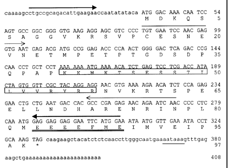
Figure 2 CTp 11 cDNA and deduced protein sequence. Primers used for PCR are
indicated by arrows (first PCR: closed arrowheads; nested PCR: open
arrowheads). Polyadenylation signal is underlined, putative nuclear
localization signal is boxed and poly-E acidic domain is double
underlined. The stop codon is marked by an asterisk.
Figure 3 (A) RT-PCR on RNA isolated from 17 different fresh normal human
tissues. Only testis is positive (297 bp cDNA band). In a few samples a
CA 02322713 2000-10-31
-14-
weak genomic DNA band is visible (1 kb). (B) Control RT-PCR of 2-
microglobulin (136 bp).
Figure 4 (A) Nested RT-PCR (188 bp) on RNA isolated from samples of normal
human skin and on RNA isolated from tissue samples containing lesions
with different stages of melanocytic tumor progression (NS= normal
skin; NN= common naevus naevocellularis; AN= atypical naevus; PM=
primary melanoma; MM= melanoma metastasis). (B) Control RT-PCR
of f32-microglobulin.
Figure 5 Western blot analysis of cell lysates from BLM (lane 1), BLM
transfected
with a construct containing only eGFP (lane 2) and BLM with a construct
containing the full length cDNA/eGFP fusion construct (lane 3). Bands
were visualized by incubation with a polyclonal antibody against eGFP.
Note that the 27 and 38 kD bands are specific and that the band at 50 kD
is aspecific.
Example 1
Materials and methods
Cell lines and primary cultures
A panel of eight different human melanoma cell lines containing 530, 1F6, MV1,
M14,
Me157, BLM, MV3, and 1F6m was described earlier (Westphal, J.R., et al., Br.
J. Cancer 76
(1997) 561-570; van Muijen, G.N., et al., Clin. Exp. Metastasis 9 (1991) 259-
272). In this
panel of cell lines 530 and 1F6 are poorly metastatic, while MV3, BLM and 1F6m
are highly
metastatic cell lines. MV1, M14, and Me157 are cell lines with an intermediate
metastatic
capacity. 1F6m is a metastatic subline of 1F6. Most other cell lines used were
described
earlier (Zendman, A.J., et al., FEBS Lett. 446 (1999) 292-298). Cell lines
RAMOS and RAJI
are from the ATCC. BLM is an HLA-A1 negative melanoma cell line. All cell
lines were
grown in Dulbecco's modified Eagle's medium as described earlier (de Vries,
T.J., et al.,
Cancer Res. 56 (1996) 1432-1439). Normal human foreskin melanocytes and human
nevus
cells were cultured as described previously (Verbeek, M.M., Am. J. Pathol. 144
(1994) 372-
382).
CA 02322713 2004-05-17
-15-
Human tissues
Lesions from all stages of melanocytic tumor progression (common nevi,
atypical nevi,
primary melanoma and melanoma metastases) and other tumor specimens were
excised
from patients at the University Hospital Nijmegen, The Netherlands. As normal
human
tissues, disease-free samples from surgically removed tissues or from
autopsies with a post-
mortem delay shorter than 4 hours were used. Tissue samples were snap-frozen
in liquid
nitrogen and stored at -80 C until use.
RNA Isolation
From cultured cells total RNA was isolated using the RNeasy kit (Qiagen,
Hilden,
Germany) following the manufacturer's protocol. From tissue samples total RNA
was
isolated (following manufacturer's protocol) by disrupting about 25 frozen
sections of
m thickness in 1 ml RNAzo1BTdA (Campro, Veenendaal, The Netherlands) using a
pestle. The RNAzo1BT"' method was followed by an additional RNeasy cleaning
step.
mRNA differential display
15 Prior to mRNA differential display PCR, DNaseI treatment was performed on
the RNA
samples using the Message-CleanTM kit (GenHunter Corporation, Nashville, TN).
For
differential display the RNAmapTM protocol (GenHunter) was used with some
minor
modifications. Differing from the original protocol, [32P] -dATP was used
instead of [35S] -
dATP. For the PCR, combinations of the four T12MN primers together with six
arbitrary
20 primers, AP1, 2, 6, 7, 11, 12 (Bauer, D., et al., Nucleic Acids. Res. 21
(1993) 4272-4280), were
used.
Northern blotting
Ten micrograms of total RNA were treated with glyoxal/DMSO (Sambrook et al.,
Molecular Cloning: A Laboratory Manual (1989), Cold Spring Harbor Laboratory
Press,
New York, USA), separated on a 1.2% agarose gel and blotted onto a Hybond N+
membrane (Amersham, Aylesbury, UK). cDNA probes were radiolabeled by [32P]-
dATP
incorporation using a random-primed DNA labeling kit (Roche Diagnostics GmbH,
Penzberg, Germany). Membranes were hybridized overnight with the radiolabeled
probes
at 65 C in a hybridization mix (0.25 M sodium phosphate buffer pH 7.2, 7% SDS,
1% BSA,
1 mM EDTA, 0.1 mg/ml single stranded Salmon sperm DNA). Afterwards membranes
* Trademark
CA 02322713 2004-05-17
-16-
were washed at 65 C with buffers containing decreasing amounts of salt (1%
SDS, 1 mM
EDTA and 125 mmol/1 sodium phosphate pH 7.2), and autoradiographed using Kodak
Xomat-S films.
cDNA library screening, sequencing and homology searching
cDNA probes were labeled as described in Sambrook et al., Molecular Cloning: A
Laboratory Manual (1989), Cold Spring Harbor Laboratory Press, New York, USA
and
hybridized to aXZAP cDNA library of a human melanoma cell line (MV3). After
isolation
of a full-length cDNA, both strands were sequenced using the Dye Terminator
Reaction
Mix (Perkin Elmer, Norwalk, CT). Homology searches were performed with BLAST
(Altschul, S.F., et al., Nucleic Acids Res. 25 (1997) 3389-3402) and other
software on all
kinds of public servers of DNA and protein databases as described earlier
(Zendman, A.J.,
et al., FEBS Lett. 446 (1999) 292-298).
RT- PCR
Synthesis of cDNA (10' at 25 C, followed by 59' at 42 C) was performed on 0.5 -
1.0 g of
total RNA using the AMV RT kit (Roche Diagnostics GmbH). The reaction mixture
was
supplemented with 0.04 U of random hexadeoxynucleotide primers, 2 l 25 mM
MgC12,
1 1 10 mM dNTPs, 1 1 of RT buffer (100 mM Tris/HCl pH 8.3, 500 M KC1), 25 U
RNasin, 10 U AMV RT and water to a final volume of 10 l. For amplification
one tenth of
the cDNA was supplemented with 2.5 0 PCR-buffer (200 mM (NH4)ZSO4, 750 mM
Tris/HC1 pH 9, 0.1% Tween), 5 l 1M dNTPs, 10 pmoles of each primer, 2.5 l 15
mM
MgC12, 0.15 U of ThermoperfectplusTM DNA polymerase (Integro, Zaandam, The
Netherlands) and water to a final volume of 25 l. PCR conditions were 45" at
94 C, 1' at
59 C and 1'30" at 72 C for 30 cycles. These cycles were preceded by 3 min.
denaturation at
94 C and followed by a 5 min. elongation step at 72 C. The primer combination
used was:
sense: 5'- CTGCCGCAGACATTGAAGAA-3' (SEQ ID NO:3)
antisense: 5'- TCCATGAATTCCTCCTCCTC-3' (SEQ ID NO:4)
The PCR product length was 297 bp. When nested PCR was performed the
conditions were
30" at 94 C, 45" at 59 C and 1' at 72 C for 30 cycles, again preceded by
denaturation and
followed by elongation steps as described for the first PCR. For this nested
PCR there were
used 2 l of 100 times diluted product from the first PCR, again in a total
volume of 25 1.
Nested primers used were:
* Trademark
CA 02322713 2004-05-17
-17-
sense: 5'-TGTGAATCCAACGAGGTGAA-3' (SEQ ID NO:5)
antisense: 5'-TTGATTCTGTTCTCTCGGGC-3' (SEQ ID NO:6)
Nested PCR product length was 188 bp.
(32-Microglobulin primers used were:
sense: 5'-CTCGCGCTACTCTCTCTTTCT-3' (SEQ ID NO:7)
antisense: 5'-TGTCGGATTGATGAAACCCAG-3' (SEQ ID NO:8)
The (3z-microglobulin PCR product length was 136 bp. DNA molecular weight
markers
were from Roche Diagnostics GmbH.
Chromosomal localization
Chromosomal localization of the gene was determined by genomic PCR on a panel
of
hamster/human and mouse/human hybrid cell lines (Kondoh, M., et al., Melanoma
Res. 3
(1993) 241-245). For this PCR the intron enclosing primers of the first PCR
shown above
were used, yielding a 1 kb PCR product.
Plasmid construction and transfection
For localization studies a fragment (bp 1-330) was cloned, containing the full
length ORF
minus the termination codon, in the Sacl-Kpnl sites of pEGFP-N3 (Clontech,
Palo Alto,
CA). This fuses eGFP C-terminally to the fragment with a linker coding for
amino acids
RSIAT. The in-frame junction was confirmed by sequencing. Transfections were
performed
using FuGENE16 transfection reagent (Roche Diagnostics GmbH). In short, BLM
cells
were seeded in 6 well plates and grown till subconfluency. Transfections were
performed
with 1 g plasmid construct and 3 l FuGENETM6 in 2 ml medium. Transient
expression of
the fusion protein was checked within 48 hours. Stable transfectants were
created under
Geneticiri (Roche Diagnostics GmbH) selection (500 g/ l).
To visualize expression of the fusion protein, cells (grown on coverslips in 6
well plates)
were fixed with 4% paraformaldehyde for 15 minutes at room temperature and
subsequently placed for 2 minutes in acetone at -20 C. Air-dried coverslips
were put on a
glass slide and mounted with 10 l Tris-buffered glycerol (per 100 ml: 90 ml
glycerol; 2 ml
Tris/HCl pH 8; 8 ml H20) containing 1:4 Vectashield~ (Vector, Burlingame, CA)
and
* Trademark
CA 02322713 2000-10-31
-18-
1:10.000 DAPI (Sigma, Zwijndrecht, The Netherlands). Fluorescent images were
obtained
using a fluorescence microscope equipped with a CCD camera.
Western blotting
Cultured cells were lysed in SDS-lysis buffer (1% SDS; 5mM EDTA; 10 g/ml
leupeptin
(Sigma); 200 g/ml AEBSF (Sigma) and 10 g/ml chymostatin (Sigma) in PBS).
After
centrifugation equal protein amounts of supernatant were diluted 1:1 with non-
reducing
sample buffer and boiled for 5 minutes. These samples were size-separated
using SDS
PAGE on a 10% gel along with a protein marker and afterwards blotted
electrophoretically
on a nitrocellulose membrane in blotting buffer (25 mM Tris/HCl pH 8.6; 192 mM
glycin;
20% methanol and 0.02% SDS). The marker-lane was separated and stained with
amidoblack (0.1% amidoblack in methanol:acetic acid:water of 45:10:45) for
size-reference.
Previous to incubations blots were washed for 15 minutes in PBST and overnight
incubated
at room temperature with blocking solution (PBST containing 5% low fat milk
powder and
0.01% antifoam A (Sigma)). The blot was incubated for 1 hour with anti-eGFP
polyclonal
rabbit antiserum as first antibody and with peroxidase-labeled swine-anti-
rabbit antiserum
(Dako, Glostrup, Denmark) as second antibody. All incubations were performed
in
blocking solution and after each step the blot was washed 3 times 10 minutes
in PBST.
Detection was done with ECL chemoluminescence (Roche Diagnostics GmbH)
according
to manufacturer's protocol. Blots were then exposed to Kodak Xomat-S films and
developed.
Example 2
Isolation and cloning of CTpl l
Comparing mRNA expression between human melanoma cell lines 1F6 and 1F6m with
differential display, using primer T12MA in combination with AP1 (Bauer, D.,
et al., Nucleic
Acids. Res. 21 (1993) 4272-4280), yielded a 300 bp differential cDNA band. The
band was
abundantly present in the 1F6m lane and absent in the 1F61ane. To study the
expression in
a broader panel of human melanoma cell lines with known metastatic behavior
after
subcutaneous inoculation into nude mice, Northern blotting was performed,
using the 300
bp cDNA as a probe. This revealed a mRNA of about 0.5 kb that was specifically
expressed
in the highly metastatic cell lines MV3, BLM and 1F6m (Fig. 1). No expression
could be
detected in the intermediate and low metastatic cell lines.
CA 02322713 2000-10-31
-19-
To isolate a full length cDNA clone, a~ZAP cDNA library of the MV3 melanoma
cell line
was screened, using the 300 bp cDNA fragment as a probe. A 408 bp cDNA was
picked up
(EMBL: AJ238277). Sequencing revealed a perfect 3' match with the probe used
and
showed an ORF coding for a protein of 97 amino acids (Fig. 2). This putative
protein
contains a possible bipartite nuclear localization signal (NLS) (a.a. 40-57),
though it is not
completely consensus (Dingwall and Laskey, Trends. Biochem. Sci. 16 (1991) 478-
481).
Another remarkable feature is its high content of glutamic acid residues (14%)
resulting in
an acidic C-terminal cluster (a.a. 83-89). Overall one third of the residues
are charged (18
negative; 14 positive) and the expected molecular weight is 11 kD. The protein
has a
calculated pI of 5Ø
Example 3
Expression profile of CTp11
In addition to Northern blotting of the panel of human melanoma cell lines
with known
metastatic behavior, RT-PCR on RNA of these cell lines was also performed. The
PCR
results confirmed the expression pattern of the melanoma cell lines seen on
Northern blots
(Table 1).
Table 1
mRNA expression determined by RT-PCR in cultured human melanoma cell lines and
in
subcutaneous xenograft lesions
Cell line Metastatic potential Cultured cells Xenograftsa
530 low - -
1 F6 low - NT
MV 1 intermediate - -
M14 intermediate - NT
Me157 intermediate - -
1F6m high + +
MV3 high + +
BLM high + +
a NT: not tested
CA 02322713 2000-10-31
-20-
A specific product could only be detected in the highly metastatic cell lines
1F6m, MV3 and
BLM. RT-PCR analysis on corresponding xenograft :material also showed an
expression
profile that completely matched with the expression profile of the cultured
cell lines.
Analysis of a larger series of human melanoma cell lines not studied in the
nude mouse
model showed expression in 3 (BRO, E10 and 518A2) out of 15 cell lines (Table
2).
Table 2
mRNA expression determined by RT-PCR in human tumor cell lines
Type of tumor cell line Expression
kidney 0/6
prostate 0/7
bladder 5/17
melanoma 6/23a
other 2/16
a melanoma cell lines listed in Table 1 are included.
Regarding expression in cell lines derived from other types of malignant
tumors (Table 2),
expression was found in 5 out of 17 bladder carcinoma cell lines whereas 6
kidney
carcinoma cell lines and 7 prostate carcinoma cell lines did not express the
gene. Finally, of
16 cell lines from other histological type than the ones already mentioned
only two were
positive (fibrosarcoma HT1080 and osteosarcoma U2OS).
Expression of the gene in normal human tissues determined by RT-PCR is shown
in Figure
3. From 17 different tissue samples tested only testis was found to be
positive.
A series of melanocytic lesions covering all stages of tumor progression for
presence of the
gene transcript was screened (Fig. 4). Nested RT-PCR analysis showed that PCR
product
was only detectable in advanced stages of melanocytic tumor progression. Three
out of 4
primary melanomas (PM) and 4 out of 6 melanoma metastases (MM) were positive.
No
expression was found in normal skin (NS), common naevus naevocellularis (NN)
and
atypical nevus (AN). Primary cultures of normal human foreskin melanocytes and
cultures
of naevus cells were also negative.
Expression was also determined in additional samples of fresh normal human
tissues and in
tumor lesions from the same types of tissue. The results are summarized in
Table 3.
CA 02322713 2000-10-31
-21-
Table 3
mRNA expression determined by nested RT-PCR in normal human tissues and in
different types of cancer
Tissue type Normal tissue Tumor tissue
pancreas 0/3 0/5
esophagus 0/3 0/6
lung 0/3 1/5
breast 0/1 1/4
colon 0/3 2/9
bladder 0/1 1/11
melanoma 0/4a 7/10
testis 3/3 10/17b
a normal skin; b positivity may be caused by contaminating normal tissue
Using nested PCR in the normal tissues expression was only seen in the three
testis samples,
which were already positive after the first PCR (30 cycles). The other normal
tissues did not
reveal any PCR product. In the tumor samples only sporadically expression was
seen: lung
(1 out of 5), breast (1 out of 4), colon (2 out of 9) and bladder (1 out of
11). Pancreas
(n=5) and esophagus (n=6) tumors were negative. Regarding the testis lesions
studied 10
out of the 17 tumor samples were positive, only after nested PCR, while three
normal testis
samples were positive already after the first round of PCR.
ExamRIg4
Molecular weight determination and cellular localization of CTpl l
To determine the molecular weight of the protein, Western blotting was
performed on
lysates from the BLM transfectant using an anti-eGFP polyclonal antibody to
detect the
fusion protein. From Figure 5 it is evident that the transfected cells express
the fusion
protein. No specific band is seen in the lane containing lysate of non-
transfected BLM cells.
From the difference in size of eGFP (27 kD) and the fusion protein (38 kD) the
size of the
protein was deduced to be about 11 kD. Based on the mRNA expression profile
and the
molecular weight, the protein was named CTp 11: cancer/testis-associated
protein of 11 kD.
To get insight into the subcellular localization of CTp 11 the complete ORF
was fused in
front of eGFP and transfected COS- 1 cells. As a control, COS-1 cells were
transfected with a
CA 02322713 2000-10-31
-22-
construct coding for eGFP alone. Fluorescence microscopy of COS-1 cells
transfected with
eGFP alone revealed the eGFP protein to be present both in the cytoplasm and
in the
nucleus as expected (Fig 6A-C), whereas COS-1 cells expressing the fusion
protein showed
specific nuclear localization of the product (Fig. 6D-F); nucleoli appear
negative for the
fusion protein. Transfection of the human melanoma cell line BLM showed
comparable
results with identical nuclear localization.
Example 5
Procedure for identification of modulators of the activity of the protein
according to the
invention
The expression vector of Example 1 is transferred into NIH 3T3 cells by
standard methods
known in the art (Sambrook et al.). Cells which have taken up the vector are
identified by
their ability to grow in the presence of the selection or under geneticin
selective conditions.
Cells which express DNA encoding CTp 11 produce RNA which is detected by
Northern
blot analysis as described in Example 1. Alternatively, cells expressing the
protein are
identified by identification of the protein by Western blot analysis using
specific antibodies.
Cells which express the protein from the expression vector will display
metastatic potential
measured according to Example 3.
Cells which express the protein are cultured with and without a putative
modulator
compound. By screening of chemical and natural libraries, such compounds can
be
identified using high throughput cellular assays monitoring cell growth (cell
proliferation
assays using as chromogenic substrates the tetrazolium salts WST- 1, MTT, or
XTT, or a cell
death detection ELISA using bromodesoxyuridine (BrdU); cf. Boehringer Mannheim
GmbH, Apoptosis and Cell Proliferation, 2nd edition, 1998, pp. 70-84).
The modulator compound will cause a decrease in the cellular response to the
CTpll
protein activity and will be an inhibitor of CTpl 1 function.
Alternatively, putative inhibitors are added to cultures of tumor cells, and
the cells display
reduced altered metastatic properties. A putative modulator compound is added
to the cells
with and without CTp 11 protein and a cellular response is monitored by growth
properties
of the cell.
CA 02322713 2000-10-31
-23-
Examgle 6
Antibodies against CTp 11
Recombinantly produced CTp 11 polypeptide is coupled to BSA. Rabbits are
interdermally
immunized separately with these immunogens in a first immunization (500 g
immunogen, Freund's adjuvant) and with further intravenous boosts (500 g
immunogen,
Freund's adjuvant). Test bleeds were done one week after each boost and
binding was
tested against the antigen of the immunogens and the full length CTp11
protein.
CA 02322713 2000-10-31
-24-
List of References
Albelda, S.M., Lab. Invest. 68 (1993) 4-17
Altschul, S.F., et al., Nucleic Acids Res. 25 (1997) 3389-3402
Ausubel I., Frederick M., Current Protocols in Mol. Biol. (1992), John Wiley
and Sons,
New York
Bauer, D., et al., Nucleic Acids. Res. 21 (1993) 4272-4280
Boel, P., et al., Immunity 2 (1995) 167-175
Boehringer Mannheim GmbH, Apoptosis and Cell Proliferation, 2nd edition, 1998,
pp.
70-84
Brinkmann, U., et al., Proc. Natl. Acad. Sci. USA 95 (1998) 10757-10762
Biiittner et al., Mol. Cell. Biol. 11 (1991) 3573-3583
Chen, M.E., et al., J. Biol. Chem. 273 (1998) 17618-17625
Chen, Y.T., et al., Cell Genet. 79 (1997) 237-249
Chen, Y.T., et al., Proc. Nat1. Acad. Sci. USA 94 (1997) 1914-1918
Chen, Y.T., et al., Proc. Natl. Acad. Sci. USA 95 (1998) 6919-6923
de Vries, T.J., et al., Cancer Res. 56 (1996) 1432-1439
Dingwall and Laskey, Trends. Biochem. Sci. 16 (1991) 478-481
Dos Santos, N.R., et al., Hum. Mol. Genet. 6 (1997) 1549-1558
Ebnet, K., et al., Annu. Rev. Immunol. 14 (1996) 155-177
EMBL Database A1962751
EMBL Database AA412605
EMBL Database AA412270
EP-A 0 063 879
EP-A 0 128 018
EP-A 0 173 251
EP-A 0 200 362
Gure, A.O., et al., Int. J. Cancer 72 (1997) 965-971
Hames, B.D., Higgins, S.G., Nucleic Acid Hybridisation - A Practical Approach
(1985) IRL
Press, Oxford, England
Hara, I., et al., Urology 53 (1999) 843-847
Kirkin, A.F., et al., Exp. Clin. Immunogenet. 15 (1998) 19-32
Kondoh, M., et al., Melanoma Res. 3 (1993) 241-245
Lethe, B., et al., Int. J. Cancer 76 (1998) 903-908
Lucas, S., et al., Cancer Res. 58 (1998) 743-752
Lurquin, C., et al., Genomics 46 (1997) 397-408
Mitchell, P.J., and Tjian, R., Science 245 (1989) 371-378
CA 02322713 2000-10-31
-25-
Muscatelli, F., et al., Proc. Natl. Acad. Sci. USA 92 (1995) 4987-4991
Pardee, A.B., Advances in Cancer Res. 65 (1994) 213-227
Robbins, J., et al., Ce1164 (1991) 615-623
Sahin, U., et al., Int. J. Cancer 78 (1998) 387-389
Sambrook et al., Molecular Cloning: A Laboratory Manual (1989) Cold Spring
Harbor
Laboratory Press, New York, USA
Shioda, T., et al., Proc. Natl. Acad. Sci. USA 93 (1996) 12298-12303
Takahashi, K., et al., Cancer Res. 55 (1995) 3478-3482
Tureci, 0., et al., Int. J. Cancer 77 (1998) 19-23
Tureci, 0., et al., Proc. Natl. Acad. Sci. USA 95 (1998) 5211-5216
USP 2915082
van den Eynde, B., et al., J. Exp. Med. 182 (1995) 689-698
van der Bruggen et al., Science 254 (1991) 1643-1647
Van Groningen, J.M., et al., Cancer Res. 55 (1995) 6237-6243
van Muijen, G.N.P., et al., Clin. Exp. Metastasis 9 (1991) 259-272
van Muijen, G.N.P., et al., Int. J. Cancer 48 (1991) 85-91
Varner, J.A., and Cheresh, D.A., Curr. Opin. Cell Biol. 8 (1996) 724-730
Verbeek, M.M., Am. J. Pathol. 144 (1994) 372-382
Wahl, G.M., et al., Proc. Natl. Acad. Sci. USA 76 (1979) 3683-3687
Wang, R.F., Mod. Med. 3 (1997) 716-731
Westphal, J.R., et al., Br. J. Cancer 76 (1997) 561-570
Weterman, M.A.J., et al., Cancer Res. 52 (1992) 1291-1296
Weterman, M.A.J., et al., Int. J. Cancer 53 (1993) 278-284
Weterman, M.A.J., et al., Int. J. Cancer 60 (1995) 73-81
WO 89/06698
WO 99/46374
Zendman, A.J., et al., FEBS Lett. 446 (1999) 292-298
CA 02322713 2000-10-31
-26-
SEQUENCE LISTING
(1) GENERAL INFORMATION:
(i) APPLICANT:
(A) NAME: F. Hoffmann-La Roche AG
(B) STREET: 124 Grenzacherstrasse
(C) CITY: Basle
(D) STATE: -
(E) COUNTRY: Switzerland
(F) POSTAL CODE (ZIP): CH-4070
(ii) TITLE OF INVENTION: Process for the Determination of CTpil and for
Determining Whether a Tumor Sample has Metastatic Potential
(iii) NUMBER OF SEQUENCES: 8
(iv) COMPUTER READABLE FORM:
(A) MEDIUM TYPE: Floppy disk
(B) COMPUTER: IBM PC compatible
(C) OPERATING SYSTEM: PC-DOS/MS-DOS
(D) SOFTWARE: PatentIn Release #1.0, Version #1.30B (EPO)
(v) CURRENT APPLICATION DATA:
(A) APPLICATION NUMBER:
(B) FILING DATE:
(vi) PRIOR APPLICATION DATA:
(A) APPLICATION NUMBER: EP 99122454.4
(B) FILING DATE: 11-NOV-1999
(2) INFORMATION FOR SEQ ID NO: 1:
(i) SEQUENCE CHARACTERISTICS:
(A) LENGTH: 408 base pairs
(B) TYPE: nucleic acid
(C) STRANDEDNESS: double
(D) TOPOLOGY: linear
(ii) MOLECULE TYPE: cDNA
(ix) FEATURE:
(A) NAME/KEY: CDS
(B) LOCATION:40..333
(xi) SEQUENCE DESCRIPTION: SEQ ID NO: 1:
CAAAAGCCTG CCGCAGACAT TGAAGAACCA ATATATACA ATG GAC AAA CAA TCC 54
Met Asp Lys Gln Ser
1 5
CA 02322713 2000-10-31
-27-
AGT GCC GGC GGG GTG AAG AGG AGC GTC CCC TGT GAA TCC AAC GAG GTG 102
Ser Ala Gly Gly Val Lys Arg Ser Val Pro Cys Glu Ser Asn Glu Val
15 20
AAT GAG ACG ATG CCG GAG ACC CCA ACT GGG GAC TCA GAC CCG CAA CCT 150
Asn Glu Thr Met Pro Glu Thr Pro Thr Gly Asp Ser Asp Pro Gln Pro
25 30 35
GCT CCT AAA AAA ATG AAA ACA TCT GAG TCC TCG ACC ATA CTA GTG GTT 198
Ala Pro Lys Lys Met Lys Thr Ser Glu Ser Ser Thr Ile Leu Val Val
40 45 50
CGC TAC AGG AGG AAC GTG AAA AGA ACA TCT CCA GAG GAA CTG CTG AAT 246
Arg Tyr Arg Arg Asn Val Lys Arg Thr Ser Pro Glu Glu Leu Leu Asn
55 60 65
GAC CAC GCC CGA GAG AAC AGA ATC AAC CCC CTC CAA ATG GAG GAG GAG 294
Asp His Ala Arg Glu Asn Arg Ile Asn Pro Leu Gln Met Glu Glu Glu
70 75 80 85
GAA TTC ATG GAA ATA ATG GTT GAA ATA CCT GCA AAG TAG CAAGAAGCTA 343
Glu Phe Met Glu Ile Met Val Glu Ile Pro Ala Lys
90 95
CATCTCTCAA CCTTGGGCAA TGAAAATAAA GTTTGAGAAG CTGAAAAAAA AAAAAAAAAA 403
AAAAA 408
(2) INFORMATION FOR SEQ ID NO: 2:
(i) SEQUENCE CHARACTERISTICS:
(A) LENGTH: 97 amino acids
(B) TYPE: amino acid
(D) TOPOLOGY: linear
(ii) MOLECULE TYPE: protein
(xi) SEQUENCE DESCRIPTION: SEQ ID NO: 2:
Met Asp Lys Gln Ser Ser Ala Gly Gly Val Lys Arg Ser Val Pro Cys
1 5 10 15
Glu Ser Asn Glu Val Asn Glu Thr Met Pro Glu Thr Pro Thr Gly Asp
25 30
Ser Asp Pro Gln Pro Ala Pro Lys Lys Met Lys Thr Ser Glu Ser Ser
35 40 45
Thr Ile Leu Val Val Arg Tyr Arg Arg Asn Val Lys Arg Thr Ser Pro
50 55 60
Glu Glu Leu Leu Asn Asp His Ala Arg Glu Asn Arg Ile Asn Pro Leu
65 70 75 80
CA 02322713 2000-10-31
-28-
Gln Met Glu Glu Glu Glu Phe Met Glu Ile Met Val Glu Ile Pro Ala
85 90 95
Lys
(2) INFORMATION FOR SEQ ID NO: 3:
(i) SEQUENCE CHARACTERISTICS:
(A) LENGTH: 20 base pairs
(B) TYPE: nucleic acid
(C) STRANDEDNESS: single
(D) TOPOLOGY: linear
(ii) MOLECULE TYPE: other nucleic acid
(A) DESCRIPTION: /desc = "Sense primer"
(xi) SEQUENCE DESCRIPTION: SEQ ID NO: 3:
CTGCCGCAGA CATTGAAGAA 20
(2) INFORMATION FOR SEQ ID NO: 4:
(i) SEQUENCE CHARACTERISTICS:
(A) LENGTH: 20 base pairs
(B) TYPE: nucleic acid
(C) STRANDEDNESS: single
(D) TOPOLOGY: linear
(ii) MOLECULE TYPE: other nucleic acid
(A) DESCRIPTION: /desc = "Antisense primer"
(xi) SEQUENCE DESCRIPTION: SEQ ID NO: 4:
TCCATGAATT CCTCCTCCTC 20
(2) INFORMATION FOR SEQ ID NO: 5:
(i) SEQUENCE CHARACTERISTICS:
(A) LENGTH: 20 base pairs
(B) TYPE: nucleic acid
(C) STRANDEDNESS: single
(D) TOPOLOGY: linear
(ii) MOLECULE TYPE: other nucleic acid
(A) DESCRIPTION: /desc = "Nested sense primer"
(xi) SEQUENCE DESCRIPTION: SEQ ID NO: 5:
TGTGAATCCA ACGAGGTGAA 20
(2) INFORMATION FOR SEQ ID NO: 6:
CA 02322713 2000-10-31
-29-
(i) SEQUENCE CHARACTERISTICS:
(A) LENGTH: 20 base pairs
(B) TYPE: nucleic acid
(C) STRANDEDNESS: single
(D) TOPOLOGY: linear
(ii) MOLECULE TYPE: other nucleic acid
(A) DESCRIPTION: /desc = "Nested antisense primer"
(xi) SEQUENCE DESCRIPTION: SEQ ID NO: 6:
TTGATTCTGT TCTCTCGGGC 20
(2) INFORMATION FOR SEQ ID NO: 7:
(i) SEQUENCE CHARACTERISTICS:
(A) LENGTH: 21 base pairs
(B) TYPE: nucleic acid
(C) STRANDEDNESS: single
(D) TOPOLOGY: linear
(ii) MOLECULE TYPE: other nucleic acid
(A) DESCRIPTION: /desc = "beta2-Microglobulin-sense
primer"
(xi) SEQUENCE DESCRIPTION: SEQ ID NO: 7:
CTCGCGCTAC TCTCTCTTTC T 21
(2) INFORMATION FOR SEQ ID NO: 8:
(i) SEQUENCE CHARACTERISTICS:
(A) LENGTH: 21 base pairs
(B) TYPE: nucleic acid
(C) STRANDEDNESS: single
(D) TOPOLOGY: linear
(ii) MOLECULE TYPE: other nucleic acid
(A) DESCRIPTION: /desc =
"beta2-Microglobulin-antisense primer"
(xi) SEQUENCE DESCRIPTION: SEQ ID NO: 8:
TGTCGGATTG ATGAAACCCA G 21