Note: Descriptions are shown in the official language in which they were submitted.
CA 02322769 2000-09-06
07-03-2000 "rtu~wH~.-r~. o.~ . '- 3- c; : ?o : sZ : cc ~ or Esc"~-~ +~o $g U S
009904951
CARDIAC PACENL~1~ LEAD
'WITH SWAGED DISTAL ELECTRODE
S This invention relates generally to cardiac stimulator leads, and more
particularly to a cardiac stimulator lead having a distal zlectrode secured
w~rith a
swaged a~?ar member.
Conventional cardiac stimulator systems consist of a cardiac stimulator
1 G and an elongated flexible cardiac lead that is connected proximally to a
header
structure on the cardiac stimulator and is implanted distally at one or more
sites
within the heart requiring cardiac stimLlation or sensing_ The cardiac
stimulator
is normally a pacemaker, a eardioverterldefibrillator, a sensing instrument,
or
some combination ofthese devices.
15 At the time of implantation, the distal end of a cardiac lead is inserted
through an. incision in the chest and manipulated by the physician to the site
requiring electrical stimulation with the aid of a flexible stylet that is
removed
prier to closure. .At the site requiring electrical stimulation, the distal
end of the
lead i$ anchored to the cndocardium by an active mechanism, such as a screw-is
20 electrode tip, or alternatively, by a passive mechanism, such as one or
mare
radially spaced tines that engage the endocardium. The proximal end of the
lead
is then eomiected to the cardiac stimulator and the incision is closed. The
implantation route and site are usually imagod in real time by fluoroscopy to
eonfirru proper manipulation and placement of the lead.
25 A conventional cardiac stimulator lead normally consists of an elcmgated,
fl..eexi-ble, tubular, electrically insulating sleeve that is connected
proximally to a
connector that is adapted to couple to the header of a cardiac stimulator, and
distally to a tip electrode. See, fur example, LT.S. Pat. No. 4,538,623,
issued on
Sep. 3, 1985 to Proctor et al, which relates to a threaded pacing electrode
30 assembly for use in irnptantable electzical leads. One or more electrodes
may be
secured to the sleeve at varioia positions along the length or at the tip of
the
sleeve. See, for example, EP 0 62z 090 A which relates to a sintered
electrode.
AMENDED SHEET
7- 3- O .ca2~23~~2769 2000-09-o6:iT.r ~cv~ +~~ s~ US 00990491
.... .......-.... .rii:EVCHEV U4
07-03-2000
2
The proximal end of the sl~:ve is connected to the connector by application of
various biocompatible adhesives applied to various portions of the connector
and
the sleeve. The tip electrode ordinarily consists of a tubular st~nict'~re
that has an
increased diameter portion that forms an annular shoulder against which the
distal end of the lead sleeve is abutted. The exterior surface of the tubular
structure is normally smooth, a.S is the intezior surface of the distal end of
the
lead sleeve.
Although a combination of crimping and adhesives is commonly
employed to secure the proximal end of a lead sleeve to the connector, the
connection between the distal rnd of the lead sleevE and the tip electrode far
most conventional cardiac leads is accomplished by use of an adhesive alone. A
bioconipatible adhesive, such as silicone based adhesive, is applied to the
exterior of the tubular structure and the distal end of the lead sleeve is
slipped
over the tubular structure.
Many conventional lead desigAS incorporate a tip electrode that is
compostd of a non-radiapaque material. Altho'sgh the motivations for selecting
a non-radiopaQue material for the tip electrode ate several, a principal
reason for
selecting such materials is their ability to resist corrosion and maintain a
relatively constant threshold voltage during long term exposure to the
relatively
hostile endocardial environment.
There are several disadvantages associated anth conventional designs for
cardiac leads, and particularly the structure of the interface between the
lead
sleeve and the tip electrode. .As noted above, a biocompatible adhesive is
used
as the dominant mechanism for securing the distal end of a load sleeve to a
tip
electrode. To ensure that ~ adequate bond is formed bctweva the adhesive and
the mating surfaces of the lead sleeve and the tip electrode, most adhesives
must
be allowed to cure for d~ations of up to eight hours or more. This represents
a
significant bottleneck. in the manufacturing and assembly process since the
partially assembled Lead must be set aside without further handling while the
adhesive is allowed to cure.
Aside from manufactuzing disadvantages, adhesiv es used for the sleeve-
to-electrode joints may experience decrease$ in bond strength over time. The
decrease may be caused by reactions with body fluids or tissues or may stem
AMENDED SHEET
CA 02322769 2000-09-06,.
. _. __ . htiJENCt~EN 04 . 7- 3- U : wu : 5z : w.C l'I'l' ECN1-~ +49 89 US
009904951
07-03-2000
3
firom inconsistent mixing and/or chemical makeup at the time of assembly. As a
result, there exists a small risk that the load sleeve niay disconnect from
the tip
electrode in circumstances where au axial force is applied to the proximal end
of
the lead sleeve, such as when the lead is removed from the patient.
A lack of radiopacity is a shortcoming associate with conventional
electrode tips that are composed of non radiopaque material. Proper
positioning
of such leads is often a di~cult task since the figs of such leads arc not
readily
visible via fluoroscopy. Fn such circumstances physicians often rely on the
radiopaque character ofthe conducting coils inside the lead as an indicator of
the
position of the lead tip. However, for more modern leads incorporating
individual small gauge conductor wires, reliance upon the fluoroscopic
visibility
of the conductor wire may be insu~cieni as such ~nc wires namzally do not
show up clearly during fluoroscopy. A possible solution to the problem
izivolves
the incorporation of one or more radiographic markers into the Iead sleeve.
This
t~hniduc involves additional expense and potentially complex manufacturing
processes.
'The present invention is directe3 to overcoming or reducing one or more
of the foregoing disadvantages.
In accordance with one aspect of the present invention, a cardiac
stimulator lead is provided. The cardiac stimulator lead includes a connector
for
connecting to a cardiac stimulator and a flexible tubular Sleeve that has a
first
end coupled to the connector and a second end. An electrode that has a
proximal
end is iBSGrted into the second end. An annular member is disposed around the
proximal end and the second end and is dsfoaned to clamp the second end to the
proximal end. A conductor wire is coupled between the comzector and the
electrode.
In aecord.a~occ with anotlier aspect of the present invention, a cardiac
stimulator lead is provided. The cardiac stimulator lead includes a connector
for
cotmecting to a cardiac stimulator and a Qeaoible tubular sleeve that has a
first
end coupled to the coruaector and a second end. An electrode is provided that
has a proximal end inserted into the second end. The proximal end has a
plurality of grooves fornied on the exterior thereof. An annularznember is
AMENDED SHEET
CA 02322769 2000-09-06 ~.L.t. ~a~~~ +4s as US 009904951
...... . . ... .~.y,~CfiEW 04 . 7- :3- U ~ :10 ~ ~S3 v
07-03-2000
4
disposed .around the prcrxirnal end and the second end and is deformed to
ciaznp
the second end to the proximal cmd such that portions of the sleeve are
deformed
into the grooves. A conductor wire is eotipled between the c~ctor and the
electrode.
rn accordance with still another aspect of the present invention, a tip
electrode ass~bly for connecting to a flexible tubular sleeve of a cardiac
stimulator lead is provided. The tip electrode assembly includes an electrode
~~ has a proximal end far insertion into the sleeve and an armular member
adapted to be positioned around the sleeve and the proximal end and deformed
to
clamp the sleeve to the proximal end.
»ie~f D~r~o~rin~
~'he foregoing and other advantages of the invention will become
apparern upon reading the following detailed description and upon reference to
the drawings in ~iliCh:
FIG. 1 is a pictorial view of an exemplary embodt~ of a cardiac
stimulator lead and a cardiac stimulator in accordance with the present
invention;
FIG. 2 is a cross-sectional view of the cardiac lead of FIG. 1 taken at
section 2-2 in accordance with the present invention;
FIG. 3 is .a closo-in pictorial view of a pordvn of the lead shown in FIG. 1
depicting the annular member' in accordance with, the present invention;
FIG. 4 is a detailed cross-sectional view of a. designated portion of the
lead shown in FIG. 2 in accordance with the present invention;
FIG. 5 is a view like FIG. 3 of an alternate embodiment of an annular
member in accordance with tlzc present invention;
FIG. 6 is a view like FIG. 4 depicting the altema'te annular rr!ember in
accordance .with the present invention;
FIG. 7 is a side view of an alternate tip electrode embodying a two-part
electrode in accordance with the present invention;
FIG. 8 is a cross-sectional view Like FIG. 2 depicting an altcmate .
cmbodim~t of the cardiac stimulator lead of FIG. 1 in accordance with the
present invention; and
FIG. g is a detailed cross-sectional view of a designated portion of the
lead shown in FIG. 8 in accordance arith the present invention_
AMENDED SHEET
-oU . ~~3 : CC 1'CT ECV1-. +.~:) ~3J
r--- w"-"'"- 11L~E\i.HE\ U-~ . T_ 3_ CCA 02322769 2000-09-06 US 009904951
07-C13-2000
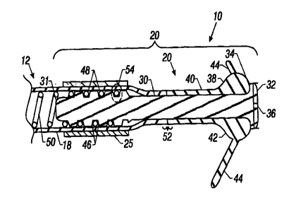
rn the drawings dcscnbed below, reference numerals are generally
repeated where identical elements appear in more than one figure. Turning now
to the drawings, and in particular tv FIG. I, there is shov~ra an exemplary
cardiac
lead 10 that includes a flexible insulating sleeve 12 that has a proximal end
14
coupled to a connector 16, and a distal end I8 ovupled to a tip electrode
assembly 20. The connector 16 is desigacd to be inserted ittto a cardiac
stimulator 2~-, and is shown highly cxaagerated in size relative to the
cardiac
stimulator 24. The cardiac stimulator 24 may be a paoomaker, a
cardioverter/denbrillator, or other type of stimulator or a sensing
instrument.
The tip electrode assembly 20 includes an annular member 25 to aid in securing
the sleeve I2 to the electrode assembly 20 as descn-bed more fully below. The
illustrated embodiment or the lead 10 is bipolar. Accordingly, the distal end
I 8
is provided with an electrode 2b located proximal to the tip electrode
assembly
24. However, unipolar arrangements are possiblt as well. A suture sleeve 28 is
-
slipped over the sleeve 12. During implantation, the suture sleeve 28 is sewn
to
body tissue at the sift oftramsvenous entry.
The sleeve 12 is a flexible tubular member that provides a rob~ist,
electrically insulating coupling between the connector 16 ;see FIG. 1) and the
electrode assembly 20. The sleeve 12 protects one or morn fine gauge conductor
wires enclosed therein from body fluids and tissues. The sleeve 12 is
advantageously compost of a biocompatible, electrically insulating material,
such as silicone, polyurcthaae, or like materials.
The detailed structure of the tip electrode assembly 20 may be
understood by referring now to FIG. 2, which is a cross-xctional view of FIG.
I
taken at section 2-2. 'Fhc electrode assembly 20 includes an elongated
conducting electrode 30 secured to tl~e sleeve 12 by the annular member 25. A
distal end 32 of the electrode 30 is provided with an expanded diameter to
establish an annular shoulder 34 facing proximally. The distal end 32 is
designed to transnut electrical signals to and from myocardial tissue. To
inesease the surface arcs of the cod. 32 exposed to myocardial tissue, and
thereby
eni~anee the ability of the end 32 to transmit electrical sigaals, the end 32
is
provided with one or more slots 36. A tine assembly 3$ is disposed over the
AMENDED SHEET
CA 0C32~769 2000-09-06,0,~~,T EC~~, +4.y Sy US 009904951
___ __ _ _ . ___ ~,jL~E~CI~E\ 04 . 7- 3- 0
r 07-0;3-2000
6
electrode 30. The tine assembly 38 includes a supporting body 40 composed of
a suitable biocompatible insulating material, such as silicone, polyurathaue,
or
like materials. The body 40 may be injection molded around the electrode 30 or
separately molded and slipped over the electcodc 30. The body 40 includes a
buloous portion 42 which abuts the atmular shoulder 34. Two or morn tines 44
prvj ect rardially outwardly from the bulbous portion 42 and may be integrally
molded with the body 40 or separately rnald~cd and coupled thereto.
The proximal end 31 of the electrode 30 is inserted into the distal end 18
of the sleeve 12 so that the proximal asd of the supparting body 40 abuts the
distal end 18 at 52. The proximal end 31 is provided with a sot of exteraal
grooves 46 that are configured much Like the external threads of a typical
bolt or
machine screw. The grooves 46 provide slsaees to receive protruding portions
of
the distal end 18 of the sleeve 12 as descrr~bed below.
T'lte distal coils 48 of a conducwr ware ~0 may be spiraled around tb.e
proximal end 31, disposed m the grooves 46 and seCUred to the dcctrode 30 by
laser welding, other like welding techniques, yr other suitable fastening
methods.
The proximal end 31 of the electrode 30 is proy-ided with a slightly larger
diameter than the inner diameter of the coils 48 of the wire 50. The distal
coils
48 may be connected to the electrode 30 by first urging the coils 48 over the
slightly larger diameter proximal end 31 and then threading the coils 48 into
the
channels 46 by rotating the wire 50 aniJor the electrode 30. Alternatively,
the
distal coils 48 may be coupled to the proximal end 31 without engaging the
channels 46, ln. either circumstance, the location and number of the welds is
a
matter of design discretion. 'the conductor wire 50 is connxted proximally to
the connector 16 shown in p'!G. 1 by welding or other suitable technique.
The con3uctor wire 50 is depic'~ed as a single individually insulated wise
~rith insulation removed from the coils 48 so that electrical contact is
established
between the wire 50 and the tip elecdrodc 30. However, the skilled artisan
will
appreciate that the conductor wire a0 may not be individually insulated if the
lead 2 0 is unipolar or if the various conductor wires in the lead 10 are
coaxi,ally
arranged or arranged in a nested configuration. Another conductor wire (not
shown) couples the electrode 26 Shown in FIG. 1 to the connector 16.
AMENDED SHEET
CA 02322769 2000-09-06~CtTT ECVi-~ +ø~ $~ US 009904951
.. _. . _.._ . n.~L~E\CHEN O t ~ ~- 3- 0 : '~'0 ~ ~g
07-03-2000
The detailed interaction bctwxn the annularmember 25, the distal end
18, add the elec~ode 30 may be understood by referring now also to FIGS. 3 and
4. FIG. 3 is a close up pictorial view of the distal end 18 and the annular
member 25, and FIG. 4 is a detailed ~icw of the po~i.on of the lead 10
circumscrihed by the dashed oval 54 in RIG. 2. The annular member 25 is
slipped over the sleeve 12 and positioned around the chaxmels 46. The annular
member 25 is then deformed to snagiy secure the distal end 18 to the electrode
30. The deformation decreases the internal diameter of the annular member 25.
The distal end 18 is pinched between the internal. surface 56 of the annular
member 25 and the external surface 58 of the electrode 30. As a result of the
pinching action dad the elastomcric character of the distal end 18, a portion
of
the distal end 18 designated generally at 60 wit protrude into each channel
46.
Dapendiixg upon whether a distal coil 48 is disposed in the groove 46, and
upon
the diameter of the distal coil 48, the protruding portion 60 may or may not
beat
against the distal coil 48. Preferably, a medical adhesive 61 has been placed
between the distal cad 18 and the electrode 30. The adhesive may be a suitable
medical grade adhesive, such as silicone based adhesive, a two part adhesive,
or
similar adhesives. When doe adhesive has cured, the ping of the annular
member 25 forces the adhesive into the groove 46. 'The adhesive forms a secure
molecular bon with the distal end I8 and a secure and a secure mechanical bond
with the electrode 30 by forming aidges 63. The presence of the annular member
thereafter prevents the distal end from expanding and keeps the ridges 63 in
the groove 46. LTse of the adhesive is important where the sleeve is formed of
low durometer material, such as silicon. It can be omitted where the sleeve is
25 formed of sti~'er material, such as polyurethane.
The objective of the deformation operation is to reduce the internal
diameter of the annular member 25 sufficieantly to pinch &nd secure the distal
end
18 to the electrode 30. The manner in which the annular member 25 is dcfotmed
to produce the desired internal diameter may be varied and is a matter of
desi~
discretion. 1n the embodiment depicted in FIGS. 3 and 4, the d.efornaation of
the
annular member 25 is advantageously provided by cr-imging. The crimping
produces radially projecting ridges 62 and b4 of material formed on oppositt
sides of the annular member 25.
AMENDED SHEET
CA 02322769 2000-09-061. gChl.., +~9 89 US 009904951
NUE~iCHEN U~~ . : - 3- 0 : ~0 ~ 53-
07-03-2000
8
The annular rnerraber 25 providas both frictional and mechanical retention
of tha distal end 18 to the eleotrod~e 30. The clamping action provides a snug
interference fit between the interiaz surface 56 of the distal and 18 and the
exterior surface 68 of the eleetmde 30. In addition, the vanous
P=°~'Sioas 60 of
the distal and 18 forcibly engage the walls 65 of their respective channels 46
to
pravidc a secuzc mechanical engagement between the distal end 18 and the
electrode 30.
The cured medical adhesive 6I completes the assembly by a very secure
mechanical engagement with the electrode 30 and a very secure molecular
engagement with the dish an 18. It is, therefarc, not necessary to rely on the
inherently weaker molecular bond between the adhesive and the metal electrode
30.
In au alternate embodiment illustxated in FIGS. 5 and 6, the defoxtnation
of the annular member, now designated 25, is produced in a different crimping
operation. FIG. 5 is a view like FIG. 3 and FIGr. 6 is a view Iike FIG. 4. In
this
embodiment, the annular azember 25 is crimped to yield several
circumfer~tiall'Y sp~~ St°°ves 66 in the exterior surface of the
a~naulaz
member 25 and an attendant decxea.se in the internal diameter of the annular
member 25 in the vicinity of the grooves 66. The number, size and spacing of
the gxooves 66 is largely a matter of design discretion. In those areas where
the
internal diameter of the azmular meatber ?$ is decreased, the distal end 18
vrill
be pinched between the internal surfaca 56 of the annular member 25 and the
external surface 58 of the electrode 30. ,As a result of the pinching a~.-tion
and the
elastomeric character of the distal end 18, a portion of the d'stal end 18
designated generally at 68 will protrude into each channel 46. Each protrusion
68 will generally be Positioned diroctly beneath the groove 66 and extend to
some distance on either side of the groove 66. Depending upon the diameter of
the distal coil 48, the protruding Po~vn 68 may or may nvt bear against the
distal coil 48. The adhesive 61 will also be forced into the groova forming
the
ridges 63, as dcscn'bad above.
Life the annular member described above, the arznuiar member 25
provides both frictional sad mechanical retention of the distal end 18 to the
electrode 30. The clamping action provides a snug interference fit between the
AMENDED SHEET
,. ., ...,......_. ~.~LiE\(:Hc.~i U4~ . 7_ 3_ UCA 0~2~ 22769 2000-09-06=C,I'f-
1' FCNt-~ +ø9 89 US 009904951
07-03-2000
9
interior surface 56 of the distal end i 8 and the exterior surface 58 of the
electrode 30. In addition, the various protrusions 68 of the distal end I8
forcibly
engage the walls 65 of their respective channels 46 to provide a secure
mechanical engagement between the distal snd 18 and the electrode 30.
The skilled artisan will appreciate that deformation techniques other than
crimping may be eased to clamp the annular members 25 and/or 25' to the distal
end 18. Far example, the d,.~sircd deformation may be accomplished by swaging.
VPhen swaged, the anaularmembers 25 andlvr 25' will increase in length
slightly
as a result of deformation into a smaller internal diameter.
The elocrrode 30 may be fabricated from a variety of biocompatible
conducting materials, such as iridium oxide coated titanium, NiP35N, stainless
steel, platinum-iridium alloy consisting of approximately 90% platinum and 10%
iridium, or some other bioeampatible conductix~.g metal, or a semiconductor
material, such as silicon, or other setnicanductor ma~terial_ The annular
member
25 tray be fabricated from the same types of materials. However, while
sufficient ductility to enable ready defo~.mation is de~rable, high electrical
conductivity is not required. The axuxular member 25 may be fabricated from a
radiopaquc material where tlse electrode 30 is not fabricated from a
radiopaque
material or where it is desired to provide the lead I O with enhanced
radiapaque
characteristics,
FIG. 7 depicts a side view of an alternate embodiment of the electrode,
now desigaatod 30. The electrode 30 includes a tip member 70 coupled to a
tubular shank 72 at 74. The eormection at 74 may be by welding, ttu~eaded
connxtion, or other suitable connection method. The tip member 70 is
composed of a non-radiopaq-ac material, such as, for example, iridium oxide
coated titanium or other suitaule conducting non-radiopaque material. The
shank 72 may be composed of a radiopaque material, such as, for example,
platinum iridium alloy (90% platinum, 10% iridium) or other suitable
conducting
radiopaque matt:ial. This embodiment provides a radiopaque electrode to
facilitate ~.uoroscopic observation wham the annular member 25 described above
is not composed of a radiopaque material or ve~hete additonal radivpaque
characteristics are desired.
AMENDED SHEET
---- A1UENCHEN 04 . 7_ 8- 0 CA 22032769 2000-09-06.Cj.1.,1. F.L~,~~ +49 89
US009904951
07-03-2000
FIG_ 8 depicts a cross-sectional view like FIG. 2 of an alternate
embodiment of the lead, now designated 10, and FIG. 9 depicts a detailed view
of the portion of the lead 10 circumscribed by the dashed oval 76_ In This
embodiment, the electrode, now designated 30, is configured with a generally
5 smooth, isodiametric exterior surface and is not provided with the
aforementioned channels or threads. The annular member 25 is positioned and
deformed as dcscribod above. The deformation yields protrusions 78 in the
distal and 18 of the sleeve 12 (one ofwhich is sho~w~a in FIG. 9j that a-re
disposed
between individual coils ~.$ and bear against the exterior surface of the
proximal
10 end 31, providing a friction engage~nacnt betv; een the proximal end 31 and
the
protrusions 78. As with the aforementioned embodiment, a suitable medical
grade adhosive may be applied between the distal end 18 and the electrode 3U
to
provide a seal against the intrusion of body fluids and to provide a
complimentary mechanism to secu~rr tile distal end 18 to the electrode 30.
AMENDED SHEET