Note: Descriptions are shown in the official language in which they were submitted.
CA 02324048 2000-10-20
1
COMPUTER ASSISTED RADIOTHERAPY DOSIMETER SYSTEM AND
SOFTWARE THEREFOR
DESCRIPTION
TECHNICAL FIELD:
The invention relates to radiotherapy dosimeter systems, especially of the
kind
which use a plurality of dosimeter sensors distributed in a region to be
irradiated and
means for monitoring radiation levels detected by the sensors.
BACKGROUND ART:
Radiotherapy treatment of cancer patients involves the use of machines which
produce high energy X-rays or high energy electrons. It is common practice to
verify
the radiation dose delivered to the patient with a dosimetry system such as
the Thomson
& Nielsen Patient Dose Verification System.
There are three different types of dosimetry system used in radiotherapy.
These
are based on (a) film or thermal luminescent dosimeters (TLD), (b) diodes and
(c)
MOSFETs. Diode and MOSFET systems use electronic dosimeter sensors together
with
electronic reading systems, whereas film or TLD use chemical or thermal
methods of
reading the detectors into an electronic reading system.
Since diode and MOSFET based dosimetry systems have the convenience of
direct electronic reading of the dosimeters, they also have the potential
advantage of
direct data communication with computer systems. The person using a patient
dosimetry
system (usually a medical physicist, dosimetrist or therapist) requires the
radiation dose
information from the system to be in a format that is suitable for good
quality assurance
records.
The state of the art with patient dose verification systems is for the dose
data to
be presented in one of three formats - (a) on a display on the reading
instrument, (b) on
a print-out from the electronic reader or (c) on a computer screen. The latter
case, the
information presented on the screens is in the form of numbers and, in some
cases,
graphs.
Thomson & Nielsen MOSFET dosimetry systems use ExcelT'''' spreadsheets for
this purpose. Sun Nuclear and Scanditronix have diode-based systems which use
WindowsTM - based systems with numerical tables and graphs of data.
A disadvantage of these known systems is that it is not easy to confirm that
the
dose values measured were taken at the proper locations on the body or
patient.
CA 02324048 2000-10-20
2
DISCLOSURE OF INVENTION:
An object of the present invention is to at least mitigate this disadvantage
and, to
this end, provides a dosimetry system having means for displaying a
representation of
the body, e.g., a patient, to be irradiated, showing locations of radiation
sensors.
According to one aspect of the present invention, there is provided a
dosimetry
system in which a plurality of sensors for disposition on, in or near a body
to be
irradiated, for example a patient, are connected to a sensor reading
instrument which is
interfaced with a display system, for example a personal computer, which is
arranged
to display, in use, one or more representations, for example drawings or
photographs,
of the body to be irradiated, along with the positions and the dose data for
those specific
sites where the dosimeter sensors were placed.
Preferably, the display system is arranged to display the representations,
prior to
irradiation, with the sensor locations and sensor identifiers and, after
irradiation, with
the measured doses associated with each sensor.
Preferably, the display system provides for adjustment of the sensor locations
relative to the irradiation, to select desired locations, and then may provide
for printing
of the representations, showing the sensor locations, prior to irradiation,
thus allowing
the print-out to be used by an operator as a guide when positioning the
sensors.
According to a second aspect of the invention, a method of using a dosimetry
system of the first aspect to monitor radiation doses at various sites on a
body comprises
the steps of:
(i) displaying one or more representations of the body to be irradiated
and points or icons representing a plurality of dosimeter sensors, and
(ii) adjusting the display to position the sensor points or icons at
preselected sites on, in or near the body at which radiation doses are to be
measured.
Preferably, the method further comprises the steps of:
(iii) irradiating the body and obtaining data of radiation measured at
each of the sensors,
(iv) displaying the data for each sensor in the same display as the one
or more representations of the body with the sensor points or icons at said
preselected
positions.
In preferred embodiments of either aspect of the invention, in the display,
the
dosimeter sensors are represented by graphical points or icons associated with
respective
identifiers, conveniently interconnected in the display by, for example, lead
lines. The
positions of the graphical sensor site points or icons may be adjusted
relative to the
representation of the body to locate them at sites on the image which
correspond to the
actual positions at which the sensors are (to be) located. In the display,
each of the
identifiers then is associated, conveniently in a table, with the
corresponding dose data.
CA 02324048 2000-10-20
3
Embodiments of the invention advantageously enable the physicist to plan the
sites
where dose measurements are required, ensure that the dosimeters are placed
according
to plan, and confirm that the body (patient) has received the correct dose to
the correct
site according to the plan.
Yet another advantageous feature is that the one or more representations of
the
body, together with the preselected dosimeter sensor pasitions, may be printed
prior to
patient treatment so as to facilitate correct positioning of the dosimeter
sensors in the
correct anatomical positions by the medical personnel performing the
radiotherapy
procedure.
Advantageously, embodiments of the present invention may provide real-time
display of data from the dosimetry system reader.
Another advantageous feature is that the patient's treatment information may
be
readily recorded (e.g. patient name, identification of radiotherapy machine
used, energy
of machine).
The one or more representations used to indicate the positions of the
dosimeter
sensors on the body, e.g. on the patient's anatomy, may comprise standard line
drawings
or custom images, such as scanned photographs or digital camera images. In the
latter
cases, the use of actual images of the body facilitates proper location of the
sensors.
Another advantageous feature of embodiments of the present invention which use
a computer display is that the software may calculate the radiation dose using
the data
input from the reading instrument and any calibration or correction factors
previously
input by the physicist, typically following a previous calibration of the
dosimetry system
in known manner. The software then may compare the dose calculations with pre
determined target doses and indicate, conveniently by highlighting in the
display, any
deviation for corrective action.
A further feature of embodiments of the present invention is the capability to
view, print or electronically save the final report with all the relevant
dosimetry data
collected during the patient's treatment.
According to a third aspect of the invention there is provided software for
interfacing a plurality of dosimeter sensors and a reader to a microcomputer
or personal
computer to provide for the specified display of an image or other
representation of the
body/patient and the corresponding doses, in a system according to the first
aspect.
BRIEF DESCRIPTION OF THE DRAWINGS:
A computer assisted dosimetry system in accordance with the invention will now
be described, by way of example, with reference to the accompanying drawings,
in
which: -
CA 02324048 2000-10-20
4
Figure 1 illustrates, partially and schematically, a dosimetry system for
irradiating
a person;
Figure 2 illustrates a portion of a display of the system;
Figure 3A illustrates a representation displayed during assignment of sensor
positions; Figure 3B illustrates a representation subsequently displayed
during assignment
of sensor positions;
Figure 4 illustrates a report provided by the system;
Figure 5 is a flowchart depicting operation of the system;
Figures 6A to 6F and Figures 7A to 7F show display screens displayed during
operation of the system.
DETAILED DESCRIPTION OF PREFERRED EMBODIMENTS:
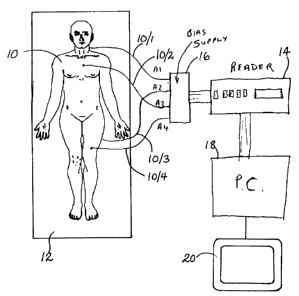
Figure 1 illustrates a patient 10 lying upon a table 12 ready for radiation
therapy.
The therapy entails irradiating the patient 10 by means of a radiation therapy
machine,
which might be an X-ray machine, a CT scanner, or other machine having means
(not
shown) for irradiating the patient. The amount of radiation to which the
patient is
subjected is monitored by a dosimetry system which comprises a set of MOSFET
radiation sensors A1...A4 positioned at predetermined locations on the
patient's body and
connected by leads 10/ 1...10/4, respectively, to a reader 14 by way of a bias
supply unit
16. The reader 14 is connected to a personal computer 18 which controls a
display
device 20. The sensors A1-A4, bias supply 16, reader 14 and computer 18 may be
of
known construction and so will not be described in detail. The personal
computer 18 is
equipped with software, such as Visual BasicT'''', or the like. It is assumed
that the
sensors A 1-A4 and, when applicable, other parts of the dosimetry system, have
been
previously calibrated using known techniques.
Figure 2 illustrates a portion of the display 20 controlled by the computer 18
and
showing, representations of the patient 10, specifically in outline, front lOF
and rear lOR
views of the patient 10 and positions for icons representing the four
dosimeter sensors
A1, A2, A3, and A4. The display also shows a table 22 listing the sensors A1-
A4 and
associated data. When the irradiation process has been carried out, the data
will include
the dose measured by each sensor.
Operation of the dosimetry system involves two main phases, namely (i)
assignment of the sensor icons to selected positions on the representations,
and (ii)
measurement and display of the measured doses. 'these two phases need not be
performed at the same time. For the first phase, the patient need not be
present and, in
fact, the first phase could be carried out remote from the radiation therapy
machine. For
convenience, however, both phases will be described as if carried out
together.
Referring now to Figure 3A, which is the type of graphic first shown to the
user on the
CA 02324048 2000-10-20
computer screen 20, the sensor icons are not assigned, but merely grouped to
the right
of the front images lOF. The sensors A1, A2, A3, A4 are represented by icons
comprising sensor dots connected by lead lines to respective labels
(identifiers).
The computer then instructs the user to assign dosimeter sensors to various
parts
5 of the anatomy. Figure 3B shows the screen displayed to the user once this
task is
completed. In the example shown, the user has dragged and dropped both the
dots and
labels of the dosimeter sensors (e.g. A1, A2 etc.) so that the dots are
located at the
required sites on the images and the identification label are conveniently
nearby. A
description of the sites is optionally recorded in the database.
On completing dosimeter assignment, the user can print out the diagram or
photo
of the patient with dosimeter locations so that the medical personnel may then
place the
dosimeters in the correct locations.
Following irradiation, the dose information from the dosimeters is read into a
computer by operating the dosimetry system connected as in Figure 1. (The
dosimeters
may be removed from the patient for this part of the procedure).
The dose measurements are stored in the computer and displayed on a final
report, along with the patient and treatment information. Figure 4 shows the
format of
the final report with the dosimetry position information as well as the dose
measurement,
target dose and deviation information.
Figure 5 shows a flow diagram of the software required to carry out the above
process. This particular software has been developed using Visual Basic TM.
4.1 Overview of the Program
This program catalogs its functions into 5 groups:
- System Setup
- Pre-Irradiation (Step 1)
- Treatment Information (Step 2)
- Measuring Dose (Step 3)
- Viewing/Printing Reports (Step 4)
The following is a description of the steps that the software carnes out in
order
to proceed from Step 2 (Treatment Information) to Step 4 (Viewing/Printing
Reports).
Step 2 Treatment Information:
The user determines the number of patients in the current treatment, and, for
each
patient, selects the position on the screen to type in the appropriate
information e.g.
Patient's ID, Treatment Plan Reference and Radiation Settings. The user
assigns
CA 02324048 2000-10-20
6
dosimeters to each patient through an on-screen table, and types in words to
describe the
locations and target doses of each dosimeters. (See Figure 2.)
There is an on-screen picture-box which accommodates an image as background
and some labels, lines and red dots as foreground. The user can select the
background
image from the software's built-in images, or use any image that has been
stored in the
computer's hard disk in BITMAP, JPEG or GIF format. For every assigned
dosimeter,
the picture-box shows on the foreground a label, a red dot, and a line to link
the label
and dot. Every label and dot can be dragged to appropriate positions to
indicate the
dosimeters' sites graphically.
The software uses a compound data type to store treatment information in this
step. For every patient, the software creates an instance of this data type
that
accommodates fields to keep Patient's ID, Treatment Plan Reference, Radiation
Settings,
Dosimeters' Positions and Target Doses. It also includes a field to keep a
reference to
the selected background image, and some fields to keep the relative
coordination of every
foreground label, dot and line.
Step 3 Measuring Dose
Dose data is inputted from the Reader 14 through a cable and placed in the
dosimeter locations on the screen. The user can activate the "Recording"
procedure to
allow the input data to overwrite the existing data, or freeze this procedure
to prevent
the recorded from being changed.
The software uses a data array to store the recorded data in this step.
Step 4 Viewing/Printing Reports
In this step, the software extracts information, that is necessary to create a
measurement report, from the inputted data in step 2 and recorded data in step
3. This
information is stored into a special array. Then, from this array, a report
summary is
composed and the corresponding picture (see Figure 4) is drawn. If the user
needs to
save this report, the software will save all fields of this array to the hard
disk of
computer 18 (next time, they can be read into the array if needed). The data
in this array
are also used to print out the report. They may also be saved to a floppy disk
or other
removable storage medium or transmitted via a network or modem connection.
Step by step operation of the system will not be described, the various
display
screens presented to the user being shown in Figures 6A to 6F and 7A to 7F.
There are
5 display/control panels to let users access these 5 function groups. They are
organized
in a straightforward style and easy to use. Every panel except the last one
has a yellow-
coloured text box to show On-Screen Prompt. The following is an overview of
these
panels.
CA 02324048 2000-10-20
7
Setting up the system: Initially, where the user can determine the
communication
port, set up the title of measurement reports, set or change the password and
determine
its protection scope, input the lists of available radiation machines and TN-
RD-50
Readers.
Step 1. Pre-Irradiation: In this step, the user can modify CFs and CRs, check
dosimeters, modify system settings, or view existing reports. In case some
parameters
need to be changed or some MOSFETs need to be replaced, the user can find them
and
take corrective actions.
Step 2. Treatment Information: In this step, the user will input treatment
information for the patients and assign dosimeters. The user can describe the
dosimeters' sites by description or graphically. The left-hand picture shows
an example
without picture.
The left-hand picture is an example to show how to describe the dosimeters'
sites
graphically. This program has 5 standard body images built into it. It also
provides a
very easy way to let the user use other images. Any BITMAP, JPEG, and GIF
images
can be used.
Step 3. Measuring Dose: In this step, the program will record the measurement
data from the TN-RD-50 Reader and calculate Dose (if the Reader's output is
set to
"mV") and Deviation from target dose.
Step 4. Viewing/Printing Reports: This is the last step of the measurement
procedure. The user can view/print/save reports. The user can also type in
your
comments on reports. There are two report styles available.
Setting up the System
The user clicks on the "TN-Dose Reporter 2.31" entry of the computer's "Start
~ Programs" menu to run the program. The "Set Up the System" panel is shown
and
the user is required to input some information or make some decisions,
including:
(1) Choosing a serial port to communicate with the TN-RD-50 Reader.
(2) Inputting the Institution Name and the Report Title. They will be printed
on the measurement reports. The default Report Title is "DOSIMETRY
REPORT" .
(3) Building up the list of radiation machines types.
(4) Building up the list of radiation machines' S/N.
(5) Building up the list of TN-RD-50 Readers' S/N.
(6) Setting or changing the user's password and determining the password-
protection's scope.
Of these, (1) is a must, (2)-(6) are option.
CA 02324048 2000-10-20
g
Usually, every computer provides at least one serial port. When the user
connects the TN-RD-50 Reader to the computer, the user should check which port
(COM1-COM4) is being used and select this port when setting up the system.
The port number (i.e., 1-4) may not be labelled on the computer, and the user
may not be sure which port is being used. In such a situation, each port
should be tried
until the correct port is selected. To do so, the user may follow the
instructions shown
on the screen.
Note:
(a) This "Set Up The System" panel will not be shown when the program is
run later. To view or change system settings, the user can select the
action of "Modify System Settings" from Step 1 panel.
(b) When the program is started, it checks the computer's hardware resources
and lists all available serial ports in the pull-down list. If there is no
port
available (for example, in case all ports being used by other applications),
the program will give out a message and automatically show the panel of
step 4 ("Viewing/Printing Reports").
c) After setup, a new folder "c:\TN-Dosimetry" is established in the
computer. In it there is a file ("MessageHistory.txt") and two sub folders
("Libs" and "Reports"). These folders should not be renamed.
Step 1. Pre-Irradiation
When the user has finished setting up their system, the panel of "Step 1. Pre-
Irradiation" will be shown. In this step, the user can modify calibration
parameters (CFs
and CRs), check dosimeters, modify system settings, or view existing reports.
About the Message Window: This window is used to display the messages from
the TN-RD-50 Reader. The user can view all messages (in the current
measurement
procedure) or just view recent messages. Every message displayed here is also
saved
into a file "c:\TN-Dosimetry\MessageHistory.txt" simultaneously.
About the CFs and CRs: The Reader can be set to read in radiation units (cGy
or R) using Calibration Factors determined by the user for each dosimeter. The
reader
can also be set to read the MOSFET voltage in mV. In order to give the user
more
flexibility, this Dose Reporter program allows the user to store the CFs in
the program
when the mV mode is used. The program also enables the user to specify
Correction
Factors (CRs) to the dose calculation.
If the Reader is set to output radiation units (cC~y or R), then the CFs and
CRs
in the program are inoperable. If the user sets the output of the Reader to
mV, then CFs
and CRs must be set, because they will be used to calculate the doses
according to the
formula "Dose = CR * (Voltage / CF)".
CA 02324048 2000-10-20
9
Note: An example of the use of a CR would be if the user wanted to determine
Dmas but was measuring doses with less than full build-up.
The user can get a hard copy of CFs and CRs by clicking the "Print" button.
The allowed CF range is 0.1 mV/cGy to 99.99 mV/cGy. If the user enters a too
large or too small value, it will be trimmed into this range. The allowed CR
range is
0.100 to 9.000. If the user enters a value beyond this range, it will be
trimmed into this
range.
When the user has finished modifying CFs or CRs, the user can set them as
defaults. Otherwise, the default CF and CR is I.OOmV/cGy and 1.000
respectively. If
the user does not like other users changing CFs or CRs (or both), the user can
set up a
password (in "Setting Up the System" Panel) and put CFs or CRs (or both) into
the
protection scope, then restart this program.
Step 2. Treatment Information
When the user has finished Step 1 and start Step 2, the user is first required
to
select the number of patients in this treatment. Then, Figure 2 appears on the
screen to
let the user input the Patient Information, Treatment Plan Reference, and
Radiation
Settings (the user can set them by importing treatment information from an
existing
measurement report by clicking "Import Existing Treatment Info"). The user
also needs
to assign dosimeters to the patient(s). The user can describe the dosimeters'
sites with
words or with pictures. To do the former, the user may type words in the
corresponding
cells of the dosimeter-Assigning Table. To do the latter, the user may click
"Show
Picture", whereupon a human body image will be displayed on screen, as Figure
4.
When the user assigns dosimeters to the current patient, the corresponding
Site
Pointer and Dosimeter Label will appear on the image area. To indicate the
dosimeter's
site, the user may simply the Site Pointer and Dosimeter Label to the
appropriate place
on the image. (The user can drag the Pointer and Label to the same place, and
the
pointer will disappear.)
Clicking the "Print" button on the picture's bottom-right corner can print out
the
picture. (If that button is not enabled, the user may click the "Apply"
button.)
The user can change the human body image. For example, 5 optional images,
called "Standard Images", may be provided. They are
#0, Unisex Body
#1, Female Chest
#2, Male Head
#3, Female Head
#4, Female Body
CA 02324048 2000-10-20
Besides the standard images, the user can use their own images, conveniently
called "Custom Images", such as those from a digital camera photo or a scanned
photo.
Any BITMAP (*.bmp), JPEG (*.jpg) and GIF (*.gifj images can be used as a
custom
image. If the image to be used has been stored in another format, some tools
(such as
5 Paint or PhotoShop) may be used to open them and save them in BITMAP or JPEG
format. There is no special requirement on the images' size.
To change the image, the current image is double-clicked, or right-clicked to
pop
up a menu and "change image" selected. An image-selection window, as in Figure
4,
will be displayed on screen.
10 To select a standard image, its preview window is clicked. To select a
custom
image, the user should click on the corresponding item in the library of
custom images
to preview it, then, click on the preview window.
When the program is run for the first time, the library of custom images is
empty. To populate it, the user may click the "Add new Custom Image" button,
then
select an image file from the open-file dialog box. That image will be copied
to the
library and can be used as a custom image by the program.
Step 3. Measuring Dose
The panel of this step is shown in Figure 5. In this step, the user is
required to
perform 3 actions:
(1) Zero MOSFETS: press the Reader's START (or ZERO) button for 1
second to initiate the procedure.
(2) Place MOSFETS ON PATIENT(s) body. (To do it correctly, it is
suggested that the user print out the dosimeter-site diagram in step 2 as
a reference. )
(3) Read MOSFETs: click the "Record" button on the screen, then follow
the prompt.
In the measurement procedure, if "N/A" appears in the "Voltage" column, it
means that the voltage is Not Available since the Reader has been set up to
output does
in the radiation units cGy or R. Voltages are only shown in this column when
the user
is using the Reader in the "mV" mode and applying ('.alibration Factors (CFs)
and/or
Correction Factors (CRs) to translate m/v to radiation units.
Step 4. Viewing/Printing Reports
In this last step, the user can review the information in the report summary
before
printing and saving. All report files have a filename with extension ".dsrpt".
The
default file name is "Patient First Name + Patient Last Name + Date + .dsrpt".
For
CA 02324048 2000-10-20
11
example, if John Smith was treated on May 10, 2000, then the default file name
would
be
JohnSmith 2000Mayl0.dsrpt
The default folder for saving reports is "c:\TN-Dosimetry\Reports". The user
can save
S the reports in any folder.
When the user wants to print out the reports, there are two styles available.
Style
#1 accommodates a picture to indicate dosimeters' sites graphically. Style #2
doesn't
print out the picture, but uses a table to provide more information about the
treatment.
Two sample reports are illustrated in Figure 4 and 7F to show their
differences.
In this step, the user can also type in comments.
Other Information
5.1 Operation Mode
This program has two operation modes. The first is "Full-Functional Mode" in
which the user can perform measurements, make reports, view existing reports,
and
modify system settings. The second one is "Report-Viewing Mode" in which the
user
can view/print existing reports, but can't perform measurements or modify
system
settings.
When the user double-clicks the program's icon to start it, it will run in
Full-
functional Mode. The initialization procedure for this mode is longer than
that of the
Report-Viewing Mode. If the user needs to view the existing reports
frequently, the user
should find the file "View Dose Report.exe" in the installation disk and copy
it to the
computer's desktop. Double-clicking its icon will start "TN-dose Reporter
v2.31" in
Report-Viewing Mode.
5.2 Using the program in Local Networks
This program can be used to view existing reports through Local Networks. To
allow the reports in the computer to be viewed from another computer, the user
needs
to set the computer's "c:\TN-dosimetry" folder as a shared folder.
5.3 Records of Message History
All messages sent by the Reader are recorded in file "c:\TN-
Dosimetry\MessageHistory. txt" . The user can view and print it using
"Notepad" . (The
attached is an example of printed History File.) Every day, when the user
first uses the
program, the user will be asked whether or not to erase the existing records.
If the
answer is "No", the new messages will be appended to the existing records. If
the user
always answers "No", the size of the "MessageHistory.txt" will increase
rapidly.
CA 02324048 2000-10-20
12
5.4 Double-click report files to view them
If some reports have been saved in the computer, the user can double-click
their
icons to view them. In such situations, the "TN-Dose Reporter v2.31" runs in
Report-
Viewing Mode.
5.5 Use patients' photos when doing measurement
Section 4.4 has described how to use an existing custom-image to indicate
dosimeters' sites. But, in fact, that image need not have to exist before the
user runs the
program. The user can use the REAL photos of the patients) in current
treatment. The
following example shows how to do so (assuming that the user has a digital
camera).
Example
(1) When in Step 2, the user should input the patient's treatment information,
but NOT draw the picture.
(2) In Step 3, after placing MOSFETs on the patient's body, the user should
take a photo, then do the measurement.
(3) The user should minimize the "Step 3" Panel, then, input the photo to the
computer, save it in the hard disk as JPEG file, and restore the "Step 3"
Panel.
(4) Going back to "Step 2" panel, the user should add the saved JPEG file to
"custom-Images Lib" and select it, then, draw the picture.
(S) The user should go to "Step 4" panel to view the report.
It should be remembered that the user can change the treatment information and
dosimeter-assignment at any time BEFORE starting a new measurement by clicking
the
red-coloured square on "Step 4" panel.
It should be appreciated that the software enabling implementation of the
invention could be used with various kinds of hardware. Hence, the invention
also
embraces software per se, conveniently carried by a suitable storage medium,
for
operating a dosimetry system as described hereinbefore.