Note: Descriptions are shown in the official language in which they were submitted.
CA 02324105 2000-09-15
WO 99/48427 PCT/N099/00093
Method and device for suturless anastomosis
The present invention relates to a support device for use in suturless
anastomosis procedures, and a method for performing suturless anastomosis.
The method according to the invention will be referred to in the present
specification as IKF-IS technique (Institutt for Kirurgisk Forskning -
Intervensjonssenteret).
The risk inherent in the performance of conventional cardiopulmonary bypass
grafting (CABG) and the relatively frequent need for reintervention after
percutaneous transluminal coronary angioplasty (PTCA) have caused a rising
interest in developing thoracoscopy assisted procedures that combine the
patient-friendly nature of PTCA with the durable benefits offered by CABG.
Three approaches are currently undergoing evaluation, none of which
eliminate the need for both cardiopulmonary bypass and thoracotomy. As these
two procedures represent the primary causes of morbidity after CABG, there is
an urgent need for developing a minimally invasive procedure that can be
performed on a beating heart, entirely under endoscopic-fluoroscopic
guidance.
The IKF-IS technique has been envisaged to meet this need.
An important feature of this technique is that its use is not limited to the
coronary arteries. An IKF-IS anastomosis can be done in any vascular area
within reach of an endoscope. The range of use includes also extravascular
tubular structures such as the esophagus, intestines, ureter, biliary ducts
and
fallopian tubes.
Suturless anastomosis of vessels is not a new concept. A large number of
anastomosis devices has been described in literature, though few of them have
passed the test of time.
Structurally sound anastomosis between vessels can be rapidly established
simply by apposing the vessel ends with interlocking external collars.
GB-B-1.413.191 describes a device for the eversion of hollow organs and a
vascular stapling instrument incorporating same. The device optionally
comprises a rigid bush with a longitudinal slot or a rigid split bush
comprising
two pivotally connected half bushes which can be mechanically disengaged
from each other. The bush forms an integral part of an instrument used to
evert
the cut edges of the limbs of the tubular organ to be anastomosed and
temporarily approximate them so as to facilitate suturing or placement of
clips
that will hold the edges together. When the clips are in place, the instrument
and the bush are removed. Thus the device simply acts as an aid to the
creation
of an anastomosis and in no way removes the drawbacks to using sutures and
clips for creating anastomoses.
CA 02324105 2000-09-15
,: , ,. ,
,;. ,',~ " , ,.
US-A-4.917.087 describes devices, kits and methods for non-suture end-to-end
and end-to-side anastomosis that employ tubular connection members having
clip retaining elements and spring clips which comprise a ring-shaped body
with separable opposed ends whereby a circular opening defined by the body
can be enlarged.
Unfortunately, these anastomosis devices and others available today were
designed for use at open surgery and are not appropriate for endoscopic
placement.
EP-A-781.528 describes a fastener for connecting severed blood vessels. The
device has a plurality of miniature barbs which pierce the wall of the blood
vessel and anchor the fastener-in place. In one embodiment the fastener
comprises a sheet provided on one of its surfaces with a plurality of barbs,
the
sheet can be rolled to a diameter smaller than that of the blood vessel,
inserted
into the blood vessel and unrolled so that the barbs pierce and anchor in the
1 S inner wall of the blood vessel. The sheet can alternatively be wrapped
round
the blood vessel so that the barbs pierce and anchor to the outer wall of the
blood vessel. The bond strength of the device as tested is not adequate for
clinical use, because the biologic response is not appropriate or the design
does
not provide the structural strength to tolerate the expected loading forces
(whether in shear or in tension is not specified in the document) at the
interface
between the device and the vessel surface. The penetration of the vessel wall
by the barbs on the device can cause separation of the layers of the vessel
wall,
which in turn can lead to thrombus formation or dissection at the site. The
damage to the vessel wall would logically be even more severe if the size of
the barbs is increased. Barbs which spontaneously retract, will leave behind
holes in the vessel wall from which bleeding could occur. From the description
provided in claims 1 and 2, it does not seem possible that these embodiments
of the device lend themselves to use via an endoscope. Besides, it is unclear
how the barbs will be prevented from inadvertedly engaging the adj acent
overlying layer as the device is being unfolded by the balloon.
Furthermore WO 98/52474, WO 94/27506, demonstrate devices for
performing anastomosis, with and without eversion of the blood vessel
respectively, while US-A-5,254,113 describes. the use of strips for
anastomosis
of intestines. None of these publications do, however, describe a sleeve and
use of a sleeve to evert the blood vessels, use of a transitional temperature
range (TTR) material in an anastomosis device and an anastomosis device
provided with metal collars. In addition, the use of these devices involves
the
retention of an intraluminal foreign body after anastomosis -creation in
direct
contradiction to the invention in the present application.
FR-A-1.518.083 describes a device for performing end to end and end to side
anastomosis. In the embodiment adapted for end to side anastomosis, the
AMEHDEp ~~,.
CA 02324105 2000-09-15
'; '~,' , ; : ; . , ; ; ,,.
device comprises a curved plate with a bore and a joint surrounding the bore.
The plate is glued to a first vessel, a hole is cut in said vessel
corresponding to
said bore in the plate, and a muff containing the second vessel is attached to
the joint. The curved plate simply provides a surface area for the adhesive
used
to attach the joint to the first vessel. Said plate does not permit fastening
to the
first vessel solely by mechanical means without an adhesive. It does not offer
any self attaching capability.. Hence, in case of adhesive failure during the
healing process, the curved plate will get dislodged with possibly
catastrophic
consequences.
An alternative method that has recently been successfully used for coronary
artery bypass grafting on a beating heart is the- Tulleken technique. By the
._
incorporation of excimer laser arteriotomy, this technique permits the
creation
of end-to-end bypass without interrupting flow in the diseased vessel. However
,- at its present stage of development, performance of a Tulleken anastomosis
via
an endoscope is not feasible. The high costs related to the use of excimer
lasers
further restrict the benefits of the Tulleken technique from the perspective
of
minimally invasive coronary artery surgery.
The object of the present invention is therefore to provide a device that
permits
creation of a suturless anastomosis between vessels via an endoscope. This
object is achieved by means of a device comprising at least one tubular
member, and characterized in that the tubular member is provided with a
longitudinal slit, which slit permits introduction of the anastomosed vessels
in
the device, and the device is adapted for attachment to the vessel without any
damage to the vessels' walls.
The need for suturing is entirely eliminated by the invention, reducing danger
of vascular trauma.
The anastomosis is externally supported by the device according to the
invention, this reduces the risk of acute structural failure, delayed aneurysm
formation and in the presence of compliance mismatch, improves the long-
term patency rate.
The invention will be explained in more detail with the help of the following
drawings, where:
Fig. 1 and 2 show first (Type Ia) and second (Type Ib) embodiments of the
invention for performing end-to-side anastomosis;
Fig. 3 shows a third (Type II) embodiment of the invention for performing
end-to-side anastomosis;
Fig. 4 shows a fourth (Type III) embodiment of the invention for performing
end-to-side anastomosis;
~uEr~D4 D StfEE l
CA 02324105 2000-09-15
;.4~, .; .' , w, ; .". ,'v
Fig. 5 shows a fifth embodiment of the invention for performing end-to-end
anastomosis;
Figs. 6A and 6B show sixth and seventh embodiments of the invention for
performing end-to-side anastomosis;
Figs. 7A and 7B show eigth and ninth embodiments of the invention for
performing end-to-side anastomosis;
Fig. 8 shows a cross-section of a first alternative joint of the slit's edges;
Fig. 9 shows a cross-section of a second alternative joint of the slit's
edges;
Fig. 10 shows a third-alternative of the slit's edges;
Fig. 11 shows a first alternative embodiment of the fixation sleeve;
Figs. 12A, 12B and 12C show second alternative embodiment of the fixation
sleeve, and an alternative embodiment of the inner collar and of the
anastomosis device;
Figs. 13-20 illustrate an externally stented end-to-side anastomosis by means
of the device according to the invention for outflow vessels that cannot be
circumferentially dissected;
'Figs. 21-24A and 24B illustrate an externally stented end-to-side anastomosis
by means of the device according to the invention for outflow vessels which
can be circumferentially dissected;
Figs. 25-32 illustrate an externally stented end-to-side anastomosis by means
of the device according to the invention.
Fig. 33-37A and 37B illustrate an externally stented end-to-side anastomosis
by means of the device according to the invention.
Fig. 38-39A and 39B illustrate an externally stented end-to-side anastomosis
by means of the device according to the invention.
Fig. 40-46 illustrate an externally stented end-to-end anastomosis by means of
the device according to the invention.
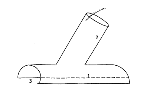
Fig. 1 shows a first embodiment of the invention to be used if circumferential
dissection of the outflow vessel is not possible (e.g. in coronary artery
bypass). This embodiment of the invention shows a first tubular member 1
and a second tubular member 2, where the first tubular member 1 is provided
with a longitudinal slit 3. The slit 3 is wide and the edges are not in
contact.
This characteristic allows use of this embodiment of the invention in cases
where the outflow vessel cannot be circumferentially dissected, by placing
the first tubular member as a "cap" on the vessel. The second tubular member
4.uF~~aLcD ~rFf:~
CA 02324105 2000-09-15
.: ,' , . . . . . : ; ;
2 is attached on the side of the first tubular member 1 opposite the slit. In
this
embodiment of the invention, the second tubular member does not show a
slit.
Fig. 2 shows a second embodiment in which the first member is a flat sheet.
Fig. 3 shows a third embodiment of the invention to be used if circumferential
dissection of the outflow vessel is possible. This embodiment of the invention
is similar to the embodiment in Fig. 1 except that the second tubular member 2
is split too and it is attached to the first tubular member 1 in such a way
that
both slits are in contact. The whole device is "hinged" round a longitudinal
line
in tubular member 1 lying opposite to the slit 3. The term "hinged" in this
case
is to be understood as a minimum separation of the slit's 3 edges, that
otherwise are in contact.
Fig. 4 shows a fourth embodiment of the invention adapted for performing
end-to-side anastomosis, and where the tubular members 1 and 2 are parallel,
and have slits 3 and 3' respectively. the tubular members l and 2 can have
different diameters (a,b) depending on the size of the vessel to be
anastomosed. Slit 3' is a longitudinal slit along the common central plane of
the device. The two halves of members 1 and 2 can be distracted perpendicular
to the longitudinal axis without plastic deformation. Edges AB and CD are not
in contact, the distance between them will vary according to the diameters of
members 1 and 2. In an embodiment adapted for use with outflow vessels that
cannot be circumferentially dissected, edges EF and GH are not in contact. In
another embodiment for use with outflow vessels that can be circunzferentially
dissected edges EF and GH are in contact or overlap each other.
Fig. 5 shows another embodiment of the invention, adapted for performing
end=to-end anastomosis. This embodiment comprises only one tubular member
1, with a slit 3:
Figs. 6A and 6B show two embodiments of the inner sleeve of the IKS-IF
anatomosis kit for performing end-to-side anastomosis. The embodiment
comprises one tubular member without a slit with (Fig. 6A) or without (Fig.
6B) parallel edges.
Figs. 7A and 7B show two embodiments of the fixation sleeve of the IKS-IF
anatomosis kit for performing end-to-side anastomosis. The embodiment
comprises one funnel shaped tubular member without a slit with (Fig. 7A) or
without (Fig. 7B) parallel edges.
Several possibilities are envisaged for the slits' edge area, with the
intention of
giving the invention high flexibility in use.
In one alternative embodiment, the opposing edges of the slit are configured
so
that they mechanically lock on the application of a centripetal radial force.
One
,;:~~~:;.~ ,-n
~-~ t ~'l 1~'i~: ~l
CA 02324105 2000-09-15
,~; . ~ ,~ , , ,
possible configuration for this purpose is shown in fig. 8, where the edges
show a Z-profile.
Fig: 9 shows another possibility for connection of the slit's edges. This
possibility consists in extending the slit's edges to form overlapping flaps.
The
surfaces of the flaps facing each other can be provided with a fastening
material, e.g. Velcro strips.
Fig. 10 shows an alternative embodiment of linear free edges of the slit
member of the anastomosis device type Ia illustrated in fig. 1.
Fig. 11 shows an alternative embodiment of the fixation sleeve. The
cylindrical
10- segment of the fixation sleeve is-reinforced with-a cylindrical mesh of a -
thermodynamic shape-memory metal (e.g. equiatomic nickel-titanium
intermetallic compound such as nitinol) with transitional temperature range
(TTR) slightly above normal body temperature. Below the TTR, the mesh is in
martensitic state and its diameter greater than that of the inner sleeve to
simplify placement. Above TTR the metal moves into austenitic state and the
cylinder shrinks in diameter to match the inner sleeve.
Figs. 12A, 12B and 12C illustrate alternative embodiment of the fixation (2")
and inner (2') sleeves and anastomosis device Ia. Metal collars 5 are embedded
in tubular member 2 of anastomosis device type Ia at its junction with tubular
member I, and in the corresponding edges of the inner collar (S') and the
fixation collar (S"). After the inner sleeve is mated to the fixation sleeve
and
the latter to tubular member 2 of anastomosis device Ia (as illustrated in
fig. 36), the collars are crimped together so that inner and fixation sleeves
with
the tubular organ sandwiched between them is secured to tubular member 2 of
the anastomosis device.
In a further embodiment of the anastomosis devices, a continuous wire/strip of
a thermodynamic shape-memory metal (e.g. equiatomic nickel-titanium
intermetallic compound such as nitinol) with transitional temperature range
(TTR) slightly above normal body temperature is embedded along the free
edge of the anastomosis device. Below the TTR, the wire frame is in
martensitic state and hence malleable so that the device can be straightened,
if
necessary, to simplify placement. Above TTR the metal moves into austenitic
state and the wire regains the shape in its memory, and the anastomosis device
recovers its original configuration.
In another embodiment of the invention, the outer surface of the fixation
sleeve
and inner surface of the side-arm of anastomosis device type II have ridges
and
troughs respectively (or vice versa) that engage when the side-arm is closed
around the fixation sleeve.
'~"L'~1_:f~a ~~ .,
CA 02324105 2000-09-15
6a
In yet another embodiment of the invention, the inner surfaces of the
anastomosis device and fixation sleeve and both surfaces of the inner sleeve
are lined with an appropriate adhesive.
In yet another embodiment of the invention, the inner surfaces of the
anastomosis device and inner sleeve are lined with appropriate pharmacologic
agents.
In another embodiment of the invention, the anastomosis device will be
reinforced with a mobile, coaxial, close-fitting collar that will be drawn
over
the device to secure its closure.
20
'°~'f :; E~;c::J ..
~r,~E: ~.
CA 02324105 2000-09-15
WO 99/48427 PCT/N099/00093
7
It will be clear that any of the above mentioned embodiments can be used
together with any embodiment of the invention.
The invention will now be illustrated by way of examples of creation of an
anastomosis. These examples are only illustrative and do not in any way limit
the scope of the invention as set forth in the attached patent claims.
Example 1 : Externally stented end-to-side anastomosis with an
anastomosis device (type I or II [Y-shaped]) (Figs. 1-3) alone, for outflow
vessels which cannot be circumferentially dissected (e.g. left anterior
descending artery)
It is assumed that the device is precoated with an single component
adhesive or the substrate of a two component adhesive. If the device is not
pre-
coated with a single component adhesive/substrate of two-component
adhesive, it is applied to the inner surface of the anastomosis device before
it is
introduced into the operative field.
1. Angiography of left anterior descending artery (LAD} is performed to
identify the best site for anastomosis, a skin marker is placed, and the
catheter
removed (Fig. 1)
2. Angiography of left internal mammary artery is performed to identify any
anomaly that will hinder use of the vessel as a bypass, and the catheter is
left
in situ (Fig. 13)
3. Left IMA is endoscopically dissected (Fig. 14)
4. The angiography catheter in left IMA is exchanged for an angioplasty
catheter.
5. The angioplastic catheter is advanced in left IMA until its tip is at the
site
selected for anastomosis, the balloon is inflated.
6. Two clip are placed on the vessel distal to the catheter tip, and the
vessel
divided in between, flush with catheter tip (Fig. 14).
7. Left anterior descending artery (LAD) is endoscopically dissected at the
site selected for anastomosis (Fig. 2).
8. The stump ofleft IMA is held with a pair of forceps and drawn into the side-
arm of a type I Y-shaped anastomosis device (Figs. 1, 15A, 15B and 15C).
(Modification : If a two component adhesive is being used, the appropriate
activator is sprayed on the stump of left IMA.)
CA 02324105 2000-09-15
WO 99/48427 PCT/N099/00093
8
9. The balloon is inflated apposing the wall of left IMA to the inner surface
of the anastomosis device (Figs. 1 SA, 15B and 15C).
{Modfication: If a photopolymerizable adhesive is being used, light of an
appropriate wavelength is beamed on the side-arm of the anastomosis device.)
10. The balloon is deflated and the catheter withdrawn a short distance. The
balloon is reinflated.
11. The stump of left IMA protruding from the sidearm of the anastomosis
device distal to the catheter tip is cut flush with the inner surface of the
anastomosis device.
12. The edges of the anastomosis device are distracted and the device is
placed on the LAD. (Fig. 16).
(Modifications
(i) If a two component adhesive is being used, the appropriate
activator is sprayed on the surface of LAD prior to
placement of the anastomosis device.)
(ii) If a photoplymerizable adhesive is being used, light
of an appropriate wavelength is beamed on the the
anastomosis device after it is placed on LAD.
(iii) If the anastomosis device is made of/reinforced with a
thermodynamic alloy, physiologic saline at
temperature higher than the TTR of the alloy is
sprayed over the anastomosis device after it is placed on
LAD.
(iv) If a type Ib anastomosis device is being used, its flat
component is tamped down over LAD and its
surrounding tissues.)
13. More adhesive is sprayed along the edges of the anastomosis device, and
on its surface (Fig. 17).
14. A guidewire or an optical fibre is passed through the angioplasty
catheter.
CA 02324105 2000-09-15
WO 99/48427 PCT/N099/00093
9
15. Using radiofrequency alternating current carried by the guidewire or a
laser beam, the outflow vessel is perforated (Fig. 18).
16. The balloon is deflated and the catheter is advanced, and the anastomosis
dilated (Fig. 18).
17. The balloon is deflated and the catheter is withdrawn into the left IMA
(Fig. 19).
18. The integrity of the anastomosis is endoscopically verified (Fig. 20).
19. The angioplasty catheter is replaced with a Doppler guidewire, and
pressure gradient across the anastomosis is measured.
20. The Doppler guidewire is replaced with an angiography catheter or
endosonography catheter and an endoluminal examination performed.
21. Depending on the findings, a spasmolytic, thrombolytic is administered, or
the anastomosis redilated at higher pressures.
1 S Example 2 : Externally stented end-to-side anastomosis with an
anastmosis device (type I or II [Y-shaped]) (Figs. 1-3) alone, for outflow
vessels which can be circumferentially dissected
Steps 1-7 are as described above in Example 1 (Fig. 21).
8. The balloon is inflated and the stump of left IMA cut flush with the tip of
the catheter (Fig. 22).
9. A type II Y-shaped anastomosis support device (Fig. 3) is slipped around
the
outflow vessel so that it fits snugly in the main stem of the support device
(Fig.
23).
10. The stump of the outflow vessel is then placed in the side-arm of the
support device so that it abuts the inflow vessel (Fig. 24A, 24B). The two
halves of the anastomosis device are approximated and held thus for a few
minutes.
CA 02324105 2000-09-15
WO 99/48427 PCT/N099/00093
(Modifications:
(i) If a two component adhesive is being used, the appropriate
activator is sprayed on the surface of both outflow and
inflow vessels prior to approximating edges of the
anastomosis device.
(ii) If a photoplymerizable adhesive is being used, light
of an appropriate wavelength is beamed on the
the anastomosis device after the edges are approximated.
(iii) If the anastomosis device is made of/reinforced with a
10 thermodynamic alloy, physiologic saline at
temperature higher than the TTR of the alloy is
sprayed over the anastomosis device after the
inflow vessel is placed in the sidearm.
(iv) Radial compressive forces are applied to the anastomosis
device if it is equipped with adhesive/fixation
strips or a locking mechanism.)
The rest of the procedure comprises steps 13-20 described above
(Example 1 ).
Example 3 : Externally stented end-to-side anastomosis with a type III
(double-barrel) anastomosis device (Fig. 4) alone
The same procedure is used irrespective of whether the outflow vessel can be
circumferentially dissected. The first four steps are the same as in Example
1.
5. The angioplasty catheter is advanced in left IMA until its tip is at the
site
selected for anastomosis. The vessel is ligated at two sites distal to the
catheter tip, and the vessel divided in between.
6. LAD is endoscopically dissected at the site selected for anastomosis.
7. The free edges of the type III anastomosis device (Fig. C) are distracted
and it is placed on the LAD (Fig. 25).
CA 02324105 2000-09-15
WO 99/48427 PCT/N099/00093
11
(Modifications
(i) If a two component adhesive is being used, the appropriate
activator is sprayed on the surface of LAD prior to
placement of the anastomosis device.
(ii) If the anastomosis device is made of/reinforced with a
thermodynamic alloy, physiologic saline at temperature
higher than the TTR of the alloy is sprayed over the
anastomosis device after it is placed on LAD.)
(iii) Radial compressive forces are applied to the anastomosis
device if it is equipped with adhesive/fixation strips or a
locking mechanism.)
8. The ligated stump of left IMA is held with a pair of forceps and drawn into
the vacant limb of the anastomosis device (Fig. 26).
(Modification:
IS (i) If a two component adhesive is being used, the
appropriate activator is sprayed on the stump of left
IMA.)
9. The balloon is inflated apposing the external surface of IMA with the
external surface of LAD and the luminal surface of the anastomosis device,
facilitating the formation of cohesive adhesive bonds between them (Fig. 27).
(Modfication:
(i) If a photopolymerizable adhesive is being used, light
of an appropriate wavelength is beamed on the
anastomosis device.
10. More adhesive is sprayed along the edges of the anastomosis device, and
on its surface (Figs. 28A and 28B).
11. The balloon is deflated and the catheter is withdrawn a short distance.
The balloon is inflated and a torque-controlled guidewire introduced through
the catheter (Fig. 29).
CA 02324105 2000-09-15
WO 99/48427 PCT/N099/00093
12
12. Using radiofrequency alternating current carried by the guidewire, the
adherent walls of IMA and LAD are perforated and the wire advanced to a
secure position in the latter (Fig. 29).
13. The balloon catheter is advanced into LAD. The balloon is inflated to
dilate the anastomosis. The balloon is deflated and the catheter is withdrawn
into left IMA (Fig. 30).
The rest of the procedure comprises of steps 13-20 described in example 1
(Figs. 31, 32).
Example 4 : Externally stented end-to-side anastomosis with an IKF-IS
anastomosis kit (Figs. 1, 6A, 6B, 7A, 7B), for outflow vessels which cannot
be circumferentially dissected (e.g. left anterior descending artery)
It is assumed that the components of the kit are precoated with an single
component adhesive or the substrate of a two component adhesive. If they are
not pre-coated with a single component adhesive/substrate of two component
adhesive, it is applied before the various components of the anatomosis kit
are
introduced into the operative field.
Steps I-7 are as in Example 1.
8. The stump of left IMA is held with a pair of forceps and drawn into an
inner sleeve (Figs. 33A, 33B, 33C).
9. The balloon is inflated apposing the wall of left IMA to the inner surface
of the inner sleeve (Figs. 6A and 6B).
I0. While the inner sleeve is held in position a fixation sleeve (Figs. 7A,
7B)
is drawn over it everting free edge of left IMA and f xing it to the outer
surface of the inner sleeve (Fig. 34).
(Modfications:
(i) If a two-component adhesive is being used, activator is
sprayed on the inner sleeve before the fixation sleeve
is placed.
CA 02324105 2000-09-15
WO 99/48427 PCT/N099/00093
13
(ii) If the fixation sleeve is made of/reinforced with a
thermodynamic alloy, physiologic saline at
temperature higher than the TTR of the alloy is
sprayed over the sleeve after it is drawn over the inner
sleeve.
(iii) If the inner and fixation sleeves have metal collars, they
are crimped securing the sleeves to each other.)
11. The fixation collar carrying the inflow vesssel is inserted into the
sidearm
of a type I anastomosis device (Fig. 36).
(Modfications:
(i) If a two-component adhesive is being used, activator is
sprayed on the fixation sleeve before it is placed in the
side-arm.
(ii) If a photopolymerizable adhesive is being used, light of
the appropriate wavelength is beamed on the
sidearm.
(iii) If the the fixation sleeve and the side-arm of the
anastomosis device have metal collars, they are
crimped securing the sleeves to each other.)
The rest of the procedure comprises steps 13-20 described under
example 1 (Figs. 35, 37A, 37B).
Example 5 : Externally stented end-to-side anastomosis with an IKF-IS
anastomosis kit (Figs. 3, 6A, 6B, 7A, 7B), for outflow vessels which can be
circumferentially dissected
Steps 1-10 are the same as for example 4.
11. A type II anastomosis device is slipped around the outflow vessel so that
it lies snugly in the stem of the anastomosis device (Fig. 38).
12. The fixation sleeve carrying the inflow vessel is then placed in the side-
arm of the anastomosis device such that it abuts the outflow vessel. The two
CA 02324105 2000-09-15
WO 99/48427 PCT/N099/00093
14
halves of the anastomosis device are approximated and held thus for a few
minutes (Figs. 39A, 39B).
(Modifications:
(i) If a two component adhesive is being used, the appropriate
activator is sprayed on the surface of both outflow and
inflow vessels prior to approximating edges of the
anastomosis device.
(ii) If a photoplymerizable adhesive is being used, light of an
appropriate wavelength is beamed on the the anastomosis
device after the edges are approximated.
(iii) If the anastomosis device is made of/reinforced with a
thermodynamic alloy, physiologic saline at
temperature higher than the TTR of the alloy is
sprayed over the anastomosis device after the
inflow vessel is placed in the sidearm.
(iv) Radial compressive forces are applied to the anastomosis
device if it is equipped with adhesive/fixation strips or a
locking mechanism).
The rest of the procedure comprises steps 13-20 of Example 1.
Example 6: Externally stented end-to-end anastomosis.
It is assumed that the anastomosis device/components of anatomosis kit
are precoated with an single component adhesive or the substrate of a two
component adhesive. If the device is not pre-coated with a single component
adhesive/substrate of two-component adhesive, it is applied to the inner
surface of the anastomosis device before it is introduced into the operative
field.
1. Angiography of the outflow vessel is performed to identify the best site
for anastomosis, skin marker placed, and catheter is removed.
2. Angiography of the inflow vessel is performed to identify any anomaly
that will hinder use of the vessel as a bypass.
CA 02324105 2000-09-15
WO 99/48427 PCT/N099/00093
1$
3. The inflow vessel is endoscopically dissected.
4. The angiography catheter is exchanged for a triple-lumen, double-balloon
catheter which is advanced till its distal balloon lies astride the site
selected
for anastomosis. The balloon is inflated and its midpoint marked on the
adventitia. A clip is placed on the vessel distal to the balloon (Fig. 40).
5. The proximal balloon is inflated and the distal balloon deflated. The
vessel
is divided at the site marked on the adventitia (Fig. 41 ).
6. The outflow vessel is endoscopically dissected and a clip placed on each
side of the site selected for anastomosis. The vessel is divided between the
clips (Fig. 42).
7. The inflow and outflow vessels are aligned along a common longitudinal
axis (Fig. 43).
8. The balloon catheter is introduced into the lumen of the outflow vessel and
advanced until the divided edges of the two vessels abut against each other
(Fig. 43). The distal balloon is inflated.
9. The anastomosis device is slipped around the vessels and gently tamped
against the inflated balloon (Fig. 44).
(Modifications
(i) If a two component adhesive is being used, the appropriate
activator is sprayed on the surface of the outflow and
inflow vessels prior to placement of the anastomosis
device.
(ii) If a photopolymerizable adhesive is being used, light of an
appropriate wavelength is beamed on the anastomosis
device after placement.
(iii) If the anastomosis device is made of/reinforced with a
thermodynamic alloy, physiologic saline at
temperature higher than the TTR of the alloy is
sprayed over the anastomosis device after it is
placed.
CA 02324105 2000-09-15
WO 99/48427 PCT/N099/00093
16
(iv) Radial compressive forces are applied to the anastomosis
device if it is equipped with adhesive/fixation strips or a
locking mechanism).
10. More adhesive is sprayed along the seam of the anastomosis device and
its edges and on its surface (Fig. 45).
11. Both balloons are deflated and the catheter withdrawn proximal to the
anastomosis (Fig. 45). The integrity of the anastomosis is endcoscopically
verified (Fig. 46). If deemed necessary the pressure gradient across the
anastomosis is measured followed by sonographic or radiographic
examination. Depending on the findings, a spasmolytic or thrombolytic will
be administered.
Based on the creation of a suturless anastomosis under videoendoscopic-
radiographic guidance, the IKF-IS technique represents an original concept
that has this far not been investigated. If the underlying hypothesis proves
to
be right, it could be the first procedure in a whole new family of minimally
invasive reconstructive procedures that could be used in coronary circulation
areas, in other vascular areas of the body and also at extravascular locations
such as the oesohagus, intestines, ureter, biliary ducts and fallopian tubes.
About 900-1000 percutaneous coronary angioplasties per million inhabitants
are performed annually in North America and Western Europe. Approximately
half of these are related to a diseased left anterior descending artery and
can be
treated by means of the IKF-IS technique. The IKF-IS procedure can also be a
substitute for coronary bypass grafting (300.000 procedures/year in the US)
when the culprit lesion lies in the left anterior descending artery. In
addition a
substantial number of patients with multivessel disease can also benefit
because the IKF-IS technique being radiographically guided can be easily
combined with percutaneous angioplasty.
The above mentioned IKF-IS technique offers a simple, inexpensive option
that can be used with endoscopic-fluoroscopic guidance. Antegrade flow in the
outflow vessel will be stopped for only a few seconds, reducing the
possibility
of ischaemic complications. Restrain of the cardiac motion at the anastomosis
is unnecessary, and thus expensive custom-made instruments or creation of
cardioplegia and cardiopulmonary bypass are avoided.
CA 02324105 2000-09-15
WO 99/48427 PCT/N099/00093
17
Ostial stenosis reported as a consequence of use of laser in e.g. the Tulleken
technique may not represent a problem because the anastomosis is created by
means of pneumatic dilation.
There is a clear need in the market for devices according to the invention
that
S make performance of suturless anastomosis in a safe and inexpensive way
possible.