Note: Descriptions are shown in the official language in which they were submitted.
CA 02324296 2000-09-19
WO 99/48417 PCT/US99/05685
-I-
ANCHORING AND POSITIONING
DEVICE AND METHOD FOR AN ENDOSCOPE
BACKGROUND OF THE INVENTION
The present invention relates to an anchoring and
positioning balloon device deployed using an endoscope. More
particularly, the present invention relates to an inflatable
balloon used to secure the position of an endoscope when the
endoscope is situated within a body cavity. The present
invention also relates to a method for anchoring and
positioning a balloon device within a body cavity.
Endoscopes are effective devices for diagnosing and
treating patients with minimal intervention and discomfort.
There are many types of endoscopes configured for different
diagnoses and treatments. For example, a duodenoscope is used
for examining the duodenum, a colonoscope for examining the
colon, and so on. Because of the nature of the operation of
an endoscope, it is necessary that the endoscope be flexible
and small in diameter in order to follow the tortuous path to
various body cavities. A major problem with conventional
endoscopes is inadequate stabilization of the endoscope tip
after placement at a position for a specific surgical
procedure. As a result, endoscopes frequently lose correct
orientation and cannulation during surgical procedures. This
problem makes the operation of the endoscope much more time-_
consuming and results in more discomfort to the patient.
CA 02324296 2000-09-19
WO 99!48417 PCT/US99/05685
-2-
Attempts have been made to alleviate this problem by
positioning a balloon about a portion of the endoscope tip to
secure its position within the duodenum close to the papilla
and to provide a leveraged force reaction during st mt
placement. In such a situation, the balloon is placed
opposite to the viewing lens and elevator at the endoscope
tip. However, placement of the balloon in this configuration
causes a viewing device in the endoscope to be pressed against
the mucosa, thus preventing a good view during the operation
of the endoscope. A donut-shaped balloon has also been tested
but prevented smooth operation of the viewing and working
devices of the endoscope.
Thus, there is a need to provide an endoscope with
an anchoring and positioning device that provides a solution
to aforementioned problems.
An object of the present invention is to provide an
endoscope with an anchoring and positioning device that
facilitates examination of and surgical procedures within body
cavities.
Another object of the present invention is to
provide a method of anchoring and positioning an endoscope
within a body cavity.
A still further object of the present invention is
to provide both a method and apparatus for anchoring and
positioning an endoscope wherein the field of view of the user
of the endoscope is not diminished.
CA 02324296 2000-09-19
WO 99/48417 PCT/US99/05685
-3-
Additional objects and advantages of the invention
will be set forth in part in the description which follows,
and in part will be obvious from the description, or may be
learned by practice of the invention. The objects and
advantages of the invention will be realized and attained by
means of the elements and combinations particularly pointed
out in the appended claims.
To achieve the objects and in accordance with the
purpose of the invention, as embodied and broadly described
herein, the present invention comprises an endoscope having a
distal end section having a section length, a major peripheral
part, and a minor peripheral part. A window section is
located on the minor peripheral part. An inflatable balloon
having an axial length corresponding to the section length is
shaped as a cradle circumscribing the major peripheral part.
The balloon is capable of spacing the window section from a
wall of a lumen surrounding the endoscope. The present
invention also comprises a means for inflating and deflating
the balloon.
The present invention further comprises a method for
anchoring and positioning an endoscope having a distal end
section having a section length, a major peripheral part, and
a minor peripheral part with a window portion, including the
steps of attaching an inflatable balloon having an axial
length corresponding to the section length over the distal end
section and a cradle portion circumscribing the major
CA 02324296 2000-09-19
WO 99/48417 PCT/US99/05685
-4-
peripheral part, and spacing the window section from a wall of
a body cavity, inserting the distal end section of the
endoscope into a body cavity, inflating the balloon in the
body cavity for anchoring and thereby positioning the
endoscope.
It is to be understood that both the foregoing
general description and the following detailed description are
exemplary and explanatory only and are not restrictive of the
invention, as claimed.
The accompanying drawings, which are incorporated in
and constitute a part of this specification, serve to explain
the principles of the invention. In the drawings,
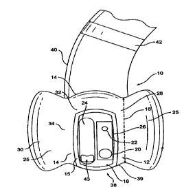
Fig. 1 is a side view of an anchoring and
positioning device in accordance with the present invention;
and
Fig. 2 is a front view of the anchoring and
positioning device shown in Fig. 1.
Reference will now be made in detail to the present
preferred embodiments of the present invention, an example of
which is illustrated in the accompanying drawings. Wherever
possible, the same reference numbers will be used throughout
the drawings to refer to the same or like parts.
In accordance with the present invention, an
endoscope is provided with an anchoring and positioning
device, such as an inflatable balloon at a distal end section
of the endoscope. The balloon is shaped to include an annular
CA 02324296 2000-09-19
WO 99/48417 PCTNS99/05685
-5-
portion and a contiguous cradle portion that provides a
balloon opening over a window section of the endoscope.
A preferred embodiment of the present invention,
shown in Figs. 1 and 2, is used with a side-view type
endoscope l0 provided with a distal end section 12. Side-view
type endoscope 10 is suitable for examining the duodenum, and
it is called a duodenoscope. The present invention can also
be used with an end-view type endoscope, suitable for
examining the colon, and called a colonoscope. As is typical,
the endoscope 10 has a flexible elongated tubular body. The
diameter of the endoscope 10 may vary, but approximates 10 mm
for most applications. The distal end section 12 is located
at the tip of the endoscope 10 and is inserted into the body
cavity to be examined. Endoscope 10, in general, includes an
illumination device 20, a viewing device 22, and a working
lumen or channel 24. The illumination 20 device provides
light for an endoscopic operation in a dark body cavity. The
viewing device 22, such as a TV camera, captures an image in
the body cavity, and the image is electrically or optically
transmitted through the tubular body of endoscope 10. The
working channel 24 extends from the distal end section 12
through the tubular body of endoscope 10, and is generally
made of tetrafluoroethylene resin. The working channel 24 is
designed to accommodate various medical instruments and
devices, such as a stmt. Moreover, the working channel 24
CA 02324296 2000-09-19
WO 99/48417 PCT/US99/05685
-6-
may be equipped with an elevator 43 capable of changing the
direction of a medical instrument inserted therein. Such an
elevator is commonly known in the art and is controlled by the
endoscope operator at the proximal end of the endoscope 10.
As illustrated in Fig. 1, the distal end section 12
has a section length 16, a major peripheral part 14, and a
minor peripheral part 15. The section length 16 is an axial
length of the distal end section 12 along the endoscope 10,
and it may vary depending.on the intended application of
endoscope 10 but approximates 20 mm for a duodenoscope. The
major peripheral part 14 is a relatively large arcuate area
portion of the distal end section 12 of the endoscope 10. On
the other hand, the minor peripheral part 15 is a relatively
small arcuate area portion of the end section.
The distal end section 12 has a window section 18 on
the minor peripheral part 15. The window section 18 is
equipped with illumination device 20, viewing device 22, and
an end of the working channel 24. The window length 26 is
defined as the length of the window section 18 in the axial
direction. Preferably, the window length 26 is smaller than
the section length 16 of the distal end section 12; therefore,
the distal end section 12 has a remaining section length 28
defined by the difference in length between the section length
16 and the window length 26 in the axial direction of the
endoscope 10. '
CA 02324296 2000-09-19
WO 99/48417 PCT/US99/05685
An anchoring and positioning device for the
endoscope 10, in the form of an inflatable balloon 30, is
attached to the distal end section 12 of the endoscope 10.
The inflatable balloon 30 is used for anchoring the endoscope
as well as for providing leverage for the operation of
medical instruments extending through working channel 24 into
a desired body cavity. Additionally, the endoscope 10 can be
precisely positioned by adjusting inflation of the balloon 30.
The balloon 30 is made of a material with a high friction
coefficient so that it attaches naturally to the endoscope 10,
and it is preferably made of ethylene vinyl acetate or
polyethylate. The balloon 30 can be attached to the endoscope
10 due to friction between the balloon material and the
endoscope. As the balloon 30 is inflated, the friction
increases, strengthening the attachment between the balloon
and the endoscope. In another embodiment, the balloon 30 may
be attached to the endoscope 10 by an adhesive suitable for
endoscopic use. The balloon 30 has an axial length
approximately the same as the section length 16, but the size
of the balloon 30 may vary depending on its application. For
a duodenoscope, preferably, the 180-degree outer diameter of
the balloon 30 is approximately 20-25 mm when it is inflated.
Balloon 30 includes a cradle portion 34, which
circumscribes the major peripheral part 14 of the distal end
section 12. The cradle portion 34 of the inflated balloon 30'
has an arcuate length corresponding to the major peripheral
CA 02324296 2000-09-19
WO 99/48417 PC'TNS99/05685
_g_
part 14 and terminates at arcuate ends 35. When the balloon
30 is inflated, the arcuate ends 35 radially extend from
endoscope 10 to provide a balloon opening 38 between the
window section 18 and a wall 27 of a passage surrounding the
endoscope 10. The wall 27 is a part of the body cavity into
which the endoscope 10 is extended. During operation of the
endoscope 10 with the balloon 30 inflated, the cradle portion
34 spaces the window section 18 from the examining area, thus
providing a good view of and a sufficient working space
relative to the wall 27. The arcuate length of the cradle
portion 34 may vary depending on the application and design of
the endoscope 10.
Preferably, the arcuate ends 35 are connected at the
proximal end of the axial length of the inflated balloon 30.
Thus, the balloon 30 has an annular portion 32 circumscribing
a remaining section 28 of the distal end section 12 that is
not occupied by the window section 18. The annular portion 32
of the balloon 30 is preferably shaped such that the balloon
30 has the same outer diameter throughout the full circular
configuration when the balloon 30 is inflated.
Additionally, as shown in Fig. 1, a cap 39 is
attached to the distal end of the balloon 30 to prevent it
from sliding in the axial direction away from the distal end
section 12 of the endoscope 10. The cap 39 is preferably made
CA 02324296 2000-09-19
WO 99/48417 PCT/US99/05685
-9-
of the same material as the balloon 30 or a similar plastic
material attached to the balloon 30. However, the cap 39 is
not inflatable.
The anchoring and positioning device of the present
invention also includes a means for inflating and deflating
the balloon 30. As shown in Fig. 1, a catheter 40 is coupled
to the balloon 30 to introduce a fluid in and out of the
balloon 30 for inflation and deflation. Preferably, the
medium used for the inflation and the deflation of the balloon
30 is air, water, or a contrast mixture for fluoroscopic
visualization. The catheter 30 is preferably made of nylon,
pebax (a plastic material known to one skilled in the art),
polyethylene, or other suitable material, and preferably has a
length of about 180 cm. As shown in Fig. 1, the catheter 40
extends from the balloon 30 along the flexible tubular body of
the endoscope 10. Preferably, the catheter 40 is fastened to
the tubular body of the endoscope 10 with a fastener 42 in
order to minimize tissue resistance in body cavities. The
fastener 42 is preferably made of silicone or latex bands.
Other embodiments of the invention will be apparent
to those skilled in the art from consideration of the
specification and practice of the invention disclosed herein.
It is intended that the specification and examples be
considered as exemplary only, with a true scope and spirit of
the invention being indicated by the following claims.