Note: Descriptions are shown in the official language in which they were submitted.
CA 02324367 2000-09-18
WO 99/47201 PCTNS99/05942
APPARATUS AND METHOD FOR THE DIALYSIS OF BLOOD
Field Of The Invention
This invention relates to the dialysis of blood in
general, and more particularly to apparatus and methods
for use in the same.
Background Of The Invention
A healthy kidney removes toxic wastes and excess
water from the blood. In End Stage Renal Disease
("ESRD"), or chronic kidney failure, the kidneys
progressively stop performing these essential functions
over a long period of time. When the kidneys fail, a
patient dies within a short period of time unless that
patient receives dialysis treatment for the rest of
that patient's life or undergoes transplantation of a
healthy, normal kidney. Since relatively few kidneys
are currently available for transplantation, the
overwhelming majority of patients with ESRD receive
dialysis treatment.
SUBSTITUTE SHEET (RULE 26)
CA 02324367 2000-09-18
WO 99J47201 PCT/US99/05942
s
Hemodialysis therapy is an extracorporeal (i.e.,
outside the body) process which removes toxins and
water from a patient's blood. A hemodialysis machine
pumps blood from the patient, through a dialyzer, and
then back to the patient. The dialyzer removes the
toxins and water from the blood by a membrane diffusion
process. Typically, a patient with chronic kidney
disease requires hemodialysis three times per week, for
3-6 hours per session. Removing blood from the body
requires a vascular access to the patient's blood
system.
One common method for accessing a patient's blood
system for hemodialysis involves the use of a
percutaneous catheter assembly. The percutaneous
catheter assembly is inserted into a major vein, such
as the femoral, subclavian or jugular vein. For long
term maintenance dialysis, the jugular vein is
generally the preferred insertion site. The catheter
assembly is percutaneous, with one end external to the
body and the other end dwelling in either the superior
vena cava or the right atrium of the heart. The
external portion of the catheter assembly has
SUBSTITUTE SHEET (RULE 26}
CA 02324367 2000-09-18
WO 99/47201 PCT/US99/05942
- 3 -
connectors permitting attachment of blood lines leading
- to and from the hemodialysis machine.
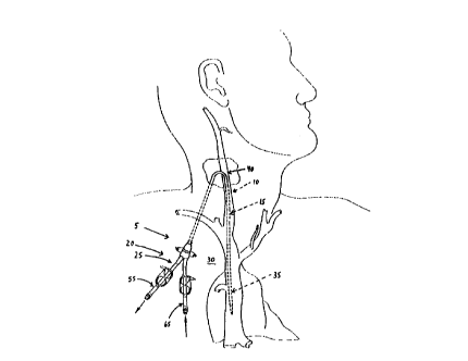
Figs. 1 and 2 show the traditional manner of
positioning a percutaneous catheter assembly 5 relative
to the body. More particularly, percutaneous catheter
assembly 5 generally comprises a catheter portion 10
comprising a dual-lumen catheter element 15, and a
connector portion 20 comprising an extracorporeal
connector element 25. The catheter assembly's
extracorporeal connector element 25 is disposed against
the chest 30 of the patient, and the distal end 35 of
catheter element 15 is passed into the patient's
internal jugular vein 40 (Fig. 2) and then down into
the patient's superior vena cava 45. More
particularly, the distal end 35 of catheter element 15
is positioned within the patient's superior vena cava
45 such that the mouth 50 of suction line 55, and the
mouth 60 of return line 65, are both located between
the patient's right atrium 70 and the patient's left
subclavia vein 75 and right subclavia vein 80.
Alternatively, the distal end 35 of catheter element 15
may be positioned so that mouth 50 of suction line 55,
SUBSTITUTE SKEET (RULE 2b)
CA 02324367 2000-09-18
WO 99/47201 PCTNS99/05942
w
- 4 -
and mouth 60 of return line 65, are located within the
patient's right atrium 70. The percutaneous catheter
assembly 5 is then left in this position relative to
the body, waiting to be used during an active dialysis
session.
When hemodialysis is to be performed on the
patient, the catheter assembly's extracorporeal
connector element 25 is appropriately connected to a
dialysis machine (not shown), i.e., suction line 55 is
connected to the input port (i.e., the suction port) of
the dialysis machine, and return line 65 is connected
to the output port (i.e., the return port) of the
dialysis machine. The dialysis machine is then
activated (i.e., the dialysis machine's blood pump is
turned on and the flow rate set), whereupon the
dialysis machine will withdraw relatively "dirty" blood
from the patient through suction line 55 and return
relatively "clean" blood to the patient through return
line 65.
It has also been proposed to use a subcutaneous
port and catheter assembly to provide vascular access
for hemodialysis.
SUBSTITUTE SHEET (RULE 26)
CA 02324367 2000-09-18
WO 99/47201 PCT/US99105942
- 5 -
More particularly, a subcutaneous port and
catheter assembly 82 is shown in Figs. 3-5. Looking
first at Fig. 3, subcutaneous port and catheter
assembly 82 generally comprises a connector portion 84
comprising a subcutaneous pork element 86, and the'
aforementioned catheter portion 10 comprising the
dual-lumen catheter element 15. As noted above, the
catheter element 15 in turn comprises the suction line
55 and the return line 65. Subcutaneous port element
86 includes a needle port 88 which is connected to
suction line 55, and a needle port 90 which is
connected to return line 65. The distal end of suction
line 55 terminates in the aforementioned mouth 50, and
the distal end of return line 65 terminates in the
aforementioned mouth 60.
Figs. 4 and 5 show subcutaneous port and catheter
assembly 82 positioned within the body. More
particularly, the assembly's port element 86 is
disposed under the skin of the patient (e.g., in the
chest area of the patient), and the assembly's catheter
element 15 is passed into the patient's internal
jugular vein 90 and then down into the patient's
SUBSTITUTE SHEET (RULE 26)
CA 02324367 2000-09-18
WO 9.9J47201 PCT/US99/05942
w
superior vena cava 45. The distal end of the
- assembly's catheter element 15 may be positioned within
the patient's superior vena cava 45 such that mouth 50
of suction line 55, and mouth 60 of return line 65, are
both located approximately between the patient's right
atrium 70 and the patient's left subclavia vein 75 and
right subclavia vein 80. Alternatively, the distal end
of catheter element 15 may be positioned so that mouth
50 of suction line 55, and mouth 60 of return line 65,
are located within the patient's right atrium 70. The
subcutaneous port and catheter assembly 82 is then left
in this position within the body, waiting to be used
during an active dialysis session.
When hemodialysis is to be performed on the
patient, the assembly's subcutaneous port element 86 is
appropriately connected to a dialysis machine, i.e.,
needle port 88 is connected to the input port (i.e.,
the suction port) of the dialysis machine with an
appropriate percutaneous needle (not shown), and the
assembly's needle port 90 is connected to the output
port (i.e., the return port) of the dialysis machine
with an appropriate percutaneous needle (not shown).
SUBSTITUTE SHEET (RULE 26}
CA 02324367 2000-09-18
WO 99/47201 PCTNS99/05942
The dialysis machine is then activated, whereupon it
will withdraw relatively "dirty" blood from the patient
through suction line 55 and return relatively "clean"
blood to the patient through return line 65.
It will be appreciated that both percutaneous
catheter assembly 5 (Figs. 1 and 2) and subcutaneous
port and catheter assembly 82 (Figs. 3-5) comprise the
catheter portion 10, which in turn comprises the
dual-lumen catheter element 15,~ with the distal end of
the catheter element normally dwelling in the patient's
vascular system.
Inasmuch as a substantial portion of catheter
element 15 dwells in the patient's vascular system
(e. g., within internal jugular vein 40 and superior
vena cava 45), it is desirable for the catheter element
to have the smallest possible outside diameter so as to
minimize interference with normal blood flow. At the
same time. however, it is also desirable for the
catheter element to have the largest possible inside
diameter so that maximum dialysis blood flow can be
achieved. Thus, from the standpoint of blood flow
SUBSTITUTE SHEET (RULE 2~
CA 02324367 2000-09-18
WO 99/47201 PCT/US99I05942
- 8 -
alone, it is desirable for the catheter element to have
the thinnest possible wall thickness.
Unfortunately, however, other considerations also
come into effect. For one thing, it is also important
that the catheter element have the highest possible
burst strength so that it will not fail when passing
blood under pressure. In addition, it is also
important that the catheter element be able to
withstand high negative pressures without collapsing,
so that blood can be withdrawn from the body at a rapid
rate. Furthermore, it is important that the catheter
element be capable of being bent at a substantial angle
without kinking, such as, for example. at the point
where the catheter element undergoes a large deflection
in order to enter internal jugular vein 40 (see Figs. 1
and 9). These and other considerations tend to
significantly limit the degree to which the catheter
element's wall thickness can be reduced.
Furthermore, the choice of materials for forming
the catheter element is also limited, since the element
is typically deployed in the patient's body for
substantial periods of time. Currently, silicone
SUBSTITUTE SHEET (RULE 26)
CA 02324367 2000-09-18
WO 99/47201 PCTIUS99/05942
_ g _
rubber is the accepted material for forming catheter
elements for use in percutaneous catheter assemblies
and subcutaneous port and catheter assemblies.
The foregoing factors have, collectively, tended
to limit either (1) the degree to which the outside
diameter of the catheter element can be reduced, and/or
(2) the degree to which the inside diameter of the
catheter element can be enlarged, and/or (3) the rate
at which blood can be introduced into the patient's
body through the catheter element, and/or (4) the rate
at which blood can be withdrawn from the patient's body
through the catheter element, and/or (5) the degree to
which the catheter element can be bent without kinking.
In U.S. Patent No. 5,041,098, issued August 20,
1991 to Loiterman et al., it was suggested that a
helically wound reinforcement wire could be
incorporated into the side wall of the catheter
element. Such a suggestion could appear to be
advantageous, since it could enable the walls of the
catheter element to be made thinner yet stronger.
Unfortunately, in practice, Applicants have found
that commercially-available coil-reinforced silicone
SUBSTITUTE SHEET (RULE 26)
CA 02324367 2000-09-18
WO 99/47201 PCTIUS99/05942
- 10 -
rubber tubes lack the smooth interior lumen desirable
for hemodialysis applications.
More particularly, Applicants have discovered that
in hemodialysis applications, smooth lumen walls are
important for (1) providing the laminar blood flows
required for high volume blood transfer, (2) avoiding
the creation of irregular blood currents and the
creation of blood stagnation areas. (3) avoiding the
formation of blood clots, (9) eliminating breeding
areas for bacteria, and (S) facilitating flush-cleaning
of the apparatus after dialysis has taken place.
Unfortunately, Applicants have also found that
commercially-available coil-reinforced silicone rubber
tubes lack the smooth interior lumen desirable for
hemodialysis applications.
It is believed that this may be due to the facts
that (1) commercially-available coil-reinforced
silicone rubber tubes are generally used for purposes
other than dialysis applications, and (2) the
importance of smooth interior lumens has not yet been
discovered by the dialysis industry.
SUBSTITUTE SHEET (RULE 26)
CA 02324367 2000-09-18
WO 99/47201 PCTIUS9910S942
- 11 -
It is also believed that commercially-available
coil-reinforced silicone rubber tubes may lack the
smooth interior lumen desirable for hemodialysis
applications due to the manner in which such tubes are
typically formed.
More particularly, it is believed that
commercially-available coil-reinforced silicone rubber
tubes are generally formed by (1) creating an outer
tube out of silicone rubber, (2) forcing that tube
open, (3) inserting the coil spring inside the
forced-open silicone rubber tube. (4) releasing the
outer tube so that it contracts back on the coil
spring, and (5) dip molding an interior layer of
silicone rubber onto the spring and the interior lumen
of the outer tube. While such a process is generally
adequate for capturing the coil spring within a body of
silicone rubber material, it also results in an
undulating interior lumen, since the process
essentially covers the coil spring and the interior
lumen of the outer tube with a substantially
SUBSTITUTE SHEET (RULE 2~
CA 02324367 2000-09-18
WO 99/47201 PCTYUS99I05942
- 12 -
constant-thickness dip layer. Coil-reinforced silicone
rubber tubes formed with the aforementioned process are
not smooth enough for good hemodialysis applications.
Objects Of The Invention
Accordingly, one object of the present invention
is to provide improved apparatus for use in the
dialysis of blood.
Another object of the present invention is to
provide an improved catheter element for use in the
dialysis of blood, wherein the catheter element may be
used in either a percutaneous catheter assembly or a
subcutaneous port and catheter assembly.
And another object of the present invention is to
provide an improved catheter element which has the
largest possible interior diameter and the smallest
possible exterior diameter, yet is resistant to
bursting, collapse and kinking.
Still another object of the present invention is
to provide an improved catheter element which
incorporates reinforcing means within the side wall of
the catheter, yet has an interior lumen which is
SUBSTITUTE SHEET {RULE 2b)
CA 02324367 2000-09-18
WO 9Q/47201 PCT/US99/05942
- 13 -
sufficiently smooth that the catheter element may be
- used in hemodialysis applications with good results.
And another object of the present invention is to
provide a support structure at the distal end of the
catheter element to help maintain openness of the flow
path through the catheter element.
Yet another object of the present invention is to
provide an improved method for fabricating apparatus
for use in the dialysis of blood.
And another object of the present invention is to
provide an improved method for the dialysis of blood.
Summary Of The Invention
These and other objects are addressed by the
present invention, which comprises improved apparatus
for the dialysis of blood, a method for making the
same, and an improved method for the dialysis of blood.
In one preferred embodiment, the present invention
comprises a catheter comprising at least one flexible
tubular element, the at least one tubular element
having an open proximal end, an open distal end and a
SUBSTITUTE SHEET (RULE 2b)
CA 02324367 2000-09-18
WO 99/47201 PCT/US99/05942
- 14 -
side wall defining a lumen extending between the open
proximal end and the open distal end, the side wall
(1) being formed of a biocompatible material,
(2) encasing reinforcing means therein for reinforcing
the side wall, and (3) having a smooth interior surface
for defining the lumen, the smooth interior surface
being sufficiently smooth that the at least one tubular
element may be used for hemodialysis applications with
good results.
In another preferred embodiment, the present
invention comprises apparatus for use in the dialysis
of the blood of a patient, the apparatus comprising a
connector portion and a catheter portion; the connector
portion comprising an outlet adapted for communication
with a line connected to the input port of a dialysis
machine, and an inlet adapted for communication with a
line connected to the output port of a dialysis
machine: and the catheter portion comprising a catheter
element comprising: a suction line and a return line,
each such line comprising a flexible tubular element,
the tubular element having an open proximal end, an
open distal end and a side wall defining a lumen
SUBSTITUTE SHEET (R.ULE 26)
CA 02324367 2000-09-18
WO 99/47201 PCTNS99/05942
- 15 -
extending between the open proximal end and the open
distal end, the side wall (1) being formed of a
biocompatible material, (2) encasing reinforcing means
therein for reinforcing the side wall, and (3) having a
smooth interior surface for defining the lumen, the
smooth interior surface being sufficiently smooth that
the tubular element may be used for hemodialysis
applications with good results: the proximal end of the
suction line being connected to the connector portion
and in communication with the outlet, and the distal
end of the suction line terminating in a suction line
mouth: the proximal end of the return line being
connected to the connector portion and in communication
with the inlet, and the distal end of the return line
terminating in a return line mouth; the suction line
and the return Line being adapted for disposition
within the body of the patient so that the suction line
mouth and the return line mouth are both disposed in
the vascular system of the patient.
In yet another preferred embodiment, the present
invention comprises a method for making a catheter, the
method comprising the steps of: (a) providing an
SUBSTITUTE SHEET (RULE 26)
CA 02324367 2000-09-18
WO 99147201 1'CTNS99/OS942
- 16 -
elongated, removable molding core; (b) forming a
tubular element of biocompatible material on the
molding core: (c) positioning reinforcing means for
reinforcing the tubular element on the outer surface of
the tubular element: (d) forming an overlayer of
biocompatible material over the tubular element and the
reinforcing means: and (e) removing the molding core
from the coated tubular element.
In another preferred embodiment, the present
invention comprises a method for the dialysis of the
blood of a patient, the method comprising the steps of:
(a) providing a dialysis machine, and
providing apparatus comprising a connector portion and
a catheter portion; the connector portion comprising an
outlet adapted for communication with a line connected
to the input port of the dialysis machine, and an inlet
adapted for communication with a line connected to the
output port of the dialysis machine; and the catheter
portion comprising a catheter element comprising: a
suction line and a return line, each such line
comprising a flexible tubular element, the tubular
element having an open proximal end, an open distal end
SUBSTITUTE SHEET (RULE 26)
CA 02324367 2000-09-18
WO 99/47201 PCT/US99105942
- 17 -
and a side wall defining a lumen extending between the
open proximal end and the open distal end, the side
wall (1) being formed of a biocompatible material, (2)
encasing a reinforcing means therein for reinforcing
the side wall, and (3) having a smooth interior surface
for defining the lumen, the smooth interior surface
being sufficiently smooth that the tubular element may
be used for hemodialysis applications with good
results: the proximal end of the suction line being
connected to the connector portion and in communication
with the outlet, and the distal end of the suction line
terminating in a suction line mouth: the proximal end
of the return line being connected to the connector
portion and in communication with the inlet, and the
distal end of the return line terminating in a return
line mouth: the suction line and the return line being
adapted for disposition within the body of the patient
so that the suction line mouth and the return line
mouth are both disposed in the vascular system of the
patient:
SUBSTITUTE SHEET {RULE 26)
CA 02324367 2000-09-18
WO 99/47201 PCTIUS99/05942
- 28 -
(b) placing the suction line mouth and the
return line mouth in the vascular system of the
patient:
(c) connecting the outlet to the input port
of the dialysis machine, and connecting the inlet to
the output port of the dialysis machine; and
(d) operating the dialysis machine.
Brief Description Of The Drawings
These and other objects and features of the
present invention will be more fully disclosed or
rendered obvious by the following detailed description
of the preferred embodiments of the invention, Which
are to be considered together with the accompanying
drawings wherein:
Fig. 1 is a schematic view of a percutaneous
catheter assembly installed in a patient:
Fig. 2 is a schematic view showing the distal end
of the catheter element of the percutaneous catheter
assembly of Fig. 1 installed in a patient, with the
direction of blood flow being indicated by appropriate
arrows;
SUBSTITUTE SHEET (RULE 2b~
CA 02324367 2000-09-18
WO 99/47201 PCT/US99/05942
- 19 -
Fig. 3 is a schematic view of a subcutaneous port
and catheter assembly;
Fig. 4 is a schematic view showing the
subcutaneous port and catheter assembly of Fig. 3
installed in a patient;
Fig. 5 is an enlarged schematic view showing the
subcutaneous port and catheter assembly of Fig. 3
installed in a patient;
Fig. 6 is a schematic view, partially in section,
of novel catheter apparatus formed in accordance with
the present invention;
Fig. 7 is a perspective view of a support
structure incorporated into the novel catheter
apparatus shown in Fig. 6;
Figs. 8-10 are schematic views illustrating how
the local surface profile of the central lumen of the
catheter apparatus may vary along the length of the
lumen;
Figs. 11-17 illustrate process steps for
fabricating the novel catheter apparatus shown in Fig.
6;
SUBSTITUTE SHEET (RULE 2~
CA 02324367 2000-09-18
WO 99/47201 PCT/US99/05942
- 20 -
Fig. 18 illustrates the distal end of an
alternative form of catheter apparatus formed in
accordance with the present invention;
Fig. 19 is a perspective view of the support
structure incorporated into the catheter apparatus
shown in Fig. 18;
Figs. 20 and 21 illustrate still other forms of
support structures which may be incorporated into
catheter apparatus formed in accordance with the
present invention;
Fig. 22 illustrates an alternative manner for
incorporating the support structure of Fig. 21 into
catheter apparatus formed in accordance with the
present invention;
Fig. 22A illustrates an alternative manner for
forming side openings near the distal end of the
catheter apparatus, wherein the side openings are in
the form of relatively narrow, longitudinally-extending
slits:
Fig. 23 illustrates catheter apparatus utilizing
an alternative form of reinforcing means: and
SUBSTITUTE SHEET (RULE 26)
CA 02324367 2000-09-18
WO 99/47201 PCT/US99/05942
- 21 -
Fig. 23A schematically illustrates an alternative
form of reinforcing means, wherein the reinforcing
means comprise a tubular, braided mesh reinforcer.
Detailed Description Of The Preferred Embodiments
Looking next at Fig. 6, novel catheter apparatus
100 is shown. Catheter apparatus 100 comprises a
silicone rubber tube 105 having a side wall 110, a
distal end wall 115 and a proximal end wall 120. The
outer surface 125 of side wall 110 has a smooth
configuration so as to minimize interference with blood
flow when catheter apparatus 100 is disposed in the
vascular system of a patient.
A central lumen 130 extends between distal end
wall 115 and proximal end wall 120. Central lumen 130
has a smooth interior surface so that blood can be
passed through that lumen during a hemodialysis session
with good results.
Catheter apparatus 100 also comprises reinforcing
means 135 encapsulated within side wall 110 for
reinforcing the side wall. In one preferred form of
the invention, reinforcing means 135 comprise a coil
SUBSTITUTE SHEET (RULE 26)
CA 02324367 2000-09-18
WO 99/4701 PCT/US99/05942
- 22 -
spring 140. Reinforcing means 135 may extend along the
entire length of tube 105, or reinforcing means 135 may
extend along only one or more selected portions of tube
105, as preferred. For example, reinforcing means 135
might extend along only an intermediate portion of tube
I05. In one form of the invention, reinforcing means
135 are formed out of a radio-opaque material, whereby
catheter apparatus 100 may be visualized while within
the body of the patient through the use of appropriate
imaging equipment, whereby to aid in the proper
deployment of the apparatus within the body.
Preferably, but not necessarily, at least one side
opening 145 is formed in side wall 110 adjacent to, but
spaced from, distal end wall 115. The at least one
side opening 195 communicates with central lumen 130,
whereby blood may enter and/or exit the distal end of
catheter apparatus 100 via either the distal end of
central lumen 130 and/or the at least one side opening
195. Preferably the at least one side opening 145 is
in the form of a substantially circular hole.
And preferably, but not necessarily, a support
structure 150 (Figs. 6 and 7) is disposed at the
SUBSTITUTE SHEET (RULE 26)
CA 02324367 2000-09-18
WO 99/47201 PG"T/US99/05942
w
- 23 -
intersection of central lumen 130 and distal end wall
115 so as to provide a stiffening element at the distal
end of catheter apparatus 100. This member can help
maintain openness of the flow path through catheter
apparatus 100. In one form of the invention, shown in
Figs. 6 and 7, support structure 150 is formed with a
cylindrical configuration, with a bore 155 opening on
the support structure's distal end surface 160, a
counterbore 165 opening on the support structure's
proximal end surface 170, and with an annular shoulder
175 formed at the intersection of bore 155 and
counterbore 165. In one preferred form of the
invention, support structure 150 is mounted to the
distal end of tube 105 so that the support structure's
distal end surface 160 lies flush with the tube's
distal end surface 115, and so that the support
structure's bore 155 is aligned with the tube's central
lumen 130 ( Fi g . 6 ) .
In one preferred form of the invention, tube 105
is formed out of 80 durometer silicone rubber, and has
an outside diameter of approximately 0.135 inch, an
inside diameter of approximately 0.105 inch, and a wall
SUBSTITUTE SHEET (RULE 26)
CA 02324367 2000-09-18
WO 99/47201 PC"fIUS99/05942
_ 29 _
thickness of approximately 0.015 inch; and coil spring
190 is formed out of titanium, with the wire forming
the coil spring being approximately 0.006 inch thick
and having a coil rate of approximately 0.031-0.036
pitch. Support structure 150 is preferably formed out
of a radio-opaque material, whereby the distal end of
catheter apparatus 100 may be visualized while within
the body of the patient through the use of appropriate
imaging equipment, whereby to aid in the proper
deployment of the apparatus within the body.
It is an important feature of the present
invention that central lumen 130 be formed smooth
enough that catheter apparatus 100 may be used for
hemodialysis applications with good results. However,
the presence of reinforcing means 135 in the side wall
110 of catheter apparatus 100, and/or the manner of
encapsulating reinforcing means 135 within side wall
110, and/or a variety of other factors, may cause
variations in the diameter of central lumen 130.
More particularly, as seen in Fig. 8, the presence
of reinforcing means 135 may cause central lumen 130 to
vary outwardly (as shown at 130A) in the region between
SUBSTITUTE SHEET (RULE 2b)
CA 02324367 2000-09-18
WO 99/47201 PCTNS99105942
- 25 -
adjacent occurrences of reinforcing means 135; or, as
seen in Figs. 9 and 10, the presence of reinforcing
means 135 may cause central lumen 130 to vary inwardly
(as shown at 1308) in the region between adjacent
occurrences of reinforcing means~135. Such variations
in the local surface profile of central lumen 130 can
have a detrimental effect when catheter apparatus 100
is used for hemodialysis applications.
In accordance with the present invention, central
lumen 130 of catheter apparatus 100 is formed smooth
enough so that catheter apparatus 100 may be used for
hemodialysis applications with good results.
In other words, central lumen 130 of catheter
apparatus 100 is formed smooth enough to (1)
substantially provide the laminar blood flows required
for high volume blood transfer, (2) substantially avoid
the creation of irregular blood currents and the
creation of blood stagnation areas, (3) substantially
avoid the creation of blood clots. (4) substantially
eliminate breeding areas for bacteria, and (5)
facilitate flush-cleaning of catheter apparatus 100
after dialysis has taken place.
SUBSTITUTE SHEET (RULE 26)
CA 02324367 2000-09-18
WO 99/47201 PCT/US99/05942
- 26 -
In one particular aspect of the present invention,
Applicants have discovered that the cumulative effects
of variations in the local surface profile of central
lumen 130 can have a significant detrimental effect on
the utility of that lumen for hemodialysis
applications.
Applicants have further discovered that good
hemodialysis results can be achieved if variations in
the local surface profile of central lumen 130 average
less than about 0.0015 inch and preferably less than
about 0.0005 inch (as measured between adjacent
occurrences of reinforcing means 135) along the length
of central lumen 130. In other words, if catheter
apparatus 100 is constructed with a 20 turn helical
element, there will be 19 local variation measuring
points along any path of measurement taken parallel to
the axis of the apparatus. The measurements at these
measuring points are then averaged, whereby to provide
an average variation in the local surface profile of
central lumen 130. Applicants have determined that
good hemodialysis results can be achieved where the
average variation in the local surface profile of
SUBSTITUTE SHEET (RULE 26)
CA 02324367 2000-09-18
WO 99/47201 PGTIUS99/05942
- 27 -
central lumen 130 is less than about 0.0015 inch, and
preferably less than about 0.0005 inch.
Thus, in one preferred form of the invention,
catheter apparatus 100 is formed so that the average
variation in the local surface profile of central lumen
130 is less than about 0.0015 inch, and preferably less
than about 0.0005 inch.
The present invention also includes a novel method
for fabricating catheter apparatus 100.
In the preferred embodiment of the invention, the
novel catheter apparatus 100 is formed as follows.
First, an elongated molding core 200 is provided
(Fig. 11). Molding core 200 is formed so that it is
removable from a structure which will be molded over
the core, as will hereinafter be discussed. In one
preferred farm of the invention, molding core 200 is
formed so that it has a reducible transverse
cross-section, whereby the molding core is removable
from a structure which will be molded over the core, as
will hereinafter be discussed. Molding core 200 has a
polished finish so that its outer surface 205 is smooth
and free from burrs and other surface irregularities.
SUBSTITUTE SHEET (RULE 26)
CA 02324367 2000-09-18
WO 99/47201 PCTIUS99105942
- 28 -
In one preferred form of the invention, molding core
200 is formed out of a teflon extrusion which may have
its transverse cross-section reduced by stretching it
along its longitudinal axis. Preferably the teflon
extrusion is formed out of virgin teflon stock which
is capable of reducing its diameter by approximately
5-10$ when subjected to longitudinal stretching.
Preferably the virgin stock is homogenous, so that
stretching occurs relatively evenly over the entire
body of the teflon.
Next, a silicone rubber element 210 is formed
about molding core 200 (Fig. 12). Preferably this is
done by co-extruding silicone rubber element 210 about
the outer surface 205 of molding core 200. Inasmuch as
molding core 200 has a smooth outer surface 205, the
inner surface 215 of silicone rubber element 210 will
therefore also be smooth and free from burrs and other
surface irregularities.
Then reinforcing means 135, preferably in the form
of coil spring 140, is loaded over silicone rubber
element 210 (Fig. 13).
SUBSTITUTE SHEET (RULE 26)
CA 02324367 2000-09-18
WO 99/47201 PCT/US99/05942
l
- 29 -
Next, support structure 150 (Figs. 6, 7, and 14)
is fit over the distal end of molding core 200 and the
distal end of silicone rubber element 210. More
particularly, support structure 150 is fit over the
distal end of molding core 200 and the distal end of
silicone rubber element 210 so that the distal end of
silicone rubber element 210 rests in counterbore 165 of
support structure 150 and against shoulder 175 of
support structure 150, and so that molding core 200
extends out through bore 155 of support structure 150
(Fig. 14). One or more side openings 220 are then
formed in silicone rubber element 210 (Fig. 14), and
corresponding molding pins (not shown) are inserted
into the one or more side openings 220.
Next, a silicone rubber overlayer 225 is molded
over reinforcing means 135 (e.g., coil spring 140) and
silicone rubber element 210 (Fig. 15). Preferably
silicone rubber overlayer 225 is applied so that it is
seamlessly integrated with silioone rubber element 210.
Preferably the distal end surface 230 of silicone
rubber overlayer 225 is aligned with the distal end
surface 160 of support structure 150. The molding pins
SUBSTTTUTE SHEET (RULE 26)
CA 02324367 2000-09-18
WO 99/47201 PGT/US99105942
- 30 -
(not shown) located in the one or more side openings
220 extend out through silicone rubber overlayer 225.
Once this has been done, molding core 200 is
removed.
In one preferred form of the invention, where
molding core 200 has a reducible transverse
cross-section, the molding core first has its
transverse cross-section reduced. causing it to
separate away from the inside wall 215 of silicone
rubber element 210 (Fig. 16). Then molding core 200 is
removed.
And in one preferred form of the invention, where
molding core 200 is formed out of a teflon extrusion
such that stretching the teflon extrusion
longitudinally will cause it to reduce in diameter,
molding core 200 is first stretched longitudinally,
causing the molding core to separate away from the
inside wall 215 of silicone rubber element 210 (Fig.
16). Then molding core 200 is removed (Fig. 17.).
After molding core 200 has been removed, the
molding pins located in the one or more side openings
220 are withdrawn, thereby yielding the finished
SUBSTITUTE SHEET (RULE 26)
CA 02324367 2000-09-18
WO 99/47201 PCT/US99/05942
- 31 -
catheter apparatus 100. Alternatively, the molding
- pins located in the one or more side openings 220 may
be withdrawn prior to removing molding core 200.
In one preferred form of the invention, silicone
rubber element 210 and silicone rubber overlayer 225
are both formed out of 80 durometer silicone rubber;
silicone rubber element 210 has a wall thickness of
approximately 0.005 inch: and silicone rubber overlayer
225 has a wall thickness (between adjacent occurrences
of reinforcing means 135) of approximately 0.010 inch.
It is to be appreciated that, when fabricating
catheter apparatus 100 by means of the foregoing
process, silicone rubber element 210 and silicone
rubber overlayer 225 together form the side wall 110 of
catheter apparatus 100. Furthermore, inasmuch as
molding core 200 has a smooth outer surface 205, the
inside wall 215 of silicone rubber element 210 (which
inside wall 215 defines the central lumen 130 of
catheter apparatus 100) also has a smooth profile.
It has been found that, by forming catheter
apparatus 100 by means of the foregoing process, the
central lumen 130 of that apparatus will have an
SUBSTITUTE SHEET (RULE 26)
CA 02324367 2000-09-18
WO 99147201 PCT/US99/05942
- 32 -
interior surface which is sufficiently smooth that
blood can be passed through that lumen during a
hemodialysis session with good results.
Among other things, it has been found that, by
forming catheter apparatus 100 by means of the
foregoing process, the average variation in the local
surface profile of central lumen 130 will be less than
about 0.0015 inch, and preferably less than about
0.0005 inch.
Two of the catheter apparatus 100 can be attached
together in ways well known in the art so as to form a
complete dual-lumen catheter element 15.
This complete dual-lumen catheter element 15 can
then be combined with the connector portion 20 of a
percutaneous catheter assembly so as to form a complete
percutaneous catheter assembly such as is schematically
shown in Figs. l and 2. Such a percutaneous catheter
assembly may then be used in the conventional manner in
the hemodialysis of a patient.
Alternatively, the complete dual-lumen catheter
element 15 can be combined with the connector portion
84 of a subcutaneous port and catheter assembly so as
SUBSTITUTE SHEET (RULE 26)
CA 02324367 2000-09-18
WO 99/47201 PCTNS99/05942
- 33 -
to form a complete subcutaneous port and catheter
assembly such as is schematically shown in Figs. 3-5.
Such a subcutaneous port and catheter assembly may then
be used in the conventional manner in the hemodialysis
of a patient.
In practice, it has been found possible to provide
a dual-lumen catheter element 15, formed out of two of
the catheter apparatus 100, which is flexible: capable
of substantial bending (e.g., capable of being bent so
as to enter the patient's internal jugular' vein)
without kinking, and resistant to collapse when
subjected to substantial negative pressures (e.g., 500
mm of mercury negative pressure).
It should be appreciated that a dual-lumen
catheter element 15 formed out of two of the catheter
apparatus 100 is easily "field trimmable" to the
desired length. In particular, in one preferred
trimming method, a scalpel or the like is first used to
cut through side wall 110 and expose reinforcing means
135, and then surgical scissors or the like are used to
cut through reinforcing means 135. Alternatively,
SUBSTITUTE SHEET (RULE 26)
CA 02324367 2000-09-18
WO 99/47201 PCT/US99/05942
- 39 -
surgical scissors could be used to cut completely
through catheter apparatus 100 in a single step.
Numerous modifications can be made to the
apparatus and method described above without departing
from the scope of the present invention.
Thus, for example, reinforcing means 135 might be
formed out of stainless steel, or a hard plastic, or
some other material which is harder than the material
used to form tube 105.
Or molding core 200 might be formed out of a
material other than teflon, where the alternative
material is also capable of having its transverse
cross-section reduced by longitudinal stretching. Or
molding core 200 might have its transverse
cross-section reduced by a method other than
stretching, e.g., depending on the material involved,
the molding core might be melted out or dissolved away
so as to separate it from silicone rubber element 210.
Furthermore, molding core 200 might be removed from
within the molded structure by a technique other than
reducing its transverse cross-section. By way of
example but not limitation, molding core 200 might
SUBSTITUTE SHEET {RULE 26)
CA 02324367 2000-09-18
WO 99/47201 PCT/US99/05942
- 35 -
comprise a sufficiently lubricious material such that
- the molding core could be removed from within the
molded structure by simply pulling the molding core
longitudinally out of the molded structure. Or molding
core 200 could be blown out of the molded structure.
Also, if desired, the distal end of catheter
apparatus 100 can be formed with an alternative
geometry.
By way of example but not limitation, a support
structure 180 (Figs. 18 and 19) can be utilized,
wherein support structure 180. is substantially
identical to the support structure 150 discussed above,
except that side slots 182 are formed in support
structure 180, and appropriate molding pins (not shown)
are used in association with side slots 180 to form
semi-circular side openings 189 (Fig. 18) in the distal
end of the catheter apparatus.
Or, as seen in Fig. 20, side windows 186 can be
formed in a support structure 188 which, when covered
with appropriate molding pins during molding, will
yield appropriate openings in the distal end of the
catheter apparatus.
SUBSTTrUTE SHEET (RULE 26)
CA 02324367 2000-09-18
WO 99/47201 PCT/US99/05942
- 36 -
Or a simple ring-shaped support structure 190
' (Fig. 21) can be used, with or without side openings
I45. With such a construction, support structure 190
can be positioned so that its distal end surface 192
resides flush with distal end wall 115 of tube 105, in
a manner generally analogous to the construction shown
in Fig. 6: or support structure 190 can be encapsulated
within the distal end of tube 105, in the manner shown
in Fig. 22.
It is also possible for the distal support
structure to be attached onto a surface of side wall
110 of tube 105, rather than embedded into the material
of side wall 110.
And the distal support structure may open on the
distal end of tube 105, and/or open on the inner lumen
of tube 105, and/or open on the outer lumen of tube
105.
Or the distal support structure may be omitted
completely from the catheter apparatus if desired.
Furthermore, side openings 195 might comprise
relatively narrow, longitudinally-extending slits
rather than holes, in which case they could act
SUBSTITUTE SHEET (RULE 26)
CA 02324367 2000-09-18
WO 99147201 PCT/US99105942
w
37 _
something like a check valve, opening under positive
internal pressure but otherwise remaining substantially
closed. See, for example, Fig. 22A, which shows side
openings 145 in the form of such relatively narrow,
longitudinally extending slits, with the slits being
shown in their closed position in solid line and in
their open position in dashed, or phantom, line.
It should also be appreciated that reinforcing
means 135 may take a form other than the coil spring
140 discussed above. For example, reinforcing means
135 might take the form of a plurality of ring-like
elements 199, such as is shown in Fig. 23.
Alternatively, reinforcing means 135 might comprise a
tubular, braided mesh reinforces. See, for example,
Fig. 23A, where reinforcing means 135 are shown,
schematically, in the form of a tubular, braided mesh
reinforces 195.
It should also be appreciated that one might form
a catheter apparatus of the sort generally described
above, including a distal support structure, but
omitting reinforcing means 135.
SUBSTITUTE SHEET (RULE 26)
CA 02324367 2000-09-18
WO 99/47201 PCT/US99/05942
- 38 -
It will, of course, be appreciated that various
other modifications may be made to the embodiments
disclosed above without departing from the spirit and
scope of the present invention. Accordingly, it is
intended that this invention be limited only by the
claims ultimately issued from this patent application
and/or from any patent applications? claiming priority
therefrom.
Advantages Of The Invention
Numerous advantages are achieved through the
provision and use of the present invention.
For one thing, the present invention provides
improved apparatus for use in the dialysis of blood.
And the present invention provides an improved
catheter element for use in the dialysis of blood,
wherein the catheter element may be used in either a
percutaneous catheter assembly or a subcutaneous port
and catheter assembly.
Also, the present invention provides an improved
catheter element which has the largest possible
interior diameter and the smallest possible exterior
SUBSTITUTE SHEET (RULE 2~
CA 02324367 2000-09-18
WO 99/47201 PCT/US99/05942
- 39 -
diameter, yet is resistant to bursting, collapse and
kinking.
And the present invention provides an improved
catheter element which incorporates reinforcing means
within the side wall of the catheter, yet has an
interior lumen which is sufficiently smooth that the
catheter element may be used in hemodialysis
applications with good results.
The present invention also provides a support
structure at the distal end of the catheter element to
help maintain openness of the flow path through the
catheter element.
Furthermore, the present invention provides an
improved method for fabricating apparatus for use in
the dialysis of blood.
And the present invention provides an improved
method for the dialysis of blood.
Still other advantages associated with the present
invention will be obvious to a person skilled in the
art.
SUBSTITUTE SHEET (RULE 26)