Note: Descriptions are shown in the official language in which they were submitted.
CA 02325169 2000-12-O1
1
PATENT
File 255.00040101
SOMATOSTATINS AND METHODS
This application claims the benefit of U.S. Provisional Application Serial
No. 60/168,934, filed December 3, 1999, which is incorporated herein by
reference in its entirety.
Statement of Government Rights
This invention was made with government support under grants from the
National Science Foundation, Grant No. OSR-9452892 and Grant No. IBN-
9723058.
Background of the Invention
Somatostatins are ubiquitous polypeptides known to affect basic
biological processes such as growth, development, metabolism, and cell
differentiation in vertebrates. Somatostatin was first isolated as a 14-amino
acid
peptide from ovine hypothalamus and found to inhibit the release of growth
hormone from the pituitary gland (Brazeau et al., Science, 179, 77-79 (1973)).
Since then somatostatins have been isolated from representatives of nearly
every
major group of vertebrates examined to date, from jawless fish to mammals
(Conlon et al., Regul~Peptides, 69, 95-103 (1997)). Somatostatins have been
found broadly in the central (e.g., cerebral cortex, cerebellum, pineal,
olfactory
lobe, hypothalamus, spinal cord) and peripheral nervous systems,
gastrointestinal
tract (e. g. , salivary glands, stomach, intestine), urogenital tract (e. g. ,
bladder,
prostate, collecting ducts of the kidney), pancreatic islets, adrenal glands,
thyroid
tissue, and placenta as well as in cerebral spinal fluid, blood, and saliva
(Reichlin, "Somatostatin," Brain peptide, Krieger et al., eds., John Wiley and
Sons, New York, pp. 712-752 (1982); Gerich, "Somatostatin and analogues,"
Diabetes mellitus: TheorX and practice, Ellenberg et al., eds., Medical
Examinations, New York (1983); Wass, "Somatostatin," EndocrinoloQV,
CA 02325169 2000-12-O1
2
DeGroot, ed., WB Saunders, Philadelphia, PA (1989); Patel, "General aspects of
the biology and function of somatostatin," Basic and clinical aspects of
neuroscience, Weil et al., eds., Springer-Verlag, Berlin (1992)). In neurons
and
cells, somatostatins are often co-localized with other factors (e.g.,
norepinephrine, CCK, neuropeptide-Y, CGRP, GABA, VIP, substance P)
(Gibbons, "Co-existence and co-function," The comparative ph. s~~
reaulator~peptides, Holmgren, ed., Chapman and Hall, London/New York
(1989)).
Somatostatins also possess a vast diversity of physiological actions. In
addition to secretotropic effects (including the effect on growth hormone
secretion for which the family was named), somatostatins have been reported to
have neurotropic and myotropic effects as wells as effects on transport,
metabolism, growth, differentiation, and modulation of functional development.
It should be noted that there is overlap between and among these somewhat
arbitrary classes of action. For example, the inhibition of growth hormone
secretion clearly affects growth and the inhibition of insulin secretion
clearly
affects metabolism. At the same time, the inhibition of growth hormone also
impacts metabolism while the inhibition of insulin has ramifications on growth
(Norman and Litwack, Hormones, Academic Press, New York (1997)). In
addition to such actions which result in physiological "cross talk,"
somatostatins
also have direct effects on the various classes of action. For example,
somatostatins have been shown to affect growth (e.g., proliferation) and
intermediary metabolism (e.g., lipolysis) directly in target cells (Patel,
"General
aspects of the biology and function of somatostatin," Basic and Clinical
Aspects
of Neuroscience, Weil et al., eds., Springer-Verlag, Berlin (1992); Sheridan,
Comp. Biochem. Physiol., 107b, 495-508 ( 1994)). Considering these various
roles, somatostatins may be of considerable importance in various diseases
including neuroendocrine tumors, diabetes mellitus, epilepsy, Alzheimer and
Huntington Diseases, and AIDS (Lamberts et al., Endocrine Rev., 12, 450-482
(1991); Patel et al., Life Sci., 57, 1249-1265 (1995)).
Most somatostatins appear to be synthesized as a long chain
prepropeptide, which can be subsequently processed to yield a propeptide
CA 02325169 2000-12-O1
(typically ranging in size from 25-28 amino acids) and, in some cases, dxrther
processed to yield a peptide of about 14 amino acids. This differential
processing introduces considerable molecular heterogeneity into somatostatins.
It is believed that in mammals, differential processing of the transcription
product of a single gene may account for the tissue-, organ- and cell-specific
activities of various somatostatins. The bioactivity of secreted somatostatins
is
mediated by cell-surface somatostatin receptors which likely differentiate
among
the various forms of somatostatin present in an organism. The molecular
heterogeneity of somatostatins appears to be even greater in some non-
mammalian organisms. Fish and some other non-mammals, for example, may
possess several somatostatin genes, each of which may be differentially
processed.
Notwithstanding the heterogeneity that characterizes the longer chain
preprosomatostatins and prosomatostatins, the somatostatin tetradecapeptide SS-
14 (Ala-Gly-Cys-Lys-Asn-Phe-Phe-Trp-Lys-Thr-Phe= Thr-Ser-Cys; SEQ ID
NO:1 ), present at the C-terminus of the longer forms, is completely conserved
among such mammals as monkeys, rats, cows, sheep, chickens and humans.
Somatostatins found in both mammals and non-mammals typically contain the
C-terminal SS-14 sequence (SEQ ID NO:1). Non-mammals, however,
frequently express additional somatostatins that contain variant C-terminal
tetradecapeptides with substitutions at one or more sites, such as (Tyr',
Gly'°]-
SS-14 (SEQ ID N0:2). This alternative somatostatin peptide has modifications
at positions 7 and 10 when compared to the mammalian sequence [Phe', Thr'o].
Somatostatins that contain the "mammalian"-type 14-mer sequence (SI:Q ID
NO:1) at the C-terminus are considered to be part of the "SS-I" family,
whereas
those that contain a 14-mer sequence having the [Tyr', Gly'°]
modification (SEQ
ID N0:2) are considered to be part of the "SS-II" family.
In mammalian systems, somatostatin is secreted into the blood and is
vascularly active. Different cells can synthesize different versions of the
polypeptide. Secreted somatostatin is also known to have a local paracrine
activity. There are a number of human diseases (e.g., growth disorder,
diabetes,
and several neurological disorders) that may be treated with somatostatin
CA 02325169 2000-12-O1
4
analogs. Also, some conditions (e.g., tumors) result from overproduction of
somatostatin, and there is no known somatostatin antagonist for treatment of
such disorders. New somatostatin analogs (both agonists and antagonists) that
have the potential to treat these and other human diseases would be a welcome
addition to current therapeutic strategies.
Summary of the Invention
The invention provides novel somatostatin polypeptides that contain
amino acid sequences found in Oncorhynchus mykiss preprosomatostatin I
(PPSS-I; SEQ ID N0:3) and/or Oncorhynchus mykiss preprosomatostatin II"
(PPSS-II"; SEQ ID NO:15). Also provided are bioactive analogs and subunits of
the somatostatin polypeptides of the invention. Preferred somatostatin
polypeptides include polypeptides having at least one amino acid sequence
selected from the group consisting of SEQ ID NOs:l, 2, 3, 4, 5, 6, 7, 9, 10,
11,
12, 13, 15, 16, 17, 18, and 19, and bioactive analogs and subunits thereof.
Polynucleotides encoding somatostatin polypeptides of the invention
and/or bioactive analogs and subunits thereof, as well as those that are
substantially complementary thereto, are also provided.
The invention further provides a method for identifying a modified
somatostatin polypeptide. The amino acid sequence of a somatostation
polypeptide of the invention is aligned with the amino acid sequence of a
reference somatostatin polypeptide, preferably a mammalian somatostatin
polypeptide, and at least one site or region of differing amino acid sequence
is
identified. Either the somatostatin polypeptide of the invention or the
reference
somatostatin polypeptide is then modified to incorporate at least one amino
acid
substitution, insertion, or deletion from the analogous site or region in the
other
somatostatin polypeptide to yield the amino acid sequence of a modified
somatostatin polypeptide. Optionally, the method furkher includes synthesizing
the modified somatostatin polypeptide and assaying the modified somatostatin
polypeptide for biological activity. Biological activity is preferably
determined
by determining whether the modified somatostatin polypeptide binds to a human
somatostatin receptor molecule or inhibits the binding of a natural ligand of
the
CA 02325169 2000-12-O1
human somatostatin receptor molecule. Preferably, the modified somatostatin
polypeptide identified according to the method of the invention is a
somatostatin
agonist or antagonist.
Also provided by the invention is a fusion polypeptide, wherein an N-
5 terminal somatostatin region is fused (i.e., covalently linked) to a
selected C-
terminal region. The N-terminal somatostatin region includes one or more first
amino acid sequences that contain an isoform or isoform fragment of PPSS-I
and/or PPSS-II" as described herein, or portion thereof. The C-terminal region
contains a second amino acid sequence that preferably encodes a bioactive
peptide moiety.
Brief Description of the Figures
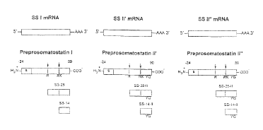
Figure 1 schematically represents the proposed biosynthesis of
somatostatins from multiple somatostatin genes in rainbow trout; arrows denote
putative cleavage sites. Processing details are set forth in the legends to
Fig. 2
and Fig. 3.
Figure 2 shows the nucleotide sequence (SEQ ID N0:8) and the deduced
amino acid sequence (SEQ ID N0:3) of the PPSS-I cDNA obtained from the
endocrine pancreas of rainbow trout. The putative N-terminal signal sequence
(amino acids -100 through -77; SEQ ID N0:7) is overlined. The presumptive
coding region for somatostatin SS-14 peptide (SEQ ID NO:1, wherein the N-
terminal Ala is denoted by a double arrow) is preceded by a putative Arg-Lys
dibasic cleavage site. An N-terminally extended 26-amino acid prosomatostatin
product (amino acids -12 through +14; SEQ ID N0:4; wherein the N-terminal
Ala is denoted by a single arrow) includes the SS-14 sequence (SEQ ID NO:l)
and is preceded by an Arg monobasic cleavage site. The 26 amino acid
proprotein (SEQ ID N0:4) contains a 12 amino acid N-terminal extension
sequence (amino acids -12 through -1; SEQ ID N0:6) linked to SS-14 (SEQ ID
NO:1). The 114 amino acid preproprotein (SEQ ID NO:3) contains an 88 amino
acid N-terminal extension sequence (amino acids -100 through -13; SEQ ID
NO:S) linked to the 26 amino acid proprotein sequence (SEQ ID NO: 4). The
CA 02325169 2000-12-O1
6
translation stop codon stop sequence is denoted by asterisks, and the putative
polyadenylation signal is underlined.
Figure 3 shows the nucleotide sequences (SEQ ID N0:14 and SEQ ID
N0:20) and the deduced amino acid sequence (SEQ ID N0:9 and SEQ ID
NO:15) of the PPSS-If cDNA and PPSS-II" cDNA, respectively, obtained from
the endocrine pancreas of rainbow trout. Nucleotides and amino acids are
numbered in right margin, and gaps are denoted with asterisks for maximum
alignment. The amino acids are shown for PPSS-II". For PPSS-If, only the
amino acids that differ from those in PPSS-II" are shown, but it should be
understood that where no amino acid is shown, it is the same as the amino acid
at
the analogous site on PPSS-II". The putative N-terminal signal sequence (PPSS-
If: amino acids -101 through -77, SEQ ID N0:13; PPSS-II": amino acids -97
through -73, SEQ ID N0:19) is underlined. The presumptive coding region for
SS-14 (SEQ ID N0:2, wherein the N-terminal Ala is denoted by a single arrow)
is preceded by a putative Arg-Lys dibasic cleavage site denoted by open
circles
and contains [Tyr',Gly'°] substitutions characteristic of the PPSS-II
family. An
N-terminally extended prosomatostatin product (PPSS-If : 28 amino acids, amino
acids -14 through +14, SEQ ID NO:10; PPSS-II": 25 amino acids, amino acids -
11 through +14, SEQ ID N0:16; wherein the N-terminal Ser is denoted by a
double arrow) includes the [Tyr',Gly'°]SS-14 sequence (SEQ ID N0:2) and
is
preceded by an Arg monobasic cleavage site. The 28 amino acid PPSS-II'
proprotein (SEQ ID NO:10) contains a 14 amino acid N-terminal extension
sequence (amino acids -14 through -1; SEQ ID N0:12) linked to
[Tyr',Gly'°]SS-
14 (SEQ ID N0:2). The 25 amino acid PPSS-II" proprotein (SEQ ID N0:16)
contains an 11 amino acid N-terminal extension sequence (amino acids -11
through -1; SEQ ID N0:18) linked to [Tyr',Gly'°]SS-14 (SEQ ID N0:2).
The
115 amino acid PPSS-If preproprotein (SEQ ID N0:9) contains an 87 amino
acid N-terminal extension sequence (amino acids -101 through -14; SEQ ID
NO:11) linked to the 28 amino acid PPSS-If proprotein sequence (SEQ ID NO:
10). The 111 amino acid PPSS-II" preproprotein (SEQ ID NO:15) contains an
86 amino acid N-terminal extension sequence (amino acids -97 through -11;
CA 02325169 2000-12-O1
7
SEQ ID N0:17) linked to the 25 amino acid PPSS-II" proprotein sequence (SEQ
ID NO: 16).
Figure 4 diagrams the sequence strategy used for 5' and 3' rapid
amplification of cDNA ends (RACE).
Figure 5 compares the amino acid (lower left) and cDNA nucleotide
(upper right) sequence identities vertebrate somatostatins. AF I denotes
anglerfish (Hobart et al., Nature, 288, 137-141 (1980)), CF I denotes catfish
I
(Minth et al., J. Biol. Chem., 257, 10372-10377 (1982)), H denotes human (Shen
et al., Proc. Natl. Acad. Sci. USA, 79, 4575-4579 (1982)), B denotes bovine
(Su
et al., Mol. Endocrinol., 2, 209-216 (1988)), M denotes monkey (Travis and
SutclifFe, Proc. Natl. Acad. Sci. USA, 85, 1696-1700(1988)), R denotes rat
(Goodman et al., J. Biol. Chem., 258, 570-573 (1983)), C denotes chicken
(Nata,
GenBank direct submission, Accession No. X60191 ( 1991 )), FR I denotes frog
(Tostivint et al., Proc. Natl. Acad. Sci. USA, 93, 12605-12610 (1996)), TR II'
denotes rainbow trout-II' (Moore et al., Gen. Comp. Endocrinol., 98, 253-261
(1995)), TR II" denotes rainbow trout-II", and TR I denotes rainbow trout-I.
Figure 6 aligns the deduced rainbow trout PPSS-I C-terminal region
amino acid sequence to other PPSS-I C-terminal region amino acid sequences
from other vertebrates. aSequences arranged for maximum alignment; identity is
greatest if it is assumed there has been a 2-amino acid deletion (designated
by
asterisks) from rainbow trout and bowfin (Wang et al., Amia calva. Regal.
Peptides, 47, 33-40 (1993). bPutative peptide deduced from cDNA. Peptide
sequence deduced from cDNA and confirmed by processing analysis for
anglerfish I (Hobart et al, Nature, 288, 137-141 (1980); Goodman et al., Proc.
Natl. Acad. Sci. USA, 77, 5869-5873 (1980); Andrews and Dixon,
Biochemistry, 26, 4853-4861 (1987)), catfish I (Andrews and Dixon, J. Biol.
Chem., 256, 8267-8270 (1981); Minth et al., J. Biol. Chem., 257, 10372-10377
(1982)), and frog (Vaudry et al., Biochem. BioPhys. Res. Commun., 188, 477-
482 (1992); Tostivint et al., Proc. Natl. Acad. Sci. USA, 93, 12605-12610
(1996)). dPeptide sequence derived directly from analysis of isolates of islet
extracts obtained from hagfish (Conlon et al., Endocrinolo~.v, 122, 1855-1859
(1988)), lamprey (Andrews et al., J. Biol. Chem., 258, 5570-5573 (1988)),
CA 02325169 2000-12-O1
torpedo (Conlon et al., Gen. Comp. Endocrinol., 60, 406-413 (1985)), ratfish
(Conlon et al., Gen. Comp. Endocrinol., 80, 314-320 (1990)), sturgeon (Nishi
et
al., Gen. Comp. Endocrinol., 99, 6-12 (1995)), eel (Conlon et al.,
Endocrinolo~v,
122, 1855-1859 (1988)), flounder and sculpin (Conlon et al., Eur. J. Biochem.,
168, 647-652 (1987a)), salmon (Plisetskaya et al., Gen. Comp. Endocrinol., 63,
242-263 (1986)), salamander (Cavanaugh et al., Gen. Comp. Endocrinol., 101,
12-20 (1996)), pigeon (Spiess et al., EndocrinoloQV, 76, 33-40 (1979)),
alligator
(Wang and Conlon, Pe-ptides, 14, 573-579 (1993)), and ovine (28-amino acid
form shown for purposes of comparison; Pradayrol et al., FEBS Lett., 109, 55-
58
(1980)).
Figure 7 aligns the deduced rainbow trout PPSS-I, PPSS-II' and PPSS-II"
amino acid sequences with PPSSs of other vertebrates; sequence identity was
maximized by inserting gaps (denoted by dashed lines j; conserved amino acids
are shaded. H denotes human (Shen et al., Proc. Natl. Acad. Sci. USA, 79, 4575-
4579 (1982)); M denotes monkey (Travis and Sutcliffe, Proc. Natl. Acad. Sci.
USA, 85, 1696-1700 (1988)); B denotes bovine (Su et al., Mol. Endocrinol., 2,
209-216 (1988)); R denotes rat (Goodman et al., J. Biol. Chem., 258, 570-573
(1983)); C denotes chicken (Nata, GenBank direct submission, Accession No.
X60191 (1991)); FR I and FR II denote frog I and frog II (Tostivint et al.,
Proc.
Natl. Acad. Sci. USA, 93, 12605-12610 (1996)); AF I denotes anglerfish I
(Hobart et al., Nature, 288, 137-141 (1980)); AF II denotes anglerfish II
(Goodman et al., Proc. Natl. Acad. Sci. USA, 77, 5869-5873 (1980); Goodman
et al., Proc. Natl. Acad. Sci. USA, 79, 1682 (1982); Hobart et al., Nature,
288,
137-141 (1980)); CF I denotes catfish I (Eilertson and Sheridan, Gen. Coma.
Endocrinol., 92, 62-70 (1993)); CF II denotes catfish II (Fujita et al., Pe-
ptides, 2,
123-131 ( 1981 )); GF I-III denotes goldfish I-III (Lin et al., Endocrinolo~v,
140,
2089-2099 ( 1999)); TRI denotes trout I; TRIP denotes trout II' (Moore et al.,
Gen. Comp. Endocrinol., 98, 253-261 (1995)); and TRII" denotes trout II".
Figure 8 graphically shows the ability of synthetic salmonid SS-25 (filled
inverted triangles) mammalian SS-14 (filled circles) and mammalian SS-28
(open circles) to inhibit the binding of I'z5-[Tyrl]-SS-14 to microsomes
prepared
from COS-7 cells transiently expressing the human SS type 1 receptor.
CA 02325169 2000-12-O1
9
Detailed Description of the Preferred Embodiments
In one aspect, the present invention provides a novel somatostatin
polypeptide or biologically active analog, subunit or derivative thereof. A
polynucleotide that encodes a novel somatostatin polypeptide or biologically
active analog, subunit or derivative thereof is also provided. As used herein,
the
term "polypeptide" refers to a polymer of two or more amino acids joined
together by peptide bonds. The terms peptide, oligopeptide, and protein are
all
included within the definition of polypeptide. In particular, the term
"somatostatin polypeptide" includes somatostatin precursor polypeptides (e.g.,
somatostatin prepropeptides, which are typically over 100 amino acids in
length),
as well as shorter polypeptides (e.g., somatostatin propeptides, typically
about
25-28 amino acid in length, and somatostatin peptides, typically about 14
amino
acids in length). A "biologically active" somatostatin analog or subunit is a
polypeptide that is able to bind to a somatostatin receptor molecule,
preferably a
human somatostatin receptor molecule. A method for evaluating binding activity
is described, for example, in Example V herein.
A biologically active "analog" of a somatostatin polypeptide includes a
somatostatin polypeptide that has been modified by the addition, substitution,
or
deletion of one or more amino acids, or that has been chemically or
enzymatically modified, e.g., by attachment of a reporter group, by an N-
terminal, C-terminal or other functional group modification or derivatization,
or
by cyclization, as long as the analog retains biological activity. Amino acid
substitutions are preferably conserved amino acid substitutions, such as
substitutions between negatively charged residues (glutamate and aspartate),
between positively charge residues (lysine, histidine and arginine), among
nonpolar residues (valine, alanine, leucine, isoleucine and phenylalanine), or
between polar residues (serine and threonine).
A biologically active "subunit" of a somatostatin polypeptide includes a
somatostatin polypeptide that has been truncated at either the N-terminus, or
the
C-terminus, or both, by one or more amino acids, as long as the truncated
CA 02325169 2000-12-O1
polypeptide retains bioactivity and contains at least 7 amino acids, more
preferably at least 10 amino acids, most preferably at least 12 amino acids.
With respect to a somatostatin polypeptide comprising the 14 amino acid
sequence SS-14 (SEQ ID NO:1) or [Tyr', Glyl°]-SS-14 (SEQ ID N0:2), a
5 preferred biologically active analog or subunit of such somatostatin
polypeptide
does not contain any amino acid substitutions, deletions or additions at
positions
6-11 of that 14 amino acid sequence, as those positions are very important for
binding to a somatostatin receptor, but may contain substitutions, deletions,
or
additions at other sites. Preferred substitutions include proline at position
+2 and
10 serine at position +5. Examples of biologically active analogs of SS-14
from
PPSS-I have been described in Reisine et al. (Endocr. Rev. 16:427-442 (1995))
and include amino acid-deleted or amino acid-substituted compounds, dicarba
analogs, bicylclic octapeptide analogs (e.g., SMS201-995A, sometimes known as
octreotide or by the tradename SANDOSTATIN) and cyclic hexapeptides (e.g.,
MK687).
A preferred polypeptide and/or polynucleotide of the invention is one that
is derived from rainbow trout (Oncorhynchus mykiss). The use of the term
"derived from" is not intended to limit the invention to a polypeptide or
polynucleotide that is physically isolated from rainbow trout, but is meant to
include biologically active somatostatin biomolecules having all or a portion
of a
trout somatostatin amino acid or nucleotide sequence, whether isolated from
trout or synthesized chemically, enzymatically, or using genetic engineering.
Trout preprosomatostatin-I (PPSS-I) is described herein in Example I and
is shown in Fig. 1 and Fig. 2. PPSS-I characterized by a 745 base pair cDNA
(SEQ ID N0:8) that encodes a precursor protein of about 114 amino acids (SEQ
ID N0:3) that appears capable of being processed into a 26 amino acid
polypeptide (SEQ ID N0:4), and further into a 14 amino acid peptide (SS-
14)(SEQ ID NO:1). Because this tetradecapeptide (SEQ ID NO:1) has the
"mammalian"-type sequence, these somatostatins are members of the SS-I
family.
Trout preprosomatostatin-If (PPSS-If) was reported by Moore et al.
(Gen. Comp. Endocrinol. 98:253-261 (1995)), and is shown in Fig. 1 and Fig. 3.
CA 02325169 2000-12-O1
11
PPSS-II' is characterized by a 624 base pair cDNA (SEQ ID N0:14) that encodes
a precursor protein of about 115 amino acids (SEQ ID N0:9) that appears
capable of being processed into a 28 amino acid polypeptide (SEQ ID NO:10),
and further into a 14 amino acid peptide (SEQ ID N0:2). Because this
tetradecapeptide has the modified [Tyr', Gly'°] sequence, these
somatostatins are
members of the SS-II family.
Trout preprosomatostatin-II" (PPSS-II") is described herein in Example
III and is shown in Fig. 1 and Fig. 3. PPSS-II" is characterized by a 600 base
pair cDNA (SEQ ID N0:20) that encodes a precursor protein of about 111 amino
acids (SEQ ID NO:15) that appears capable of being processed into a 25 amino
acid polypeptide (SEQ ID N0:16), and further into a 14 amino acid peptide
(SEQ ID N0:9). Because this tetradecapeptide has the modified [Tyr',
Gly'°]
sequence, these somatostatins are members of the SS-lI family.
Preferred somatostatin polypeptides of the invention include the different
"isoforms" of PPSS-I and PPSS-II" derived from trout, as well as "isoform
fragments" that result from actual or putative N-terminal processing of such
isoforms. Preferred PPSS-I polypeptides thus include preprosomatostatin I (114
amino acid isoform) (SEQ ID N0:3); the N-terminal pre-sequence of PPSS-I (88
amino acid isoform fragment) (SEQ ID NO:S); prosomatostatin I (26 amino acid
isoform) (SEQ ID N0:4); and the N-terminal pro-sequence of PPSS-I (12 amino
acid isoform fragment) (SEQ ID N0:6) Preferred PPSS-II" polypeptides thus
include preprosomatostatin II" (111 amino acid isoform) (SEQ ID NO:15); the
N-terminal pre-sequence of PPSS-II" (86 amino acid isoform fragment) (SEQ ID
N0:17); prosomatostatin II" (25 amino acid isoform) (SEQ ID N0:16); and the
N-terminal pro-sequence of PPSS-II" (11 amino acid isoform fragment) (SEQ ID
N0:18)
Also preferred are polypeptides that include all or a portion of one or
more PPSS-I and/or PPSS-II" amino acid sequences derived from trout. If only a
portion of a PPSS-I and/or PPSS-II" sequence is included in the polypeptide,
the
portion so included contains at least 7, and preferably at least 10, more
preferably at least 12, contiguous amino acids of a PPSS-I and/or PPSS-II"
sequence. Furthermore, if the included portion of the PPSS-I and/or PPSS-II"
CA 02325169 2000-12-O1
sequence contains all or a portion of the C-terminal 14-mer SEQ ID NO:1, the
C-terminal 14-mer SEQ ID N0:2, or the C-terminal 25-mer SEQ ID N0:16, then
said included portion also includes at least an additional 7, and preferably
an
additional 10, more preferably at least 12 contiguous amino acids of a PPSS-I
and/or PPSS-II" sequence. The additional contiguous amino acids need not be,
but may be, contiguous to the included portion of the C-terminal 14-mer. An
example of a polypeptide that includes all or a portion of a PPSS-I and/or
PPSS-
II" sequence is a chimeric polypeptide that contains the prosomatostatin
sequence of human somatostatin (SEQ ID N0:21) and the presequence of PPSS-
I derived from trout (SEQ ID NO:S).
Preferred biologically active analogs of PPSS-I and/or PPSS-II"
sequences derived from trout include (1) analogs of PPSS-I and/or PPSS-II"
isoform sequences that are at least 85% identical, more preferably at least
90%
identical, most preferably at least 95% identical to PPSS-I and PPSS-II"
isoform
sequences SEQ ID NOs:3, 4, 15 or 16; and (2) analogs of PPSS-I and/or PPSS-
II" isoform fragment sequences that are at least 90% identical, more
preferably at
least 95% identical to PPSS-I and PPSS-II" isoform fragment sequences SEQ ID
NOs:3, 4, 15 or 16. Such analogs contain one or more amino acid deletions,
insertions, and/or substitutions relative to the reference PPSS-I and/or PPSS-
II"
sequence, and may further include chemical and/or enzymatic modifications
and/or derivatizations as described above.
Percent identity is determined by aligning the residues of the two amino
acid or nucleotide sequences to optimize the number of identical amino acids
or
nucleotides along the lengths of their sequences; gaps in either or both
sequences
are permitted in making the alignment in order to optimize the number of
identical amino acids or nucleotides, although the amino acids or nucleotides
in
each sequence must nonetheless remain in their proper order. Preferably, two
amino acid sequences are compared using the Blastp program, version 2Ø9, of
the BLAST 2 search algorithm, as described by Tatiana, et al. (FEMS Microbiol.
Lett., 174, 247-250 (1999)), and available at
http://www.ncbi.nlm.nih.gov/blast.html. Preferably, the default values for all
BLAST 2 search parameters are used, including matrix = BLOSUM62; open gap
12
CA 02325169 2000-12-O1
penalty = 11, extension gap penalty = 1, gap x dropoff = 50, expect = 10,
wordsize = 3, and filter on. Likewise, two nucleotide sequences are compared
using the Blastn program, version 2Ø11, of the BLAST 2 search algorithm,
also
as described by Tatiana, et al. (FEMS Microbiol Lett, 174, 247-250 (1999)),
and
available at http://www.ncbi.nlm.nih.gov/blast.html. Preferably, the default
values for all BLAST 2 search parameters are used, including reward for match
=
1, penalty for mismatch = -2, open gap penalty = 5, extension gap penalty = 2,
gap x dropoff = 50, expect = 10, wordsize = 11, and filter on.
It should be understood that a polynucleotide that encodes a novel
somatostatin polypeptide derived from Oncorhynchus mykiss according to the
invention is not limited to a naturally occurring polynucleotide derived from
Oncorhynchus mykiss, such as a polynucleotide that includes all or a portion
of a
PPSS-I and/or PPSS-II" genomic or cDNA nucleotide sequence, but also
includes the class of polynucleotides that encode such polypeptides as a
result of
the degeneracy of the genetic code. For example, the naturally occurring
nucleotide sequence SEQ ID N0:8 is but one member of the class of nucleotide
sequences that encodes a polypeptide having amino acid SEQ ID N0:3. This
class of nucleotide sequences that encode a selected polypeptide sequence is
large but finite, and the nucleotide sequence of each member of the class can
be
readily determined by one skilled in the art by reference to the standard
genetic
code, wherein different nucleotide triplets are known to encode the same amino
acid. Likewise, a polynucleotide of the invention that encodes a biologically
active analog or subunit of a somatostatin polypeptide includes the multiple
members of the class of polynucleotides that encode the selected polypeptide
sequence.
Moreover, a polynucleotide that "encodes" a polypeptide of the invention
optionally includes both coding and noncoding regions, and it should therefore
be understood that, unless expressly stated to the contrary, a polynucleotide
that
"encodes" a polypeptide is not structurally limited to nucleotide sequences
that
encode a polypeptide but can include other nucleotide sequences outside (i.e.,
5'
or 3' to) the coding region.
13
CA 02325169 2000-12-O1
The polynucleotides of the invention can be DNA, RNA, or a
combination thereof, and can include any combination of naturally occurring,
chemically modified or enzymatically modified nucleotides. As noted above, the
polynucleotide can be equivalent to the polynucleotide fragment encoding a
somatostatin polypeptide, or it can include said polynucleotide fragment in
addition to one or more additional nucleotides. For example, the
polynucleotide
of the invention can be a vector, such as an expression or cloning vector. A
vector useful in the present invention can be circular or linear, single-
stranded or
double-stranded, and can include DNA, RNA, or any modification or
combination thereof. The vector can be a plasmid, a cosmid, or a viral vector,
such as baculovirus. Preferably, the polynucleotide of the invention takes the
form of an expression vector that is capable of expression in an organism or
in a
cell of an organism, in culture or in vivo. An organism or cell in which the
coding sequence of the vector can be expressed can be a vertebrate, and
preferably a veterinary mammal or a human. Preferably, the vector is
expressible
in bacterial expression system, such as E. coli, yeast, mammalian cell culture
or
insect cells.
It should be understood that the polynucleotide of the invention can be
single-stranded or double-stranded, and further that a single-stranded
polynucleotide of the invention includes a polynucleotide fragment having a
nucleotide sequence that is complementary to the nucleotide sequence of the
single-stranded polynucleotide. As used herein, the term "complementary"
refers
to the ability of two single-stranded polynucleotide fragments to base pair
with
each other, in which an adenine on one polynucleotide fragment will base pair
with a thymidine (or uracil, in the case of RNA) on the other, and a cytidine
on
one fragment will base pair with a guanine on the other. Two polynucleotide
fragments are complementary to each other when a nucleotide sequence in one
polynucleotide fragment can base pair with a nucleotide sequence in a second
polynucleotide fragment. For instance, S'-ATGC and 5'-GCAT are fully
complementary, as are 5'-GCTA and 5'-TAGC.
Further, the single-stranded polynucleotide of the invention also includes
a polynucleotide fragment having a nucleotide sequence that is substantially
14
CA 02325169 2000-12-O1
complementary to (a) a nucleotide sequence that encodes a novel somatostatin
polypeptide according to the invention, or (b) the complement of such
nucleotide
sequence. "Substantially complementary" polynucleotide fragments can include
at least one base pair mismatch, such that at least one nucleotide present on
a
second polynucleotide fragment, however the two polynucleotide fragments will
still have the capacity to hybridize. For instance, the middle nucleotide of
each
of the two DNA fragments 5'-AGCAAATAT and 5'-ATATATGCT will not base
pair, but these two polynucleotide fragments are nonetheless substantially
complementary as defined herein. Two polynucleotide fragments are
substantially complementary if they hybridize under hybridization conditions
exemplified by 2X SSC (SSC: 150mM NaCI, 15 mM trisodium citrate, pH 7.6)
at 55°C. Substantially complementary polynucleotide fragments for
purposes of
the present invention preferably share at least one region of at least 20
nucleotides in length which shared region has at least 60% nucleotide
identity,
preferably at least 80% nucleotide identity, more preferably at least 90%
nucleotide identity and most preferably at least 95% nucleotide identity.
Particularly preferred substantially complementary polynucleotide fragments
share a plurality of such regions. Locations and levels of nucleotide sequence
identity between two nucleotide sequences can be readily determined using
CLUSTALW multiple sequence alignment software (J. Thompson et al., Nucleic
Acids Res., 22:4673-4680 (1994)), available at http://www.ebi.ac.uk/clustalw/.
In another aspect, the invention provides methods of making the novel
somatostatin polypeptides of the invention, as well as methods of making the
multiple polynucleotides that encode them. The methods include biological,
enzymatic, and chemical methods, as well as combinations thereof, and are well-
known in the art. For example, a somatostatin polypeptide can be expressed in
a
host cell from using standard recombinant DNA technologies; it can be
enzymatically synthesized in vitro using a cell-free RNA based system; or it
can
be synthesized using chemical technologies such as solid phase peptide
synthesis, as is well-known in the art.
In yet another aspect, the invention provide a method for identifying
novel polypeptides that have somatostatin activity. This method is based on
CA 02325169 2000-12-O1
comparative analysis of (a) a somatostatin amino acid sequence derived from
trout PPSS-I or PPSS-II", preferably an amino acid sequence of at least one
isoform of PPSS-I (SEQ ID NO: 3, 4, or 1 ) or PPSS-II" (SEQ ID NO:15, 16 or 2)
or isoform fragment of PPSS-I (SEQ ID NO:S or 6) or PPSS-II" (SEQ ID N0:17
or 18) and (b) the amino acid sequence of an analogous region of a
somatostatin
polypeptide of another organism, preferably a mammal, more preferably a
human. The reference somatostatin polypeptide can be either the trout
polypeptide or the polypeptide from the other organism. The sequences are
aligned, and sites having different amino acids are identified. Then, a novel
candidate somatostatin sequence is postulated that is represented by the
reference
polypeptide modified to contain one or more amino acid substitutions,
modifications, a deletions as suggested by the other polypeptide to which it
is
compared. The candidate somatostatin polypeptide is synthesized, assayed for
somatostatin activity (i.e., binding to a somatostatin receptor of interest),
and,
optionally, further assayed for any desired therapeutic effect.
Using this method, novel somatostatin polypeptides can be identified that
function as either agonists or antagonist of the reference polypeptide or
other
naturally occurnng somatostatin, or that have altered binding specificity or
selectivity when compared to the reference polypeptide or other naturally
occurring somatostatin. For example, binding of longer somatostatin isoforms
to
receptor molecules is likely affected by the amino acid sequence of the N-
terminal region (e.g., the region upstream from the C-terminal 14 amino acid
peptide). All or a portion of an N-terminal trout somatostatin amino acid
sequence according to the invention can, for example, be fused to the C-
terminal
portion of another somatostatin or somatostatin analog in order to target the
analog or affect binding of the analog or modulate the binding activity of the
analog. For example, a novel somatostatin that contains a trout PPSS-II"
presequence (SEQ ID N0:17) joined to the mammalian SS-28 prosomatostatin
sequence (SEQ ID N0:21 ) can be postulated and evaluated for somatostatin
activity according to the method. Likewise, a small scale substitution of the
alternative SS-14 residues Tyr' and Gly'° into the C-terminus of trout
PPSS-I
16
CA 02325169 2000-12-O1
which contains the Phe' and Thr'° (SEQ ID N0:2) yields a novel
somatostatin
polypeptide that can also be evaluated according to the method of the
invention.
Advantageously, the method of the invention can be used to identify
novel somatostatin polypeptides that will bind to the human somatostatin
receptor and thus be useful for research, therapeutics or diagnostics. Such
uses
include clinical uses in both medical and veterinary applications. Thus, a
somatostatin polypeptide of the invention, or a bioactive analog or subunit
thereof, as well as those identified via the method of the invention, can be
administered to an organism to function therapeutically as a somatostatin
agonist
or antagonist. The potential pharmacological uses of such somatostatins are
numerous. For example, hypersecretion from endocrine tumors in the pituitary
(e.g., acromegaly, TSH-secreting) or gastroenteropancreatic tissues (e.g.,
gastrinoma, VIPoma, glucagonoma, carcinoid syndrome) can be treated with
somatostatin. In addition to the inhibition of hormone secretion, somatostatin
analogs also may cause tumor shrinkage via their effects on cell proliferation
and
apoptosis. Another potential use of novel somatostatins or analogs is a
adjuncts
in the treatment of diabetes mellitus (via inhibition of growth hormone and
glucagon). In addition, dysfunctional somatostatin secretion has been
associated
with AIDS and various neurological disorders (e.g., epilepsy, Alzheimer and
Huntington diseases) and a somatotstatin antagonist might be effective in the
treatment of such conditions. Nucleic acids encoding the somatostatin
polypeptides of the invention, including bioactive analogs and subunits
thereof,
are potentially useful in gene therapy.
The invention also envisions fusing a plurality of N-terminal amino acids
of a PPSS-I or PPSS-II" isoform or isoform fragment to peptides other than
somatostatin so as to target them to somatostatin receptor molecules. The C-
terminal peptide of the resulting fusion polypeptide preferably contains a
bioactive peptide or other moiety. Different cell types in an organism are
known
to express different somatostatin receptors, making tissue specific targeting
of
bioactive moieties possible. For example, the fusion peptide could be targeted
to
neoplasms and their metastases, inhibiting the release of their secretory
products
and, possibly, providing access to the interior of the cell via
internalization of the
17
CA 02325169 2000-12-O1
somatostatin receptor-ligand complex. The plurality of N-terminal amino acids
of a PPSS-I or PPSS-II" isoform or isoform fragment preferably includes at
least
7 contiguous amino acids, more preferably at least 10 contiguous amino acids,
and most preferably at least 12 contiguous amino acids. The fusion protein is
preferably made using recombinant DNA technology, but can be synthesized
enzymatically or chemically as well. The invention thus includes a method for
making the fusion peptide, as well as the resulting fusion peptide.
EXAMPLES
The present invention is illustrated by the following examples. It is to be
understood that the particular examples, materials, amounts, and procedures
are
to be interpreted broadly in accordance with the scope and spirit of the
invention
as set forth herein.
Ezam~le I Isolation, Cloning and Sequencing of PPSS-I from Rainbow
Trout
Experimental Animals
Juvenile rainbow trout (Oncorhynchus mykiss), approximately 12 months
of age, were obtained from the Garrison National Hatchery near Riverdale, ND.
The fish were maintained in dechlorinated, well-aerated municipal water at a
temperature of 14°C and were placed on a 12L:12D photoperiod. The fish
were
fed Glenco Mills (Glenco, MN) trout chow ad libitum twice daily and fasted for
24 hours before experimentation. The animals were anesthetized with
0.01 %(w/v) tricaine methanesulfonate (MS-222) and sacrificed by a sharp blow
to the head. Principal islets (Brockmann Bodies) as well as other tissues
(brain,
stomach, intestine, pyloric cecum, esophagus, kidney and liver) were removed
from animals of both sexes. Tissues (ca. 50-100 mg) were placed in 2-ml
microfuge tubes, frozen immediately on dry ice, then stored at -90°C
until
subjected to RNA extraction (usually within two weeks).
18
CA 02325169 2000-12-O1
RNA Extraction
Total RNA was prepared by a modification of the RNAzoI method
(Chomczynski et al., Anal. Biochem., 162(1):156-159 (1987)). Five hundred
microliters of RNAzoI (Cinna/Biotecx Laboratories, Friendswood, TX) was
added to 2-ml microfuge tubes containing frozen tissue (approximately 25 mg),
and the tissue was homogenized. Fifty microliters of chloroform were added to
the tubes, and the mixture was vortexed for 30 seconds and incubated at
4°C for
minutes. Following centrifugation at 1200 g for 15 minutes at 4°C, the
aqueous phase was carefully removed and transferred to a sterile 1.5 ml
10 microfuge tube. An equal volume of isopropanol was added, and the RNA was
precipitated for 2 hours at -20°C. RNA was recovered as a pellet by
centrifugation at 1200 g at 4°C for 20 minutes and resuspended in 100
~.1 of
sterile water. A second precipitation was performed by adding 50 ~1 of 5 M
NaCI and 250 pl of absolute ethanol, followed by an incubation of the mixture
at
15 -20°C overnight. Following the second precipitation, RNA was again
recovered
by centrifugation for 20 minutes at 1200 g at 4°C. The RNA pellet was
vacuum
dried for 2-5 minutes to remove any residual ethanol and resuspended in 75 ~1
of
sterile water. Total RNA was quantified by UV Az6o spectrophotometry and
diluted to 15 ~.g/~1. RNA samples were stored at -90°C until used.
Oligonucleotide Primers and cDNA Probes
National Biosciences (Plymouth, MN) custom synthesized the gene-
specific oligonucleotides used in reverse transcription and polymerase chain
reaction (PCR). The GSP-1 primer was designed from degenerate conserved
regions of the cDNAs encoding human (Shen et al., Proc. Natl. Acad. Sci. USA,
79, 4575-4579 (1982), rat (Goodman et al., J. Biol. Chem., 258, 570-573
(1983),
anglerfish I (Goodman et al., Proc. Natl. Acad. Sci. USA, 77, 5869-5873
(1980);
Hobart et al., Nature, 288, 137-141, (1980), and catfish I (Minth et al., J.
Biol.
Chem., 257, 10372-10377 (1982) somatostatins. Additional primers used for
PCR were obtained from Gibco/BRL (Gaithersburg, MD).
The full-length SS-I cDNA probe was made by reverse transcription PCR
using primers designed from the SS-I cDNA sequence and purified by
19
CA 02325169 2000-12-O1
ultrafiltration using a 100,000 M.W. cutoff filter (Millipore, Bedford, MA)
followed by ethanol precipitation (1/4 volume SM NaCI, 2 volumes absolute
ethanol) at -20°C overnight. After the cDNA probe was recovered by
centrifugation (12,000 x g, for 20 minutes at 4°C), it was resuspended
in 100 ~.1
sterile water and quantitated by UV A26o spectrophotometry. The full-length SS-
I cDNA probe was radiolabeled with [a32P)-CTP by nick translation (Nick
Translation System; Promega) according to the manufacturer's protocol. The
probe was purified over Elutip-D columns (Schleicher and Schuell) according to
the manufacturer's protocol.
Isolation and Sequence Analysis of Preprosomatostatin cDNAs
A two-phase rapid amplification of cDNA ends (RACE) PCR-based
approach (Fig. 4) was used for the isolation and characterization of selected
cDNA sequences as described previously (Moore et al., Gen. Comp. Endocrinol.,
98, 253-261 (1995). In phase I, endogenous poly-A RhIA was reverse
transcribed from 15 ~g of trout pancreatic total RNA with Superscript II
reverse
transcriptase (GibcoBRL, Gaithersburg, MD) and a 37 nucleotide antisense
adapter primer 5'-GGCCACGCGTCGACTAGTAC(T)17-3' (SEQ ID N0:22)
(GibcoBRL). Five microliters of the reverse transcription reaction were used
as
template for 3'-RACE PCR with a 21-base somatostatin gene-specific primer 5'-
AAGAACTTCTTCTGGAAGAC-3' (GSP-1; SEQ ID N0:25) and the universal
amplification primer 5'-CUACUACUACUAGGCCACGCGTCGACTAGT AC-
3' (UAP; SEQ ID N0:23). After an initial denaturation cycle of 94°C for
5
minutes, 35 PCR cycles were performed, each consisting of 1-minute annealing
(42°C), 1-minute extension (72°C), and 1-minute denaturation
(94°C). In the
last cycle, the extension time was increased to 10 minutes to ensure complete
extension. The resulting PCR product (350 bp) was identified by
electrophoresis
on an agarose gel containing 1 % (w/v) agarose (Gibco/BRL) and 1 % (w/v)
NuSeive GTG agarose (FMC Bioproducts, Rockland, ME) in 1X TBE Buffer,
followed by ethidium bromide staining and UV transillumination. Amplified
fragments were directly cloned into the TA cloning vector PCR 2000
(Invitrogen, San Diego, CA). Positive colonies were identified by agarose gel
CA 02325169 2000-12-O1
electrophoresis of restriction enzyme digests (EcoRI; Promega, Madison, WI) of
purified plasmid preparations (Del Sal et al., BioTech., 7, 514-519 (1989)).
One
to 2 ~.g of plasmid were denatured and sequenced by the dideoxy chain-
termination method (Sequenase Kit; U.S. Biochemicals Corp., Cleveland, OH)
according to the manufacturer's protocol. All sequences were confirmed by
sequencing multiple colonies from at least three independent PCR reactions and
with two or more different primers in both directions, with dGTP didoexy
nucleotides. Sequencing gels were made with 30% formamide to eliminate the
possibility of G/C compressions.
In phase II (Fig. 4), isolation of the 5' cDNA sequence was accomplished
by 5'-RACE PCR (GibcoBRL). Somatostatin mRNA was exclusively reverse
transcribed from pancreatic total RNA using a 20-base antisense
oligonucleotide
primer complementary to a region of the 3' fragment isolated in phase I 5'-
ATTCATTAACACGATGTAAA-3' (GSP-2; SEQ ID N0:26). The resulting
cDNA was purified twice over Glass Max spin columns (GibcoBRL) to remove
unincorporated dNTPs and primer and "tailed" at the 3' end with dCTP using
terminal deoxynucleotidyl transferase (GibcoBRL). Five microliters of the
tailing reaction were used as template for 5'-RACE PCR with GSP-2 (SEQ ID
N0:26) and anchor primer 5'-
CUACUACUACUAGGCCACGCGTCGACTAGTACGGGIIGGGIIGGGIIG-3'
(SEQ ID N0:24) (Gibco/BRL). Thirty-five PCR cycles were performed as in 3'-
RACE PCR, except Taq polymerase (Perkin-Elmer, Norwalk, CT) was pipetted
beneath the layer of mineral oil after the initial 5-minute denaturation cycle
(Mullis, PCR Meth. A~pl., l, 1-4 (1991). The amplified product (452 bp) was
identified by agarose gel electrophoresis, cloned, and sequenced.
Data analysis
Nucleotide and deduced amino acid sequences were aligned and analyzed
with the OMIGA 1.0 DNA/protein analysis program (Oxford Molecular Group,
Campbell, CA).
21
CA 02325169 2000-12-O1
Characterization of cDNA Clones
Sequence analysis of the 350-by 3' RACE PCR product revealed a region
of 33 nucleotides that is 87.8% identical to the last 33 nucleotides of the
human
somatostatin coding region (Shen et al., Proc. Natl. Acad. Sci. USA, 79, 4575-
4579 (1982); the rest of the 350-by fragment consisted of the 3'-untranslated
region, including a polyadenylated tail at the most 3' end. The presence of a
portion of the somatostatin coding region in the amplified product suggested
the
successful isolation of a pancreatic preprosomatostatin gene 1 fragment.
Sequence analysis of the 452-by 5' RACE PCR product revealed the complete
somatostatin coding region and the full 5'-untranslated region. Overlapping
sequence of the two fragments identified a 745-by cDNA containing the
complete 5'-untranslated region, a single initiation site 118 bases from the
most
5' end, and a single putative polyadenylation site 17 bases from the most 3'
end
that was terminated with a polyadenylated tail. The existence of only one PPSS-
I mRNA was indicated after exhaustive screening; some 15-10 colonies from
each of three independent PCR reactions were sequenced.
Analysis of Deduced Protein
Fig. 2 shows a nucleotide sequence contained an open reading frame of
342 bases that encodes for a preprosomatostatin molecule 114 amino acids in
length (SEQ ID N0:3). The predicted preprosomatostatin molecule possesses a
putative signal sequence of 24 amino acids (SEQ ID NO: 7, overlined in Fig.
2).
The deduced protein contains a number of putative processing sites,
potentially
yielding a 26-amino acid peptide (SEQ ID N0:4) that could be processed further
to a 14-amino acid peptide (SEQ ID NO:1 ) identical in structure to mammalian
SS-14, confirming that the precursor is a PPSS-I.
A comparison of rainbow trout PPSS-I cDNA with other cDNA
nucleotide sequences (Fig. 5) reveals that rainbow trout PPSS-I (TRI) is most
similar to catfish PPSS-I (CFI) with a percent identity of 77.0%. Notably,
rainbow trout PPSS-I is more similar to the preprosomatostatin I cDNAs than to
the rainbow trout PPSS-II' and PPSS-II" cDNAs.
22
CA 02325169 2000-12-O1
The deduced PPSS-I protein (SEQ ID N0:3) produced in rainbow trout
islet cells contains 114 amino acids, the same number of amino acids as
catfish
PPSS-I but slightly shorter than the 121-amino acids precursor of anglerFsh I.
The deduced amino acid sequence of rainbow trout PPSS-I exhibits 73.5%
identity with catfish I and chicken. Rainbow trout PPSS-I was the least
similar
to anglerfish I, with an identity of 58.1%. Amino acid identities between
rainbow trout PPSS-I and rainbow trout PPSS-If and PPSS-II" were lower;
identities were 49.0% and 48.2%, respectively. It would appear that
evolutionary
selection has acted to conserve the structure of the whole preprosomatostatin
I
molecule (Argos et al., J. Biol. Chem., 258, 8788-8793 (1983)).
While the details concerning the processing of rainbow trout
preprosomatostatin I are not known, a basic pattern emerges from the deduced
amino acid sequence. Analysis of the first 25 amino acids of the molecule
indicates that this segment fulfills all of the criteria for a signal sequence
established by Pugsley, Protein Targeting, Academic Press, New York (1989).
The putative signal sequence of rainbow trout preprosomatostatin I is similar
to
the known signal sequences reported for human and rat preprosomatostatin I
(Conlon et al., Biochem. J., 248, 123-127 (1987); Goodman et al., J. Biol.
Chem., 258, 570-573 (1983)) and to other leading sequences reported for
preprosomatostatin I. Based on the presence of Arg monobasic and Arg-Lys
dibasic cleavage sites (Fig. 2), we propose that rainbow trout prosomatostatin
I
gives rise to peptides 26 amino acids (SEQ ID N0:4) and/or 14 amino acids
(SEQ ID NO:I) in length.
Fig. 6 shows a comparison of rainbow trout SS-I with other somatostatin
gene 1 peptide sequences either isolated from islet tissue or deduced from
cDNAs. There has been strong conservation of the C-terminal regions (up to 19
residues); only the sequence of hagfish differs with two amino acid
substitutions
of glycine for proline and proline for alanine at positions 18 and 20. Perhaps
most interesting is the difference in the number of amino acids. All PPSS-Is
examined possess cleavage sites potentially yielding a 28-amino acid peptide
with SS-14 at its C-terminus. Rainbow trout PPSS-I is unique because it
potentially gives rise to a 26-amino acid peptide containing SS-14 at its C-
23
CA 02325169 2000-12-O1
terminus. This difference was due to a 6-nucleotide deletion in the
somatostatin
coding region. Bowfin, a non-teleost ray-finned fish, has been reported to
possess a modified SS form with 26 amino acids that contains [Sers]-SS-14 at
its
C-terminus (Wang et al., Regul. Peptides, 47, 33-40 (1993)).
Example II Differential Expression of PPSS-I Expression in Tissues of
Rainbow Trout
The distribution of PPSS-I mRNA in various tissues was investigated by
northern blot analysis. Total RNA from rainbow trout brain, pancreas, stomach,
intestine, esophagus, pyloric ceacum, kidney, and liver was isolated by the
RNAzoI method (Chomczynski et al., Anal. Biochem., 162(1):156-159 (1987)).
Ten micrograms of total RNA from each tissue were separated on a
formaldehyde-agarose denaturating gel and transferred onto a 0.45-pm nylon
support (Micron Separations Inc.) by diffusion overnight. The membrane was
baked under a vacuum at 85°C for 2 hours and prehybridized in
hybridization
solution (SX SSPE, SX Denhardt's solution, 0.1% SDS) containing 0.1 mg/ml
denatured calf thymus for 2 hours at 37°C. The prehybridization mixture
was
removed, and the membrane was hybridized at 37°C overnight in
hybridization
solution containing a full-length SSI cDNA radiolabeled (1x10⁶ CPM/ml) probe.
The blot was washed twice with 2X SSPE containing 0.2% (v/v) SDS for 20
minutes at 65°C and once with O.1X SSPE at 65°C for 20 minutes.
Autoradiography was performed by exposing the blot to Fuji RX film for 48
hours at -90°C.
Hybridization with a SS-I full-length cDNA probe revealed a single
transcript approximately 750-by in length in the pancreas, brain, stomach, and
intestine. There was no apparent signal in the other tissues examined. Thus,
Northern analysis revealed that PPSS-I was expressed in the pancreas, stomach,
intestine, and brain of rainbow trout. This result is the first report of PPSS-
I in
extrapancreatic tissues of fish. The presence of the precursor molecule in the
various tissues noted, however, is consistent with the brain-gut distribution
of SS
observed in mammals (Gerich, in: Diabetes Mellitus: Theory and Practice,
24
CA 02325169 2000-12-O1
Medical Examinations Publishing, New York, 225-254 (1983)). The presence of
a single band suggests that the expression of SS gene 1 in rainbow trout
results
in the production of a single mRNA species. The existence of a single
transcript
also suggests the expression of single SS gene 1. This result is noteworthy
since
it has been inferred from cDNA evidence that rainbow trout has two SS genes
giving rise to PPSS-II, SS-f, and SS-II", presumably because of the tetraploid
(Ohno, Evolution by Gene Duplication, Springer-Verlag, Berlin, 1970) nature of
the species. The lack of a second somatostatin gene 1 in extant trout could be
explained by an incomplete duplication associated with the autotetraploidation
event that gave rise to the two somatostatin gene 2s or by the secondary loss
of
the alternate somatostatin gene 1.
This present study contributes to the growing body of evidence that
suggests the existence of multiple somatostatin genes in vertebrates. Based on
cDNA and peptide sequence information, multiple somatostatin genes appear to
exist in lamprey, teleost fish, and frogs (Conlon et al., Re~ul. Peptides, 69,
95-
103 (1997)). Whether these genes arose form a single duplication event prior
to
the emergence of lamprey or from separate duplication events is uncertain
(Conlon et al., Regul. Peptides, 69, 95-103 (1997); Sheridan et al., Advances
in
Comparative EndocrinoloQV, 1, 291-294 (1997)). Regardless, the widespread
distribution of PPSS I and PPSS II in teleosts indicate the emergence of
separate
genes for these precursors prior to the divergence of this group. Future
research
on other taxa will provide additional insight into the evolution of the
somatostatin gene family.
Example III Isolation Cloning and Sequencing of PPSS-II" From
Rainbow Trout
Experimental Animals
Juvenile rainbow trout, Oncorhynchus mykiss, were obtained from the
Garrison National Fish Hatchery near Riverdale, North Dakota. Fish were
maintained at North Dakota State University in well-aerated, dechlorinated
municipal fresh water (14°C) under 12L:12D photoperiod and fed to
satiety
CA 02325169 2000-12-O1
twice daily with Supersweet Feeds (Glenco, MN) trout grower except 24 hours
prior to experiments. In the nutritional state experiment, fish were either
fed as
usual or fasted for two weeks prior to sample collection.
RNA Extraction
Tissues were removed from rainbow trout of both sexes after the animals
had been anesthetized with 0.01% (w/v) 3-aminobenzoic acid ethyl ester (MS-
222, Sigma) buffered with 0.2% (w/v) sodium bicarbonate. Tissue samples
(approximately 25 mg) were placed in 2-ml microfuge tubes and immediately
frozen on dry ice. Total RNA was extracted by a modification of the RNAzoI
method (CinnaBiotecx Laboratories, Friendswood, TX) described previously in
Moore et al., Gen. Comb. Endocrinol., 98, 253-261 (1995). Total RNA was
quantified by LTV A26o spectrophotometry and diluted to 15 ~g/~1. RNA samples
were stored at -90°C until used.
Primers and Probes
Oligonucleotides were either custom synthesized by National Biosciences
(Plymouth, MN) or supplied with GibcolBRL 3'- and 5'-RACE kits.
Oligonucleotides used as probes were 5'-end labeled with [y32P]-ATP
(Amersham) using T4-polynucleotide kinase (Promega) as previously described
in Molecular Cloning: A Laboratory Manual, 2"d Edition, Plainview, New York,
Cold Spring Harbor Laboratory Press (1989). The full-length SS-II cDNA probe
was radiolabeled with [a3zP]-CTP by random priming (Prime-a-Gene; Promega)
according to the manufacturer's protocol. All radiolabeled probes were
purified
over Elutip-D columns (Schleicher and Schuell) according to the manufacturer's
protocol.
Isolation and sequence analysis of preprosomatostatin cDNA
A two-phase rapid amplification of cDNA ends (RACE) PCR-based
approach (Fig. 4) was used for the isolation and characterization of selected
cDNA sequences as described previously in Moore et al., Gen. Comn.
Endocrinol., 98, 253-261 (1995). Briefly, in phase I, endogenous poly-A RNA
26
CA 02325169 2000-12-O1
was reverse transcribed from 15 ~g of trout pancreatic total RNA with
Superscript II reverse transcriptase (GibcoBRL, Gaithersburg, MD) and a 37-
nucleotide antisense adapter primer (GibcoBRL). Five microliters of the
reverse transcription reaction were used as template for 3'-RACE PCR with a 21-
base somatostatin gene-specific primer (GSP-1; 5'
GGCTGCAAGAATTTCTTCTCG 3') (SEQ ID N0:33) and the universal
amplification primer (UAP; SEQ ID N0:23; Gibco/BRL). After an initial
denaturation cycle of 94°C for 5 minutes, 39 PCR cycles were performed,
each
consisting of 1 minute denaturation (94°C), 1 minute annealing
(42°C), and 1
minute extension (72°C). In the last cycle, the extension time was
increased to
10 minutes to ensure complete extension. The resulting PCR product was
identified by electrophoresis on an agarose gel containing 1% (w/v) agarose
(GibcoBRL) and 2% (w/v) NuSeive GTG agarose (FMC Bioproducts,
Rockland, ME) in 1X TBE followed by ethidium bromide staining and UV
transillumination. Amplified fragments were directly cloned into the TA
cloning
vector PCR 2000 (Invitrogen, San Diego, CA). Positive colonies were identified
by agarose gel electrophoresis, as described above, of restriction enzyme
digests
(EcoRI; Promega, Madison, WI) of purified plasmid preparations as previously
described in Del Sal et al., Biotechniques, 7, 514-519 (1989). One to 2 ~g of
plasmid DNA was denatured and sequenced by the dideoxy chain-termination
method (Sequenase Kit; U.S. Biochemicals Corp., Cleveland, OH) according to
the manufacturer's protocol. All sequences were confirmed by sequencing
multiple colonies from at least three independent PCR reactions and with two
or
more different primers in both directions.
In phase II (Fig. 4) isolation of the 5' cDNA sequence was accomplished
by 5'-RACE PCR (GibcoBRL). Somatostatin mRNA was exclusively reverse
transcribed from pancreatic total RNA using a 20-base antisense
oligonucleotide
primer complementary to a region of the 3' fragment isolated in phase I (GSP-
2;
5' GTTGGCGGTGTGACGTGATTG 3') (SEQ ID N0:34). The resulting cDNA
was purified twice over Glass Max spin columns (GibcoBRL) to remove
unincorporated dNTPs and primer and then "tailed" at the 3' end with dCTP
using terminal deoxynucleotidyl transferase (Gibco/BRL). Five microliters of
27
CA 02325169 2000-12-O1
the tailing reaction were used as template for 5'-RACE PCR with GSP-2 (SEQ
ID N0:34) and anchor primer (SEQ ID N0:24; Gibco/BRL). Thirty-nine PCR
cycles were performed as in 3'-RACE PCR, except Taq polymerise (Perkin-
Elmer, Norwalk, CT) was pipetted beneath the layer of mineral oil after the
initial 5-min denaturation cycle as previously described in Mullis, PCR
Methods
ADDI., 1, 1-4 (1991). The 243 by amplified product was identified by agarose
gel electrophoresis, cloned, and sequenced as described above.
Data analysis
Nucleotide and deduced amino acid sequences (coding regions only)
were aligned and analyzed with the DOS-based PsiNine DNA/protein analysis
program (North Dakota State University, Department of Biochemistry) and
OMIGA 1.0 for Windows 95/NT (Oxford Molecular Group, Campbell, CA).
Quantitative data are expressed as means ~ S.E.M. The two-tailed Student t-
test
was used to estimate differences between treatment groups. A probability level
of 0.05 was used to indicate significance. All statistics were performed using
SigmaStat (Jandel Scientific, Palo Alto, CA).
Rainbow trout possess two cDNAs encoding preprosomatostatins that contain
~Tyr;Gly'°J somatostatin-14
Sequence analysis of the 243 by 3' fragment revealed six codons followed
by a stop codon with 100% identity to the last six codons (+9 to +14) of trout
PPSS-II containing [Tyr',Gly'°]-SS-14 recently identified and reported
by our
laboratory (Moore et al., Gen. Comp. Endocrinol., 98, 253-261 (1995); the
remainder of the fragment consisted of 3'-untranslated region, including a
polyadenylated tail at the most 3' end. Reverse transcription and 5'-RACE PCR
with the GSP-2 primer resulted in the amplification of a 561-by fragment
identical in sequence to that which we reported previously in Moore et al.,
Gen.
Comp. Endocrinol., 98, 253-261 (1995). Reverse transcription and 5'-RACE
PCR with a newly-designed antisense primer unique to the new 3' fragment
resulted in the amplification of a 544-by fragment. Overlapping sequence of
the
243-by 3'-RACE and 544-by 5'-RACE fragments identified a novel 600-by
28
CA 02325169 2000-12-O1
cDNA (SEQ ID N0:20) encoding for a second preprosomatostatin containing
[Tyr', Gly'°]-SS-14, which we have designated PPSS-II" (SEQ ID NO:15),
with
a single putative initiation site 101 bases downstream from the most 5' end
and
two putative polyadenylation signal sites. Exhaustive screening of 18-23
colonies from each of three independent 3'RACE and 5'RACE PCRs confirmed
the existence of only two cDNAs, one encoding PPSS-II" (SEQ ID N0:20) and
one identical to our previously reported sequence (SEQ ID N0:14) (Moore et
al.,
Gen. Comb Endocrinol., 98, 253-261 (1995)) which encodes for the precursor
we now designate PPSS-If.
A comparison between PPSS-II" cDNA (SEQ ID N0:20) and our
previously reported PPSS-I' cDNA sequence (Moore et al., Gen. Comn.
Endocrinol., 98, 253-261 (1995)) (SEQ ID N0:14) is shown in Fig. 3. While
PPSS-If is a 115-amino acid protein (SEQ ID N0:9) containing numerous
putative recognition sites for post-translational modification by converting
enzymes, potentially yielding a 28-amino acid somatostatin peptide (SEQ ID
NO:10) with [Tyr', Gly'°]-SS-14 at its C-terminus, PPSS-II" is a 111-
amino acid
protein (SEQ ID NO:15) potentially processed to a 25-amino acid somatostatin
peptide (SEQ ID N0:16) containing [Tyr', Gly'°]-SS-14 at its C-
terminus.
Somatostatin-If and SS-II" share 82.3% nucleotide and 80.5% amino acid
identity.
Despite the similarity of sequence between SS-If and SS-II", we took
advantage of a 50 base region immediately upstream from the C-termini of the
SS coding regions to design three 20-base oligonucleotides that would
specifically bind to SS-If mRNA, SS-II" mRNA, or to both SS-If and SS-II"
mRNAs (the specificity of these probes was verified by hybridization to in
vitro
synthesized RNA). Northern analysis using these probes revealed that there was
a single transcript encoding PPSS-II' and a single transcript encoding PPSS-
II".
The present study characterized two cDNAs that encode
preprosomatostatins containing [Tyr', Gly'°]-SS-14 at their C-terminus
(designated PPSS-If, SEQ ID N0:14, and PPSS-II", SEQ ID N0:20) and
demonstrated that the two PPSS-II mRNAs are differentially expressed. This is
the first report of the coexistence of two different PPSS-IIs. The nucleotide
29
CA 02325169 2000-12-O1
identity between the two cDNAs is 82.3%; the position and extent of the
differences suggests the existence of two nonallelic PPSS-II genes. The two
PPSS-Its in rainbow trout (SEQ ID NOs:9 and 15) are in addition to a single
PPSS-I (SEQ ID N0:3) containin SS-14 at its N-terminus, which also
presumably arise from a separate gene as described in Kittilson et al., Gen.
Comp. Endocrinol., 114, 88-96 (1999).
The deduced PPSS-If (SEQ ID N0:9) and PPSS-II" (SEQ ID NO:15)
proteins in rainbow trout Brockmann bodies contain 115 and 111 amino acids,
respectively, both slightly shorter than the precursors of anglerfish (Goodman
et
al., J. Biol. Chem., 258, 570-573 (1983); Goodman et al., Proc. Natl. Acad.
Sci.
USA, 77, 5869-5873 (1980); and Hobart et al., Nature, 288, 137-141 (1980)),
and goldfish (Lin et al., Endocrinoloev, 140, 2089-2099), the only other known
PPSS-Its containing (Tyr', Glyl°]-SS-14. Rainbow trout PPSS-If
shared 43.5
amino acid identity with anglerfish PPSS-II and 51.3 % amino acid identity
with
goldfish PPSS-II. The amino acid identity between rainbow trout PPSS-II" and
anglerfish PPSS-II was 38.7 % while the identity between trout PPSS-II" and
goldfish PPSS-II was 41.4%. Amino acid identities between rainbow trout
PPSS-Its and precursors derived from gene 1 were lower, between 37.9 % and
22.5 %. Rainbow trout PPSS-Its were least similar to the preprosomatostatin
giving rise to catfish SS-22. Although the evidence is limited, it appears
that
evolutionary selection has acted to conserve the biologically active C-
terminal
domain of PPSSs (see Fig. 7).
A comparison of nucleotide and predicted amino acid sequences between
SS-If and SS-II" of rainbow trout also helps to resolve questions surrounding
the
heterogeneity of the SS gene 2 family of peptides among teleosts. For example,
25-amino acid peptides with [Tyr', Gly'°]-SS-14 at their C-terminus
were
isolated from eel, Conlon et al., Gen. Comp. Endocrinol., 72, 181-189 (1988),
and coho salmon, Plisetskaya et al., Gen. Comp. Endocrinol., 63, 252-263
(1986), whereas 28-amino acid peptides with [Tyr', Gly'°]-SS-14 have
been
isolated from anglerfish, Hobart et al., Nature, 288, 137-141 (1980),
flounder,
Conlon et al., Gen. Comp. Endocrinol., 72, 181-189 (1988), goldfish, Uesaka et
al., Gen. Comb Endocrinol., 99, 298-306 (1995), sculpin, Conlon et al., Gen.
CA 02325169 2000-12-O1
Comp. Endocrinol., 72, 181-189 (1988), and tilapia, Nguyen et al., Comn.
Biochem. Physiol. C. Pharmacol. Toxicol. Endocrinol., 111C, 33-44 (1995).
The present findings in trout, in which PPSS-II' possesses a putative Arg
processing site that would give rise to a 28-amino acid peptide containing
[Tyr',
Gly'°]-SS-14 and in which PPSS-II" possesses a putative Arg processing
site that
would give rise to a 25-amino acid peptide containing [Tyr', Gly'°]-SS-
14,
suggest that the difference between the 28- and 25-amino acid forms results
from
a nine nucleotide deletion in the SS coding region.
Example IV. Differential Distribution of PPSS-II" in Tissues of Rainbow
Trout
RNA template-specific PCR
The expression of PPSS-II' and PPSS-II" mRNAs was qualitatively
evaluated in various tissues using RNA template-specific PCR (RS-PCR)
because of its high specificity (amplification of false positives derived from
contaminating genomic DNA is excluded) and high sensitivity as previously
described in Shuldiner et al., Biotechnidues, 11, 760-763 (1991). A
dl.,t3° primer
(5'CATGTACCTTGATCAACCGTCACGTGGCAGCCAGTAGAA
GTTCTTGC 3') (SEQ ID NO:50), containing 17 bases at its 3' end
complementary to both SS-II' and SS-II" (dl~), and 30 bases of non-specific
tagging sequence at its 5' end (t3°), was used to co-reverse transcribe
PPSS-If and
PPSS-II" mRNA in total RNA isolated from tissues. Five microliter (15 fig)
duplicate aliquots of total RNA were placed in 0.5-ml microfuge tubes and
either
stored at 4°C or incubated with 5 units of RNase-A (Sigma) for 30
minutes at
37°C. Following RNase-A pretreatment, the remaining reaction components
were added to both tube sets (20 ~l total volume) so that the final
composition
was 20 mM Tris-HCl (pH 8.4), 50 mM KCI, 2.5 mM MgCl2, 100 ~g/ml BSA, 10
mM DTT, 0.5 ~M primer, 2 mM dNTP's, and 5 units of AMV reverse
transcriptase (Promega). The reactions were incubated at 37°C for 1
hour and
stored on ice until used as template for PCR. Five microliters of the reverse
31
CA 02325169 2000-12-O1
transcription reaction were used as template for PCR in a final reaction
containing 50 mM KCI, 10 mM Tris-HCl (pH 8.3 at 25°C), 1.5 mM MgCl2,
0.01
mg/ml gelatin, 200 ~.M of each dNTP, 0.5 ~M upstream somatostatin u3°
primer
(5' ATTTGCAGCCAAGGAGCCGCCTCGCAGCC 3') (SEQ ID NO:51), 0.5
~M downstream t3° primer (identical to the t3° region of the
d,.,t3° primer; 5'
CATGTACCTTGATCAACCGTCTCGTGGCAG 3') (SEQ ID N0:52), and
0.04 units of Taq DNA polymerase (Perkin Elmer) overlaid with 50 ~l of sterile
mineral oil. To increase specificity, the annealing temperature was raised to
65°C, and thirty-nine PCR cycles were performed as described
previously.
The resulting RS-PCR products were subjected to Southern blot analysis.
The amplified cDNAs were separated by agarose gel electrophoresis as
described above and the gel was blotted by capillary transfer to 0.45 ~m
nitrocellulose membrane (Schleicher and Schuell) overnight as previously
described in Sambrook et al., Molecular Cloning: A Laboratory Manual,
2°a
Edition, Plainview, New York, Cold Spring Harbor Laboratory Press (1989).
The membrane was baked in a vacuum oven (80°C) for 2 hours and pre-
hybridized in hybridization solution [5 X SSPE (20 X solution: 3 M NaCI, 0.2 M
NaH2P04, 0.02 M EDTA-Na2), 5 X Denhardt's solution (100 X solution: 10 g
polyvinylpyrrolidone, 10 g BSA, 10 g Ficoll 400, H20 to 500 ml), 0.5% (v/v)
sodium dodecyl sulfate] containing 0.1 mg/ml denatured salmon sperm DNA for
2 hours at 37°C. The prehybridization mixture was removed and the
membrane
was hybridized at 37°C overnight in hybridization solution containing
353-base
SS-II cDNA radiolabeled (1 x 10⁶ CPM/ml) probe. The blot was washed twice
with 2X SSPE containing 0.2% (v/v) SDS for 20 minutes at 65°C and
autoradiography was performed (30 hours exposure at -90° C using Fuji
RX
film).
Slot -blot quantitation of mRlVA
To determine which of the two mRNA species (PPSS-If and PPSS-II")
were expressed within various tissues, RS-PCR products were subjected to slot-
blot analysis as previously described in Celi et al., Gen. Comp. Endocrinol.,
95,
169-177 (1994), a technique similar to RNase protection assay in that it
relies
32
CA 02325169 2000-12-O1
upon reference to in vitro-synthesized RNA standards and has a sensitivity of
ca.
106 molecules, but lends itself more readily to the analysis of numerous
samples.
cRNA standards were made by first cloning full-length SS-If and SS-II" cDNAs
in the sense orientation into the PCR 2000 cloning vector (Invitrogen). After
linearization with ecoRV (Promega; for SS-II' inserts) or BamHI (Promega; for
SS-II" inserts), in vitro RNA synthesis was performed using T7 RNA polymerase
(40 units; Promega), according to the manufacturer's protocol. Full-length
cRNA was separated from unincorporated NTP's by ultrafiltration (100,000
M.W. cutoff; Millipore, Bedford, MA) followed by ethanol precipitation (1/4
volume NaCI, 2 x volume absolute ethanol) at -20°C overnight. After
recovery
of RNA by centrifugation ( 12,000 x g, for 20 minutes at 4°C), RNA was
resuspended in 100 ml sterile water and quantitated by UV A26o
spectrophotometry. The homogeneity of cRNA standard preparations was
assessed by electrophoresis on a 6% polyacrylamide/ 8.0 M urea gel and
verified
by sequence analysis. Northern analysis was performed as previously described
in Kittilson et al., Gen. Comp. Endocrinol., 114, 88-96 (1999) to evaluate the
number and size of transcripts as well as to verify that the specific
oligonucleotide probes hybridized only with SS-II" and SS-II" transcripts in
the
total RNA extracted from the Brockmann bodies of trout. Four hundred-fifty
microliter replicate dilutions of standards [serially diluted in sterile water
containing yeast tRNA (10 ~.g/ml) and RNasin (80 units/ml; Promega)] and
pancreatic total RNA samples [10 ~g were initially diluted with sterile water
to a
final volume of 50 ~1 to which was added 201 of 37 % formaldehyde and 30 ~l
of 20X SSC (3 M NaCI, 0.3 M Na3C6H50.;2H20, pH 7.0). After incubation at
65°C for 15 minutes, the RNA samples were immediately placed on ice and
diluted further with 1000 ~1 of ice-cold l OX SSC.] were slotted directly onto
0.2
pm Nytran membrane (Schleicher and Schuell) and hybridized, individually,
with either SS-If-specific, SS-II"-specific or SS-If/SS-II"-common (standards
only; for normalization of RNA amount) radiolabeled oligonucleotide probes as
described above. The resulting autoradiograms were quantified by scanning
laser densitometry (Molecular Dynamics, Sunnyvale, CA). Statistical
differences were estimated by a two-tailed Student t-test (n=12; p<0.05).
Briefly,
33
CA 02325169 2000-12-O1
~l of RS-PCR product were boiled for 5 min in a 1.5-ml microfuge tube and
then immediately placed on ice and diluted with 1000 ~1 ice cold SX SSPE.
Four hundred-fifty microliters were then slotted in duplicate directly to 0.2
~,m
Nytran membrane (Schleicher and Schuell) using a Minifold II slot-blot
apparatus (Schleicher and Schuell) under weak vacuum. The wells were washed
twice with 500 ~l of SX SSPE and the membrane was allowed to air dry. The
duplicate blots were baked, prehybridized, and hybridized with either SS-If-
specific or SS-II"-specific radiolabeled (1 x 10⁶ CPM/ml) oligonucleotide
probes. The blots were then washed and autoradiographed as described above.
Two PPSS II mRlVAs are differentially expressed in various tissues
RNA from various tissues was extracted and reverse transcribed. The
resulting cDNAs encoding for PPSS-If and PPSS-II" were co-amplified by RS-
PCR, electrophoresed on agarose, and subjected to Southern blot analysis using
a
full-length SS-II cDNA probe (which does not distinguish between SS-If and
SS-II"). With this approach, PPSS-II mRNA was detected in brain, esophagus,
pyloric caeca, stomach, upper and lower intestine, and Brockmann bodies.
Duplicate samples pre-treated with RNase demonstrated that amplified products
were exclusively derived from RNA templates and not false positives derived
from contaminating genomic DNA.
When slot-blot analysis of RS-PCR products was performed using gene-
specific oligonucleotide probes that distinguish PPSS-II' and PPSS-II" mRNA,
we detected the presence of PPSS-If and PPSS-II" mRNA in esophagus, pyloric
caeca, stomach, upper and lower intestine, and Brockmann bodies, while only
PPSS-II" mRNA was present in brain.
Abundance of PPSS-ll mRlVAs is different in various tissues
Hybridization of the gene-specific oligonucleotide probes to replicate
slot-blots containing known quantities of in vitro-synthesized PPSS-If and
PPSS-II" cRNA standards, in the range of 6.5 x 10⁸ to 5.0 x 10⁹ molecules, and
RNA extracted from selected tissues allowed for the accurate evaluation of the
amounts of PPSS-If and PPSS-II" mRNAs. We used this approach to examine
34
CA 02325169 2000-12-O1
the expression of PPSS-II' and of PPSS-II" mRNAs in Brockmann bodies
(endocrine pancreas) and stomachs removed from animals under normal (fed to
satiety twice per day except 24 hours before sampling) physiological
conditions.
Under these conditions, pancreatic SS-II" mRNA levels were nearly three-fold
higher than those of SS-If, estimated to be 8.7 x 10g molecules/p.g total RNA
and 3.2 x 10g molecules/pg total RNA, respectively. The concentrations of
PPSS-II mRNAs were lower in stomach than in pancreas. In addition, the
relative abundance PPSS-II mRNA species in the stomach was opposite that in
the pancreas such that the levels of PPSS-If mRNA were ca. 10-fold higher than
those of PPSS-II" mRNA.
Abundance of PPSS II" mRNA is modulated by nutritional state
Nutritional state modulated the pattern of pancreatic PPSS-II mRNA
expression. Fish that were fasted for two weeks displayed levels of PPSS-II"
mRNA that were 2-fold higher than their continuously fed counterparts. The
levels of PPSS-If mRNA, however, were not affected by food deprivation.
The present study revealed that two PPSS-II mRNAs of rainbow trout are
differentially expressed. This conclusion is based on several observations.
First,
the pattern of PPSS-If mRNA and PPSS-II" mRNA is tissue-specific. For
example, only PPSS-II" mRNA was detected in the brain of rainbow trout,
whereas both PPSS-If and PPSS-II" mRNA were detected in pancreas and
various regions of the gut. Brain-specific expression of the mRNA encoding the
alternate form of SS in frogs (denoted PSS2) (Tostivint et al., Proc. Natl.
Acad.
Sci. USA, 93, 12605-12610 (1996)) and cortistatin (DeLecea et al., Nature,
381,
242-245 (1996)) also has been reported. Previous immunocytochemical studies
support a similar distribution of (Tyr',Gly'°]-somatostatin-14-
containing peptides
in the intestine (Beorlegegui et al., Gen. Comp. Endocrinol., 86, 483-495
(1992))
and stomach (Barrenechea et al., Tissue Cell, 26, 309-321 (1994)) of rainbow
trout. Second, the abundance of PPSS-II mRNAs was different with specific
tissues. Within the Brockmann body of rainbow trout, the predominant message
form was that encoding for PPSS-II", whereas in the stomach the predominant
form was that encoding PPSS-If. Lastly, the pattern of PPSS-II expression
CA 02325169 2000-12-O1
within the endocrine pancreas of rainbow trout was modulated by nutritional
state. Together, these results suggest that rainbow trout produce two forms of
gene 2 SS peptides and that there exists mechanisms to independently regulate
the expression of each.
The alternate forms of somatostatin (containing (Tyr', Gly'°)-SS-
14) in
rainbow trout are in addition to SS-14 as previously described in Kittilson et
al.,
Gen. Comp. Endocrinol., 114, 88-96 (1999). The functions of the various
somatostatin peptides remains to be fully elucidated; however, previous
research
has suggested that distinctive roles for the gene 1 and gene 2 forms exist.
For
example, peptides derived from gene 1 (e.g., SS-14, SS-28) were equipotent in
their ability to inhibit the release of growth hormone from goldfish pituitary
fragments in vitro, whereas peptides derived from alternate genes (e.g.,
salinonid
SS-25, catfish SS-22) had no effect on growth hormone release as previously
described in Marchant et al., Fish PhXsiol. Biochem., 7, 133-139 (1989).
Similarly, salmonid SS-25 (from gene 2) inhibited insulin in rainbow trout,
but
SS-14 (from gene 1) did not as previously described in Eilertson et al., Gen.
Comp. Endocrinol., 92, 62-70 (1993).
Example V. Competitive Binding of Somatostatins to Cloned Somatostatin
Receptors
Somatostatin receptor cDNA was cloned into an expression vector,
transfected into a
eukaryotic cell line, and expressed as a peptide on the cell's surface to
characterized the binding properties of the receptor for various somatostatin
ligands.
Human Receptor Cloning
Human somatostatin receptor subtype I cDNA was graciously obtained
from Dr. Graeme Bell at the University of Chicago. Clones were prepared
substantially as described in Yamada et al., Proc. Natl. Acad. Sci. USA, 89,
251-
255 (1992). Briefly, a cDNA library was created by inserting total cDNA into a
36
CA 02325169 2000-12-O1
cloning vector. Clones containing the cDNA inserts were digested with
restriction endonuclease BgIII and subsequently subcloned in the pCMV6b
expression vector (Stratagene Zap Express cDNA Synthesis Kit, La Jolla, CA).
The resulting expression vector containing the cloned somatostatin receptor
was
transfected into COS-7 (American Tissue Culture Collection, Manassas, VA)
cells for peptide expression.
Trout Receptor Cloning
Trout somatostatin receptor cDNA libraries can also be prepared
substantially in accordance with the manufacturer's protocol (Stratagene Zap
Express cDNA Synthesis Kit instruction manual; La Jolla, CA). Briefly, cDNA
libraries are created by reverse transcribing mRNA (Stratagene kit) from
rainbow
trout brain. XhoI and EcoRI linkers are ligated to the ends of the cDNAs. The
resulting cDNAs are then unidirectionally cloned into the pBK-CMV phagemid
vector which is inserted into the ZAP Express lambda phage.
The phage are plated onto 150-mm petri dishes and lifted using a circular
nitrocellulose membrane. The membranes are subsequently fixed by soaking
them for approximately 2 minutes in 1.SM NaCI and O.SM NaOH solution.
Membranes axe transferred to a 1.SM NaCI and O.SM Tris-HCl solution at pH
8.0 for approximately 5 minutes. Finally, the membranes are soaked in a 0.2M
Trish-HCl (pH 7.5) and 2X SSC solution for about 30 seconds.
The phage are screened by hybridization to rainbow trout somatostatin
receptor probes. Trout somatostatin receptor sequence fragments are
radiolabeled with a-32P dCTP (Amersham Pharmacia Biotech, Piscataway, NJ)
using a nick translation kit from Promega (Madison, WI). After hybridization,
blots are exposed to Kodak X-ray film for 72 hours at -90°C and
developed.
Once positive colonies are found, the pBK-CMV phagemid contained
within the phage is isolated by mass excision according to the Stratagene
instruction manual. The resulting plasmid contains the cloned somatostatin
receptor cDNA which can then be transfected into a eukaryotic cell line and
the
receptor peptide expressed on the cell surface.
37
CA 02325169 2000-12-O1
Transfection of Eukaryotic Cell Lines
Twenty-four hours after splitting cells (e.g., COS-7) into new T-75
culture flasks (Nalgene, Rochester, N~, the cells were washed with phosphate-
buffered saline (PBS) and Tris-buffered saline-dextrose (TBS-D) solutions. A
supercoiled or circular DNA/DEAE-dextran/TBS-D solution was prepared by
mixing 0.1-4 ~g/ml and 1.0 ~g/ml DEAE-dextran in TBS-D. The DNA solution
is removed and the cells are washed again with TBS-D and PBS solutions.
Prewarmed medium (37°C) supplemented with fetal bovine serum (Gibco
BRL,
Rockville, MD) was added to the culture, along with chloroquine diphosphate
(100 ~M concentration) (Sigma, St. Louis, MO) and incubated for 3-5 hours in a
humidified 37°C incubator at 5% COz. The medium was removed and the
cells
are washed three times with serum-free medium. Medium supplemented with
fetal bovine serum was added again and the cells were incubated for 30-60
hours
in a humidified 37°C incubator at 5% CO2.
Membrane Preparation
The transfected eukaryotic cells were scraped from the culture flasks in a
homogenizing
buffer containing sucrose, Tris-HCI, phenylmethylsulfonyl fluoride, and
aprotinin in distilled water. The cells remain on ice until homogenized. The
cell
suspension was homogenized in two, 15 second bursts. The homogenate was
then centrifuged at 100,000 x g for 20 minutes in a Beckman SW-28 rotor at
4°C. The supernatant was discarded and the pellet resuspended in a
buffer
containing Tris-HCl and sucrose in distilled water. A protein assay was
performed on the resuspended cell suspension and used in radio-receptor
binding
assays.
Radio-Receptor Assay
The binding of synthetic salmonid SS-25 (SEQ ID N0:16), mammalian
SS-14 (Sigma) SEQ ID NO:1) and mammalian SS-28 (Ser-Ala-Asn-Ser-Asn-
Pro-Ala-Met-Ala-Pro-Arg-Glu-Arg-Lys-Ala-Gly-Cys-Lys-Asn-Phe-Phe-Trp-
Lys-Thr-Phe-Thr-Ser-Cys) (SEQ ID N0:21) to cloned human somatostatin
38
CA 02325169 2000-12-O1
receptor subtype 1 was evaluated. The receptor binding assays were performed
substantially as described in Pesek et al., J.of Endocrinol., 150, 179-186
(1996).
Briefly, cell membranes, 25-1000 fig, were added to two sets of tubes labeled
for
total and non-specific binding (in triplicate) containing the following
reagents in
a final volume of 300 ~1. The total binding tubes contained the microsomal
membrane preparation,'ZSI-[Tyrl]-SS-14, and assay buffer, whereas the non-
specific binding tubes contained the microsomal membrane preparation,'ZSI-
[Tyrl l]-SS-14, non-radiolabeled hormone, and assay buffer.
All tubes were incubated for 30-60 minutes at 37°C while shaking.
Reactions were stopped by adding 1 ml of ice-cold assay buffer and centrifuged
at 20,000 x g for 15 minutes. The supernatant is aspirated off. The resulting
pellets were washed once with ice-cold assay buffer and centrifuged again at
20,000 x g for 15 minutes. Again, the supernatant was aspirated off. The
resulting pellets were counted in a gamma counter to determine the binding
properties of the various ligands to the somatostatin receptor.
The results, shown in Fig. 8, indicate that the human somatostatin
receptor type 1 has a greater affinity for salinonid SS-25 (SEQ ID N0:16) than
for either mammalian SS-14 (SEQ ID NO:1) or mammalian SS-28 (SEQ ID
N0:53).
The complete disclosures of all patents, patent applications including
provisional patent applications, and publications, and electronically
available
material (e.g., GenBank amino acid and nucleotide sequence submissions) cited
herein are incorporated by reference. The foregoing detailed description and
examples have been provided for clarity of understanding only. No unnecessary
limitations are to be understood therefrom. The invention is not limited to
the
exact details shown and described; many variations will be apparent to one
skilled in the art and are intended to be included within the invention
defined by
the claims.
39