Note: Descriptions are shown in the official language in which they were submitted.
CA 02325842 2004-02-27
Title : Methods for making and delivering Rho-antagonist tissue adhesive
formulations to the injured mammalian central and peripheral nervous systems
and uses thereof
The present invention provides methods for making, delivering and using
formulations that
combine a therapeutically active agent(s) (such as for example a Rho
antagonist(s)) and a
flowable carrier component capable of forming a therapeutically acceptable
matrix in vivo
(such as for exa ple tissue adhesives), to injured nerves to promote repair
and regeneration
and regrowth of injured mammalian neuronal cells, e.g. for facilitating axon
growth at a
desired lesion site. active agents are known Rho antagonists such as for
example C3,
chimeric C3 proteins, etc. (see blow) or substances selected from among known
trans-4-
amino(alkyl)-1-pyridylcarbamoylcyclohexane compounds (also see below) or Rho
kinase
inhibitors. The system for exmple may deliver an antagonist(s) in a tissue
adhesive such as
for example, a fibrin glue or a collagen gel to create a delivery matrix in
situ. A kit and
methods of stimulating neuronal regeneration are also included.
Field of the Invention
The present invention pertains to the field of mammalian nervous system repair
(e.g. repair of
a central nervous system (CNS) lesion site or a peripheral nervous system
(PNS) lesion site),
axon regeneration and axon sprouting. The present invention in particular
relates to a method
of delivery of C3 or other Rho antagonists to repair damage in the nervous
system. The
invention also pertains to use of the delivery system for toxicity testing of
compounds applied
to the injured CNS (i.e. to a central nervous system (CNS) lesion site or a
peripheral nervous
system (PNS) lesion site).
2
CA 02325842 2004-02-27
In the following by way of example only reference will generally be made to
axon growth at a
central nervous system (CNS) lesion site.
Background
Traumatic injury of the spinal cord results in permanent functional
impairment. Most of
the deficits associated with spinal cord injury result from the loss of axons
that are damaged
in the central nervous system (CNS). Similarly, other diseases of the CNS are
associated
with axonal loss and retraction, such as stroke, HIV dementia, prion diseases,
Parkinson's
disease, Alzheimer's disease, multiple sclerosis and glaucoma. Common to all
of these
diseases is the loss of axonal connections with their targets, and the ability
to stimulate
growth of axons from the affected or diseased neuronal population would
improve recovery
of lost neurological functions. For example, following a white matter stroke,
axons are
damaged and lost, even though the neuronal cell bodies are alive. Treatments
that are
effective in eliciting sprouting from injured axons are equally effective in
treating some types
of stroke. Similarly, although the following discussion will generally relate
to delivery of
Rho antagonists, etc. to a traumatically damaged nervous system, this
invention also pertains
to damage from unknown causes, such as during multiple sclerosis, HIV
dementia,
Parkinson's disease, Alzheimer's disease, prion diseases or other diseases of
the CNS were
axons are damaged in the CNS environment.
It has been proposed to use various agents to stimulate regeneration of cut
axons, i.e.
nerve lesions. Please see for example canadian Patent application nos.
2,304,981
(McKerracher et al) and 2,300,878 (Stittmatter). These documents propose the
use of known
Rho antagonists such as for example C3, chimeric C3 proteins, etc. (see below)
as well as
substances selected from among known trans-4-amino (alkyl)-1-
pyridylcarbamoylcyclohexane compounds (also see below) or Rho kinase
inhibitors for use
in the regeneration of axons.
Several major advances in our understanding of axon regeneration have led to
the ability
to stimulate some axon regeneration and functional repair in animal models of
spinal cord
injury. In the 1980's experiments by Aguayo and colleagues to use peripheral
nerve grafts
that were inserted into the brain or spinal cord showed that CNS neurons have
the capacity to
3
CA 02325842 2000-11-29
regrow, and these studies highlighted that diverse classes of CNS neurons have
the potential
to regenerate when given a permissive growth environment (Aguayo, et al.
(1981)J Exp
Biol.95:231-40). However, this technique cannot be used to rewire the complex
circuitry of
the CNS. Another major advance in our understanding of axon regeneration in
the central
nervous system was the discovery by Schwab and colleagues that the CNS
environment did
not simply lack growth promoting molecules, but that growth inhibitory
molecules existed to
block axon growth (Schwab, et al. (1993)Annu.Rev.Neurosci.16:565-595). Long
distance
regeneration in the CNS by blocking growth inhibitory molecules with
antibodies was first
achieved in juvenile rats by neutralization of inhibitory protein activity
with the IN-I antibody
in spinal cord (Schnell and Schwab (1990)Nature.343:269-272) and optic nerve
(Weibel, et
al. (1994)Brain Res.642:259- 266). However, this technique suffers from the
problem that
only a single growth inhibitory protein is targeted, and delivery by the
application of
hybridoma cells or by infusing antibodies with pumps. There have been
investigations on the
use of growth factors to promote regeneration in the CNS, some with notable
success (Ramer,
et al. (2000)Nature.403:312-316, Liu, et al. (1999)J Neurosci.19:4370-87,
Blesch, et al.
(1999)J Neurosci. 19:3556-66). Typically infusion pumps or gene therapy
techniques are used
to deliver growth factors to injured neurons. In general, trophic factors do
not stimulate long
distance regeneration, but stimulate more of a local sprouting response
(Schnell, et al.
(1994)Nature.367:170-173, Mansour-Robaey, et al.
(1994)Proc.Natl.Acad.Sci.91:1632-1636).
A more recent advance is the demonstration that increasing the intrinsic
growth capacity
of neurons is sufficient to allow axon regeneration in the CNS, and that
neurons primed for
regeneration with neurotrophins, a conditioning lesion, or treatment with Rho
antagoinsts
have a better chance to grow on inhibitory substrates (Neumann
(1999)Neuron.23:83-91, Cai,
et al. (1999)Neuron.22:89-101, Lehmann, et al. (1999)J. Neurosci.19:7537-
7547). Targeting
intracellular signalling mechanisms is likely to be the most efficient way to
promote axon
regeneration, and it has been found that Rho antagonists are able to stimulate
regeneration in
the optic nerve of adult rats (Lehmann et al (1999) IBID). However,
preliminary experiments
to apply Rho antagonists to the injured spinal cord were not successful. It is
believed that the
infused protein was not sufficiently retained at the injury site, either by
syringe application or
the use of Gelfoam. This suggested that the delivery of compounds that act
with low affinity
4
CA 02325842 2000-11-29
(compared to high affinity neurotrophins) posed unique problems in delivery.
As shall be
discussed in greater detail below the present invention relates to a tissue-
adhesive delivery
system whereby the Rho antagonist is added to the adhesive solution before
application of the
solution with a syringe, and polymerization of the adhesive within the lesion
cavity in the
CNS.
While neurons in the peripheral nervous system regenerate naturally, there are
many
techniques used to enhance and help the repair process. Most of these
techniques are not
aimed at stimulating the rate of axonal regeneration, but in helping to guide
axons back
towards their target regions. For example, severed nerve are sewn or glued
together with a
fibrin glue enhance the repair process. While the following discussion will
generally relate or
be directed at repair in the CNS, the techniques described herein may be
extented to use in
PNS repair. Treatment with Rho antagonists in the adhesive delivery system
could be used to
enhance the rate of axon growth in the PNS. This is first use of Rho
antagonists in the PNS.
Growth inhibitory proteins cause growth cone collapse (Li, et al. (1996)
J.Neurosci.Res.46:404-414, Fan, et al. (1993)J.Cell Biol.121:867-878) and it
has become
clear that GTPases of the Rho family that comprise Rho, Rac and Cdc42 are
intracellular
regulators of growth cone collapse (Lehmann, et al. (1999)J. Neurosci.19:7537-
7547, Tigyi,
et al. (1996)Journal ofNeurochemistry.66:537-548, Kuhn, et al. (1999) J.
Neurosci. 19:1965-1975, Jin and Strittmatter (1997)J.Neurosci.17:6256-
6263).These small
GTPases exist in inactive (GDP) and active (GTP) forms, and the cycling
between active
GTP-bound and inactive GDP-bound states is tightly regulated. The guanine
nucleotide
exchange factors (GEFs) accelerate the release of GDP, thereby facilitating
GTP binding. The
GTPase activating proteins (GAPs) catalyze GTP hydrolysis and conversion of
the inactive
form. The GDP dissociation inhibitors (GDIs) act to maintain Rho in a GDP-
bound form.
GEFs for Rho all have a domain homologous with the Dbl oncoprotein, and more
than 20
such proteins have been identified, including Tiam-1 which is highly expressed
in brain
(Zheng and Li (1999)J. Biol. Chem.272:4671-4679, van Leeuwen, et al. (1997)J.
Cell
Bio1.139:797-807). Once in the active form, Rho GTPases typically stimulate
ser/thr kinases,
such as ROK (Rho kinase), PAK (p21-activated kinase) and downstream effectors
that act on
the cytoskeleton.
5
CA 02325842 2000-11-29
The Rho family members that regulate the cytoskeleton and motility include
Rho, Rac and
Cdc42 (Nobes and Hall (1995)Cell 1995.81:53-62). Rho is an important link
between
signaling through integrins and signaling cascades of trophic factors
(Laudanna, et al.
(1996)Science.271:981-983, Hannigan, et al. (1996)Nature.379:91-96, Kuhn, et
al. (1998)J.
Neurobiol.37:524-540). Cdc42 is important for the regulation of filopodia
(Nobes and Hall
(1995)Cell 1995.81:53-62). Both Rac and Rho regulate growth cone motility and
axon
growth. In non-neuronal cells a hierarchy of signaling between Rho, Rac and
Cdc42 exists
(Hall (1996)Ann.Rev.Cell Biol.10:31-54). In neurons Rac and Rho may have
opposite effects
(van Leeuwen, et al. (1997)J. Cell Biol.l39:797-807, Kozma, et al.
(1997)Molec. Cell.
Biol. 17:1201-1211). Activation of Rac stimulates outgrowth of neurites from
NIE-115
neuroblastoma neurons whereas activation of Rho causes neurite retraction (van
Leeuwen, et
al. (1997)J. Cell Biol.139:797-807, Albertinazzi, et al. (1998)J. Cell
Biol.l42:815-825). In
PC 12 cells, dominant negative Rac disrupts neurite outgrowth in response to
NGF (Hutchens,
et al. (1997)Molec.Biol.Cell.8:481-500, Daniels, et al. (1998)EMBO
Journal.l7:754-764)
whereas treatment of PC 12 cells with lysophosphatidic acid (LPA), a mitogenic
phospholipid that activates Rho, causes neurite retraction (Tigyi, et al.
(1996)Journal of
Neurochemistry.66:537-548). The p21- activated kinase(PAK) is activated by
Rac, and PAK
can also induce PC12 cell neurite outgrowth (Daniels, et al. (1998) EMBO
Journal. 17:754-764). It has been shown that inactivation of Rho is sufficient
to promote PC12
cell neurite outgrowth on growth inhibitory substrates (Lehmann, et al.
(1999)J.
Neurosci. 19:7537-7547). A recent study of activating and null mutations of
Rho expressed
in PC 12 cells suggests that the differentiation state is an important
parameter for the effect of
Rho on neurite outgrowth, and that priming PC 12 cells with NGF can alter the
responsiveness
to activating and null mutations (Sebok, et al. (1999)J. Neurochem.73:949-
960). This result is
in agreement with the finding that priming neurons increases intracellular
cAMP (Cai, et al.
(1999)Neuron.22:89-101), which can in turn influence the activation of Rho
(Lang, et al.
(1996)EMBO J.15:510-519, Dong, et al. (1998)J. Biol. Chem.273:22554-22562).
In primary neurons Rac and Rho regulate both dendrite and axon growth and cone
morphology and collapse. By immunocytochemistry it has been demonstrated that
Rho is
concentrated in growth cones, and some colocalizes at sites of point contact
(Renaudin, et al.
(1998)J. Neurosci. Res.55:458-471). Experiments with activating and dominant
negative
6
CA 02325842 2000-11-29
mutations have demonstrated that activation of Rac is important in maintaining
a spread
morphology after challenge with growth cone collapsing factors (Kuhn, et al.
(1999)J.
Neurosci.19:1965-1975, Jin and Strittmatter (1997)J.Neurosci.17:6256-6263).
The activation
of Rho induces growth cone collapse, and collapse can be prevented by
treatment with
Clostridium botulinum C3 exotransferase (hereinafter simply referred to as C3)
(Tigyi, et al.
(1996)Journal of Neurochemistry.66:537-548, Jin and Strittmatter (1997) J.
Neurosci.17:6256-6263). C3 inactivates Rho by ADP-ribosylation and is fairly
non-toxic to
cells (Dillon and Feig (1995)Methods in Enzymology: Small GTPases and their
regulators
Part. B.256:174-184).
An important downstream target of activated Rho is p 160ROK, a Rho kinase
(Kimura and
Schubert (1992)Journal of Cell Biology. 116:777-783, Keino-Masu, et al. (1996)
Ce11.87:175-185, Matsui, et al. (1996)EMBO J.15:2208-2216, Matsui, et al.
(1998)J. Cell
Biol.140:647-657, Ishizaki (1997)FEBS Lett.404:118-124). Among other effects,
ROK
phosphorylates myosin phosphatase to regulate actin-myosin based motility
(Matsui, et al.
(1996)EMBO J.15:2208-2216) and regulates proteins of the ezrin family (Vaheri,
et al.
(1997)Curr. Opin. Cell Biol.9:659-666), which are concentrated in neuronal
growth cones
(Goslin, et al. (1989)J. Cell Biol.109:1621-1631). Activation of ROK also
induces growth
cone collapse, which can be prevented by the addition of the ROK inhibitor Y-
27632
(Hirose, et al. (1998)J. Cell Biol.141:1625-1636).
The above studies showed that Rho antagonists can stimulate regeneration in
the CNS. It
has been demonstrated that Rho kinase is an important downstream target of Rho
signaling
(Matsui, et al. (1996)EMBO J.15:2208-2216, Bito (2000)Neuron.26:431-441).
Among other
effects, inactivation of Rho kinase stimulates neurite outgrowth in tissue
culture (Bito
(2000)Neuron.26:431-441) as does inactivation of Rho (Lehmann, et al. (1999)J.
Neurosci. 19:7537-7547). Therefore, inactivation of Rho kinase should induce
the same
biological effects in vivo as inactivation of Rho.
The Rho kinase inhibitory Y-27632 compound is a trans-4-amino(alkyl)-1-
pyridylcarbamoylcyclohexane compound; this compound is for example described
in US
7
CA 02325842 2004-02-27
patent no. 4,997,834 this patent refers for example to compounds which may be
selected from
the group consisting of trans-4-aminomethyl-l-(4-pyridylcarbamoyl)
cyclohexane,
trans-4-aminomethyl-trans-l-methyl-1 -(4- pyridylcarbamoyl) cyclohexane,
trans-4-aminomethyl-cis-2-methyl-l-(4-pyridylcarbamoyl) cyclohexane,
trans-4-aminomethyl -1-(2-pyridylcarbamoyl) cyclohexane, trans-4-
aminomethyl-l-(3-pyridylcarbamoyl) cyclohexane,
trans-4-aminomethyl-1 [(3-hydroxy-2-pyridylcarbamoyl)] cyclohexane,
trans-4-aminomethyl-l-(3-methyl-4pyridylcarbamoyl) cyclohexane, 4-
(trans-4-aminomethylcyclohexylcarboxamido)-2, 6-dimethyl-pyridine-N-oxide,
trans-4-aminomethyl- 1 -(2-methyl-4-pyridylcarbamoyl)cyclohexane,
trans-4-(2-aminoethyl)-1-(4-pyridylcarbamoyl) cyclohexane,
trans-4-(1-amino-l-methylethyl)1-(4-pyridylcarbamoyl) cyclohexane,
trans-4-(1-aminopropyl)-1-(4-pyridylcarbamoyl)cyclohexane, and
pharmaceutically
acceptable acid addition salts thereof.
Please also see also Ishizali et al. 2000. Molecular Pharmacology 57:976-983 3
which refers
to Y-27632 in the dihydrochloride form as well as to a related compound Y-
30141, namely
(R)-trans-4-(1-aminoethyl)-N-(1 H-pyrrolo[2,3] pyridin-4-yl)
cyclohexanecarboamide
dihydrochloride. A patent application comprising Rho kinase inhibitor has been
submitted
(EPO 956 865 Al). This inhibitor has not been tested for efficacy in CNS
injury, nor has the
company who patented this compound discovered how it might be applied to a
region of CNS
injury in a kit form. Such a kit is provided in our invention. Please see also
European Patent
application no. 97934756.4; PCT/JP97/02793; International publication # WO
98/06433
(19.02.1998/07).
The compound Y-27632 has the following structure
8
CA 02325842 2000-11-29
/ 2 -0
CHCH3
N NH
The above structrure is used herein in a pharmaceutically aceptable salt form
(e.g
dihydrochloride salt).
The above mentioned related compound Y-30141 which may be exploited in
accordance with
the present invention has the following structure:
\ % H2
CHCH3
N\ / NH
HN /
Agiain the above structrure may also be used herein in a pharmaceutically
aceptable salt
form (e.g dihydrochloride salt).
The compound (R)-(+)-trans-N-(4-pyridyl)-4-(I -aminoethyl)-
cyclohexanecarboamide
9
CA 02325842 2000-11-29
(Y-27632) inhibits Rho kinase at sub-micromolar concentrations (Uehata, et al.
(1997)Nature.389:990-994). Y- 27632, made by a Yoshitoma,, affects calcium
sensitization
of smooth muscles to affect hypertension. It was reported that the cellular
target of Y-27632
is Rho-associated protein kinase, p160ROCK (Uehata, et al.
(1997)Nature.389:990-994,
Somlyo (1997)Nature.389:908-91 1).
Different methods have been used for local delivery of drugs in the CNS,
however none
of these methods have been developed as a kit with biological component that
have proven
effective in the promotion of the regeneration of injured axons. IN-1 is an
antibody that
promotes regeneration in the CNS. One method of delivery is the implantation
of cells that
secrete the active antibody (Schnell et al (1994) Nature 367:170). The use of
fibrin adhesive
for the delivery of IN- I antibody was not found to be effective (Guest
(1997)J. Neurosci.
Res.50:888-905). Another method is the use of pumps to infuse and deliver
continuously over
time compounds that stimulate regeneration. (Ramer, et al. .2000, Nature.
403:312-316,
Verge, et al. .1995. Journal of Neuroscience. 15:2081-2096).
Fibrin adhesives per se have been used in studies of CNS regeneration. It has
been used in
replacement of sutures to graft peripheral nerves into the damaged CNS (Cheng,
et al.
(1996)Science.273:510-513). A fibrin glue has also been used for the delivery
of fibroplast
growth factor (FGF) to damaged corticospinal neurons (Guest (1997)J. Neurosci.
Res.50:888-905). The use of fibrin glue plus FGF did not promote long distance
regeneration.
Collagen per se has been tested for its ability to promote regeneration after
injury (Joosten
(1995)J. Neurosci. Res.41:481-490.). Collagen has also been used for the
delivery of
neurotrophins to injured corticospinal axons (Houweling (1998)Expt. Neurol.
153:49-59).
Neither of the conditions was able to support long distance regeneration. In
tissue culture,
collagen gels can maintain gradients of small molecules important in axon
guidance
(Kennedy, et al. (1994)Cell.78:425-435). Moreover, it had been reported that
collagen gels by
themselves could foster some axon regeneration after spinal cord injury
(Joosten (1995)J.
Neurosci. Res.41:481-490.).
Many different protein-based tissue adhesives exist Examples include collagen
gels, fibrin
CA 02325842 2004-02-27
~~../
tissue adhesives, matrigel , laminin networks, and adhesives based on a
composition of
basment membrane proteins that contain collagen. Perhaps the most popular are
the fibrin
adhesives.
Fibrin sealant has three basic components: fibrinogen concentrate, calcium
chloride and
thrombin. Other components can be added to affect the properties of the gel
formation. Added
components are used to modulate time it takes for the fibrin gel to form from
the soluble
components, the size of the protein network that is formed, the strength of
the gel, and
protease inhibitors slow down the removal of the gel after it is place in the
body. Several
* *
different commercial preparations are available as kits.These include
Tissucol/Tisseel,
(Immuno AG, Vienna, now marketed by Baxter), Beriplast P, (Hoechst, West
Germany),
and Hemaseel (Hemacure Inc. Kirkland, Quebec).
To make a fibrin gel soluble thrombin and fibrinogen are mixed in the presence
of calcium
chloride. When the components mix, a fibrin adhesive gels is formed because
the fibrinogen
molecule is cleaved by thrombin to form fibrin monomers. The fibrin monomers
spontaneously will polymerize to form a three-dimensional network of fibrin, a
reaction that
mimics the final common pathway of the clotting cascade, i.e. the conversion
of fibrinogen to
fibrin sealant. The key to the preparation of commercial preparations is to
keep the frinogen
and thrombin components separate until use, so that the poymerization can be
controlled with
the desired timing before or after application to the body.
Today such use of fibrin as a biologic adhesive has been widely accepted and
found
* *
application in many fields of surgery. HEMASEELJ or Tisseel VH are used as an
adjunct to
hemostasis in surgeries involving cardiopulmonary bypass and treatment of
splenic injuries
due to blunt or penetrating trauma to the abdomen, when control of bleeding by
conventional
surgical techniques, including suture, ligature and cautery is ineffective or
impractical. The
action iof these fibrin gels is also used to stop bleeding in surgical
procedures involving
cardipulmonary bypass and repair of the spleen. Tisseel VH has also been shown
to be an
effective sealant as an adjunct in the closure of colostomies.
Collagen gels have been used in tissue culture studies to main gradients of
diffusible
11
* : Trademark
CA 02325842 2000-11-29
molecules. The use of collagen gels has permitted the identification and
testing of neuronal
guidance factors such as netrins (Kennedy, et al. (1994)Cell.78:425-435). When
collagen
polymerized it forms a dense protein network. Therefore, like fibrin, it has
the potential to act
as a tissue adhesive. Moreover, collagen is easy to purify in large
quantities.
There are many different types of collagens, and it is a major component of
basement
membranes in many different body tissues. The form of collagen often used for
experimental
studies in rodents is type IV collagen because it is easily purified from rat
tails.
Not only is collagen a component of the basement membrane in the peripheral
nervous
system, but it is known that neurons express receptors for collagen. Receptors
for collagens
are receptors of the integrin class of proteins. One important collagen
receptor expressed by
neurons is the alphl betal receptor (McKerracher, et al. .1996.
Molec.Neurobiol. 12:95-116);
this receptor is involved in the promotion of neurite outgrowth. When PC 12
cells, a neuronal
cell line, are plated on collagen substrates in tissue culture, collagen helps
promote neurite
growth in an integrin-dependent fashion. The addition of anit-integrin
antibodies block
neurite ourgrowth. Therefore, the ability of collagen, by itself, has been
tested for its ability to
promote axon regeneration after spinal cord injury. It was reported that
collagen gels by
themselves could foster some axon regeneration after spinal cord injury
(Joosten (1995)J.
Neurosci. Res.41:481-490.). However, the observed growth was more of a
sprouting response
with out any long distance regeneration past the glial scar and site of the
lesion. In addition,
collagen has been tested for its ability to promote regeneration after injury
in conjunction with
the delivery of neurotrophins to injured corticospinal axons (Houweling
(1998)Expt.
Neurol. 153:49- 59). This treatment was not able to support long distance
regeneration,
althought the treated animals had a better sprouting response than the
controls.
It would be advantageous to have a means for the direct delivery to and
maintenance at a
lesion site of an agent able to facilitate axon growth at the lesion site.
Summary of the invention
12
CA 02325842 2004-02-27
The present invention provides in one aspect thereof, an axon growth
stimulation kit
which may comprise a first container means for containing a flowable carrier
component or two or more separate components capable once intermingled of
forming a flowable carrier component, the flowable carrier components each may
be
capable of forming a therapeutically acceptable matrix in vivo at a nerve
lesion site
and a second container means for containing a therapeutically active agent for
facilitating axon growth at the lesion site, wherein the therapeutically
active agent
may be releasable from the in vivo matrix into the adjacent external
environment.
More particularly, the axon growth stimulation kit may comprise
a first container means which may have a first matrix forming element, and;
a second container means which may have a second matrix forming elements,
the first and second matrix forming elements may be capable once intermingled
of
forming a flowable carrier component and the first and second matrix forming
elements may be capable of forming a therapeutically acceptable in vivo fibrin
matrix
at a nerve lesion site,
and one of the first and second container means may further comprise a
therapeutically active agent selected from the group consisting of C3 and Y-
27632 for
facilitating axon growth at the lesion site and wherein the therapeutically
active agent
may be releasable from the therapeutically acceptable in vivo fibrin matrix
into an
adjacent external environment.
The axon growth stimulation kit may comprise means for dispersing the
therapeutically active agent in the flowable carrier component so as to form a
flowable axon growth stimulation composition
and means for delivering the flowable axon growth stimulation composition to
the
lesion site.
In a further aspect, the present invention provides a biocompatible
composition which
may comprise: (i) at least one supplement selected from the group consisting
of
therapeutically active agents for facilitating axon growth; and (ii) a
flowable carrier
component which may be capable of forming a therapeutically acceptable matrix
in
vivo at a nerve lesion site, wherein the supplement may be releasable from the
matrix
into the adjacent external environment.
12a
CA 02325842 2004-02-27
More particularly, in accordance with the present invention, the biocompatible
composition may comprise: (i) a therapeutically active agent selected from the
group
consisting of C3 and Y-27632 for facilitating axon growth, and (ii) a first=
and second
matrix forming elements which may be capable once intermingled of forming a
flowable carrier component and the first and second matrix forming elements
may be
capable of forming a therapeutically acceptable in vivo fibrin matrix at a
nerve lesion
site, wherein the therapeutically active agent may be releasable from the in
vivo fibrin
matrix into an adjacent external environment.
In yet a further aspect, the present invention provides a method for the
preparation of
a flowable biocompatible composition which may comprise admixing (i) at least
one
supplement selected from the group consisting of therapeutically active agents
for
facilitating axon growth and (ii) a flowable carrier component which may be
capable
of forming a therapeutically acceptable matrix in vivo at a nerve lesion site;
wherein
the supplement may be releasable from the matrix into the adjacent external
environment.
More particularly, in accordance with the present invention, the method for
the
preparation of a flowable biocompatible composition may comprise admixing (i)
a
therapeutically active agent selected from the group consisting of C3 and Y-
27632 for
facilitating axon growth, and (ii) a first and second matrix forming elements
which
may be capable once intermingled of forming a flowable carrier component and
the
first and second matrix forming elements being capable of forming a
therapeutically
acceptable in vivo fibrin matrix at a nerve lesion site, wherein the
therapeutically
active agent may be releasable from the in vivo fibrin matrix into the
adjacent external
environment.
The present invention also relates to a flowable biocompatible composition
obtained
from the method described herein.
In accordance with the present invention, the therapeutically acceptable
matrix may
be a collagen matrix or a fibrin matrix.
12b
CA 02325842 2004-04-27
Also, more particularly the present invention provides an axon sprouting
stimulation
kit which may comprise
- a first container means which may have a first matrix forming element, and;
- a second container means which may have a second matrix forming element,
the first and second matrix forming elements being capable once intermingled
of forming a flowable carrier component and the first and second matrix
forming elements further being capable of forming a therapeutically
acceptable in vivo fibrin matrix at a nerve lesion site, and;
-a third container means which may comprise a therapeutically active agent
selected from the group consisting of C3 and Y-27632 for facilitating axon
(growth) sprouting at the lesion site,
wherein the therapeutically active agent may be releasable from the
therapeutically acceptable in vivo fibrin matrix into an adjacent external
environment.
12c
CA 02325842 2004-02-27
As discussed herein in accordance with the present invention a therapeutically
active agent
for facilitating axon growth may be delivered (in a flowable matrix forming
substance) to a
(nerve) lesion site, for example, by injection using known syringe type glue
or sealant devices
modified as necessary or desired (e.g. by addition of a further substance
container); examples
of known delivery devices, systems, mechanisms, matrix forming compositions,
and the like
are shown for example in U.S. patent no. 5,989,215, U.S. patent no. 4,978,336,
U.S. patent
no. 4,631,055, U.S. Pat. No. 4,359,049, U.S. patent no. 6,121,422, U.S. patent
no. 6,047,861,
U.S. patent no. 6,036,955, U.S. patent no. 5,945,115, U.S. patent no.
5,900,408, U.S. patent
no. 6,124,273, U.S. patent no. 5,922,356, and in particular U.S. patent no.
6,117,425.
A sufficient amount of a therapeutically active agent for facilitating axon
growth may be
dispersed in a stable flowable (known) type of (proteinaceous) matrix forming
material.
Once delivered to the desired lesion site the resulting in situ or in vivo
matrix (e.g. gel or
crosslinked substances) inhibits the migration or diffusion of the agent from
the site of
injection, so as to maintain the primary effect of the agent in the region of
injection, i.e. in the
area of the lesion. In any event the active agent is to be present in an
amount effective to
facilitate axon growth.
A substantially uniform dispersion of the active agent may initially be formed
so as to
provide a concentrated amount of active agent in a physiologically acceptable
matrix forming
material. The matrix forming material may be comprised of any (known)
individual or
combination of peptides, proteins etc. which provides for stable placement, or
combinations
thereof. Of particular interest is a collagen material, a fibrinogen material,
or derivatives
thereof; other high molecular weight physiologically acceptable biodegradable
protein matrix
forming materials may if desired be used. The active agent may, for example,
be
incorporated in a sufficient concentration so as to provide the desired or
effect the desired
sustained release.
Typically when estimating doses in different animal species, the same weight
ratio is used. It
is for example possible to apply 40 ug protein per 20 gm mouse. Therefore, we
anticipate
13
CA 02325842 2000-11-29
that the ideal dose should be approximately 3 gm per 60 kg person. We expect
that the dose
necessary will depend on the size of the lesion and the time of application
(acute or chronic)
spinal cord injury. In cases of chronic injury, there is often a necrotic
center in the spinal cord,
and higher doses may be required.
The matrix forming material may be a one-component adhesive or sealant type
material (e.g.
collagen material); alternatively it may be a mult-component adhesive or
sealant (e.g. a
fibrinogen based material). The matrix may be a human protein matrix or if
necessary or
desired a non-human protein matix; preferably a human protein matrix.
The (proteinaceous) matrix forming material is flowable for injection, but
once in vivo it
provides for stable placement, of the active agent in the lesion area; i.e.
after injection, the
active agent is released into the immediate environment the matrix providing a
medium for
prolonged contact between a lesion site and the active agent (i.e. axon growth
facilatator or
stimulant).
The matrix forming material(s) is (are) of course to be chosen on the basis
that the materials
and resultant formed matrix will be capable on the one hand of holding the
active agent for
release in situ and on the other without preventing the therapeutic effect
thereof, i.e. the
matrix is to be therapeutically acceptable. The choice of active agent may be
determined
empirically through appropriate or suitable assays keeping in mind that the
matrix etc. are to
to be therapeutically acceptable.
The present invention in an aspect relates to a biocompatible, (supplemented
tissue sealant or
adhesive) composition comprising: (i) at least one supplement selected from
the group
consisting of therapeutically active agents for facilitating axon growth; and
(ii) a flowable
carrier component capable of forming a pharmaceutically or therapeuticallly
acceptable
matrix (in vivo) - i.e. a nerve lesion site; wherein said supplement is
releasable from said
matrix into the adjacent external environment (e.g. for a sustained period of
time).
The present invention in another aspect relates a method for the preparation
of a flowable
biocompatible composition comprising admixing (i) at least one supplement
selected from
14
CA 02325842 2000-11-29
the group consisting of therapeutically active agents for facilitating axon
growth and (ii) a
flowable carrier component capable of forming a therapeuticallly acceptable
matrix in vivo
at a nerve lesion site; wherein said supplement is releasable from said matrix
into the
adjacent external environment.
By way of example only in accordance with the present invention a method of
applying an
supplemented solution of polymerizable fibrin to a desired lesion site, may
comprise a)
affixing a cartridge containing immobilized thrombin to a syringe containing a
solution of
fibrinogen, b) contacting the solution of fibrinogen with immobilized thrombin
under
conditions resulting in an activated solution of polymerizable fibrin by
passing the solution of
fibrinogen through the cartridge containing immobilized thrombin, c) adding to
the fibrinogen
solution or to the activated solution a supplement (i) at least one supplement
selected from
the group consisting of therapeutically active agents for facilitating axon
growth; and c)
delivering the supplemented activated solution of polymerizable fibrin to the
desired lesion
site (e.g. a central nervous system (CNS) lesion site or a peripheral nervous
system (PNS)
lesion site) under conditions which result in polymerized fibrin at the lesion
site having
dispersed therein the supplement wherein said supplement is released from said
fibrin matrix
into the adjacent external environment.
In accordance with another aspect the present invention relates to a kit
comprising, in
suitable container means (e.g. separate means): (a) a first pharmaceutical
composition or
substance comprising a biological agent capable of facilitating axon growth;
and (b) a second
pharmaceutically or therapeutically acceptable component comprising a single
flowable
carrier component or two or more separate components capable once intermingled
of forming
a flowable carrier component, said flowable carrier components each being
capable of
forming a pharmaceutically or therapeutically acceptable matrix (e.g.
proteinaceous matrix,
i.e. a proteinaceous glue, proteinaceous sealant, proteinaceous gel, etc.;
e.g. a human derived
proteinaceous matrix) in vivo at a (nerve) lesion site.
In particular the present invention provides a (axon growth stimulation) kit
comprising
a) a first container means (e.g. one or more separate containers) for
containing a flowable
CA 02325842 2000-11-29
carrier component(s) or two or more separate components capable once
intermingled of
forming a flowable carrier component, said flowable carrier components each
being capable
of forming a pharmaceutically or therapeutically acceptable matrix (e.g.
proteinaceous matrix,
i.e. a proteinaceous glue, proteinaceous sealant, proteinaceous gel, etc.;
ie.g. a human derived
proteinaceous matrix) in vivo at a (nerve) lesion site (e.g. a central nervous
system (CNS)
lesion site or a peripheral nervous system (PNS) lesion site) and
b) a second container means for containing a therapeutically active agent for
facilitating axon
growth at the lesion site
wherein said therapeutically active agent supplement is releasable from said
in vivo matrix
into the adjacent external environment (e.g. for a sustained period of time).
Alternatively, if
desired or as necessary, the first and second container means may be the same,
(e.g. a
container may hold collagen and C3). The kit may if desired or necessary
additionally
comprise means for dispersing (i.e. co-mingle, blend, etc.) the
therapeutically active agent in
said flowable carrier component so as to form a flowable axon growth
stimulation
composition as well as means for delivering the flowable axon growth
stimulation
composition to the lesion site (e.g. syringe needle). The pharmaceutically
acceptable
matrix may as discussed herein be a collagen matrix or a fibrin matrix.
In accordance with the present invention the therapeutically active agent for
facilitating axon
growth may for example be a Rho antagonist which may be identified by an assay
method
comprising the following steps:
a) culturing neurons on inhibitory substrate or a substrate that incorporates
a
growth-inhibitory protein.
b) Exposing the cultured neuron of step a) to a candidate Rho antagonist in an
amount and for
a period sufficient to permit growth of neurites , and determining if the
candidate has elicited
neurite growth from the cultured neurons of step a), the appearance of
neurites being
suggestive or indicative of a Rho antagonist.
A compound can be confirmed as a Rho antagonist in one of the following ways:
a) Cells are cultured on a growth inhibitory substrate as above, and exposed
to the candidate
Rho antagonist;
b) Cells of step a) are homogenized and a pull-down assay is performed. This
assay is based
16
CA 02325842 2000-11-29
on the capability of GST-Rhotektin to bind to GTP-bound Rho. Recombinant GST-
Rhotektin
or GST rhotektin binding domain (GST-RBD) is added to the cell homogenate made
from
cells cultured as ina). It has been found that inhibitory substrates activate
Rho, and that this
activated Rho is pulled down by(GST-RBD). Rho antagonists will block
activation of Rho,
and therefore, an effective Rho antagonist will block the detection of Rho
when cell are
cultured as described by a) above;
c) An alternate method for this pull-down assay would be to use the GTPase
activating
protein, Rho-GAP as bait in the assay to pull down activated Rho, as described
(Diekmann
and Hall, 1995. In Methods in Enzymology Vol. 256 part B 207-215).
Another method to confirm that a compound is a Rho antagonist is as follows:
When added to living cells antagonists that inactivate Rho by ADP-ribosylation
of the
effector domain can be identified by detecting a molecular weight shift in Rho
(Lehmann et
al, 1999 Ibid). The molecular weight shift can be detected after treatment of
cells with Rho
antagonist by homogenizing the cells, separating the proteins in the cellular
homogenate by
SDS polyacrylamide gel electrophoresis. The proteins are transferred to
nitrocellulose paper,
then Rho is detected with Rho-specific antibodies by a Western blotting
technique.
Another method to confirm that compound is a Rho-kinase antagonist is as
follows:
a) Recombinant Rho kinase tagged with myc epitope tag, or a GST tag is
expressed in Hela
cells or another suitable cell type by transfection.
b) The kinase is purified from cell homogenates by immunoprecipation using
antibodies
directed against the myc tag or the GST tag.
c) The recovered immunoprecipitates from b) are incubated with [32P] ATP and
histone type
2 as a substrate in the presence or absence of the Rho kinase. In the absence
of Rho kinase
activity the Rho kinase antigens is able to block the phosphorylation activity
of Rho kinase
(i.e. phosphorylation of hislore), and as such identified the compound as a
Rho kinase
antagonist.
The present invention is in particular, concerned with a delivery system and
kit to apply for
example, known C3, chimeric C3, or Y-27632 type compounds (e.g. Y-27632, Y-
30141 and
17
CA 02325842 2004-02-27
the like) or a Rho kinase inhibitor to injured regions of the CNS that include
injured spinal
cord or brain, and regions of the CNS injured by stroke. The nature of C3 is
discussed herein;
Y-27632 is for example mentioned above.
In the context of the present invention, the ability of C3 to stimulate (axon)
regeneration in
vivo was examined. Thus adult rat optic nerves were crushed an C3 applied at
the same time,
directly at the lesion site (Lehmann, et al. (1999)J. Neurosci. 19:753 7-
7547). It was found that
large numbers of axons traversed the lesion to grow in the distal optic nerve.
In particular
there was for example examined the delivery of C3 to optic nerve through the
use of gelfoam
*
an Elvax, a slow release matrix (Lehmann, et al. (1999)J. Neurosci.19:7537-
7547).
It has also been found that the combination of collagen gels and C3 was able
to allow axons
to into the site of the glial scar. Based on experiments with fibrin glue (see
below), it is
believed that delivery of C3 in collagen may be improved by the addition of
protease
inhibitors to prevent lysis of the gel and C3.
However, the present invention as mentioned above is directed to the delivery
system of a
therapeutically active agent (such as for example a Rho antogonist - C3, Y-
27632, etc.) in a
protein matrix that holds the active agent (e.g. Rho antogonist) at the site
of application. This
delivery system retains the active agent (e.g. Rho antagonist) at the site of
CNS injury, allows
large doses to be given at the site of injury,andprevents large amounts of the
active agent (e.g.
Rho antagonist) from leaking into the systemic circulation. The protein matrix
can either be
based on the fibrin, a protein of the coagulation pathway, or it can be based
on collagen, a
protein of the extracellular matrix. Both proteins when applied under specific
conditions form
protein networks when polymerized. These proteins can be applied in soluble
form with the
additional components necessary for polymerization, together with the Rho
antagonist. When
the components are mixed immediately before use, polymerization occurs after
application to
the body site, in our case after application t the CNS.
The present invention as mentioned above in particular relates to a kit
suitable for use in the
above-described method of delivering fibrin sealant components to a wound
site. The kit
comprises individually packaged component solutions provided in separate
bottles to prevent
18
* : Trademark
CA 02325842 2004-02-27
mixing before use, and an applicator designed so as to permit mixing of the
fibrinogen/Factor
XIII and thrombin with C3 at the body site. The kit provides pre-measured
amounts of the
fibrinogen and factor XIII in one bottle, the thrombin in another bottle, a C3
solution in
another bottle. The contents of the bottles would be mixed in a prescribed
order, as detailed
in the example below. The kit can also include one or more other storage
containers which
are any necessary reagents including solvents, buffers, calcium chloride,
protease inhibitors
etc. The kit could be sold as lyophilized or frozen components to preserve the
activity of C3
or other Rho antagonist added to the kit.
Rho antagonist delivery system may be used in conjunction with cell
transplantation. Many
different cell transplants have been extensively tested for their potential to
promote
regeneration and repair. These include, but are not restricted to, Schwann
cells (Xu, et al.
(1996)Exp.Neurol.134:261-272, Guest (1997)Exp. Neurol. 148:502-522, Tuszynski,
et al.
(1998)Cell Transplant. 7: 187-96), fibroblasts modified to express trophic
factors (Liu, et al.
(1999)J Neurosci. 19:4370-87, Blesch, et al. (1999)J Neurosci. 19:3556-66,
Tuszynski, et al.
(1994)Exp Neurol. 126:1-14, Nakahara, et al. (1996)Cell Transplant. 5:191-
204), fetal spinal
cord transplants (Diener and Bregman (1998)J. Neurosci. 18: 779-793, Bregman
(1993)Exp.
Neurol. 123:2-16), macrophages (Lazarov-Spiegler, et al. (1996)FASEB. J.
110:1296-1302),
embryonic stem cells (McDonald, et al. (1999) Nat Med. 5: 1410-2), and
olfactory
ensheathing glia ( Li, et al. (1997)Science 277:2000-2002, Ramon-Cueto, et al.
(1998) J
Neurosci. 18: 3803-15, Ramon-Cueto, et al. (2000) Neuron. 25:425-35).
Brief description of the figures which illustrate the example embodiments of
the present
invention:
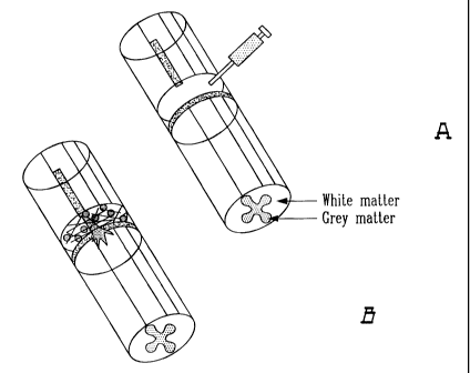
Figure 1A is a schematic diagram of adhesive delivery system of C3 applied to
an injured
spinal cord wherein a tissue adhesive plus Rho antagonist (i.e. C3) is
injected
into the site of injury;
Figure 1B is a schematic diagram of adhesive delivery system of C3 applied to
an injured
19
CA 02325842 2004-02-27
spinal cord wherein the injection is shown as resulting in axon regeneration
through the supplemented adhesion matrix and into the distal spinal cord;
Figure 2 Schematically illustrates the model used to show efficacy in vivo. A
dorsal
hemisection was made in adult mice. Three to four weeks later the
anterograde tracer WGA-HRP was injected into the cortex to label the neurons
of the corticospinal tract. Two days later the spinal cord was removed and
HRP enzymatic activity revealed to detect the CST axons. The corticospinal
tract of adult mice was lesioned at the T6 level, and the fibrin glue/C3 was
added at the time of lesion with a syringe. The expression CST refers to
cortical spinal tract.
Figure 3 Illustrates a longitudinal section of an untreated adult mouse spinal
cord 3
weeks after lesion of the corticospinal tract viewed by darkfield microscopy.
The fibers were anterogradely labeled from the motor cortex and appear
fluorescent. The fibers retract back from the site of lesion and do not
regenerate with treatment.
Figure 4A Illustrates a low magnification view of a control animal treated
with collagen
gel without C3; axons retract from the site of lesion;
Figure 4B Illustrates a higher magnification view of a spinal cord treated
with collagen
gel without C3; axons do not regenerate;
Figure 4C Illustrates a low magnification view of labeled corticospinal axons
near the
lesion site after treatment with collagen gel with C3 as a Rho antagonist;
axons
do not retract back from the lesion site; they extend into the region of
increased cellularity which is the scar;
Figure 4D Illustrates a higher magnification view of Figure C showing that
treatment
with Rho antagonist in a collagen gel allows some axons to sprout into the
lesion site;
CA 02325842 2004-02-27
Figure 5A Illustrates a low magnification view of a spinal cord following
treatment with
fibrin adhesive with C3 as a Rho antagonist; the section is viewed by
darkfield
to show the anterogradely-labeled fibers that appear white;
Figure 5B Illustrates a high magnification view of the lesion site shown in
Figure 5A
showing that axons grow through the scar region; the scar appears as the
vertical line;
Figure 5C Illustrates a high magnification view approximately 7 mm distal of
the lesion
site of the spinal cord shown in Figures 5A and 5B; the regenerating fibers
(arrows) grow long distances;
Figure 6A Illustrates a darkfield microscopy of a spinal cord section after
treatment with
Rho antagonist C3 in a fibrin adhesive showing long distance regeneration;
axons sprout into the white matter and cross the lesion site;
Figure 6B Illustrates a section of the same spinal cord shown in Figure 6A to
show axons
that have regenerated a distance of 10 mm from the lesion site;
Figure 7A Illustrates an untreated mouse two days after spinal cord injury;
the control
mouse is mobile but uses its front paws to drag itself forward ant it shows
some movement of hindlimb joints;
Figure 7B Illustrates an animal 2 days after spinal cord injury and treatment
with
C3/matrix; the animal is able to walk with weight support two days after
treatment;
Figure 7C Illustrates a comparison of fibrin, collagen, Gelfoam* and Elvax*
methods of
C3 delivery on long-distance regeneration. Animals were treated with the test
delivery system without (-C3) or with (+C3) Rho antagonist. Distance of
growth of the longest axon was scored by blind examination of at least five
sections from each animal. The longest distance of axon growth was scored.
* Trademark
21
1
CA 02325842 2004-04-27
Not shown is that the animals that were not treated with Rho antagonist always
showed axon retraction back from the site of lesion. When axon growth was
measured, the distance was measured from from the proximal edge of the lesion
site. Each point represents data from one animal (approximately 5 sections
per animal);
Figure 8 Is illustrative of open field test of behavioral recovery. Mice were
scored for
recovery of function by the 21 point BBB open field test (see experimental
section). Two phase of recovery are seen. An early phase, observed in all
mice,
although the BBB score is higher in the C3-treated mice. The later phase of
recovery of coordinated forelimb-hindlimb movement was only observed after
treatment with C3. The C3-treated mice regain almost normal walking behavior;
and
Figure 9 Is a Schematic diagram of a system exploiting a kit in accordance
with the present
invention.
Figure 10 Is a schematic diagram of another embodiment of a system exploiting
a kit in
accordance with the present invention.
As used herein it is to be understood that a number of words and/or
expressions are to have the
meanings as hereinafter described.
The term "fibrin glue" or " fibrin clot" is meant to include any formulations
used to make a
fibrinclot: eg tisseel* VH or see (Herbert (1998)J. Biomed. Mater Res.40:551-
559, Cheng, et al.
(1996)Science.273:510-513, Guest (1997)J. Neurosci. Res. 50: 888-905). Another
definition is
any fibrin glue composition not sold as Tisseel*, but made by combining
fibrinogen, thrombin
calcium ions, with or without other components such as factor XIII or
apoprotinin.
The term "Rho antagonist" includes, but is not restricted to (known ) C3,
including C3 chimeric
proteins, Y276321, or other Rho antagonists delivered in the delivery system.
The term "Y276321" is defined as a Rho kinase inhibitor that stimulated
neurite outgrowth
through its ability to inactivate the Rho signaling pathway (Uehata, et al.
* Trademark
22
CA 02325842 2000-11-29
(1997)Nature.389:990-994, Bito (2000)Neuron.26:431-441).
The term "nerve injury site" refers to a site of traumatic nerve injury or
nerve injury caused by
disease. The nerve injury site may be a single nerve (eg sciatic nerve) or a
nerve tract
comprised of many nerves (eg. damaged region of the spinal cord). The nerve
injury site may
be in the central nervous system of peripheral nervous system in any region
needing repair.
The nerve injury site may form as a result of damage caused by stroke. The
nerve injury site
may be in the brain as a result of surgery, brain tumour removal or therapy
following a
cancerous lesion. The nerve injury site may result from Parkinson's disease,
Alzheimer's
disease, Amyotrophic lateral sclerosis, diabetes or any other type of
neurodegenerative
disease.
Rho GTPases include members of the Rho, Rac and Cdc42 family of proteins. Our
invention
concerns Rho family members of the Rho class. Rho proteins consist of
different variants
encoded by different genes. For example, PC 12 cells express RhoA, RhoB and
RhoC
(Lehmann et al 1999 IBID). To inactivate Rho proteins inside cells, Rho
antagonists of the C3
family type are effective because they inactivate all forms of Rho (eg. RhoA,
Rho B etc). In
contrast, gene therapy techniques, such as introduction of a domainant
negative RhoA family
member into a diseased cell, will only inactivate that specific RhoA family
member.
Compounds of the C3 family from closteridium botulinum inactivate Rho by
ADP-ribosylation.
Recombinant C3 proteins, or C3 proteins that retain the ribosylation activity
are also effective
in our delivery system and are covered by this invention. In addition, Rho
kinase is a
well-known target for active Rho, and inactivating Rho kinase has the same
effect as
inactiving Rho, at least in terms of neurite or axon growth (Kimura and
Schubert
(1992)Journal of Cell Biology. 116:777-783, Keino-Masu, et al.
(1996)Ce11.87:175-185,
Matsui, et al. (1996)EMBO J.15:2208-2216, Matsui, et al. (1998)J. Cell
Biol.140:647-657,
Ishizaki (1997)FEBS Lett.404:118-124), the biological activity that concerns
this invention.
Therefore, chemical compounds such as Y-27632, any other compound are covered
by this
invention as a preferred delivery in a tissue adhesive system. Numerous
references
23
CA 02325842 2000-11-29
describing C3 type compounds can be found in Methods in Enzymology, Vol. 256,
Part B,
Eds.: W.E. Balch, C.H. Der, and A. Hall; Academic Press, 1995, for eg. Pgs.
196-206, 207 et
seq, 184-189, and 174 et seq.. In any event C3 may for example be selected
from the group
consisting of ADP-ribosyl transferase derived from Closteridum botulinum and a
recombinat
ADP-ribosyl transferase.
On the other hand any compound or molecule that does not have a direct action
on Rho itself
but works to decrease the function of Rho such as anti-sense oligos to Rho,
anti-Rho kinase
antibodies, and the like. Such Rho antagonists that can be delivered in a
tissue adhesive
system are also covered by our invention. The C3 polypeptides of the present
invention
include biologically active fragments and analogs of C3; fragments encompass
amino acid
sequences having truncations of one or more amino acids, wherein the
truncation may
originate from the amino terminus, carboxy terminus, or from the interior of
the protein.
Analogs of the invention involve an insertion or a substitution of one or more
amino acids..
Fragments and analogs will have the biological property of C3 that is capable
of inactivation
Rho GTPases. Also encompassed by the invention are chimeric polypeptides
comprising C3
amino acid sequences fused to heterologous amino acid sequences. Said
heterologous
sequences encompass those which, when formed into a chimera with C3 retain one
or more
biological or immunological properties of C3. A host cell transformed or
transfected with
nucleic acids encoding C3 protein or c3 chimeric protein are also encompassed
by the
invention. Any host cell which produces a polypeptide having at least one of
the biological
properties of a C3 may be used. Specific examples include bacterial, yeast,
plant, insect or
mammalian cells. In addition, C3 protein may be produced in transgenic
animals.
Transformed or transfected host cells and transgenic animals are obtained
using materials and
methods that are routinely available to one skilled in the art. Host cells may
contain nucleic
acid sequences having the full-length gene for C3 protein including a leader
sequence and a
C-terminal membrane anchor sequence (see below) or, alternatively, may contain
nucleic
acid sequences lacking one or both of the leader sequence and the C-terminal
membrane
anchor sequence. In addition, nucleic acid fragments, variants and analogs
which encode a
polypeptide capable of retaining the biological activity of C3 may also be
resident in host
expression systems.
24
CA 02325842 2000-11-29
The Rho antogaonist that is a recombinant proteins can be made according to
methods present
in the art. The proteins of the present invention may be prepared from
bacterial cell extracts,
or through the use of recombinant techniques. In general, C3 proteins
according to the
invention can be produced by transformation (transfection, transduction, or
infection) of a
host cell with all or part of a C3-encoding DNA fragment in a suitable
expression vehicle.
Suitable expression vehicles include: plasmids, viral particles, and phage.
For insect cells,
baculovirus expression vectors are suitable. The entire expression vehicle, or
a part thereof,
can be integrated into the host cell genome. In some circumstances, it is
desirable to employ
an inducible expression vector.
Those skilled in the field of molecular biology will understand that any of a
wide variety of
expression systems can be used to provide the recombinant protein. The precise
host cell used
is not critical to the invention. The C3 protein can be produced in a
prokaryotic host (e.g., E.
coli or B. subtilis) or in a eukaryotic host (e.g., Saccharomyces or Pichia;
mammalian cells,
e.g., COS, NIH 3T3, CHO, BHK, 293, or HeLa cells; or insect cells).
Proteins and polypeptides can also be produced by plant cells. For plant cells
viral expression
vectors (e.g., cauliflower mosaic virus and tobacco mosaic virus) and plasmid
expression
vectors (e.g., Ti plasmid) are suitable. Such cells are available from a wide
range of sources
(e.g., the American Type Culture Collection, Rockland, Md.). The methods of
transformation
or transfection and the choice of expression vehicle will depend on the host
system selected.
The host cells harbouring the expression vehicle can be cultured in
conventional nutrient
media adapted as need for activation of a chosen gene, repression of a chosen
gene, selection
of transformants, or amplification of a chosen gene. One expression system is
the mouse 3T3
fibroblast host cell transfected with a pMAMneo expression vector (Clontech,
Palo Alto,
Calif.). pMAMneo provides an RSV-LTR enhancer linked to a dexamethasone-
inducible
MMTV-LTR promotor, an SV40 origin of replication which allows replication in
mammalian
systems, a selectable neomycin gene, and SV40 splicing and polyadenylation
sites. DNA
encoding a C3 protein would be inserted into the pMAMneo vector in an
orientation
designed to allow expression. The recombinant C3 protein would be isolated as
described
below. Other preferable host cells that can be used in conjunction with the
pMAMneo
CA 02325842 2000-11-29
expression vehicle include COS cells and CHO cells (ATCC Accession Nos. CRL
1650 and
CCL 61, respectively).
C3 polypeptides can be produced as fusion proteins. For example, expression
vectors can be
used to create lacZ fusion proteins. The pGEX vectors can be used to express
foreign
polypeptides as fusion proteins with glutathione S-transferase (GST). In
general, such fusion
proteins are soluble and can be easily purified from lysed cells by adsorption
to
glutathione-agarose beads followed by elution in the presence of free
glutathione. The pGEX
vectors are designed to include thrombin or factor Xa protease cleavage sites
so that the
cloned target gene product can be released from the GST moiety. Another
stategy to make
fusion proteins is to use the His tag system.
In an insect cell expression system, Autographa californica nuclear
polyhedrosis virus
AcNPV), which grows in Spodoptera frugiperda cells, is used as a vector to
express foreign
genes. A C3 coding sequence can be cloned individually into non-essential
regions (for
example the polyhedrin gene) of the virus and placed under control of an AcNPV
promoter,
e.g., the polyhedrin promoter. Successful insertion of a gene encoding a C3
polypeptide or
protein will result in inactivation of the polyhedrin gene and production of
non-occluded
recombinant virus (i.e., virus lacking the proteinaceous coat encoded by the
polyhedrin gene).
These recombinant viruses are then used to infect spodoptera frugiperda cells
in which the
inserted gene is expressed (see, Lehmann et al for an example of making
recombinant MAG
protein).
In mammalian host cells, a number of viral-based expression systems can be
utilised. In cases
where an adenovirus is used as an expression vector, the C3 nucleic acid
sequence can be
ligated to an adenovirus transcription/translation control complex, e.g., the
late promoter and
tripartite leader sequence. This chimeric gene can then be inserted into the
adenovirus
genome by in vitro or in vivo recombination. Insertion into a non-essential
region of the viral
genome (e.g., region El or E3) will result in a recombinant virus that is
viable and capable of
expressing a C3 gene product in infected hosts.
Specific initiation signals may also be required for efficient translation of
inserted nucleic
26
CA 02325842 2000-11-29
acid sequences. These signals include the ATG initiation codon and adjacent
sequences. In
cases where an entire native C3 gene or cDNA, including its own initiation
codon and
adjacent sequences, is inserted into the appropriate expression vector, no
additional
translational control signals may be needed. In other cases, exogenous
translational control
signals, including, perhaps, the ATG initiation codon, must be provided.
Furthermore, the
initiation codon must be in phase with the reading frame of the desired coding
sequence to
ensure translation of the entire insert. These exogenous translational control
signals and
initiation codons can be of a variety of origins, both natural and synthetic.
The efficiency of
expression may be enhanced by the inclusion of appropriate transcription
enhancer elements,
transcription terminators.
In addition, a host cell may be chosen which modulates the expression of the
inserted
sequences, or modifies and processes the gene product in a specific, desired
fashion. Such
modifications (e.g., glycosylation) and processing (e.g., cleavage) of protein
products may be
important for the function of the protein. Different host cells have
characteristic and specific
mechanisms for the post-translational processing and modification of proteins
and gene
products. Appropriate cell lines or host systems can be chosen to ensure the
correct
modification and processing of the foreign protein expressed. To this end,
eukaryotic host
cells that possess the cellular machinery for proper processing of the primary
transcript,
glycosylation, and phosphorylation of the gene product can be used. Such
mammalian host
cells include, but are not limited to, CHO, VERO, BHK, HeLa, COS, MDCK, 293,
3T3,
W13 8, and in particular, choroid plexus cell lines.
Alternatively, a C3 protein can be produced by a stably-transfected mammalian
cell line. A
number of vectors suitable for stable transfection of mammalian cells are
available to the
public; methods for constructing such cell lines are also publicly available.
In one example,
cDNA encoding the C3 protein can be cloned into an expression vector that
includes the
dihydrofolate reductase (DHFR) gene. Integration of the plasmid and,
therefore, the C3
protein-encoding gene into the host cell chromosome is selected for by
including 0.01-300
M methotrexate in the cell culture medium (as described in Ausubel et al.,
supra). This
dominant selection can be accomplished in most cell types.
27
CA 02325842 2000-11-29
Recombinant protein expression can be increased by DHFR-mediated amplification
of the
transfected gene. Methods for selecting cell lines bearing gene amplifications
are known in
the art; such methods generally involve extended culture in medium containing
gradually
increasing levels of methotrexate. DHFR-containing expression vectors commonly
used for
this purpose include pCVSEII-DHFR and pAdD26SV(A). Any of the host cells
described
above or, preferably, a DHFR-deficient CHO cell ligne (e.g., CHO DHFR cells,
ATCC
Accession No. CRL 9096) are among the host cells preferred for DHFR selection
of a
stably-transfected cell line or DHFR-mediated gene amplification.
A number of other selection systems can be used, including but not limited to
the herpes
simplex virus thymidine kinase, hypoxanthine-guanine
phosphoribosyltransferase, and
adenine phosphoribosyltransferase genes can be employed in tk, hgprt, or aprt
cells,
respectively. In addition, gpt, which confers resistance to mycophenolic acid
; neo, which
confers resistance to the aminoglycoside G-418; and hygro, which confers
resistance to
hygromycin. can be used.
Alternatively, any fusion protein can be readily purified by utilising an
antibody specific for
the fusion protein being expressed. For example, a system described in
Janknecht et al.
(1981) Proc. Natl. Acad. Sci. USA 88, 8972, allows for the ready purification
of
non-denatured fusion proteins expressed in human cell lines. In this system,
the gene of
interest is subcloned into a vaccinia recombination plasmid such that the
gene's open reading
frame is translationally fused to an amino-terminal tag consisting of six
histidine residues.
Extracts from cells infected with recombinant vaccinia virus are loaded onto
Ni2+
nitriloacetic acid-agarose columns, and histidine-tagged proteins are
selectively eluted with
imidazole-containing buffers.
Alternatively, C3 or a portion thereof, can be fused to an immunoglobulin Fc
domain. Such
a fusion protein can be readily purified using a protein A column.
It is envisioned that small molecule mimetics of the above described
antagonists are also
encompassed by the invention.
28
CA 02325842 2000-11-29
In the following a method to identify active Rho antagonists will be
discussed.
To test Rho antagonists for activity, a tissue culture bioassay system was
used. This bioassay
is used to define acitivity of Rho antagonists that will be effective in
promoting axon
regeneration in spinal cord injury, stroke or neurodegenerative disease.
Neurons do not grow neurites on inhibitory niyelin substrates. When neurons
are placed on
inhibitory substrates in tissue culture, they remain rounded. When an
effective Rho antagonist
is added, the neurons are able to grow neurites on myelin substrates. The time
that it takes for
neurons to growth neurites upon the addition of a Rho antagonist is the same
as if neurons
had been plated on growth permissive substrate such as laminin or polylysine,
typically 1 to 2
days in cell culture. The results can be scored visually. If needed, a
quantitative assessment of
neurite growth can be performed. This involved measuring the neurite length in
a) control
cultures where neurons are plated on myelin substrates and left untreated b)
in positive
control cultures, such as neurons plated on polylysine c) or treating cultures
with different
concentrations of the test antagonist.
To test C3 in tissue culture, it has been found that the best concentration is
25-50 ug/ml.
Thus, high concentrations of this Rho antagonist are needed as compared to the
growth
factors used to stimulate neurite outgrowth. Growth factors, such as nerve
growth factor
(NGF) are used at concentrations of 1- 100 ng/ml in tissue culture. However,
growth factors
are not able to overcome growth inhibition by myelin. Our tissue culture
experiments are all
performed in the presence of the growth factor BDNF for retinal ganglion
cells, or NGF for
PC 12 cells. When growth factors have been tested in vivo, typically the
highest
concentrations possible are used, in the ug/ml range. Also they are often
added to the CNS
with the use of pumps for prolonged delivery (eg. Ramer et al, IBID). For in
vivo experiments
the highest concentrations possible was used when working with C3 stored as a
frozen
1 mg/ml solution. The concentration that was chosen does not prevent the
fibrin matrix from
polymerizing.
For test purposes it was decided to dilute a 1 mg/ml solution of C3 with 1/3
volume thrombin
and 1/3 volume fibrinogen solutions (contain calcium and aprotinin). In order
to increase the
29
CA 02325842 2000-11-29
concentration of C3, it would be possible to lyophylize C3 and then resuspend
it in the
fibrinogen solution. Lyophilized C3 has been tested and found to be active.
The Rho antagonist C3 is stable at 37 C for at least 24 hours. The stability
of C3 was
tested in tissue culture with the following experiment. The C3 was diluted in
tissue culture
medium, left in the incubator at 37C for 24 hours, then added to the bioassay
system
described above, using retinal ganglion cells as the test cell type. These
cells were able to
extend neurites on inhibitory substrates when treated with C3 stored for 24
hours at 37C.
Therefore, the minimun stability is 24 hours. This is in keeping with the
stability projection
based on amino acid composition (see sequence data, below).
In the following various tissue Adhesives and Formulations used to make them
will be
discussed.
Different types of tissue adhesive can be made. Examples include collagen
gels, fibrin tissue
adhesives. Other examples are matrigel, laminin networks, and adhesives based
on a
composition of basment membrane proteins that contain collagen.
Fibrin sealant has three basic components: fibrinogen concentrate, calcium
chloride and
thrombin. Other components can be added to affect the time of clot formation,
and the size of
the protein network that is formed. Generally when the components mix, a
fibrin coagulum is
formed in that the fibrinogen molecule is cleaved through the action of
thrombin to form
fibrin monomers which spontaneously will polymerize to form a three-
dimensional network
of fibrin, largely kept together by hydrogen bonding. This corresponds to the
last phase of the
natural blood clotting cascade, the coagulation rate being dependent on the
concentration of
thrombin used. In order to improve the tensile strength, covalent crosslinking
between the
fibrin chains is provided for by including Factor XIII in the sealant
composition. In the
presence of calcium ions, thrombin activates factor XIII to factor XIIIa.
Activated factor
XIIla together with thrombin catalyzes the cross-linkage of fibrin and
increases the strength of
the clot. The strength of the fibrin clot is further improved by the addition
of fibronectin to
the composition, the fibronectin being crosslinked and bound to the fibrin
network formed.
During wound healing the clot material undergoes gradual lysis and is
completely absorbed.
CA 02325842 2004-02-27
~' \y
To prevent a too early degradation of the fibrin clot by fibrinolys, the
fibrin sealant
composition may comprise a plasminogen activator inhibitor or a plasmin
inhibitor, such as
aprotinin. Such an inhibitor will also reduce the fibrinolytic activity
resulting from any
residual plasminogen in the fibrinogen composition. Similarly, compositions
may include
hyaluronic acid (or other polysaccharides), and these may also comprise a
hyaluronidase
inhibitor such as one or more flavonoids (or corresponding inhibitors for
other
polysaccharides) in order to prevent degradation (i.e. to prolong the
duration) ofthe.
hyaluronic acid component by hyaluronidase which is always present in the
surrounding
tissues. The hyaluronic acid may, as mentioned above, be crosslinked, a
commercially
available example being Hylan® (trademark, available from Biomatrix,
Ritchfield, N.Y.,
USA). The hyaluronic acid compositions may e.g. have the form of gels,
solutions, etc.
Fibrin clots in any one of the above described embodiments, may be used for
the
application of a pharmaceutically active substance. By incorporating a drug,
such as an
antibiotic, a growth factor, etc. into the tissue adhesive it will be enclosed
in the fibrin
network formed upon application of the tissue adhesive. It will thereby be
ensured that the
drug is kept at the site of application while being controllably released from
the composition.
Fibrin sealant products prepared from human plasma fibrinogen/Factor XIII are
available
*
commercially. One product is a tissue glue called Tisseel Fibrin Sealant
(Baxter Hyland
* * *
Immuno Corporation). (Tissucol/Tisseel, Immuno AG, Vienna) and another
Beriplast P,
Hoechst, West Germany. A frozen formution of a fibrin glue delivered with a 2
syringe
system is Hemaseelinade by Hemacure Inc. (Kirkland, Quebec).
ln the following methods for making Tissue Adhesive Delivery kits will be
discussed.
In a preferred embodiment, the kit includes the solutions provided in separate
bottles to
prevent mixing before use, and an applicator designed so as to permit mixing
of the
fibrinogen/Factor XIII and thrombin with C3 at the body site. The kit would
provide
pre-measured amounts of the fibrinogen and factor XIII in one bottle, the
thrombin in another
bottle, a calcium chloride solution in third bottle, and a C3 solution in a
fourth bottle. The
contents of the bottles would be mixed in a prescribed order , as detailed in
the example
31
* : Trademark
CA 02325842 2000-11-29
below. The kit can also include one or more other storage containers which are
any necessary reagents including solvents, buffers, etc. The kit could be sold
as lyophilized or
frozen components to preserve the activity of C3 or other Rho antagonist added
to the kit.
The applicator can, for example, take the form of a glass or plastic syringe
with disposable
needles. With a single syringe system, the components of the kit would be
mixed immediately
before application to the injury site.
A more elaborate system would allow two syringes to be attached, so that the
mixing could
take place in the syringe or a mixing compartment of the syringe, before
injection. One
example of a two syringe system is a Luer lock syringe, such as used for
mixing adjuvants..
For this a 3-way stopcocks, such as commercially available (Bio-Rad cat
#7328103) is
attached to the syringe so that the solution can be passed back and forth
beore attaching the
injection needle to the third port of the 3-way stopcock. These are plastic,
sterile, and
disposable.
Another method of application could be through the use of a clip to hold two
syringes, and the
clip would have a common plunger to ensure that equal volumes of the thrombin
and
fibrinogen components are mixed in a chamber with the calcium chloride and C3,
before
being ejected trough the needle.
Other Ingredients for the Tissue Adhesive Rho Antagonist Delivery System are
discussed
hereinafter,
Other components can be added to the tissue adhesive to improve efficacy of
the treatments.
Such additions include growth factors, protease inhibitors, cytokines, anti-
inflammatory
compounds, cell transplant systems. Agents that prevent cell death, such as
agents that affect
the apoptosis pathway could be added components to the delivery system.
Methods of Packaging Delivery System are discussed hereinafter.
In the preferred formulation, Rho antagonist, fibrinogen and thrombin are
mixed together just
32
CA 02325842 2004-02-27
before application, so that polymerization of the gel occurs in the injured
CNS. Therefore, it
is important that the fibrinogen and thrombin are packaged separately.
However, the C3 can
be packaged separately, or added to either the thrombin or fibrinogen bottles.
In another
formulation, the fibrinogen, thrombin and C3 are packaged together, but held
at low pH,
which prevents polymerization of the gel. Polymerization would be induced by
mixing this
formulation with a basic component that would neutralize the pH to induce
coagulation of the
adhesive. In another formulation, the Rho antagonist could be added separately
to the
fibrinogen/thrombin mix in the form of liposomes or other similar delivery
system. Living
cells that could secrete C3 could be added as Rho antagonist.
A method of Applying Rho antagonist in vivo is discussed hereinafter.
Tissue adhesive formulations are typically applied to wound sites with a
syringe and needle.
The shape of the needle determines the type of surface that is formed when the
adhesive
polymerizes. In some cases, adhesives can be sprayed onto the wound surface,
or into the
desired region. This invention covers all types of syringes and needles used
to apply fibrin
plus Rho antagonists to injured regions of the CNS. In addition, it covers the
addition of
previously polymerized tissue adhesives with C3 to the wound. For example,
fibrin can be
polymerized in a test tube, and forceps used to remove the gel and place it in
the body cavity.
Similarly, collagen can be applied by pre-polymerization and application by
using forceps to
place the gel in the injured spinal cord. One example of this is more fully
explained in the
example section of this application.
Tests were done with Gelfoam(TM), a surgical collagen-based sponge, and
Elvax*, a slow
release plastic (Lehmann et al 1999, IBID) for the ability to deliver
biologically effective
concentrations of C3. Neither of these two delivery systems was effective.
Therefore, only
tissue adhesive formulations (i.e. the matrix forming formulations discussed
herein) have
efficacy in the delivery of C3 to the injured CNS in vivo.
Therapeutic Applications/Medical Uses will be discussed below.
The tissue adhesive system for the delivery of Rho antagonists may be useful
in many other
* Trademark
33
CA 02325842 2004-02-27
conditions that affect the central and peripheral nervous system. Treatments
that are effective
in eliciting sprouting from injured axons are equally effective in treating
some types of
stroke. Since it has been determined that it is possible to elicit sprouting
(using a kit of the
present invention), it is obvious that the treatments can be extended to
stroke. Similarly,
although the subject of this invention is related to delivery of Rho
antagonists to the
traumatically damaged nervous system, this invention also pertains to damage
from
neurodegeneration, such as during Parkinson's disease, Alzheimer's disease,
prion diseases
or other diseases of the CNS where axons are damaged, in the CNS environment.
In such
cases, small volumes of the tissues adhesive with C3 could be injected into
the affected
region with the use of a syringe. The treatment will cause local sprouting to
restore function
of neurons whose axon processes had retracted in the course of the
neurodegeneration.
Testing example Formulation(s) and Delivery System(s) will be discussed below.
Test of invention formulation were conducted in mice after injury of the
corticospinal tract.
All mice were tested for anatomical regeneration of lesioned axons by
anterograde tracing
techniques. Some of the mice were also assessed for recovery of locomotion.
The details of
these experiments are given in the experimental section, the example sections,
and the results
are shown in the figures.
EXAMPLES
EXAMPLE 1 A kit for a tissue adhesive system.
The kit contains:
1 vial fibrinogen
1 vial aprotinin solution for reconstitution of fibrinogen
1 vial thrombin
1 vial calcium chloride solution for reconstitution of thrombin
34
CA 02325842 2000-11-29
1 vial C3 solution
1.1 Lyophilized fibrinogen (75mg/ml) in glycine buffer (2mg/ml NaCl, 4 mg/ml
trisodium
citrate, 15 mg/ml glycine) was reconstituted in an aprotinin solution 3000
KIU/ml and
heated to 37 C. For ease of handling, a combined heating and stirring device
was used
(appropriate vials contain a maganetic stirrer. This is called solution I.
1.2 A thrombin solution is prepared; the solution comprisin Lyophilized
thrombin
500 IU/ml , 2.4 mg/ml glycine, 8 mg/mi sodium chloride. The calcium chloride
solution (40
umol CaC12) and thrombin are mixed and heated to 37C. This is called solution
II.
1.3 A solution of C3 (1 mg/ml) is heated to 37C
1.4 Equal amounts of solution I, II, and III are mixed, and immediately drawn
up in a syringe, and added to the injury site where polymerization occurs.
Thus the C3 is
added as part of the fibrin glue solution that is placed in the lesion cavity
to polymerize.
A combined heating and stirring device can be used in conjunction with the
kit. For this,
small magnetic stirrers are included in each of the mixing vials. The vials
are then placed in
the combined mixing and warming device where the magnetic stirrer keeps the
solution
stirred while the solution is warming.
Mice that received a dorsal hemisection were treated with the fibrin/C3
adhesive. In some
experiments, 10 l of Img/ml C3 in phosphate buffered saline was added to the
lesion site
before applying the C3/fibrin. Behavior recovery was assessed in an open field
environment
as described by Beattie, Basso and Breshnahan (1995) J. Neurotrauma 12:1-20.
Anatomical
regeneration was assessed by anterograde labeling of the corticospinal
fibres.Three weeks to
three months after injury, the corticospinal fibres were labeled by inject the
anterograde tracer
WGA-HRP into the motor cortex as described in the art (Huang
(1999)Submitted.). Two days
CA 02325842 2004-02-27
later the animals were killed, the spinal cord removed, and longitudinal
sections cut and
reacted for HRP enzymatic activity, as described (Huang (1999)Submitted.). The
labeled
fibres were observed by microscopy to extend many mm past the lesion site (see
figures 5
and 6) after treatment with C3/fibrin.
EXAMPLE 2. Modification of the kit in example 1.
The formulation given in example 1 was used with the following modifications.
Solution II is
made with the addition of recombinant C3 directly to the solution II vial. In
other words,
solution II contains thrombin, calcium chloride and C3. Solution I is loaded
in one syringe,
solution II is loaded in a second syringe. A syringe with a plunger that
simultaneously loads
both solutions is used. Thus the solutions are mixed as they enter a small
chamber before the
needle, and the polymerization occurs in situ in the injured region of the CNS
where the
solution is applied. The system describe here is the Duploject system from
Baxter
Phamaceuticals U.S.A..
EXAIvIPLE 3. Modification of the kit in example 1.
As example 2, but the C3 solution is mixed in vial 1 with the fibrinogen. Vial
one and vial II
are heated and prepared as described in example 1, and injected into the
injured CNS with the
Duploject system.
EXAMPLE 4 : Collagen gels used a a tissue adhesives.
First collagen is purified. Collagen can be purified from any source, human or
mammalian.
One source of collagen is the EHS tumor cell line which is passed in mice.
Collagen was
purified from rat tails. The tails were soaked in 70% alcohol for about 20
minutes. The
remaining steps were performed under aseptic conditions. The tails are broken
about 2 cm
36
* : 'I',rademark
CA 02325842 2000-11-29
from the tip with a hemostat and the tendon is slowly pulled out and placed in
a sterile dish.
The tendons are cut into small pieces and soaked in acetic acid-water (1:1000)
for 48 hours in
the cold. 150 ml of solution is used per tail. The solution is centrifuged at
15,0000 rpm, 30
min. and stored in aliquots at B l OC.
Collagen gels with C3 as Rho antagonist are formed in vivo as follows. For
treatment of one
mouse, 40 g of C3 was lyophilized. The C3 protein was reconstituted in 10 1
of 7.5%
NaHCO3. Collagen at 0.7 mg/ml was used, and 25 1 collagen was added to the
C3 solution.
A mouse that had received a dorsal hemisection of the spinal cord was treated
with 10 1 of
1 mg/ml C3 in the collagen (i.e. at the lesion site). The time it takes for
the collagen to
polymerize may be modified by varying the NaHCO3 solution. Anatomical
regeneration of
transected cortical spinal fibres was assessed as described in the detailed
description of the
invention.
EXAMPLE 5. Procedure to make recombinant C3 as a Rho antoagonist. Recombinant
C3
protein was made as follows. The plasmid pGEX2T-C3 coding for the
glutathione-S-transferase (GST)-C3 fusion protein was obtained from N.
Lamarche (McGill
Univ.). Bacteria were transformed with pGEX2T-C3, allowed to grow overnight
induced with
IPTG, and sonicated to break open the cells. The recombinant protein was
purified by affinity
chromotography as described (Ridley and Hall (1992)Cel1.70:389- 399). The GST
fusion
protein was cleaved by thrombin, and thrombin was removed by incubation with
100 1 of
p-aminobenzamidine agarose-beads (Sigma). The C3 solution was dialyzed against
PBS, and
sterilized with a 0.22 m filter. The C3 concentration was evaluated by
protein assay (DC
assay, BioRad Labs, Missassauga, Ont.) and C3 purity was controlled by SDS-
PAGE
analysis.
Example 6: Testing the Fibrin-Rho-Antagonist Formulation using the Delivery
System
To test the tissue adhesive system a rodent model of spinal cord injury was
used. For this,
37
CA 02325842 2000-11-29
Balb-c mice were anaesthetized with 0.6 ml/kg hypnorm, 2.5 mg/kg diazepam
and35 mg/kg
ketamine. A section of the thoracic spinal cord was exposed using fine rongers
to remove the
bone. A dorsal hemisection was made to cut the dorsal columns at level T6. The
fibrin/C3
adhesive was injected immediately after injury. As control another group of
animals received
fibrin alone, and a third group received no treatment. The following day
behavioural testing
began, and continued for three weeks. The animals were placed in an open field
environment
that consisted of a rubber mat approximately 4' X 3' in size. The animals were
left to move
randomly, the movement of the animals were videotaped. For each test two
observers scored
the animals for ability to move ankle, knee and hip joints in the early phase
of recovery. In the
intermediate phase, the ability to support weight and correct placement of the
feet was
assessed (dorsal or plantar placement). In the late phase of recovery, the
animals were
assessed for correct foot position, trunk stability , and foot drag. Only
animals that received
C3/fibrin reached the late phase of recovery of coordinated forelimb-hindlimb
movement.
Untreated control animals did not typically pass beyond the early phase of
recovery.
Additional Experimental activity will be discussed below.
Spinal cord injury
To study The CST was cut bilaterally by a dorsal hemisection extending past
the central canal
(1 (Fig.2) at the T6 level. Balb-c mice were anaesthetized with 0.6 ml/kg
hypnorm, 2.5
mg/kg diazepam and 35 mg/kg ketamine. A section of the thoracic spinal cord
was exposed
using fine rongers to remove the bone, and a dorsal hemisection was made at
level T6. Fine
sissors were used to cut the dorsal half of the spinal cord, and it was recut
a second time with
fine knife to ensure all lesions extended past the central canal. Three weeks
to four weeks
after injury, the corticospinal fibres were labeled by injection the
anterograde tracer
WGA-HRP into the motor cortex as into 6 sites. For injection into the motor
cortex a pulled
glass pipette was used . Two days later the animals were perfused
transcardially with saline
then 4% paraformaldehyde and the spinal cords and brains were removed.
C3 toxin was delivered locally to the site of the lesion by a fibrin-based
tissue adhesive
delivery system (Figure 1). Recombinant C3 was mixed with fibrinogen and
thrombin in the
38
CA 02325842 2000-11-29
presence of CaClz. Fibrinogen is cleaved by thrombin, and the resulting fibrin
monomers
polymerize into a three- dimensional matrix. C3 was added as part of a fibrin
adhesive,
which polymerized within about 10 seconds after being placed in the injured
spinal cord.
Anterograde tracing with WGA-HRP was used to study anatomical regeneration
past the site
of lesion in three groups of animals: animals treated with fibrin plus C3
(C3/fibrin), animals
treated with fibrin alone, and animals that did not received treatment after
injury (see figure
7). With no treatment, transected CST axons retract back from the site of
lesion from 500
um to 1 mm (Fig. 3). Animals treated with fibrin alone showed less axon
retraction, and
sprouting of axons was observed to extend towards the scar. Application of C3
to the injured
spinal cord elicited an extensive sprouting of CST axons into the dorsal white
matter, and the
axons grew into the scar and and extended past past the lesion (Fig. 4). A
long distance
regeneration of individual CST axons and axon bundles was elicited by C3 (Fig.
5), but not
in untreated or fibrin controls. This regeneration was significantly different
from any growth
observed following treatment with fibrin alone.
Several different tissue adhesive delivery systems were tested. When C3 was
delivered in
collagen gels less axon retraction was observed, but the same extent of axon
regeneration was
not observed as with fibrin. Gelfoam(TM) , a surgical collagen sponge, was
also tested.
Gelfoam was not as effective as fibrin as promoting long-distance regeneration
(Fig. 7). A
non-biological material, Elvax, was also tested which is a polymer-based
artificial release
system (see Lehmann et al, 1999 IBID). This system was not effective in
allowing cut axons
access to C3.
To test functional recovery following treatment of injured spinal cord with
C3, three
groups of animals were score for locomotor behaviour in an open field
environment
according to the 21 point BBB scale (Basso et al. ). The animals were examined
by two
reviewers and were placed alone in an open field environment that consisted of
a rubber mat
approximately 4' X 3' in size. Each animal was videotaped for approx. 3 min.
For the early
and intermediate phases, the BBB scores were derived following observation,
and confirmed
by video analysis. In the late phase of recovery, the BBB score was determined
from the
videos projected on a computer at 3 speed from sequences of 4 steps or more.
The BBB test
includes three phases of recovery: an early phase (scores 1-7) of joint
movement, an
intermediate phase (score 8-13) where weight support and foot placement
(dorsal or plantar)
39
CA 02325842 2000-11-29
are assessed, and a late phase of coordinated movements (scores 14-21) where
correct foot
position, and foot drag are examined. The C3 treated animals rapidly regained
the ability to
support weight (Fig. 7B ) while control animals moved mostly by the action of
their forelimbs
(Fig. 7A ). The control groups entered the intermediate recovery phase with
the ability to
support weight within one weeks, at which point they obtained their recovery
plateau.
Animals that received C3 treatment continues to recover over the 1 month
period of
observation, and recovered coordinated movement and almost normal stepping
(Fig. 8).
In rats that receive a contusion injury the recovery period depends on the
severity and
location of the lesion. Typically, rats reach a plateau of recovery by about
two week, whereas
after dorsal hemisection in mice it was found that the plateau of recovery is
reached within
about 1 week. The remarkable improvement in C3-treated mice within one day of
spinal
cord lesion is likely due to changes in the local spinal cord circuitry. These
local changes
might result from the robust sprouting immediately after application of C3 is
applied to the
transected axons. Rates of axon growth in vivo are known to be approximately
the same as
the slow axonal transport rate of 50-200 um/hr. It is also possible that the
local effects on the
spinal cord are mechanistically different by acting on central pattern
generators implicated in
walking behaviors or by neuroprotection immediatley after treatment. Most
importantly,
treated mice performed better immediately after lesion, and they recovered
almost normal
walking patterns by one month (Figure 8). This slower phase of recovery is
attributed to the
long-distance regeneration of axons that was induced by C3 (Figure 4).
Moreover, while we
only flowed the CST axons in this study, our treatments also are likely
stimulate growth from
other transected axonal populations.
In the following Production of recombinant C3 will be discussed.
C3 is a protein product made by the bacteria Clostridium botulinum. The
fragment
containing the C3 gene was cloned into a pGEX vector (from Amersham Pharmacia
Biotech
inc. Baie D'rfe, Quebec, Canada), now referred to as pGEX2T-C3, and this vetor
was
obtained from Nathalie Lamarche of McGill University. To confirm the C3
sequence
corresponded to that reported in the literature the insert was sequenced (see
sequence below).
The C3- containing pGEX vector was transformed into the RRl strain of E. coli
(GIBCO).
CA 02325842 2000-11-29
Bacteria were grown in L-Broth (IOg/L Bacto-Tryptone, 5g/L Yeast Extract,
lOg/L NaCI
(Fisher Scientific) with Ampicillin(BMC-Roche) at 50ug/ml in a shaking
incubator for 1 hr at
37 C. Isopropyl 13-D-thiogalactopyranoside(IPTG), ( GIBCO) was added to a
final
concentration of 0.5mM to induce production of recombinant protein and the
culture was
grown for a further 6 hrs at 37 C. Bacterial pellets were obtained by
centrifugation, in 250 ml
centrifuge bottles, at 6000rpm at 4 C for 5min. Pellets can be kept frozen at -
80 C at this
time.
5 mis of Buffer A(50mM Tris, pH7.5, 50mM NaCI, 5mM MgC12, 1mM DTT) + 1mM PMSF
was added to each pellet. Pellets were resuspended and transferred to a 50m1
plastic beaker
on ice, and a further 5 mls of buffer A was used to wash the centrifuge
bottles. Total volume
of buffer A + pellets from a 2 L culture is usually 30-40mis. The pellets, on
ice, were
sonicated 5 X 30 secs using a BRANSON SONIFIER 450 probe sonicator. Bacteria
were
cooled on ice 1 minute between sonications. The sonicate was centrifuged in a
Sorvall SS-34
rotor at 10,000 rpm for 10 min at 4 C to clarify supernatant.
Glutathione-agarose beads (SIGMA#G-45 10) were purchased as a lyophilized
powder and the
beads were swollen in deionized water, then stored in llvl NaCI at 4 C. Five
ml of the beads
(50%v/v) were washed in a 50ml tube filled with buffer A (no PMSF). Tube was
centrifuged
at 2000 rpm (500g) for 5min, water was removed, and replaced with buffer A.
These beads
were added to the cleared bacterial supernatant, and mixed for 1-2hrs at 4 C.
The beads were
washed 4 times with buffer B (buffer A, NaCI is 150mM, no PMSF), then 2X with
buffer C
(buffer B+2.5mM CaC12). Washes were poured out and the beads retained each
time. Next,
5mis of thrombin(Bovine, Plasminogen-free, CALBIOCHEM #605160) 20U at (50%v/v)
was added to the beads to cleave the C3 from the GST affinity purification tag
(see cleavage
site in the nucleotide sequnce given below). This reaction was left overnight,
with mixing, at
4 C.
The beads are loaded into an empty 10 ml column, PBS (phosphate buffered
saline) was
added to the column and 20 1 ml aliquots were collected. To determine the
location of the
protein peak, 0.5 l spots were put on a nitrocellulose sheet, from each
aliquot, and this is
stained with Amido Black(Bio-rad) as a protein dot-blot. Aliquots containing
C3 were
41
CA 02325842 2004-02-27
pooled and 20 uls p-Aminobenzamidine (SIGMA#A7155) was added. The solution was
mixed for 30min at 4 C to remove thrombin. The C3 is centrifuged to remove the
p-
aminobenzamidine, and then concentrated using CENTRIPREP*-10 concentrator
(AMICON). The concentrated C3 is then passed through a PD-10 column
(PHARMACIA,
containing Sephadex* G-25M) and 10 0.5 ml aliquots are collected. A dot-blot
is done on
these aliquots, and the appropriate aliquots (usually 3 tubes) are pooled
(total volume about
1.5mis). The purified recombinant protein was filter-sterilized, aliquoted,
and stored at 80 C.
A protein assay was done on a small amount to determine precisely the
concentration. Purity
of the recombinant C3 was evaluated by SDS polyacrylamide gel electrophoresis.
Bioactivity was assessed with a bioassay using either retinal ganglion cells
or PC 12 cells.
Turning now to Figure 9, this figure illustrates in schematic fashion a system
exploiting a kit
of the present invention for mixing and delivering a supplemented matrix
forming material.
An actual apparatus may for example be of multi-cartridge syringe type as
known or
modified as necessary or desired.
The kit portion of the illustrated system comprises a container means 1 for
fibrinogen
material, a container means 2 for thrombin material and a container means 3
for a
therapeutically active agent for facilitating axon growth (e.g. C3 or a
modified or hybrid C3).
If desired or necessary the kit portion may include additional containers for
the separate
containment of other desired or necessary components; as shown the system in
figure 9
includes in dotted outline an additional container means 4 for the flowable
matrix forming
part of the kit. The system also includes a mixing container 6 wherein the C3
(hybrid) is
mixed with the matrix forming elements to form the supplemented flowable
matrix forming
carrier. The feed line 8 is indicative of the addition of C3 to the container
6 whereas the feed
line 10 is indicative of the addition of the flowable matrix forming elements
from containers
1 and 2 and which is formed from the merging of feed lines 12 and 13. The
mixing in the
container means 6 may be effected or carried out in any suitable (known)
fashion, (e.g.
simple stirring with a magnetic stirrer). The output line 15 of the mixing
container is
indicative of the delivery of the supplemented mixture to the lesion site
(e.g. by needle (e.g.
syringe), pipette, etc.).
Although in figure 9 the therapeutically active agent for facilitating axon
growth (e.g. C3) is
* Trademark
42
,
CA 02325842 2004-04-27
shown as being associated with a separate container 4, if so desired or as
necessary the
therapeutically active agent may be associated with a container holding a
flowable carrier
component (e.g. a container may hold fibrinogen and C3).
Turning now to figure 10, this figure illustrates in schematic fashion another
embodiment of a
system exploiting a kit of the present invention for mixing and delivering a
supplemented
matrix forming material. The containers means are as described in Figure 9
with the
exception that the "container means for therapeutically active agent"
(container means 3) has
been interchanged with the "container means for second matrix forming element"
(container
means 1). Feed line 10 is indicative of the addition of a first matrix forming
element from
container 2 with C3 from container 3 into a mixing container 6. Feed line 8 is
indicative of
the addition of a second matrix forming element into container 6. Mixing may
be effected as
described for figure 9.
43
CA 02325842 2000-11-29
SEQUENCE LISTING
(1) GENERAL INFORMATION:
(i) APPLICANT: LISA MCKERRACHER
(ii) TITLE OF INVENTION: Methods for making and delivering Rho-antagonist
tissue
adhesive formulations to the injured mammalian central
and peripheral nervous systems and uses thereof
(iii) NUMBER OF SEQUENCES: 3
(iv) CORRESPONDENCE ADDRESS:
(A) ADRESSEE: BROULLETTE KOSIE
(B) STREET: I 100 RENE-LESVEQUE BLVD WEST
(C) PROV/STATE: QUEBEC
(D) COUNTRY: CANADA
(E) POSTAL/ZIP CODE: H3B 5C9
(v) COMPUTER READABLE FORM:
(A) MEDIUM TYPE: Floppy disk
(B) COMPUTER: IBM PC compatible
(C) OPERATING SYSTEM: PC-DOS/MS-DOS
(D) SOFTWARE:ASCII (TEXT)
(vi) CURRENT APPLICATION DATA:
(A) APPLICATION NUMBER:
(B) FILING DATE:
(C) CLASSIFICATION:
(vii) ATTORNEY/AGENT INFORMATION:
(A) NAME: BROULLETTE KOSIE
(B) REGISTRATION NO.:
(C) REFERENCE/DOCKET NO.: 06447-003-CA-2
(D) TEL. NO.: (514) 397 8500
(E) FAX NO.: (514) 397 8515
(2) INFORMATION FOR SEQ ID NO: 1:
(i) SEQUENCE CHARACTERISTICS:
(A) LENGTH:
(B) TYPE:
(C) STRANDEDNESS:
1
CA 02325842 2000-11-29
(C) STRANDEDNESS:
(D) TOPOLOGY:
(ii) MOLECULE TYPE:
(v) FRAGMENT TYPE:
(vi) ORIGINAL SOURCE:
(A) ORGANISM:
(vii) IIVIlVIEDIATE SOURCE:
(ix) FEATURE: -
(A) NAME/KEY:
(B) LOCATION:
(D) OTHER INFORMATION:
(x) PUBLICATION INFORMATION:
(A) AUTHORS:
(B) TITLE:
(C) JOURNAL:
(D) VOLUME:
(E) ISSUE:
(F) PAGES:
(G) DATE:
(H) DOCUMENT NO.:
(I) FILING DATE:
(J) PUBLICATION DATE:
(K) RELEVANT RESIDUES IN SEQ ID NO:
(xi) SEQUENCE DESCRIPTION: SEQ ID NO: 1:
GTG GCG ACC CTT CCC AAA TCG GAT CTG GTT CCG CGT GGA TCC TCT AGA
1 5 10 15
GTC GAC CTG CAG GCA TGC AAT GCT TAT TCC ATT AAT CAA AAG GCT TAT TCA
20 25 30
AAT ACT TAC CAG GAG TTT ACT AAT ATT GAT CAA GCA AAA GCT TGG GGT AAT
35 40 45 50
GCT CAG TAT AAA AAG TAT GGA CTA AGC AAA TCA GAA AAA GAA GCT ATA
2
CA 02325842 2000-11-29
55 60 65
GTA TCA TAT ACT AAA AGC GCT AGT GAA ATA AAT GGA AAG CTA AGA CAA
70 75 80
AAT AAG GGA GTT ATC AAT GGA TTT CCT TCA AAT TTA ATA AAA CAA GTT GAA
85 90 95
CTT TTA GAT AAA TCT TTT AAT AAA ATG AAG ACC CCT GAA AAT ATT ATG TTA
100 105 110 115
TTT AGA GGC GAC GAC CCT GCT TAT TTA GGA ACA GAA TTT CAA AAC ACT
120 125 130
CTT CTT AAT TCA AAT GGT ACA ATT AAT AAA ACG GCT TTT GAA AAG GCT AAA
135 140 145
GCT AAG TTT TTA AAT AAA GAT AGA CTT GAA TAT GGA TAT ATT AGT ACT TCA
150 155 160 165
TTA ATG AAT GTT TCT CAA TTT GCA GGA AGA CCA ATT ATT ACA AAA TTT AAA
170 175 180
GTA GCA AAA GGC TCA AAG GCA GGA TAT ATT GAC CCT ATT AGT GCT TTT CAG
185 190 195 200
GGA CAA CTT GAA ATG TTG CTT CCT AGA CAT AGT ACT TAT CAT ATA GAC GAT
205 210 215
ATG AGA TTG TCT TCT GAT GGT AAA CAA ATA ATA ATT ACA GCA ACA ATG
220 225 230
ATG GGC ACA GCT ATC AAT CCT AAA TAA
235 240
(2) INFORMATION FOR SEQ ID NO: 2:
(i) SEQUENCE CHARACTERISTICS:
(A) LENGTH:
(B) TYPE:
(C) STRANDEDNESS:
(D) TOPOLOGY:
3
CA 02325842 2000-11-29
(vi) ORIGINAL SOURCE:
(A) ORGANISM:
(ix) FEATURE:
(D) OTHER INFORMATION:
(xi) SEQUENCE DESCRIPTION: SEQ ID NO: 2:
GGATCCTCTA GAGTCGACCT GCAGGCATGC AATGCTTATT CCATTAATCA 50
AAAGGCTTAT TCAAATACTT ACCAGGAGTT TACTAATATT GATCAAGCAA 100
AAGCTTGGGG TAATGCTCAG TATAAAAAGT ATGGACTAAG CAAATCAGAA 150
AAAGAAGCTA TAGTATCATA TACTAAAAGC GCTAGTGAAA TAAATGGAAA 200
GCTAAGACAA AATAAGGGAG TTATCAATGG ATTTCCTTCA AATTTAATAA 250
AACAAGTTGA ACTTTTAGAT AAATCTTTTA ATAAAATGAA GACCCCTGAA 300
AATATTATGT TATTTAGAGG CGACGACCCT GCTTATTTAG GAACAGAATT 350
TCAAAACACT CTTCTTAATT CAAATGGTAC AATTAATAAA ACGGCTTTTG 400
AAAAGGCTAA AGCTAAGTTT TTAAATAAAG ATAGACTTGA ATATGGATAT 450
ATTAGTACTT CATTAATGAA TGTTTCTCAA TTTGCAGGAA GACCAATTAT 500
TACAAAATTT AAAGTAGCAA AAGGCTCAAA GGCAGGATAT ATTGACCCTA 550
TTAGTGCTTT TCAGGGACAA CTTGAAATGT TGCTTCCTAG ACATAGTACT 600
TATCATATAG ACGATATGAG ATTGTCTTCT GATGGTAAAC AAATAATAAT 650
TACAGCAACA ATGATGGGCA CAGCTATCAA TCCTAAATAA
(2) INFORMATION FOR SEQ ID NO: 3:
(i) SEQUENCE CHARACTERISTICS:
(A) LENGTH:
(B) TYPE:
(C) STRANDEDNESS:
(D) TOPOLOGY:
(vi) ORIGINAL SOURCE:
(A) ORGANISM:
(ix) FEATURE: (D) OTHER INFORMATION:
(xi) SEQUENCE DESCRIPTION: SEQ ID NO: 3:
4
CA 02325842 2000-11-29
GSSRVDLQAC NAYSINQKAY SNTYQEFTNI DQAKAWGNAQ YKKYGLSKSE 50
KEAIVSYTKS ASEINGKLRQ NKGVINGFPS NLIKQVELLD KSFNKMKTPE 100
NIMLFXGDDP AYLGTEFQNT LLNSNGTINK TAFEKAKAKF LNXDRLEYGY 150
ISTSLMNVSQ FAGRPIITKF KVAKGSKAGY IDPISAFQGQ LEMLLPRHST 200
YHIDDMRLSS DGKQIIITAT MMGTAINPK