Note: Descriptions are shown in the official language in which they were submitted.
CA 02327268 2006-12-15
60412-2830
BACKGROUND OF THE INVENTION
The Invention Relates to Endoscopes.
Surgical procedures for treating incompetent
perforating veins and for harvesting saphenous veins
generally require long incisions to be made along the leg of
the patient.
Perforating veins run substantially
perpendicularly through layers of subcutaneous fat and
muscle fascia (i.e., the fibrous layer attached to
underlying softer tissue) into the muscle to connect the
deep and superficial venous systems. When the perforating
veins become diseased (e.g., varicose), it may be necessary
to surgically remove portions of the vein, or strip out the
entire vein. In milder cases, merely tying off (ligate) the
veins to relieve pressure may suffice.
One conventional approach for ablating such
perforating veins in the leg is to make a relatively long
incision extending from the knee down to below the ankle.
However, patients having incompetent perforating veins
(particularly the elderly) may suffer from chronic venous
insufficiency (CVI), a condition in which the skin becomes
ulcerated and often infected. Incisions made through skin in
this condition have a relatively high wound complication
rate. At best, patient recovery is significantly increased
and, in some cases, a new, morbid wound is created.
Saphenous vein harvesting is typically performed
in conjunction with coronary (heart) or peripheral artery
bypass. Under endoscopic visualization, the saphenous vein
1
CA 02327268 2006-12-15
60412-2830
is harvested from the leg and used to bypass a clogged
artery in the heart or leg. In conventional approaches for
removing the saphenous vein, a single long incision or
several separate and spaced incisions are made along the
length of the leg. The vein is then freed by severing and
ligating the branches of the vein, after which the vein is
removed from the patient. The single long incision or series
of spaced incisions is then closed using, for example,
suture or stapes.
A new approach, known as subfascial endoscopic
perforator surgery (SEPS), has recently developed as an
alternative procedure for performing perforator ligation. In
general, the SEPS procedure allows a working instrument to
be introduced through a small incision and, with the aid of
an endoscope, guided below the fascia to the surgical work
area. This is particularly advantageous for patients
suffering from chronic venous insufficiency since the SEPS
approach allows incisions to be made in healthy tissue
remote from the morbid tissue; one incision is generally
required for the working instrument, another for the
endoscope used to visualize the procedure. Thus, the SEPS
approach reduces wound complications often associated with
procedures involving long incisions through compromised
tissue.
SUMMARY OF THE INVENTION
The invention features an endoscope having a
working channel through which a working instrument is
introduced for use at a worksite, and a detachable sheath
for creating and maintaining a working space for the working
instrument at the worksite.
2
CA 02327268 2006-12-15
60412-2830
In a general aspect of the invention, the
endoscope includes a housing having the working channel
extending therethrough; an elongated insertion section,
mounted to the housing and having a distal end for insertion
into an object; and a sheath configured to be attached to
the housing so as to extend along the optical axis of the
endoscope and cover a portion of the elongated insertion
section. The sheath defines a working space that
communicates with the working channel of the housing.
Thus, the endoscope provides visualization of a
surgical site while facilitating access for handheld
surgical instruments to the site through the working space.
The endoscope is adapted to receive one or more multipurpose
detachable sheaths. The sheaths primarily create and
maintain a working space at the surgical site to improve
visualization by the endoscope. The sheaths also protect the
elongated insertion section of the endoscope and the
surgical instrument extending therethrough. In certain
applications the sheaths may be used to perform limited
dissection of tissue.
Because no two patients and procedures are
identical, the sheaths used with the endoscope are of
different sizes and shapes. Thus, a family of reusable
instruments is provided, each instrument being easily
attachable and detachable from the endoscope and
individually constructed for use in a particular anatomical
situation. Advantageously, only a single incision is
required for providing access to the surgical worksite for
the working instrument and visualization of the worksite by
the endoscope.
3
CA 02327268 2006-12-15
60412-2830
Embodiments of the invention may include one or
more of the following features.
A distal end of the sheath is sized and shaped to
temporarily displace portions of the object (e.g., tissue)
when inserted therein. For example, the distal end has a
radius of curvature relative to the optical axis of the
sheath which is greater than a radius of curvature of an
elongated shaft portion of the sheath. In one embodiment,
the distal end extends outwardly away from the optical axis.
In certain embodiments, the detachable sheath has
an open medial portion, extending substantially the length
of the sheath, allowing greater maneuverability of the
surgical instrument and reducing trauma to the tissue during
its introduction through tissue. The opening defines a wall
having in cross-section a C-shape. Alternatively, in other
embodiments, the detachable sheath has a closed medial
section forming a tube to enclose the elongated insertion
section, thereby creating a sealed working space for
procedures requiring gas insufflation.
The endoscopic instrumentation system utilizes a
combination of a tapered mount with a bayonet locking
mechanism to mechanically couple the housing and detachable
sheath. In particular, the distal end of the housing has a
tapered outer surface which mates with a corresponding
tapered inner surface of the proximal end of the sheath.
This mounting arrangement is mechanically robust and
provides a quick and reliable approach for attaching and
detaching the sheaths from the endoscope. Where gas
insufflation is required, an airtight sealing ring can be
4
CA 02327268 2006-12-15
60412-2830
provided between the housing of the endoscope and the
detachable sheath.
The endoscope includes a handle connected to the
housing and extending in a direction offset from the optical
axis defined by the elongated insertion section. In certain
embodiments, the handle extends in a direction substantially
transverse to the optical axis. Offsetting the handle in
this manner provides an unobstructed space which is in-line
with the longitudinal axis of the insertion section, thereby
facilitating manipulation of the surgical instruments
introduced through the working channel of the endoscope.
The elongated insertion section and handle of the
endoscope includes an optical system. The handle includes a
rotatable manipulator coupled to a mechanism for focusing
the optical system. With this arrangement, the endoscope is
easily rotated about the optical axis of the endoscope
without cables and working instruments used with the
endoscope becoming entangled. In addition, this arrangement
allows the surgeon to hold and manipulate (e.g., reposition
and focus) the endoscope with one hand while freeing the use
of the other hand for manipulating the working instrument.
During manipulation of the endoscope and working
instruments extending therethrough, significant forces can
be imparted both longitudinally and radially to the distal
end of the sheath. The rugged construction of the sheaths
and the manner in which the sheath is mounted to the
endoscope avoids bending to reduce the risk of impingement
on the elongated insertion section with its optical elements
and working instrument.
5
CA 02327268 2006-12-15
60412-2830
The working channel has an exit port having, in
cross section, a semi-circular (sector or pie-shaped)
opening to increase lateral movement of the working
instrument passing therethrough. The elongated insertion
section includes light transmissive elements (e.g. a fiber
optic bundle) for conveying light from an external light
source to the object. The housing further includes an
insufflation port which, when used with a closed sheath,
permits delivery of gas or fluid insufflation to the
worksite.
Another aspect of the invention relates to a
method of visualizing a surgical procedure on a body using
an endoscope of the type described above. The method
includes attaching a sheath on the housing to extend
generally along and in parallel with the optical axis to
cover a portion of the elongated insertion section;
positioning the insertion section and sheath through an
incision port in the body and to a surgical worksite; and
introducing a working instrument to the surgical worksite
through the working channel of the housing. The sheath
defines a working space that communicates with the working
channel of the housing.
Embodiments of this aspect of the invention may
include one or more of the following features.
Positioning the insertion section and sheath
includes manipulating a handle which is attached to the
housing and extends in a direction substantially transverse
to the optical axis of the endoscope. The handle is
manipulated by the user using one hand while the working
instrument is introduced using the other hand. The endoscope
6
j. ...
CA 02327268 2007-11-27
60412-2830
is focused by actuating a focusing mechanism disposed on the
handle.
The method further includes introducing gas
insufflation to the surgical worksite.
According to one aspect of the present invention,
there is provided an endoscope for internal inspection of an
object comprising: a housing having a distal end and a
working channel extending therethrough to the distal end,
the working channel configured to allow passage of a
surgical instrument; an elongated insertion section having a
proximal end mounted to the housing and a distal end to be
inserted into the object, the elongated insertion section
defining an optical axis of the endoscope; and a sheath,
detachable from the distal end of the housing, and having a
proximal end configured to be directly attached to the
housing and a distal end, the sheath extending along the
optical axis, covering a portion of the elongated insertion
section, and defining a working space for the surgical
instrument, the working space extending substantially from
the proximal end of the sheath to the distal end of the
sheath and communicating with the working channel of the
housing, wherein a first portion of the working space
occupied by the insertion section and a second portion of
the working space occupied by the surgical instrument are
contiguous with each other, the endoscope being configured
to allow visualization, via the insertion section, of the
surgical instrument inserted through the working channel and
into the working space.
According to another aspect of the present
invention, there is provided a use of an endoscope, the
endoscope comprising an elongated insertion section which
7
CA 02327268 2007-11-27
60412-2830
defines an optical axis of the endoscope, the insertion
section having a distal end; and a housing attached to a
proximal end of the insertion section and having a distal
end and a working channel extending therethrough to the end
of the housing and substantially in parallel with the
optical axis; for insertion into a body, wherein: a
detachable sheath is adapted to be attached directly to the
distal end of the housing, the sheath having a proximal end
and a distal end on the housing so as to extend in parallel
with the optical axis and cover a portion of the elongated
insertion section, the sheath defining a working space
extending substantially from the proximal end of the sheath
to the distal end of the sheath, the working space
communicating with the working channel of the housing; the
insertion section and sheath are adapted to be positioned
through an incision port in the body and to a surgical
worksite; a working instrument is adapted to be introduced
to the surgical worksite through the working channel of the
housing into the working space, wherein a first portion of
the working space occupied by the insertion section and a
second portion of the working space occupied by the working
instrument are contiguous with each other; and the working
instrument is adapted to be visualized, via the insertion
section, at the surgical worksite after introduction through
the working channel and into the working space.
Other features and advantages of the invention
will become apparent from the following detailed
description, and from the claims.
8
CA 02327268 2006-12-15
60412-2830
BRIEF DESCRIPTION OF THE DRAWINGS
FIG. 1 shows an endoscope according to the
invention and a handheld instrument positioned for use in a
surgical procedure.
FIG. 2 is a rear perspective view of the endoscope
of FIG. 1.
FIG. 3 is a front perspective view of the
endoscope of FIG. 1.
FIG. 4 is a front perspective view of a housing of
the endoscope of FIG. 1.
FIG. 5 is a cross-sectional side view of the
endoscope of FIG. 1.
FIG. 6 is a cross-sectional side view of the
housing of the endoscope of FIG. 1.
FIG. 7 is a front view of the housing of the
endoscope having an insufflation channel.
FIG. 8 is a perspective view of the bayonet mount
used to attach the interchangeable sheath to the endoscope.
FIG. 9 is a perspective view of a family of
interchangeable sheaths for use with the endoscope of FIG.
1.
9
CA 02327268 2006-12-15
60412-2830
FIGS. 10A and lOB are cross-sectional side and
front views, respectively, of the distal end of one of the
interchangeable sheaths of FIG. 9.
FIG. 11 is a side view of a portion of the
endoscope of FIG. 1 having a closed sheath and gas seal
attachment.
FIG. 12 shows the endoscope of FIG. 1 in use.
DETAILED DESCRIPTION
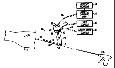
Referring to FIG. 1, a video endoscope 10 is shown
prior to being inserted within an incision port 12 in the
body, here a leg 14 of a patient. Endoscope 10 is of the
type including an optical system (described in detail below)
for conveying an optical image from a distal end of the
endoscope to a video camera 28 attached to the endoscope.
Referring also to FIGS. 2-3, endoscope 10 includes
a housing 16 having a working channel 18 for allowing a
handheld instrument 20 (e.g., ligator. dissector, cutter,
abrader) to extend through the housing for use in a surgical
procedure being viewed with endoscope 10. An elongated
insertion section 22 (which defines an optical axis 25 of
the endoscope) and a detachable sheath 24 extend from
housing 16.
As will be described in greater detail below in
conjunction with FIG. 9, endoscope 10 can be used with any
of a family of detachable sheaths, each of which is sized
and shaped to create and maintain a working space at a
surgical worksite for working instruments introduced through
CA 02327268 2006-12-15
60412-2830
working channel 18 of housing 16. Each detachable sheath 24
also serves to protect elongated insertion section 22,
particularly during advancement through tissue.
A handle 26 for manipulating endoscope 10 extends
in a direction substantially perpendicular to optical axis
25 to provide an unobstructed space, in-line with optical
axis 25 of the endoscope, thereby facilitating manipulation
of surgical instruments introduced through working channel
18. In addition, a rounded heel region 27 of handle 26 has a
low profile to facilitate introduction and manipulation of a
second working instrument within incision port 12 but
beneath insertion section 22. A video camera 28 having an
electro-optical sensor (not shown) is mounted to the upper
end of handle 26. The electro-optical sensor may be, for
example, a charge-coupled device (CCD) for converting
optical images received by the endoscope to electrical image
signals.
Electrical image signals from video camera 28 are
conveyed to a camera control unit 30, via a cable 32, for
view on a display monitor, such as color CRT 34. The upper
end of handle 26 also includes a fitting 36 (FIG. 2) which
receives a fiber optic cable 38 connected to a light source
40. A second fitting 42 (FIG. 2), adjacent fitting 36,
receives a tube 44 connected to a gas (e.g., CO2) or
fluid insufflation source 46. As will be described in
greater detail below, gas provided to fitting 36 is passed
to the surgical worksite through handle 26, housing 16 and a
cylindrically-shaped closed sheath. A focusing ring 48 is
positioned at the upper end of handle 26 to allow the user
to focus endoscope 10. This arrangement provides several
advantages. Arranging handle 26 to be offset from optical
11
CA 02327268 2006-12-15
60412-2830
axis 25 provides an unobstructed space for manipulating
handheld instrument 20. Attachments to endoscope 10 are also
located away from optical axis 25 so that the surgeon can
manipulate the endoscope and working instruments extending
therethrough without interference from cables 32, 38, 44.
Further, because the optical, illumination and gas
insufflation systems are all arranged along the same plane
of handle 26, endoscope 10 can be easily rotated around
optical axis 25 without the cables becoming entangled with
each other or any working instruments used with the
endoscope. Further still, the contour of handle 26 allows
its use by a surgeon with either hand (i.e., left to right
or vice versa).
Referring to FIG. 4, insertion section 22 is shown
with sheath 24 removed to reveal an optical support tube 50
disposed between a pair of illumination support tubes 52,
54. Each tube has a length of approximately 230 mm. Fiber
optic bundles 55 (FIG. 7) are positioned within and extend
the length of support tubes 52, 54, and through housing 16
to fitting 36 on handle 26.
Referring to FIG. 5, an objective lens assembly 56
is disposed within a distal end 58 of optical support tube
50 to receive and convey the image of the object being
viewed to a series of relay lens systems 60 within the
support tube. Objective lens assembly 56 is positioned
within distal end 58 to provide a direction of view pointing
downward toward the open portion of the sheath and at an
angle of about 12 degrees from optical axis 25. For this
reason, distal 58 of support tube 50 is bevelled to hood
objective lens system 60 and provides an unobstructed view
12
CA 02327268 2006-12-15
60412-2830
of the worksite. Objective lens assembly 56 provides a field
of view of about 85 degrees.
With a detachable sheath of the type shown in
FIGS. 1-3, the range of movement at the distal end of a
working instrument passing through endoscope 10 is
substantial (e.g., as much as 2 inches). However, because
the view provided by objective lens assembly 56 is directed
slightly downward, if endoscope 10 is required to be moved
at all, it is most likely moved so that the distal end tilts
upwardly, consistent with the direction endoscope 10 tilts
due to its own weight and the weight of cables 32, 38, 44.
Relay lens systems 60 convey images to a prism 62,
positioned within housing 16, which translates the image to
an axis 64 defined by handle 26. In particular, the image is
received by a series of a relay lenses 66 disposed within a
vertical tube 68 within handle 26. An ocular lens 70 is
positioned at the proximal end of vertical tube 68 to
receive and convey the image to a focusing lens 72. Focusing
lens 72 is supported within a sleeve 74 mechanically coupled
to focusing ring 48 which, when rotated, moves focusing lens
72 along axis 64 of handle 26. An endoscope mechanism
suitable for use in endoscope 10 is described in U.S. Pat.
No. 5,575,757, entitled "Endoscope with Focusing Mechanism",
assigned to the assignee of the present invention and
incorporated herein by reference. Handle 26 includes, at its
proximal end, a centering mount ring 76 for receiving video
camera 28.
Referring again to FIG. 4, as well as FIGS. 6-7,
working channel 18 is positioned adjacently below optical
support tube 50 and illumination support tubes 52. Working
13
CA 02327268 2006-12-15
60412-2830
channel 18 has a working length between about 200 and 230 mm
and a diameter in a range between about 5.5 mm and 7 mm for
an endoscope 10 having an insertion section with a diameter
between 10 and 14 mm. A working channel of this dimension is
sufficiently sized to receive handheld working instruments
having shafts of 5 to 6 mm diameter. Working channel 18
includes a sector or pie-shaped port 80 (FIG. 5) to allow
greater side-to-side maneuverability of instruments used
through the working channel. Due to the shape of port 80 and
the length of the insertion section 22, a relatively small
movement of working instrument 20 at the proximal end of
endoscope translates to a much larger movement at the
worksite, with sheath 24 providing better visibility by
moving tissue away.
Referring to FIG. 6, housing 16 includes a distal
end 84 having a tapering outer surface 86a for mating with a
corresponding tapering inner surface 86b of a locking ring
90. Locking ring 90 is attached to and rotates freely about
the proximal end of detachable sheath 24. In one embodiment,
outer surface 86a is tapered relative to optical axis 25 at
an angle of about 8 degrees.
For applications requiring gas insufflation,
housing 16 also includes a gas port 59 (FIG. 7) which
connects to a conduit (not shown) extending through handle
26 to fitting 36. As will be discussed in greater detail
below, when a cylindrical detachable sheath 24e (FIG. 9.) is
attached to distal end 84 of housing 16, gas insufflation
flows to the surgical worksite through gas port 59 and along
the length of sheath 24e. Because sheath 24e is "closed"
(i.e., does not have an open side wall) it serves as a
conduit between gas port 59 and the surgical worksite. In
14
CA 02327268 2006-12-15
60412-2830
such applications, a fitting 85 is shown permanently
attached to entrance port 80 for receiving a gas seal member
81 (FIG. 11) to provide an air-tight seal between a working
instrument passing through working channel 18.
Referring to FIG. 8, the coupling between
detachable sheath 24 and housing 16 is accomplished using a
bayonet mount. In particular, a pin 92 projects upwardly
from outer surface 86a of housing 16 and is received within
an L-shaped slot 94 of locking ring 90. L-shaped slot 94
includes a longitudinal groove 96 terminating at a groove
98. To lock detachable sheath 24 to housing 16, pin 92 is
slid within longitudinal groove 96 until it reaches groove
98. A projecting stem 100 formed on locking ring 90 is then
rotated counterclockwise to draw surfaces 86a, 86b of
respective ones of housing 16 and locking ring 90 together
in a self-locking manner. The bayonet mount also includes an
0-ring 102 (FIG. 6) to seal the interface between housing 16
and sheath 24 in the event that gas insufflation is
required.
FIG. 9 shows an exemplary set of detachable
sheaths 24a-24e, each being approximately 230 mm in length
and having shafts llla-llle extending between locking ring
90 and corresponding distal end members 1l0a-110e. The
shafts llla-llld of some of the sheaths 24a-24d are open. By
"open" it is meant that shafts llla-llld have C-shaped walls
(in cross section) which define open sides 112a-112d and
extend over a predetermined arc of curvature along
substantially the entire length of the sheaths. In contrast,
"closed" in this context means that shaft forms a complete
tube. For example, sheath 24e has a closed tube-like shaft
lila having a distal end member 110e with a C-shaped open
CA 02327268 2006-12-15
60412-2830
wall. Of course, all sheaths 24a-24e have open distal ends,
as shown in FIG. 9.
In general, because shafts lila-llid include open
sides 112a-112d pressure and trauma inflicted upon the
surrounding anatomy is minimized as endoscope 10 and sheath
24 are advanced through tissue. Sheaths 24a-24d with open
sides 112a-112d are particularly well-suited in procedures
in which more than one handheld instrument is being used at
the same time. For example, one handheld instrument is used
through the sheath while the other instrument is used along
side the sheath. Open sides 112a-112d also permit a larger
range of movement (particularly lateral movement) of
handheld instruments introduced through working channel 18
and hooded by the sheaths.
In general, each of open sheaths 24a-24d include
distal end members 1l0a-110d shaped to create and maintain a
working space at the surgical site. Specifically, distal end
members ll0a-110c are shaped with radii of curvature greater
than that of shafts llla-lllc. The larger radii of distal
end members 110a-110c serve to push surrounding tissue away
at the distal end of the sheaths, thereby creating an
expanded working space. However, distal end members 110a-
110d are sized and configured differently to adapt to
anatomical differences between patients as well as the
particular worksite within a patient.
For example, distal end member 110a is
cylindrically shaped and extends coaxially and in parallel
with shaft illa a distance of approximately 50 mm from the
tip of sheath 24a.
16
CA 02327268 2006-12-15
60412-2830
Sheath 24b and sheath 24c are particularly well-
suited for surgical procedures involving the lower leg or
thigh because each sheath maintains a working channel
generally parallel with the leg while providing an exposed,
fuller view at the distal end of each sheath. Distal end
member 110b of sheath 24b has a cylindrically shaped
proximal portion 113 which extends distally and in parallel
with shaft lllb and then flares outwardly to provide an
enlarged working space.
Referring to FIGS. 10A-10B, outwardly flaring
distal end member 110b has a length of about 0.75 inches and
extends outwardly, relative to optical axis 25, at an angle
of 25 degrees. End portion 114, in cross-section, extends
about 58 degrees to either side of a vertical plane 116
passing through optical axis 25 and perpendicular to a
horizontal axis 117. With this configuration, a working
instrument passed through working channel 18 and hooded by
sheath 24 is capable of being maneuvered at the distal end
of the sheath by as much as 1 inch to either side of plane
116.
Referring again to FIG. 9, distal end member 110c
has a shape which gradually tapers outwardly from a proximal
end to a distal end. Sheath 24d, on the other hand, has an
integrally formed distal end member 110d which is not
enlarged (i.e., distal end member has the same radius of
curvature as shaft llld). Distal end member 110d has a C-
shaped wall 117 (in cross-section) similar to that of shaft
llld, but having a smaller opening between edges 119
defining C-shaped wall 117. C-shaped wall 117 provides
greater overall structural rigidity to sheath 24d in the
event that endoscope 10 and sheath 24d are lifted. In
17
CA 02327268 2006-12-15
60412-2830
addition, edges 119 of distal end member 110d provides
support surfaces which rest upon underlying tissue and allow
endoscope 10 and sheath 24d to remain in place with little
or no support by the operator at handle 26.
Distal ends l10a-110d can be provided as a
separate member permanently attached (e.g., soldered) to the
end of the sheaths, as is the case for sheaths 24a-24c.
Alternatively, as is the case with sheath 24d, distal end
member 110d may be integrally-formed to the sheath.
In applications (e.g., SEPS), where gas
insufflation is required at the surgical site, a cylindrical
sheath 24e having a "closed" shaft 112e is provided. Sheath
24e communicates with gas port 59 in housing 16 to provide a
supply channel for gas provided from insufflation source 46
(FIG. 1) through handle 26, housing 16 and through the
cylindrical sheath. Because gas insufflation is relied upon
to maintain a working space at the surgical worksite, sheath
24e does not require a flared distal end typical of open
sheaths 24a-24d. In essence, sheath 24e creates a common
working channel endoscope through which a wide variety of
working surgical instruments can be introduced therethrough
while allowing simultaneous viewing of the surgical worksite
at the end of the sheath. The inner surface of closed sheath
24e also serves to guide the surgical instrument to the
worksite. Distal end member 110e of sheath 24e also includes
a longitudinal slot 121 which is used to provide simple
dissection of tissue. For example, as sheath 24e is being
advanced through tissue, veins which may require dissection
are encountered. In these situations, slot 121 is used to
peel away tissue surrounding the vein to determine, for
example, whether the vein requires dissection.
18
CA 02327268 2006-12-15
60412-2830
Referring to FIG. 11, a threaded seal 120 is slid
over and positioned at the proximal end of closed sheath
24e. Seal 120 is secured in place using locking ring 122
and, in use, is threaded into incision port 12 to prevent
escape of the gas from the incision port. Gas seal member 81
is placed over fitting 85 of housing 16 to provide an air-
tight seal between a working instrument passing through
working channel 18.
Referring again to FIG. 1, endoscope 10 having an
open sheath 24 is shown in use in a procedure for harvesting
a saphenous vein. Prior to placing endoscope 10 within
incision port 12, a dissector 20 is used at incision port 12
to separate the fascia from the tissue. Dissection by direct
visualization is generally limited to an area of only about
5 cm from incision port 12.
Referring to FIG. 12, after this initial
dissection procedure, endoscope 10 with sheath 24 are
inserted together through port 12 with the sheath held
generally parallel to leg 14. Dissector 20 is then
introduced through working channel 18 of endoscope 10 and
separation of the fascia and tissue is continued with
endoscope 10 used to visualize the dissection. Dissection
continues in this manner, with sheath 24 maintaining a
working space for dissector 20, until the surgical site is
reached. In some surgical procedures, endoscope 10 is not
required to be removed during this dissection procedure.
However, in other procedures, endoscope 10 may be
used first with a detachable sheath 24 having a distal end
member 110 with a relatively small cross-section (e.g.,
19
CA 02327268 2006-12-15
60412-2830
sheath 24d) to advance the endoscope to the target worksite.
Upon arriving at the surgical worksite, endoscope 10 can be
removed and a detachable sheath 24 having a larger distal
end member (e.g., sheath 24a or 24b) can be used to
temporarily displace tissue surrounding the worksite,
thereby allowing better visualization of the worksite when a
handheld instrument is passed through working channel 18 of
endoscope. Alternatively, a larger sheath may be required to
accommodate a different working instrument (e.g., ligator).
In still other procedures, a closed detachable sheath (e.g.,
sheath 24e) may be exchanged for an open sheath and gas
insufflation provided to displace tissue and expand the
worksite.
Other embodiments and applications are within the
claims. For example, although endoscope 10 is described as
being useful for a saphenous vein harvesting procedure, it
can be used in wide variety of surgical applications,
including treatment of patients having incompetent
perforating veins in a leg and suffering from chronic venous
insufficiency.
In addition, use of endoscope 10 is not limited to
vascular procedures, but has application in other surgical
procedures where a working space needs to be maintained and
simultaneous visualization is required. For example,
endoscope 10 may be used to examine the thoracic cavity or
to perform certain plastic surgical procedures.