Note: Descriptions are shown in the official language in which they were submitted.
CA 02327314 2000-11-O1
WO 99/60120 PCTlGB99/01588
SOLUBLE T CELL RECEPTOR
This invention relates to non-membrane bound recombinant T cell
receptors (TCRs) and to methods and reagents for producing them.
General background
1. Antigen presentation on the cell surface
io MHC molecules are specialised protein complexes which present short
protein fragments, peptide antigens, for recognition on the cell surface by
the cellular arm of the adaptive immune system.
Class I MHC is a dimeric protein complex consisting of a variable heavy
is chain and a constant light chain, ~i2microglobulin. Class I MHC presents
peptides which are processed intracellularly, loaded into a binding cleft in
the MHC, and transported to the cell surface where the complex is
anchored in the membrane by the MHC heavy chain. Peptides are usually
8-11 amino acids in length, depending on the degree of arching introduced
2o in the peptide when bound in the MHC. The binding cleft, which is formed
by the membrane distal a1 and a2 domains of the MHC heavy chain, has
°closed" ends, imposing quite tight restrictions on the length of
peptide
which can be bound.
2s Class II MHC is also a dimeric protein consisting of an a (heavy) and a ~
(light) chain, both of which are variable glycoproteins and anchored in the
cell by transmembrane domains. Like Ciass I MHC, the Class II molecule
forms a binding cleft in which longer peptides of 12-24 amino acids are
inserted. Peptides are taken up from the extracellular environment by
3o endocytosis and processed before loading into the Class II complex, which
is then transported to the cell surface.
CA 02327314 2000-11-O1
WO 99/60120 PCT/GB99/01588
2
Each cell presents peptides in up to six different Class I molecules and a
similar number of Class I! molecules, the total number of MHC complexes
presented being in the region 105-10g per cell. The diversity of peptides
presented in Class I molecules is Typically estimated to be between 1,000-
s 10,000, with 90% of these being present in 100-1,000 copies per cell (Hunt,
Michel et al. 1992; Chicz, Urban et al. 1993; Engelhard, Appella et al. 1993;
Huczko, Bodnar et al. 1993). The most abundant peptides are thought to
constitute between 0.4-5% of the total peptide presented, which means that
up to 20,000 identical complexes could be present. However, an average
to number for the most abundant single peptide complexes is likely to be in
the region of 2,000-4,000 per cell, and typical presentation levels of
recognisable T cell epitopes are in the region of 100-500 complexes per
cell (for review see (Engelhard 1994)).
is 2. Recognition of antigen presenting cells
A wide spectrum of cells can present antigen, as MHC-peptide, and the
cells that have that property are known as antigen presenting cells (APC).
The type of cell which presents a particular antigen depends upon how and
2o where the antigen first encounters cells of the immune system. APCs
include the interdigitating dendritic cells found in the T cell areas of the
lymph nodes and spleen in large numbers; Langerhans cells in the skin;
follicular dendritic cells in B cell areas of the lymphoid tissue; monocytes,
macrophages and other cells of the monocyte/macrophage lineage; B cells
2s and T cells; and a variety of other cells such as endothelial cells and
fibroblasts which are not classical APCs but can act in the manner of an
APC.
APCs are recognised by a subgroup of lymphocytes which mature in the
3o thymus (T cells), where they undergo a selection procedure designed to
ensure that T cells which respond to self-peptides are eradicated (negative
selection). In addition, T cells which do not have the ability to recognise
the
CA 02327314 2000-11-O1
WO 99/60120 PCT/GB99/01588
3
MHC variants which are presented (in man, the HLA haplotypes) fail to
mature (positive selection).
Recognition of specific MHC-peptide complexes by T cells is mediated by
the T cell receptor (TCR) which consists of an a- and a a-chain, both of
which are anchored in the membrane. In a recombination process similar
to that observed for antibody genes, the TCR a and p genes rearrange
from Variable, Joining, Diversity and Constant elements creating enormous
diversity in the extracellular antigen binding domains {10'3 to 10'5 different
io possibilities). TCRs also exist in a different form with y and 8 chains,
but
these are only present on about 5% of T cells.
Antibodies and TCRs are the only two types of molecules which recognise
antigens in a specific manner, and thus the TCR is the only receptor for
is specific for particular peptide antigens presented in MHC, the alien
peptide
often being the only sign of an abnormality within a cell,
TCRs are expressed in enormous diversity, each TCR being specific for
one or a few MHC-peptide complexes. Contacts between TCR and MHC-
2o peptide ligands are extremely short-lived, usually with a half life of less
than
a second. Adhesion between T cells and target cells, presumably
TCR/MHC-peptide, relies on the employment of multiple TCRI MHC-
peptide contacts as well as a number coreceptor-ligand contacts.
2s T cell recognition occurs when a T-cell and an antigen presenting cell
{APC) are in direct physical contact and is initiated by ligation of antigen-
spec~c TCRs with pMHC complexes. The TCR is a heterodimeric cell
surface protein of the immunoglobulin superfamily which is associated with
invariant proteins of the CD3 complex involved in mediating signal
3a transduction. TCRs exist in a~i and y8 forms, which are structurally
similar
but have quite distinct anatomical locations and probably functions. The
extracellular portion of the receptor consists of two membrane-proximal
CA 02327314 2000-11-O1
WO 99/60120 PCT/GB99/01588
4
constant domains, and two membrane-distal variable domains bearing
highly polymorphic loops analogous to the complementarity determining
regions (CDRs) of antibodies. It is these loops which form the MHC-
binding site of the TCR molecule and determine peptide spec~city. The
MHC class I and class II ligands are also immunoglobulin superfamily
proteins but are specialised for antigen presentation, with a highly
polymorphic peptide binding site which enables them to present a diverse
array of short peptide fragments at the APC cell surface.
io Recently, examples of these interactions have been characterised
structurally (Garboczi, Ghosh et al. 1996; Garcia, Degano et al. 1996; Ding,
Smith et al. 1998). Crystallographic structures of murine and human Class
I pMHC-TCR complexes indicate a diagonal orientation of the TCR over its
pMHC ligand and show poor shape complementarity in the interface.
is CDR3 loops contact exclusively peptide residues. Comparisons of
liganded and unliganded TCR structures also suggest that there is a
degree of flexibility in the TCR CDR loops (Garboczi and Biddison 1999).
T cell activation models attempt to explain how such protein-protein
2o interactions at an interface between T cell and antigen presenting cell
(APC) initiate responses such as killing of a virally infected target cell.
The
physical properties of TCR-pMHC interactions are included as critical
parameters in many of these models. For instance, quantitative changes in
TCR dissociation rates have been found to translate into qualitative
2s differences in the biological outcome of receptor engagement, such as full
or partial T cell activation, or antagonism (Matsui, Boniface et al. 1994;
Rabinowitz, Beeson et al. 1996; Davis, Boniface et al. 1998).
TCR-pMHC interactions have been shown to have low affinities and
3o relatively slow kinetics. Many studies have used biosensor technology,
such as Biacore~"" (Wllcox, Gao et al. 1999; Wyer, Wrllcox et al. 1999),
which exploits surface plasmon resonance (SPR) and enables direct affinity
CA 02327314 2000-11-O1
WO 99/60120 PCT/GB99/01588
and real-time kinetic measurements of protein-protein interactions (Garcia,
Scott et al. 1996; Davis, Boniface et al. 1998). However, the receptors
studied are either alloreactive TCRs or those which have been raised in
response to an artificial immunogen.
s
3. TCR and CD8 interactions with MHC-peptide complexes
The vast majority of T cells restricted by Class I MHC-peptide complexes
also require the engagement of the coreceptor CD8 for activation, while T
to cells restricted by Class II MHC require the engagement of CD4. The exact
function of the coreceptors in T cell activation is not yet entirely
clarified.
Neither are the critical mechanisms and parameters controlling activation.
However, both CD8 and CD4 have cytoplasmic domains which are
associated with the kinase p56~~' which is involved in the very earliest
is tyrosine phosphorylation event which characterises T cell activation. CD8
is a dimeric receptor, expressed either in an as form or, more commonly, in
an a~i form. CD4 is a monomer. In the CD8 receptor, only the a-chain is
associated with p56~~'.
2o Recent determinations of the physical parameters controlling binding of
TCR and CD8 to MHC, using soluble versions of the receptors, has shown
that binding by TCR dominates the recognition event. TCR has
significantly higher affinity for MHC than the coreceptors ~Ilcox, Gao et
al. 1999; Wyer, Willcox et al. 1999).
2s
The individual interactions of the receptors with MHC are very short-lived at
physiological temperature, i.e. 37°C. An approximate figure for the
half life
of a TCR-MHC/peptide interaction, measured with a human TCR specific
for the influenza virus matrix" peptide presented by HLA-A*0201 (HLA A2),
3o is 0.7 seconds. The half life of the CDBaa interaction with this
MHC/peptide
complex is less than 0.01 seconds, i.e. at least 18 times faster.
CA 02327314 2000-11-O1
WO 99/60120 PGT/GB99/01588
6
4. Production of soluble MHC-peptide complexes
Soluble MHC-peptide complexes were first obtained by cleaving the
molecules of the surface of antigen presenting cells with papain (Bjorkman,
Strominger et al. 1985). Although this approach provided material for
crystallisation, it has, for Class I molecules, in recent years been replaced
by individual expression of heavy and light chain in E.coli followed by
refolding in the presence of synthetic peptide (Garboczi, Hung et al. 1992;
Madden, Garboczi et al. 1993; Garboczi, Madden et al. 1994; Reid,
to McAdam et al. 1996; Reid, Smith et al. 1996; Smith, Reid et al. 1996;
Smith, Reid et al. 1996; Gao, Tormo et al. 1997; Gao, Gerth et al. 1998).
This approach has several advantages over previous methods in that a
better yield is obtained at a lower cost, peptide identity can be controlled
very accurately, and the final product is more homogeneous. Furthermore,
is expression of modified heavy or light chain, for instance fused to a
protein
tag, can be easily performed.
5. Soluble TCR
2o At present, there are no published data on human TCR-pMHC interactions,
and no studies arfalysing naturally selected TCRs specific for natural (e.g.
viral) epitopes. This may reflect difficulties in obtaining protein which is
suitable for SPR i.e. protein which is homogenous, monomeric, correctly
folded, available in milligram quantities and stable over a range of
2s concentration.
It would be an advantage to be able to produce a recombinant TCR in non-
membrane bound (or soluble) form, not only for the purpose of investigating
specific TCR-pMHC interactions, but also as a diagnostic tool to detect
3o infection or to detect autoimmune disease markers, or to detect the
efficacy
of T cell vaccines. Soluble TCR would also have applications in staining,
for example to stain cells for the presence of a particular viral antigen
CA 02327314 2000-11-O1
WO 99/60120 PCT/GB99/01588
7
presented in the context of the MHC. Similarly, the soluble TCR could be
used to deliver a therapeutic agent, for example a cytotoxic compound or
an immunostimulating compound, to cells presenting a particular antigen.
s Proteins which are made up of more than one polypeptide subunit and
which have a transmembrane domain can be difficult to produce in soluble
form because in many cases the protein is stabilised by its transmembrane
region. This is the case for the TCR.
to Production of soluble TCR has only recently been described by a number
of groups. In general, all methods describe truncated forms of TCR,
containing either only extracellular domains or extracellular and
cytoplasmic domains. Thus, in all cases, the transmembrane domains
have been deleted from the expressed protein. Although many reports
is show that TCR produced according to their methods can be recognised by
TCR-specific antibodies (indicating that the part of the recombinant TCR
recognised by the antibody has correctly folded), none has been able to
produce a soluble TCR at a good yield which is stable at low
concentrations and which can recognise MHC-peptide comptexes
The first approach to yield crystallisable material made use of expression in
eukaryotic cells but the material is extremely expensive to produce (Garcia,
Degano et al. 1996; Garcia, Scott et al. 1996). Another approach which
has produced crystallisable material made use of an E.coli expression
2s system similar to what has previously been used for MHC-peptide
complexes (Garboczi, Ghosh et al. 1996; Garboczi, Utz et al. 1996). The
latter method, which expresses the extracellular portions of the TCR chains
truncated immediately before the cysteine residues involved in forming the
interchain disulphide bridge, followed by refolding in vitro, has turned out
3o not to be generally applicable. Most heterodimeric TCRs appear to be
unstable when produced in this fashion due to low affinity between the a
and ~3 chains.
CA 02327314 2000-11-O1
WO 99/60120 PCT/GB99/01588
8
In addition, a number of other descriptions of engineered production of
soluble TCR exist. Some of these describe only the expression of either
the a or (i chain of the TCR, thus yielding protein which does not retain
s MHC-peptide specific binding (Calaman, Carson et al. 1993; Ishii, Nakano
et al. 1995). ~3 chain crystals have been obtained without a chain, either
alone or bound to superantigen (Boulot, Bentley et al. 1994; Bentley, Boulot
et al. 1995; Fields, Malchiodi et al. 1996).
to Other reports describe methods for expression of heterodimeric y/S or a/(i
TCR (Gregoire, Rebai et al. 1991; Necker, Rebai et al. 1991; Eilat, Kikuchi
et al. 1992; Weber, Traunecker et al. 1992; Corr, Slanetz et al. 1994; Ishii,
Nakano et al. 1995; Gregoire, Malissen et al. 1996; Romagne, Peyrat et al.
1996). In some cases, the TCR has been expressed as a single chain
is fusion protein (Brocker, Peter et al. 1993; Gregoire, Malissen et al. 1996;
Schlueter, Schodin et al. 1996). Another strategy has been to express the
TCR chains as chimeric proteins fused to Ig hinge and constant domains
(Eilat, Kikuchi et al. 1992; Weber, Traunecker et al. 1992). Other chimaeric
TCR proteins have been expressed with designed sequences which form
2o coiled-coils which have high affinity and specificity for each other, thus
stabilising TCR a-(i contacts and increasing solubility. This approach was
taken by Chang, Bao et al. (1994) who replaced the transmembrane region
of the protein with a leucine zipper protein consisting of two synthetic
peptide sequences, an acid peptide and a base peptide, that specifically
2s interact to create a heterodimeric coiled coil. The authors employed a
bacculovirus expression system in eukaryotic cells to secrete heterodimeric
TCR protein. The artificial leucine zipper peptides assist
heterodimerisation of the TCR a and ~i chains, which are also linked by an
interchain disulphide band just above the fusion point with the zipper
3o peptides. However, these techniques have not since proved successful
and there is no evidence that the soluble TCR described can recognise a
CA 02327314 2000-11-O1
WO 99/60110 PCT/GB99/01588
9
TCR ligand. Similarly, Golden, Khandekar et al (1997) described the
production of heterodimeric T cell receptors as leucine zipper fusion
proteins. The soluble TCR was expressed in E. coli as a secreted
heterodimer with the a-~i interchain disulphide bond as in Chang et al.
s Again, there is no evidence that this ready-folded TCR heterodimer is
capable of interacting with its ligand.
There is therefore a need for a soluble version of the membrane bound
TCR, which is correctly folded so that it is capable of recognising its native
to ligand. A soluble form of a TCR which is stable over a period of time, and
a
method for producing it in reasonable quantities, would also be useful. The
present invention aims to meet some or all of these requirements.
The Invention
is
The invention provides in one aspect a refolded recombinant T cell receptor
(TCR) which comprises:
i) a recombinant TCR a or y chain extracellular domain having a
first heterologous C-terminal dimerisation peptide; and
2o ii) a recombinant TCR (i or b chain extracellular domain having a
second C-terminal dimerisation peptide which is specifically
heterodimerised with the first dimerisation peptide to form a
heterodimerisation domain.
2s The TCR according to the invention, which is refolded after being
expressed, rather than secreted as a heterodimer, is conformationally
superior to soluble TCR previously available. This is indicated by its ability
to recognise MHC-peptide complexes. It is also stable at relatively low
concentrations. It may be stable at concentrations below 1 mglml and
3o preferably about 10 pglml.
CA 02327314 2000-11-O1
WO 99/60120 PCT/GB99/01588
In a further aspect, the invention provides a biologically-active recombinant
T cell receptor (TCR) which comprises:
i) a recombinant TCR a or y chain extracellular domain having a
first heterologous C-terminal dimerisation peptide; and
s ii) a recombinant TCR ø or 8 chain extracellular domain having a
second C-terminal dimerisation peptide which is specifically
heterodimerised with the first dimerisation peptide to form a
heterodimerisation domain. Such a TCR will bind spec~cally to MHC-
peptide complexes, preferably in a manner similar to the native TCR from
to which it is derived.
In another aspect, the invention provides recombinant nucleic acid
sequences encoding the recombinant TCR chains as described herein.
Such nucleic acid sequences may be isolated from T-cell clones.
is Alternatively, they may be produced synthetically, for example by inserting
a heterologous nucleic acid sequence in a nucleic acid sequence encoding
a TCR chain, or by mutating (by insertion, deletion or substitution) a nucleic
acid sequence encoding a TCR chain. Thus, the invention includes within
its scope synthetic peptides which have the activity and/or binding
2o specificity of native TCRs.
In yet another aspect, the invention provides a method of making a
recombinant non membrane bound T cell receptor, which method
comprises:
2s expressing a recombinant TCR oc or y chain extracellular
domain having a first heterologous C-terminal dimerisation peptide, and a
recombinant TCR ø or b chain extracellular domain having a second C-
terminal dimerisation peptide which specifically heterodimerises with the
first dimerisation peptide to form a heterodimerisation domain; and
3o refolding the chains together in vitro to produce a TCR
heterodimer.
CA 02327314 2000-11-O1
WO 99/60120 PCf/GB99/01588
11
Recombinant TCRs in accordance with the invention may be for
recognising Class I MHC-peptide complexes and Class II MHC-peptide
complexes.
s
The heterodimerisation domain of the recombinant TCR according to the
invention is preferably a so-called °coiled coil" or "leucine zipped'.
These
terms are used to describe pairs of helical peptides which interact with
each other in a specific fashion to form a heterodimer. The interaction
to occurs because there are complementary hydrophobic residues along one
side of each zipper peptide. The nature of the peptides is such that the-
formation of heterodimers is very much more favourable than the formation
of homodimers of the helices. Leucine zippers may be synthetic or
naturally occurring. Synthetic leucines can be designed to have a much
is higher binding affinity than naturally occurring leucine zippers, which is
not
necessarily an advantage. In fact, preferred leucine zippers for use in the
invention are naturally occurring leucine zippers or leucine zippers with a
similar binding affinity. Leucine zippers from the c jun and c-fos protein are
an example of leucine zippers with a suitable binding affinity. Other
2o suitable leucine zippers include those from the myc and max proteins
(Amati, Dalton, et al 1992). Other leucine zippers with suitable properties
could easily be designed (O'Shea et al 1993).
It is preferred that the soluble TCRs in accordance with the invention have
2s approximately 40 amino acid leucine zipper fusions corresponding to the
heterodimerisation domains from c jun (achain) and c-fos (chain)
(O'Shea, Rutkowski et al 1989, O'Shea, Rutkowski et al, 1992, Glover and
Harrison, 1995). Longer leucine zippers may be used. Since
heterodimerisation specificity appears to be retained even in quite short
so fragments of some leucine zipper domains (O'Shea, Rutkowski et al, 1992),
it is possible that a similar benefit could be obtained with shorter c jun and
c-fos fragments. Such shorter fragments could have as few as 8 amino
CA 02327314 2000-11-O1
WO 99/60120 PCT/GB99/01588
12
acids for example. Thus, the leucine zipper domains may be in the range
of 8 to 60 amino acids long.
The molecular principles of specificity in leucine zipper pairing is well
s characterised (Landschulz, Johnson et al, 1988; McKnight, 1991 ) and
leucine zippers can be designed and engineered by those skilled in the art
to form homodimers, heterodimers or trimeric complexes (Lumb and Kim,
1995; Nautiyal, Woolfson et al, 1995; Boice, Dieckmann et al, 1996, Chao,
Houston et al, 1996). Designed leucine zippers, or other
io heterodimerisation domains, of higher affinity than the c jun and c-fos
leucine zippers may be beneficial for the expression of soluble TCRs in
some systems. However, as mentioned in more detail below, when soluble
TCR is folded in vitro, a solubilising agent is preferably included in the
folding buffer to reduce the formation of unproductive protein aggregates.
is One interpretation of this phenomenon is that the kinetics of folding of
the
leucine_ zipper domains are faster than for the TCR chains, leading to
dimerisation of unfolded TCR a and ~i chain, in tum causing protein
aggregation. By stowing the folding process and inhibiting aggregation by
inclusion of solubilising agent, the protein can be maintained in solution
2o until folding of both fusion domains is completed. Therefore,
heterodimerisation domains of higher affinity than the c-fos and c jun
leucine zippers may require higher concentrations of solubilising agent to
achieve a yield of soluble TCRs comparable to that for c jun and c-fos.
2s Different biological systems use a variety of methods to form stable homo-
and hetero protein dimers, and each of these methods in principle provide
an option for engineering dimerisation domains into genetically modified
proteins. Leucine zippers (Kouzarides and Ziff 1989) are probably the most
popular dimerisation modules and have been widely used for production of
so genetically designed dimeric proteins. Thus, the leucine zipper of GCN4, a
transcriptional activator protein from the yeast Saccharomyces cerevisiae,
has been used to direct homodimerisation of a number of heterologous
CA 02327314 2000-11-O1
WO 99/60120 PCT/GB99/01588
13
proteins (Hu, Newell et al. 1993; Greenfield, Montelione et al. 1998). The
preferred strategy is to use zippers that direct formation of heterodimeric
complexes such as the Jun/Fos leucine zipper pair (de Kruif and
Logtenberg 1996; Riley, Ralston et al. 1996).
s
The heterodimerisation domain of the recombinant TCR according to the
present invention is not limited to leucine zippers. Thus, it may be provided
by disulphide bridge-forming elements. Alternatively, it may be provided by
the SH3 domains and hydrophobidproline rich counterdomains, which are
1o responsible for the protein-protein interactions seen among proteins
involved in signal transduction (reviewed by Schlessinger, (Schlessinger
1994). Other natural protein-protein interactions found among proteins
participating in signal transduction cascades rely on associations between
post-translationally modified amino acids and protein modules that
1s specifcally recognise such modified residues. Such post-translationally
modified amino acids and protein modules may form the heterodimerisation
domain of the recombinant TCR in accordance with the invention. An
example of a protein pair of this type is provided by tyrosine
phosphorylated receptors such as Epidermal Growth Factor Receptor or
2o Platelet Derived Growth Factor Receptor and the SH2 domain of GRB2
(Lowenstein, Daly et al. 1992; Buday and Downward 1993). As in all fields
of science, new dimerisation modules are being actively sought (Chevray
and Nathans 1992) and methods for engineering completely artificial
modules have now successfully been developed (Zhang, Murphy et al.
2s 1999).
In a preferred recombinant TCR according to the invention, an interchain
disulphide bond which forms between two cysteine residues in the native a
and ~3 TCR chains and between the native Y and b TCR chains, is absent.
3o This may be achieved for example by fusing the dimerisation domains to
the TCR receptor chains above the cysteine residues so that these
residues are excluded from the recombinant protein. In an alternative
CA 02327314 2000-11-O1
WO 99/60120 PCT/GB99/01588
14
example, one or more of the cysteine residues is replaced by another
amino acid residue which is not involved in disulphide bond formation.
These cysteine residues may not be incorporated because they may be
detrimental to in vitro folding of functional TCR.
s
Refolding of the a and ~i chains or Y and b chains of the refolded
recombinant TCR according to the invention takes place in vitro under
suitable refolding conditions. In a particular embodiment, a recombinant
TCR with correct conformation is achieved by refolding solubilised TCR
io chains in a refolding buffer comprising a solubilising agent, for example
urea. Advantageously, the urea may be present at a concentration of at
least 0.1 M or at least 1 M or at least 2.5M, or about 5M. An alternative
solubilising agent which may be used is guanidine, at a concentration of
between 0.1 M and 8M, preferably at least 1 M or at least 2.5M. Prior to
is refolding, a reducing agent is preferably employed to ensure complete
reduction of cysteine residues. Further denaturing agents such as DTT
and guanidine may be used as necessary. Different denaturants and
reducing agents may be used prior to the refolding step (e.g. urea, (i-
mercaptoethanol). Alternative redox couples may be used during refolding,
2o such as a cystamine/cysteamine redox couple, DTT or (i-
mercaptoethanoUatmospheric oxygen, and cysteine in reduced and
oxidised forms.
The monomeric TCR described herein may be labelled with a detectable
2s label, for example a label which is suitable for diagnostic purposes. Thus,
the invention provides a method for detecting MHC-peptide complexes
which method comprises contacting the MHC-peptide complexes with a
TCR in accordance with the invention which is specific for the MHC-peptide
complex; and detecting binding of the TCR to the MHC-peptide complex.
3o In tetrameric TCR formed using biotinylated heterodimers, fluorescent
streptavidin (commercially available) can be used to provide a detectable
label. A fluorescently labelled tetramer will be suitable for use in FACS
CA 02327314 2000-11-O1
WO 99/60120 PCT/GB99/01588
analysis, for example to detect antigen presenting cells carrying the peptide
for which the TCR is speck.
Another manner in which the soluble TCRs may be detected is by the use
of TCR-specific antibodies, in particular monoclonal antibodies. There are
many commercially available anti-TCR antibodies, such as ~iFl and aFl,
which recognise the constant regions of the (i and a chain, respectively.
The TCR may alternatively or additionally be linked to a therapeutic agent
io which may be for example a toxic moiety for example for use in cell
killing,
or an immunostimulating agent such as an interleukin or a cytokine. Thus,
the invention provides a method for delivering a therapeutic agent to a
target cell, which method comprises contacting potential target cells with a
TCR in accordance with the invention under conditions to allow attachment
is of the TCR to the target cell, said TCR being specific for the MHC-peptide
complexes and having the therapeutic agent associated therewith.
In particular, the soluble TCR could be used to deliver therapeutic agents to
the location of cells presenting a particular antigen. For instance, a toxin
could be delivered to a tumour thereby helping to eradicate it. This would
2o be useful in many situations and in particular against tumours because not
all cells in the tumour present antigens and therefore not all tumour cells
are detected by the immune system. Wth the soluble TCR, a compound
could be delivered such that it would exercise its effect locally but not only
on the cell it binds to. Thus, one particular strategy envisages anti-tumour
2s molecules linked to T cell receptors specific for tumour antigens.
Many toxins could be employed for this use, for instance radioactive
compounds, enzymes (perforin for example) or chemotherapeutic agents
(cis-platin for example). To ensure that toxic effects are exercised in the
3o desired location the toxin could be inside a liposome linked to
streptavidin
so that the compound is released slowly. This will prevent damaging
effects during the transport in the body and ensure that the toxin has
CA 02327314 2000-11-O1
WO 99/60120 PCT/GB99/01588
16
maximum effect after binding of the TCR to the relevant antigen presenting
cells.
An alternative way to label or attach another moiety such as a toxic moiety
s to the soluble TCR is by including the label or other moiety in a mixed
molecule multimer. An example of such a multimeric molecule is a
tetramer containing three TCR molecules and one peroxidase molecule.
This could be achieved by mixing the TCR and the enzyrrae at a molar ratio
of 3:1 to generate tetrameric complexes and isolating the desired complex
to from any complexes not containing the correct ratio of molecules. Mixed
molecules could contain any combination of molecules, provided that steric
hindrance does not compromise or does not significantly compromise the
desired function of the molecules. The positioning of the binding sites on
the streptavidin molecule is suitable for mixed tetramers since steric
is hindrance is not likely to occur.
Preferably, the recombinant TCR chains according to the invention have a
flexible linker located between the TCR domain and the dimerisation
peptide. Suitable flexible linkers include standard peptide linkers
2o containing glycine, for example linkers containing glycine and serine. C-
terminal truncations close to the cysteine residues forming the interchain
disulphide bond are believed to be advantageous because the a and ~i
chains are in close proximity through these residues in cellular TCRs.
Therefore only relatively short linker sequences may be required to supply
2s a nondistortive transition from the TCR chains to the heterodimerisation
domain. It is preferred that the linker sequences Pro-Gly-Gly or Gly-Gly are
used. However, the linker sequence could be varied. For instance, the
linker could be omitted completely, or reduced to a single residue, the
preferred choice in this case being a single Glycine residue. Longer linkers
3o variations are also likely to be tolerated in the soluble TCR, provided
that
they could be protected from protease attack which would lead to
CA 02327314 2000-11-O1
WO 99/60120 PCT/GB99/01588
17
segregation of the dimerisation peptides from the extracellular domains of
the TCR w~h ensuing loss of a-~ chain stability.
The soluble TCR according to the invention is not necessarily a-~TCR.
s Molecules such as y-8, a-S and ~-~iTCR molecules, as well as TCR
molecules (pre-TCR) containing invariant alpha chains which are only
expressed early in development are also included. Pre-TCR specifies the
cell lineage which will express a-~ T cell receptor, as opposed to those
cells which will express ~-8 T cell receptor (for reviews, see (Aifantis,
to Azogui et al. 1998; von Boehmer, Aifantis et al. 1998; Wurch, Biro et al.
1998)). The Pre-TCR is expressed with the TCR (3 chain pairing with an
invariant Pre-TCR a chain (Saint Ruf, Ungewiss et al. 1994; Wilson and
MacDonald 1995) which appears to commit the cell to the a-~i T cell
lineage. The role of the Pre-TCR is therefore thought to be important
is during thymus development (Ramiro, Trigueros et al. 1996).
Standard modifications to the recombinant proteins according to the
invention may be made as appropriate. These include for example altering
an unpaired cysteine residue in the constant region of the ~i chain to avoid
2o incorrect intrachain or interchain pairing.
The signal peptide may be omitted since it does not serve any purpose in
the mature receptor or for its ligand binding ability, and rnay in fact
prevent
the TCR from being able to recognise ligand. In most cases, the cleavage
2s site at which the signal peptide is removed from the mature TCR chains is
predicted but not experimentally determined. Engineering the expressed
TCR chains such that they are a few, i.e. up to about 10 for example,
amino acids longer or shorter at the n-terminal end will have no significance
for the functionality of the soluble TCR. Certain additions which are not
3o present in the original protein sequence could be added. For example, a
short tag sequence which can aid in purification of the TCR chains could be
CA 02327314 2000-11-O1
WO 99/60120 PCT/GB99/01588
18
added provided that it does not interfere with the correct structure and
folding of the antigen binding site of the TCR.
For expression in E.coli, a methionine residue may be engineered onto the
s N-terminal starting point of the predicted mature protein sequence in order
to enable initiation of translation.
Far from all residues in the variable domains of TCR chains are essential
for antigen specificity and functionality. Thus, a significant number of
to mutations can be introduced in this region without affecting antigen
specificity and functionality.
By contrast, certain residues involved in forming contacts to the peptide
antigen or the HLA heavy chain polypeptide, i.e. the residues constituting
is the CDR regions of the TCR chains, may be substituted for residues that
would enhance the affinity of the TCR for the ligand. Such substitutions,
given the low affinity of most TCRs for peptide-MHC ligands, could be
useful for enhancing the specificity and functional potential of soluble
TCRs. In the examples herein, the affinities of soluble TCRs for peptide-
2o MHC ligands are determined. Such measurements can be used to assay
the effects of mutations introduced in the TCR and thus also for the
identification of TCRs containing substitutions which enhance the activity of
the TCR.
2s Far from all residues in the constant domains of TCR chains are essential
for antigen specificity and functionality. Thus, a significant number of
mutations can be introduced in this region affecting antigen specificity.
In Example 14 below, we have shown that two amino acid substitutions in
the constant domain of a TCR ~i chain had no detectable consequences for
3o the ability of the TCR to bind a HLA-peptide ligand.
CA 02327314 2000-11-O1
WO 99/60120 PCT/GB99/01588
19
The TCR (3 chain contains a cysteine residue which is unpaired in the
cellular or native TCR. Mutation of this residue enhances the efficiency of
in vitro refolding of soluble TCR. Substitutions of this cysteine residue for
serine or alanine has a significant positive effect on refolding efficiencies
in
vitro. Similar positive effects, or even better effects, may be obtained with
substitutions for other amino acids.
As mentioned previously, it is preferred that the cysteine residues forming
the interchain disulphide bond in native TCR are not present so as to avoid
to refolding problems. However, since the alignment of these cysteine
residues is the natural design in the TCR and also has been shown to be
functional with this alignment for the c jun and c-fos ieucine zipper domains
(O'Shea et al, 1989), these cysteine residues could be included provided
that the TCR could be refolded.
is
Because the constant domains are not directly involved in contacts with the
peptide-MHC ligands, the C-terminal truncation point may be altered
substantially without loss of functionality. For instance, it should be
possible to produce functional soluble TCRs excluding the entire constant
2o domain. in principle, it would be simpler to express and fold soluble TCRs
comprising only the variable regions or the variable regions and only a
short fragment of the constant regions, because the polypeptides would be
shorter. However, this strategy is not preferred. This is because the
provision of additional stability of the a-(i chain pairing through a
2s heterodimerisation domain would be complicated because the engineered
C-termini of the two chains would be some distance apart, necessitating
long linker sequences. The advantage of fusing heterodimerisation
domains just prior to the position of the cysteines forming the interchain
disulphide bond, as is preferred, is that the a and ~ chains are held in close
3o proximity in the cellular receptor. Therefore, fusion at this point is less
likely to impose distortion on the TCR structure.
CA 02327314 2000-11-O1
WO 99/60120 PCT/GB99/01588
It is possible that functional soluble TCR could be produced with a larger
fragment of the constant domains present than is preferred herein, i.e. they
constant domains need not be truncated just prior to the cysteines forming
the interchain disulphide bond. For instance, the entire constant domain
s except the transmembrane domain could be included. It would be
advantageous in this case to mutate the cysteine residues forming the
interchain disulphide bond in the cellular TCR.
In addition to aiding interchain stability through a heterodimerisation
to domain, incorporation of cysteine residues which could form an interchain
disulphide bond could be used. One possibility would be to truncate the a
and (i chains close to the cysteine residues forming the interchain
disulphide bond without removing these so that normal disulphide bonding
could take place. Another possibility would be to delete only the
is transmembrane domains of the a and ~ chains. If shorter fragments of the
a and ~i chains were expressed, cysteine residues could be engineered in
as substitutions at amino acid positions where the folding of the two chains
would bring the residues in close proximity, suitable for disulphide bond
formation.
Purification of the TCR may be achieved by many different means.
Alternative modes of ion exchange may be employed or other modes of
protein purification may be used such as gel filtration chromatography or
affinity chromatography.
In the method of producing a recombinant TCR in accordance with the
invention, folding efficiency may also be increased by the addition of certain
other protein components, for example chaperone proteins, to the refolding
mixture. Improved refolding has been achieved by passing protein through
3o columns with immobilised mini-chaperones (Altamirano, Golbik et al. 1997;
Altamirano, Garcia et al. 1999).
CA 02327314 2000-11-O1
WO 99/60120 PCT/GB99/01588
21
In addition to the methods described in the examples, alternative means of
biotinylating the TCR may be possible. For example, chemical biotinylation
may be used. Alternative biotinylation tags may be used, although certain
amino acids in the biotin tag sequence are essential (Schatz et al, 1993).
s The mixture used for biotinylation may also be varied. The enryme requires
Mg ATP and low ionic strength although both of these conditions may be
varied e.g. it may be possible to use a higher ionic strength and a longer
reaction time. It may be possible to use a molecule other than avidin or
streptavidin to form multimers of the TCR. Any molecule which binds biotin
to in a multivalent manner would be suitable. Alternatively, an entirely
different linkage could be devised (such as poly-histidine tag to chelated
nickel ion (Quiagen Product Guide 1999, Chapter 3 "Protein Expression,
Purification, Detection and Assay" p. 35-37). Preferably, the tag is located
towards the C-terminus of the protein so as to minimise the amount of
is steric hindrance in the interaction with potential peptide-MHC complexes.
Examples of suitable MHC-peptide targets for the TCR according to the
invention include, but are not limited to, viral epitopes such as HTLV-1
epitopes (e.g. the Tax peptide restricted by HLA A2; HTLV-1 is associated
2o with leukaemia), HIV epitopes, EBV epitopes, CMV epitopes; melanoma
epitopes and other cancer-specific epitopes; and epitopes associated with
autoimmune disorders, such as rheumatoid arthritis.
A multitude of disease treatments can potentially be enhanced by localising
2s the drug through the specificity of soluble TCRs.
Viral diseases for which drugs exist, e.g. HIV, SIV, EBV, CMV, would
benefit from the drug being released in the near vicinity of infected cells.
For cancer, the localisation in the vicinity of tumours or metastasis would
3o enhance the effect of toxins or immunostimulants. In autoimmune
diseases, immunosuppressive drugs could be released slowly, having more
local effect over a longer time-span while minimally affecting the overall
immuno-capacity of the subject. In the prevention of graft rejection, the
CA 02327314 2000-11-O1
WO 99/60120 PCT/GB99/01588
22
effect of immunosuppressive drugs could be optimised in the same way.
For vaccine delivery, the vaccine antigen could be localised in the vicinity
of
antigen presenting cells, thus enhancing the effcacy of the antigen. The
method can also be applied for imaging purposes.
s
Preferred features of each aspect of the invention are as for each of the
other
aspects mufafis mutandis. The prior art documents mentioned herein are
incorporated to the fullest extent permitted by law.
to The invention is further described in the following examples, which do not
limit the scope of the invention in any way.
Reference is made in the following to the accompanying drawings in which:
is Figure 1 is a schematic view of a T cell Receptor-leucine zipper fusion
protein. Each chain consists of two immunoglobulin superfamily domains,
one variable (~ and one constant (C). The constant domains are
truncated immediately n-terminal of the interchain cysteine residues, and
fused to a leucine zipper heterodimerisation motif from c-Jun (a) or c-Fos
20 (~3) of around 40 amino acids at the C-terminal via a short linker. The a-
Jun
and ~i-Fos each contain two intrachain disulphide bonds and pair solely by
non-covalent contacts. The alpha chain is shorter than the beta chain due
to a smaller constant domain.
2s Figure 2 is a photograph of a reducing/non-reducing gel analysis of
heterodimeric JM22zip receptor. Identical samples of purified TCR-zipper
were loaded onto a 15% acryfamide SDS gel, either under reducing
conditions (lane 2) and non-reducing conditions (lane 4). Marker proteins
are shown in lanes 1 and 3. Molecular weights are shown in kiiodaltons.
3o Under both sets of conditions, the non-covalently associated heterodimer is
dissociated into alpha and beta chains. In lane 4, each chain runs with a
higher mobility and as a single band, indicating a single species of intra-
CA 02327314 2000-11-O1
WO 99/60120 PCT/GB99/01588
23
chain disulphide bonding is present. This is compatible with correct
disulphide bond formation.
Figure 3 is a graph showing the specific binding of JM22zip TCR to HLA A2
s Flu matrix (M58-66) complexes. HLA-A2 complexes, refolded around
single peptides and biotinylated on ~2-microglobulin have been
immobilised onto three streptavidin-coated flow cells: 3770 Resonance
Units (RU) of HLA-AZ POL control onto flow cell (FC) 3, and two different
levels of HLA A2 M58-6fi FLU (2970 RU on FC1 and 4960 RU on FC2).
to JM22zip has been injected in the soluble phase sequentially over all three
flow cells at a concentration of 43 NM for 60 seconds. During the injection,
an above-background increase in the response of both HLA-A2 FLU-
coated flow cells is seen, with approximately 1000 RU and 700 RU of
specific binding of JM22zip to flow cells 1 and 2 respectively.
is
Figure 4 shows the sequences of synthetic DNA primers used for "anchor
amplification of TCR genes. Recognition sites for DNA restriction enzymes
used for cloning are underlined. A: poly-C "anchor primer". B: TCR a
chain constant region specific primer. C: TCR ~ chain constant region
2o specific primer.
Figure 5 shows the sequences of synthetic DNA primers used for PCR
amplification of DNA fragments encoding the 40 amino acid coiled-coil
("leucine zipper") regions of c jun and c-fos. Recognition sites for DNA
2s restriction enzymes used for cloning are underlined. A: c jun 5' primer. B:
c jun 3'primer. C: c-fos 5' primer. D: c-fos 3' primer.
Figure 6 shows the respective DNA and amino acid (one letter code)
sequences of c-fos and c jun fragments as fused to TCRs (inserts in
3o pBJ107 and pBJ108). A: c jun leucine zipper as fused to TCR a chains. B:
c-fos leucine zipper as fused to TCR ~i chains.
CA 02327314 2000-11-O1
WO 99/60120 PCT/GB99/01588
24
Figure 7 shows the sequences of the synthetic DNA primers used for
mutating the unpaired cysteine residue in TCR ~ chains. The primers were
designed for used with the "QuickchangeTM° method for mutagenesis
s (Stratagene). A: Mutation of cysteine to serine, forwards (sense) primer,
indicating amino acid sequence and the mutation. B: mutation of cysteine
to serine, backwards (nonsense) primer. C: mutation of cysteine to
alanine, forwards (sense) primer, indicating amino acid sequence and the
mutation. D: mutation of cysteine to alanine, backwards (nonsense)
io primer.
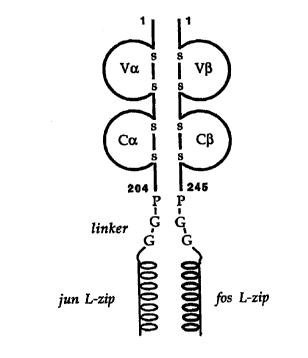
Figure 8 is a schematic representation of a TCR-zipper fusion protein. The
four immunoglobulin domains are indicated as domes, with the intrachain
disulphide bridges between matching pairs of cysteine residues shown.
is The numbers indicate amino acid positions in the mature T-cell receptor
chains; due to slight variation in chain length after recombination, the
lengths of the chains can vary slightly between different TCRs. The
residues introduced in the linker sequences are indicated in the one-letter
code.
Figure 9 shows the sequences of the synthetic DNA primers used for PCR
amplification of TCR a and ~i chains. Recognition sites for DNA restriction
enzymes are underlined and the amino acid sequences corresponding to
the respective TCR chains are indicated over the forward primer
2s sequences. Silent DNA mutations relative to the TCR gene sequences and
other DNA sequences which do not correspond to the TCR genes are
shown in lower case letters. A: 5' PCR primer for the human Va10.2 chain
of the JM22 Influenza Matrix virus peptide-HLA-A0201 restricted TCR. B:
5' PCR primer for the human V~317 chain of the JM22 Influenza Matrix virus
3o peptide-HLA-A0201 restricted TCR. C: 5' PCR primer for the mouse Va4
chain of the Influenza nucleoprotein peptide-H2-Db restricted TCR. D: 5'
CA 02327314 2000-11-O1
WO 99/60120 PCT/GB99/01588
PCR primer for the mouse V~11 chain of the Influenza nucleoprotein
peptide=H2-Db restricted TCR. E: 5' PCR primer of the human Va23 chain
of the 003 HIV-1 Gag peptide-HLA A0201 restricted TCR. F: 5' PCR
primer of the human V~i5.1 chain of the 003 HIV-1 Gag peptide-HLA-A0201
s restricted TCR. G: 5' PCR primer of the human Va2.3 chain of the HTLV 1
Tax peptide-Hl~-A0201 restricted A6 TCR. H: 5' PCR primer of the
human V(i12.3 chain of the HTLV 1 Tax peptide-HLA-A0201 restricted A6
TCR. I: 5' PCR primer of the human Va17.2 chain of the HTLV-1 Tax
peptide-HLA A0201 restricted B7 TCR. J: 5' PCR primer of the human
to V~i12.3 chain of the HTLV 1 Tax peptide-HLA A0201 restricted B7 TCR.
K: 3' PCR primer for human Ca chains, generally applicable. L: 3' PCR
primer for human C~ chains, generally applicable.
Figure 10 shows the predicted protein sequence (one letter code, top) and
is DNA sequence (bottom) of the soluble HLA A2/flu matrix restricted TCR a
chain from JM22, as fused to the "leucine zipper" domain of c jun.
Mutations introduced into the 5' end of the DNA sequence to enhance
expression of the gene in E. coli are indicated in small letters, as is the
linker sequence between the TCR and c jun sequences
Figure 11 shows the predicted protein sequence (one letter code, top) and
DNA sequence (bottom) of the soluble HLA-A2/flu matrix restricted TCR (3
chain from JM22, as fused to the "leucine zipper" domain of c-fos. The
linker sequence between the TCR and o-fos sequences is indicated in
2s small letters.
Figure 12 shows the predicted protein sequence (one fetter code, top) and
DNA sequence (bottom) of the soluble H2-Db/lnfluenza virus nucleoprotein
restricted TCR a chain from murine F5 receptor, as fused to the "leucine
3o zipper" domain of o-jun. Mutations introduced into the 5' end of the DNA
sequence to enhance expression of the gene in E. coli are indicated in
CA 02327314 2000-11-O1
WO 99/60120 PCT/GB99/01588
26
small letters, as is the linker sequence between the TCR and c jun
sequences.
Figure 13 shows the predicted protein sequence (one letter code, top) and
s DNA sequence (bottom) of the soluble H2-Db/lnfluenza virus nucleoprotein
restricted TCR ~ chain from murine F5 receptor, as fused to the "leucine
zipper" domain of o-fos. The linker_sequence between the TCR and c-fos
sequences is indicated in small letters.
to Figure 14 shows the predicted protein sequence (one letter code, top) and
DNA sequence (bottom) of the soluble HLA-A2/HIV-1 Gag restricted TCR a
chain from patient 003, as fused to the "leucine zipper" domain of o-jun.
Mutations introduced into the 5' end of the DNA sequence to enhance
expression of the gene in E. coli are indicated in small letters, as is the
is linker sequence between the TCR and c jun sequences.
Figure 15 shows the predicted protein sequence (one letter code, top) and
DNA sequence (bottom) of the soluble HLA-A2/HIV-1 Gag restricted TCR ~i
chain from patient 003, as fused to the "leucine zipper" domain of c-fos.
2o The linker sequence between the TCR and c-fos sequences is indicated in
small letters.
Figure 16 shows the predicted protein sequence (one letter code, top) and
DNA sequence (bottom) of the soluble HTLV-1 Tax/HLA A2 restricted TCR
2s a chain clone A6 (Garboczi, Utz et al, 1996; Garboczi, Ghosh et al, 1996),
as fused to the "leucine zipper" domain of o-jun. Mutations introduced into
the 5' end of the DNA sequence to enhance expression of the gene in E.
coli are indicated in small letters, as is the linker sequence between the
TCR and c jun sequences.
Figure 17 shows the predicted protein sequence (one letter code, top) and
DNA sequence (bottom) of the soluble HTLV-1 TaxIHLA-A2 restricted TCR
CA 02327314 2000-11-O1
WO 99/60120 PCT/GB99101588
27
~ chain from clone A6 (Garboczi, Utz et al, 1996; Garboczi, Ghosh et al,
1996), as fused to the "leucine zipper" domain of c-fos and the biotinylation
tag which acts as a substitute for BirA (Barker and Campbell, 1981; Barker
and Campbell, 1981; Howard, Shaw et al, 1985; Schatz, 1993;
s O'Callaghan, Byford, 1999). The linker sequence between the TCR and c-
fos sequences is indicated in small letters. Mutation of the DNA sequence
which substitutes a cysteine residue for an alanine residue is indicated in
bold and underlined.
1o Figure 18 shows the predicted protein sequence (one letter code, top) and
DNA sequence (bottom) of the soluble HTLV-1 TaxIHLA-A2 restricted TCR
a chain from clone M10B7/D3 (Ding et al, 1998), as fused to the "leucine
zipper" domain of c jun. The linker sequence between the TCR and c jun
sequences is indicated in small letters.
1s
Figure 19 shows the predicted protein sequence (one letter code, top) and
DNA sequence (bottom) of the soluble HTLV-1 Tax/HLA A2 restricted TCR
(i chain from clone M10B7/D3 (Ding et al, 1998), as fused to the "leucine
zipper" domain of c-fos and the biotinylation tag which acts as a substitute
2o for BirA. The linker sequence between the TCR and c-fos sequences is
indicated in small letters. Mutation of the DNA sequence which substitutes
an alanine for a cysteine residue is indicated in bold and underlined. Two
silent mutations (P-G codons) introduced for cloning purposes and to
remove a Xmal restriction site are also indicated in small letters.
Figure 20 shows the predicted protein sequence (one letter code, top) and
DNA sequence (bottom) of mutated soluble HTLV-1 Tax/HLA-A2 restricted
TCR p chain from clone A6 (Garboczi, Utz et al, 1996; Garboczi, Ghosh et
al, 1996), as fused to the "leucine zipper" domain of c-fos and the
3o biotinylation tag which acts as a substitute for BirA (Barker and Campbell,
1981; Barker and Campbell, 1981; Howard, Shaw, 1985; Schatz, 1993;
O'Callaghan, Byford, 1999). The linker sequence between the TCR and c-
CA 02327314 2000-11-O1
WO 99/60120 PCT/GB99/01588
28
fos sequences is indicated in small letters. Mutation of the DNA sequence
which substitutes a cysteine residue for an alanine residue is indicated in
bold and underlined. Also indicated in bold and underlined is a substitution
of an asparagine residue for an aspartic acid, a mutation in the constant
s region which had no detectable functional effect on the soluble TCR.
Figure 21 shows the predicted protein sequence (one letter code, top) and
DNA sequence (bottom) of the c-fos - biotinylation fusion partner used for
TCR p chains. Recognition sites for DNA restriction enzymes are
to underlined and the borders of the two fusion domains are indicated. Linker
sequences are shown in lower case letters.
Figure 22 shows the sequence of a synthetic DNA primer used for PCR
amplification of the V(3-c-fos leucine zipper fragment of the human JM22
is Influenza Matrix peptide-HLA A0201.
Figure 23 is a set of photographs of gels. a. Preparation of denatured
protein for the TCR specific for the 003 HIV gag peptide - HLA A2 complex
analysed by SDS-PAGE. Lane 1: broad-range molecular weight markers
20 (Bio-Rad), lanes 2 & 3: bacteria after induction of protein expression with
0.5 mM IPTG, lanes 4 & 5: purified inclusion bodies solubilised in 6M
guanidine buffer. b. Preparation of denatured protein for the biotin-tagged
TCR specific for the influenza matrix peptide - HLA A2 complex analysed
by SDS-PAGE. Lane 1: broad-range molecular weight markers (Bio-Rad),
2s lanes 2 & 3: a- & ~3-chain purified inclusion bodies solubilised in 6M
guanidine buffer. c. Preparation of denatured protein for the biotin-tagged
TCR specific for the HTLV tax peptide - HLA-A2 complex analysed by
SDS-PAGE. Lanes 1 & 5: broad-range molecular weight markers (Bio-
Rad), lanes 2, 3 & 4: a-, ~- 8~ mutant ~-chain expression in bacteria after
3o induction of protein expression with 0.5 mM IPTG, lanes 6, 7 8~ 8: a-, ~- &
mutant f3-chain purified inclusion bodies solubilised in 6M guanidine buffer.
CA 02327314 2000-11-O1
WO 99!60120 PCT/GB99/01588
29
Figure 24 is a chromatogram showing the elution of the JM22z heterodimer
from a POROS 10HQ anion exchange column. Dashed line shows the
conductivity which is indicative of a sodium chloride concentration, the solid
s line shows optical density at 280 nm which is indicative of protein
concentration of the eluate. Peak protein containing fractions were pooled
for further analysis. Insert shows a chromatogram of elution of purified
JM22z from a Superdex 200 HR column. Arrows indicate the calibration of
the column with proteins of known molecular weight. By comparison with
to these proteins, the refolded JM22z protein has a molecular weight of
approximately 74 kDA which is compatible with a heterodimeric protein.
Figure 25 is a photograph showing an SDS-polyacrylamide gel
electrophoresis (Coomassie-stained) of the purified JM22z protein. Lanes
is 1 & 3: standard proteins of known molecular weight (as indicated), lane 2:
JM22z protein treated with SDS-sample buffer containing reducing agent
(DTT) prior to sample loading, lane 4: JM22z protein treated with SDS-
sample buffer in the absence of reducing agents.
2o Figure 26. a. Pur~cation of the refolded biotin-tagged TCR specific for the
influenza matrix peptide - HLA-A2 complex. i. Chromatogram of the
elution of the protein from a POROS 10HQ column. Line x indicates
absorbance at 280 nm and line y indicates conductivity (a measure of
sodium chloride gradient used to elute the protein). Fraction numbers are
2s indicated by the vertical lines ii. SDS-PAGE of the fractions eluting off
the
column as in i. Lane 1 contains broad-range molecular weight markers
(Bio-Rad) and lanes 2 -13 contain 5 NI of fractions 6 -15 respectively. iii.
SDS-PAGE analysis of pooled fractions from i. containing biotin-tagged flu-
TCR. Lane 1: broad-range molecular weight markers (Bio-Rad), lane 2:
3o biotin-tagged flu-TCR protein. b. Purification of the refolded biotin-
tagged
TCR specific for the HTLV-tax peptide - HLA-A2 complex. i.
Chromatogram of the elution of the protein from a POROS 10HQ column.
CA 02327314 2000-11-O1
WO 99/60120 PCT/GB99/01588
Line x indicates absorbance at 280 nm and line y indicates conductivity (a
measure of sodium chloride gradient used to elute the protein). Fraction
numbers are indicated in by the vertical lines. ii. SDS-PAGE of the fractions
eluting off the column as in i. Lane 1 contains broad-range molecular
s weight markers (Bio-Rad) and lanes 2 -10 contain 5NI of fractions 3 -11
respectively. iii. SDS-PAGE analysis of pooled fractions from i. of biotin-
tagged tax-TCR. Lane 1: broad-range molecular weight markers (Bio-
Rad), lane 2: biotin-tagged tax-TCR protein, lane 3: mutant biotin-tagged
tax-TCR protein.
io
Figure 27 is a chromatogram showing elution of biotin-tagged soluble TCR
after biotinylation with BirA enzyme from a Superdex 200 HR column
equilibrated in PBS. The biotinylated TCR elutes at around 15-16 minutes
and the free biotin elutes at around 21 minutes. Fractions containing
is biotinylated soluble TCR are pooled for future use.
Figure 28 is a set of photographs of gels. Assessment of biotinylation of the
biotinylated TCRs. a. SDS-PAGE of refolded TCRs and inclusion body
preparations. Lane 1: broad-range molecular weight markers (Bio-Rad),
20 lane 2: Biotinylated flu-TCR, lane 3: Biotinylated tax-TCR, lane 4:
Biotinylated mutant tax-TCR, lane 5: HIV gag-TCR, (not biotin-tagged); b.
Western blot of a gel identical to a. except that the broad-range markers
were biotin labelled (Bio-Rad). Staining was with avidin-HRP conjugate to
show biotinylated proteins and visualisation was with Opti-4CN (Bio-Rad).
2s
Figure 29 illustrates JM22z binding to different HLA-A2-peptide complexes.
(a inset) The specificity of the interaction between JM22z and HLA-A2-flu
is demonstrated by comparing the SPR response from passing the TCR
over a flow cell coated with 1900 RU of HLA-A2-flu to the responses from
3o passing the TCR over two other flow cells one coated with 4200 RU of
HLA-A2-pol, the other coated with 4300 RU of CDS. Background
responses at different JM22z concentrations were measured on 1700 RU
CA 02327314 2000-11-O1
WO 99/60120 PCT/GB99/01588
31
of HLA A2-pol (a). The background value was subtracted from the specific
response measured on 1900 RU of HLA-A2-flu (b) and plotted against
concentration (c). The Kd of 13 NM, estimated by non-linear curve fitting
was in accordance with the Kd of 12 NM calculated on basis of a Scatchard
s plot of the same data.
Figure 30 is a graph showing the result of Biacore 2000T"" analysis of wild-
type and mutant soluble biotinylated tax TCR. 5 pl of wild-type tax TCR at
a concentration of 2.2 mg/ml and then mutant fax TCR at a concentration of
l0 2.4 mg/ml was flowed over four flow cells with the following proteins
attached to the surface: A: tax-pMHC complex, B/C: flu-pMHC complex, D:
OX68 control protein. Both wild type and mutant proteins bind similarly to
the specific pMHC complex.
is Figure 31 shows the effect of soluble CDBaa binding on soluble TCR
binding to the same HLA-A2-flu complex. (A) TCR or TCR plus 120 NM
soluble CD8 were injected into a control flow cell coated with 4100 RU of
an irrelevant protein (CD5) and a probe flow cell coated with 4700 RU of
HLA A2-flu. After subtraction of the background, the calculated equilibrium
2o response values at different concentrations of TCR alone {open circles) or
in combination with 120 NM soluble CD8 (closed circles) is shown. Also
shown is the value of CD8 alone (open triangles) and the calculated
difference between TCR + CD8 and TCR alone (open squares). (B) The
time-dependence of the responses on 4700 RU of immobilised HLA-A2-flu
2s of 49NM TCR alone (open circles) or in combination with 120 NM CD8
(closed circtes) at 25° C and a flow rate of 5 NI/min is shown (The
values
are corrected for background contributions measured on 4100 RU of
immobilised CD5); the off rate of TCR is not affected by the simultaneous
CD8 binding.
Figure 32 shows the protein sequence (one-letter code, top) and DNA
sequence (bottom) of the soluble, HLA-A2/flu matrix restricted TCR alfa
CA 02327314 2000-11-O1
WO 99/60120 PCT/GB99/01588
32
chain from JM22, as fused to the °leucine zipper" domain of c~un.
Mutations introduced in the 5' end of the DNA sequence to enhance
expression of the gene in E.coli are indicated in small letters as is the
linker
sequence between the TCR and c-jun sequences.
s
Figure 33 show the protein sequence (one-letter code, top) and DNA
sequence (bottom) of the soluble, HLA-A2lflu matrix restricted TCR beta
chain from JM22, as fused to the °leucine zipper" domain of c-fos. The
linker sequence between the TCR and c-fos sequences is indicated in
1o small letters. Mutation of the DNA sequence which substitutes a Serine
residue for a Cysteine residue is indicated in bold and underlined. This
mutation increases the folding efficiency of the TCR.
Figure 34 shows the protein sequence (one-letter code, top) and DNA
1s sequence (bottom} of the soluble, HtJa A2/flu matrix restricted TCR beta
chain from JM22, as fused to the Nleucine zipper" domain of c-fos and the
biotinylation tag which acts as a substrate for BirA. The linker sequence
between the TCR and c-fos sequences, and between c-fos and the
biotinylation tag, are indicated in small letters. Mutation of the DNA
2o sequence which substitutes a Serine residue for a Cysteine residue is
indicated in bold and underlined. This mutation increases the folding
efficiency of the TCR.
Figure 35 is a schematic diagram of TCR-zipper-biotinylation tag fusion
2s protein
Figure 36. Elution of refolded TCR from POROS 10HQ column with a
gradient of sodium chloride. TCR elutes as a single peak at approximately
100 mM NaCI. Fractions containing protein with an
3o OD(280 nm) of more than 0.1 were pooled and concentrated for
biotinylation.
CA 02327314 2000-11-O1
WO 99/60120 PCT/GB99/01588
33
Figure 37: Separation of biotinylated TCR from free biotin by gel filtration
on a Superdex 200HR 10/30 column (Pharmacia). TCR-biotin elutes at
around 15 ml, corresponding to a molecular weight of 69 kDa. (Standard
proteins and their elution volumes: Thyroglobulin (669 kDa) 10.14 ml,
s Apoferritin (443 kDa) 11.36 ml, beta-amylase (200 kDa) 12.72 ml, BSA
dimer (134 kDa) 13.12 ml, BSA monomer (67 kDa) 14.93 ml, ovalbumin (43
kDa) 15.00 ml, chymotrypsinogen A (25 kDa) 18.09 ml, RNase A (13.7
kDa) 18.91 ml).
to Figure 38. Protein sequence (one-letter code, top) and DNA sequence
(bottom) of the soluble, HTLV-1 Tax/HLA-A2 restricted TCR alfa chain from
clone A6 (Garboczi et al., 1996; Garboczi et al., 1996), as fused to the
"leucine zipper" domain of o-jun. Mutations introduced in the 5' end of the
DNA sequence to enhance expression of the gene in E.coli are indicated in
is small letters as is the linker sequence between the TCR and o-jun
sequences.
Figure 39. Protein sequence (one-letter code, top) and DNA sequence
(bottom) of the soluble, HTLV-1 TaxIHLA-A2 restricted TCR beta chain
2o from clone A6 (Garboczi et al., 1996; Garboczi et al., 1996), as fused to
the
"leucine zipper" domain of c-fos and the biotinylation tag which acts as a
substrate for BirA. The linker sequence between the TCR and c-fos
sequences is indicated in small letters. Mutation of the DNA sequence
which substitutes an Alanine residue for a Cysteine residue is indicated in
2s bold and underlined.
Figure 40. Protein sequence (one-letter code, top) and DNA sequence
(bottom) of the soluble, HTLV-1 TaxIHLA-A2 restricted TCR alfa chain from
clone M10B7/D3 (Ding et al., 1998), as fused to the "leucine zipper" domain
30 of c jun. The linker sequence between the TCR and c jun sequences is
indicated in small letters.
CA 02327314 2000-11-O1
WO 99/60110 PCT/GB99/01588
34
Figure 41. Protein sequence (one-letter code, top) and DNA sequence
(bottom) of the soluble, HTLV-1 Tax/HLA-A2 restricted TCR beta chain
from clone m10B7/D3 (Ding et al., 1998), as fused to the Nleucine zipper"
domain of o-fos and the biotinylation tag which acts as a substrate for BirA.
s The linker sequence between the TCR and o-fos sequences is indicated in
small letters. Mutation of the DNA sequence which substitutes an Alanine
residue for a Cysteine residue is indicated in bold and underlined. Two
silent mutations (P-G colons) introduced for cloning purposes and to
remove a Xmal restriction site are also indicated in small letters.
io
EXAMPLES
In the following examples, the general methods and materials set out below
were used.
~s
Materials
Restriction enzymes (Ndel, BamHl, Hindlll, Bsu361, Xmal) were from New
England Biolabs.
2o Tris pH 8.1 was made up as a 2M stock solution from equal parts of Tris
base and Tris-HCI both from USB.
EDTA (Sigma) was made up as a 0.5M stock solution and the pH was
adjusted to 8.0 using 5M NaOH (Sigma).
Glutathione in oxidised and reduced forms was from Sigma.
2s Cystamine and cysteamine were from Sigma.
Sodium Chloride was from USB and was made up to a 4M stock solution.
Miniprep kits for plasmid purification were from Quiagen.
PCR purification kits were from Quiagen.
DTT was from Sigma.
3o Guanidine was from Fluka.
Urea was from Sigma.
RPMI medium was from Sigma.
CA 02327314 2000-11-O1
WO 99/60120 PCT/GB99/01588
PBS was made up from tablets from Oxoid.
Glycerol was from BDH.
General Methods
s
Bacterial media (TYP media) were prepared as follows:
160 g Yeast Extract (Difco), 160 g Tryptone (Difco), 50 g NaCI (USB) and
25 g K2HP04 (BDH) wen: dissolved in 2 L demineralised water. 200 ml
aliquots of this solution were measured into 10 x 2 L conical flasks and
io made up to 1 L by adding 800 ml demineralised water. Flasks were
covered with four layers of aluminium foil, labelled and autoclaved. After
cooling, the flasks were stored at room temperature out of direct sunlight
prior to use.
is Protein concentrations were measured using a Pierce Coomassie-binding
assay and BSA as a standard protein. Briefly, 0-25 pg BSA standards in a
volume of 1 ml water were prepared from a stock 2 mg/ml BSA (Pierce) in
4 ml plastic cuvettes. Approximately 10 ~g of unknown protein was made
up to 1 ml with water in the same way. 1 ml Pierce Coomassie reagent
2o was added to each cuvette and the contents were thoroughly mixed. The
optical density was measured within 15 minutes at 595 nm using a
Beckman DU-530 UV spectrophotometer. A linear regression was
performed on the results from the BSA standards (linearity was good up to
25 p.g BSA) and the unknown protein concentration was estimated by
2s interpolation with these results.
Gel filtration chromatography was performed on a Pharmacia FPLC system
equipped with a computer controller. Protein elution was monitored using a
UV-M II system measuring absorbance at 280 nm wavelength. For small-
so scale separations, a Superdex 200 HR 10/30 column was employed and
sample was loaded using a 1 ml loop. Prior to running the column was
equilibrated with 30 ml of PBS and the sample was run at 0.5 mllmin with 1
CA 02327314 2000-11-O1
WO 99/60120 PCT/GB99/01588
36
ml fractions being collected. For large-scale separations, a Superdex 75 or
200 PG 26/60 column was used with a 10 ml superloop. In this case 5 or
ml samples were collected and the column was run at 4 ml/min. All
separations were performed at room temperature.
s
Ion exchange chromatography was performed on a Biocad Sprint system
(Perkin-Elmer). For cation exchange, a 20 HS or a 50 HS column was
employed. For anion exchange, a 10 HQ, 20 HQ or a 50 HQ column was
employed. Columns were run using the recommended buffers attached to
io a 6-way mixer. Small samples ( 5 - 25 ml ) were injected using a 5 ml
injection loop. Larger samples ( > 100 ml ) were injected using one of the
buffer lines. 1 ml fractions were collected during the elution phase of the
column run. Protein elution was measured by in-line absorbance at 280
nm.
is
SDS polyacrylamide gel eletrophoresis (SDS-PAGE) was performed using
a Bio-Rad Mini-Protean II gel set. Gels were poured prior to use using the
following procedure. The gel plate assembly was prepared and checked to
ensure against leakage. Then the following mixture was prepared: 12
2o acrylamide / bisacrylamide (from a 30 % acrylamide I 0.8 % bisacrylamide
stock solution (National Diagnostics)), 0.375 M Tris pH 8.8 (from a 1.5 M
stock of the same pH), 0.1 % SDS (from a 10 % SDS stock solution), 0.05
Ammonium persulphate (from a 10% stock of the same, stored at 4 C)
and 0.1 % TEMED (Sigma). The mixture was immediately poured into the
2s gel plate assembly and water-saturated butanol was layered on top to
ensure a flat upper surface. After the gel had set (10 -15 minutes
minimum), the stacking gel was mixed as follows. 4 % acrylamide (from
stock as before), 0.125 M Tris pH 6.8 (from 0.5 M stock of the same pH),
0.1 % SDS, 0.05 % Ammonium persulphate, and 0.2 % TEMED. The
3o butanol was removed from the surface of the resolving gel by absorption
onto a tissue and the stacking gel mixture was poured on top of the
resolving gei. A gel comb was immediately inserted taking care to avoid
CA 02327314 2000-11-O1
WO 99/60120 PCT/GB99/01588
37
introducing air bubbles into the gel and the stacking gel was allowed to set
for a minimum of 5 minutes.
The gel was then assembled into the gel apparatus and running buffer (3
s g/L Tris-base, 14.4 g/L glycine, 1 gIL SDS (diluted from a 10x concentrated
stock solution) was poured into the apparatus at the anode and the
cathode. After removing the gel comb, the wells were washed out with
running buffer to prevent residual acrylamide mixture from setting in the
bottom of the wells. Samples were prepared by mixing protein 1:1 with the
to following mixture: 4 % SDS, 0.125 M Tris pH 6.8, 20 % glycerol, 10 % [3-
mercaptoethanol, 1 % bromophenol blue (Sigma). Samples were then
heated to 95 °C for 2 minutes and cooled prior to loading up to 25 ~I
into
the wells in the stacking gel. Approximately 1 -10 ~g of protein was
usually loaded to ensure good staining and running of the gel. After
is loading, the gels were run at a constant voltage of 200 V for approximately
40 minutes or until the bromophenol blue dye was approximately 5 mm
from the end of the gel.
After completing of the electrophoresis, the gels were removed from the
2o apparatus and carefully dropped into a 0.1 % solution of Coomassie R-250
(Sigma) in 10 % acetic acid, 40 % methanol, 50 % water. Gels were then
gently agitated for at least 30 minutes prior to destaining in several
changes of 10 % acetic acid, 40 % methanol, 50 % water until the gel
background was clear. Gels were then stored in water and recorded using
2s a UVP gel documentation system consisting of a light box, a digital camera
and a thermal printer.
Example 1 - Recombinant Soluble TCR
3o A recombinant soluble form of the heterodimeric TCR molecule was
engineered as outlined in Figure 1. Each chain consists of membrane-
CA 02327314 2000-11-O1
WO 99/60120 PCT/GB99/01588
38
distal and -proximal immunoglobulin domains which are fused via a short
flexible linker to a coiled coil motif which helps stabilise the heterodimer.
The TCR constant domains have been truncated immediately before
s cysteine residues which in vivo form an interchain disulphide bond.
Consequently, the two chains pair by non-covalent quaternary contacts,
and this is confirmed in Figure 2b. As the Fos-Jun zipper peptide
heterodimers are also capable of forming an interchain disulphide
immediately N-terminal to the linker used (O'Shea et al 1989), the
to alignment of the two chains relative to each other was predicted to be
optimal. Fusion proteins need to be joined in a manner which is compatible
with each of the separate components, in order to avoid disturbing either
structure.
15 cDNA encoding alpha and beta chains of a TCR specific for the influenza-
matrix protein 58-66 epitope in HLA-A2 was obtained from a V~i17+ human
CTL clone (JM22) by anchored PCR as described previously (Moss et al
1991 ).
2o Alpha and beta TCR-zipper constructs pJM22a-Jun and pJM22~-Fos were
separately constructed by amplifying the variable and constant domain of
each chain using standard PCR technology and splicing products onto
leucine zipper domains from the eukaryotic transcription factors Jun and
Fos respectively (See Figure 1). These 40 amino acid long sequences
2s have been shown to specifically heterodimerise when refolded from
synthetic peptides, without the need for a covalent interchain linkage
(O'Shea et al 1989).
Primers were designed to incorporate a high AT content immediately 3' to
3o the initiation codon (to destabilise mRNA secondary structure) and using
E.coli codon preferences, in order to maximise expression (Gao et a~. The
CA 02327314 2000-11-O1
WO 99/60120 PCT/GB99/01588
39
spare cysteine in the TCR beta constant domain was mutated to serine to
ensure prevention of incorrect disulphide bonding during refolding.
DNA constructs were ligated separately into the E.coli expression vector
s pGMT7. Plasmid digests and DNA sequencing confirmed that the
constructs were correct.
In detail the procedures used were as follows.
io Expression of TCR zipper chains and purification of denatured inclusion
bodies: GFG020 and GFG021, the pGMT7 expression plasmids containing
JM22a-Jun and JM22~i-Fos respectively were transformed separately into
E.coli strain BL21pLysS, and single ampicillin-resistant colonies were
grown at 37°C in TYP (ampicillin 100~g/ml) medium to OD~o of 0.4 before
is inducing protein expression with 0.5mM IPTG. Cells were harvested three
hours post-induction by centrifugation for 30 minutes at 4000rpm in a
Beckman J-6B. Cell pellets were resuspended in a buffer containing 50mM
Tris-HCI, 25% (wlv) sucrose, 1 mM NaEDTA, 0.1 % (w/v) NaAzide, 1 OmM
DTT, pH 8Ø After an overnight freeze-thaw step, resuspended cells were
2o sonicated in 1 minute bursts for a total of around 10 minutes in a Milsonix
XL2020 sonicator using a standard 12mm diameter probe. Inclusion body
pellets were recovered by centrifugation for 30 minutes at 13000rpm in a
Beckman J2-21 centrifuge. Three detergent washes were then carried out
to remove cell debris and membrane components. Each time the inclusion
2s body pellet was homogenised in a Triton buffer (50mM Tris-HCI, 0.5%
Triton-X100, 200mM NaCI, 10mM NaEDTA, 0.1% (w/v) NaAzide, 2mM
DTT, pH 8.0) before being pelleted by centrifugation for 15 minutes at
13000rpm in a Beckman J2-21. Detergent and salt was then removed by a
similar wash in the following buffer: 50mM Tris-HCI, 1 mM NaEDTA, 0.1
30 (w/v) NaAzide, 2mM DTT, pH 8Ø Finally, the JM22a-Jun and JM22~-Fos
inclusion body pellets were dissolved separately in a urea solution (50mM
MES, 8M urea, 10mM NaEDTA, 2mM DTT, pH 6.5) for 3 to 4 hours at
4°C.
CA 02327314 2000-11-O1
WO 99/60120 PCT/GB99/01588
Insoluble material was pelleted by centrifugation for 30 minutes at
13000rpm in a Beckman J2-21, and the supernatant was divided into 1ml
aliquots and frozen at -70°C. Inclusion bodies solubilised in urea were
quantitated with a Bradford dye-binding assay (Biorad). For each chain a
yield of around 100mg of purified inclusion body was obtained from one
litre of culture. Each inclusion body (JM22a-Jun, JM22~i-Fos) was
solubilised in urea solution at a concentration of around 20mg/ml, and was
estimated from gel analysis to be around 90% pure in this form (data not
shown).
to
Co-refolding of TCR-zipper fusion proteins:
is Initial refolding experiments using a standard refolding buffer (100mM Tris
pH 8.5, 1M L-Arginine, 2mM EDTA, 5mM reduced Glutathione, 0.5rnM
oxidised Glutathione, 0.2mM PMSF) resulted in severe protein precipitation
which was dependent upon the presence of the zipper domains. The fact
that this phenomenon occurred at concentrations below the dissociation
2o constant of zipper dimerisation (i.e. when most zipper helices are expected
to be monomeric) suggested additional forces were stabilising misfolded
species. The most likely explanation is that the entirely alpha-helical zipper
domains fold first and that their transient heterodimerisation induces inter
chain aggregation of partially folded intermediates of the more complex
2s immunoglobulin domains. The refolding buffer was therefore altered to
include 5M urea in order to prevent hydrophobic interactions between
partially folded immunoglobulin domains and allow individual chains to fold
completely before heterodimerisation. This step is sufficient to prevent
precipitation occurring, and allows correctly folded TCR-zipper
3o heterodimers to assemble with acceptable yields using the following
protocol.
CA 02327314 2000-11-O1
WO 99/60120 PCT/GB99/01588
41
Urea-solubilised stocks of TCR-zipper chains JM22a-Jun and JM22~i-Fos
were renatured by dilution co-refolding. Approximately 30mg (i.e. 1 Mole)
of each solubilised inclusion body chain was thawed from frozen stocks
and a further pulse of DTT (4~,moles/ml) was added to ensure complete
s reduction of cysteine residues. Samples were then mixed and the mixture
diluted into 15m1 of a guanidine solution (6 M Guanidine-hydrochloride,
10mM Sodium Acetate, 10mM EDTA), to ensure complete chain
denaturation. The guanidine solution containing fully reduced and
denatured TCR-zipper chains was then injected into I litre of the following
1o refolding buffer: 100mM Tris pH 8.5, 400mM L-Arginine, 2mM EDTA, 5mM
reduced Glutathione, 0.5mM oxidised Glutathione, 5M urea, 0.2mM PMSF.
The solution was left for 24 hrs. The refold was then dialysed twice, firstly
against 10 litres of 100mM urea, secondly against 10 litres of 100mM urea,
10mM Tris pH 8Ø Both refolding and dialysis steps were carried out at 6-
is 8°C.
Purification of refolded TCR-zipper.
TCR-zipper JM22zip was separated from degradation products and
2o impurities by loading the dialysed refold onto a POROS 10HQ analytical
anion exchange column in seven 200m1 aliquots and eluting bound protein
with a gradient of 0-400mM NaCI over 50 column volumes using a BioCad
workstation (Perceptive Biosystems). Non-covalently associated
heterodimer eluted in a single peak at approximately 100mM NaCI. Peak
2s fractions (typically containing heterodimer at a concentration of 100-
300wg/ml) were stored at 4°C before being pooled and concentrated. The
yield of heterodimer is approximately 15%.
Characterisation of the refolded TCR-zipper JM22zip:
The JM22zip heterodimer purified by anion exchange elutes as an
approximately 70kDa protein from a Superdex 200 gel filtration sizing
CA 02327314 2000-11-O1
WO 99/60120 PC'T/GB99101588
42
column (Pharmacia). It is especially important to include gel filtration steps
prior to surface plasmon resonance binding analysis since accurate affinity
and kinetic measurements rely on monomeric interactions taking place. In
this way, higher order aggregates can be excluded from the soluble protein
s fraction used for analysis. In particular, aggregates cause artifactually
slow
association and dissociation rate constants to be detected.
The oxidation state of each chain has been examined by a reducinglnon-
reducing gel analysis in Fig 2. In the presence of SDS, the non-covalently
to associated heterodimer is dissociated into alpha and beta chains. If DTT is
used in loading buffer, the two chains run either side of the 31 kDa marker.
In the absence of such denaturants both chains still behave as a single
species, but the mobility of each increases, which suggests each chain has
fom~led a single, disulphide-bonded species (Garboczi et al 1996).
is
The antibody reactivity of refolded receptor has been tested using surface
plasmon resonance on a Biacore 2000 machine (Biacore). The TCR-zipper
JM22z was immobilised to a dextran matrix (CM chip} binding surface at
pH 5.5 using standard amine coupling methods. A variable region antibody
2o specific for the beta chain (V(i17) spec~cally binds to the immobilised
receptor, implying correct conformation.
Stability:
2s The soluble TCRs expressed as alpha jun and beta-fos leucine zipper
fusions are stable over periods of months and are therefore suitable for the
detection of specific antigens presented by class I MHC.
Example 2 - Kinetics and Affinity Study of human TCR-viral peptide-
3o MHC
Specific binding of refolded TCR-zipper to peptide-MHC complexes:
CA 02327314 2000-11-O1
WO 99/60120 PCT/GB99/01588
43
A surface plasmon resonance biosensor (Biacore) was used to analyse the
binding of a TCR-zipper (JM22zip, specific for HLA A2 influenza matrix
protein M58-66 complex) to its peptide-MHC ligand (set Fig. 3). We
s facilitated this by producing single pMHC complexes (described below)
which can be immobilised to a streptavidin-coated binding surface in a
semi-oriented fashion, allowing efficient testing of the binding of a soluble
T-cell receptor to up to four different pMHC (immobilised on separate flow
cells) simultaneously. Manual injection of HLA complex allows the precise
io level of immobilised class I molecules to be manipulated easily.
Such immobilised complexes are capable of binding both T-cell receptors
(see Fig. 3) and the coreceptor CDBaa, both of which may be injected in
the soluble phase. Specific binding of TCR-zipper is obtained even at low
is concentrations (at least 40~g/ml), implying the TCR zipper is relatively
stable. The pMHC binding properties of JM22z are observed to be
qualitatively and quantitatively similar if TCR is used either in the soluble
or
immobilised phase. This is an important control for partial activity of
soluble species and also suggests that biotinylated pMHC complexes are
2o biologically as active as non-biotinylated complexes.
Preparafion of chemically biofinylafed HLA complexes:
Methods for the production of soluble, recombinant single peptide class I
2s HLA complexes have already been described (Garboczi et al 1992). These
have been modified in order to produce HLA complexes which have ~-2-
microglobulin domains chemically biotinylated and may therefore be
immobilised to a streptavidin coated binding chip and used for surface
plasmon binding studies.
CA 02327314 2000-11-O1
WO 99/60120 PGT/GB99/01588
44
~i-2-microglobulin was expressed and 40mg refolded in a standard refolding
buffer (100mM Tris pH 8.0, 400mM L-Arginine, 2mM EDTA, 5mM reduced
Glutathione, 0.5mM oxidised Glutathione, 0.1 mM PMSF) essentially as
described (Garboczi ef al 1992). After an optional gel filtration step,
protein
s was exchanged to 0.1 M Sodium Borate pH 8.8, and finally concentrated to
5-10mg/ml. ~i-2-microglobulin was also quantitated using the Bradford
assay (Biorad). A 5 molar excess of biotin hydroxysuccinimide (Sigma)
was added from a stock made up at l0mglml in DMSO. The reaction was
left for 1 hour at room temperature, and stopped with 20p1 of 1 M
to Ammonium Chloride/250~.g of biotin ester used. Refolded HLA complex
was separated from free biotin and free biotinylated beta-2-microglobulin
using a Superdex 200 gel filtration sizing column (Pharmacia). Streptavidin
was immobilised by standard amine coupling methods.
~ s Conclusions:
Thus, the protein refolding methods described in Example 1 produce a
stable, correctly folded, functional recombinant receptor fusion protein
which is suitable for biophysical analysis using an optical biosensor. This
2o has provided a reagent used to cant' out a detailed affinity and kinetic
analysis of a human TCR-pMHC interaction. The effects of T-cell co-
receptor-MHC and TCR-pMHC interactions on each other have also been
studied. The recombinant techniques used are applicable in principle to
both murine and human TCRs, both class I and class II - restricted, and
2s will enable similar analyses of a range of TCRs. This would allow various
questions to be addressed, such as the span of TCR affinities within an
antiviral response, the properties of dominantly selected receptors and the
kinetic requirements for receptor triggering. The methods also provide a
way of verifying the ligand specificity of a TCR prior to crystallization
trials,
3o and may also have implications for the recombinant production of other cell
surface receptors.
CA 02327314 2000-11-O1
WO 99/60120 PCT/GB99/01588
Example 3 - Biotinylation and tetramerisation of soluble T-cell
receptors
2.5 ml purified soluble TCR prepared as described in Example 1 (~ 0.2
s mg/ml) was buffer exchanged into biotinylation reaction buffer (10 mM Tris
pH 8.0, 5 mM NaCI, 7.5 mM MgCl2) using a PD-10 column (Pharmacia).
The eluate (3.5 ml) was concentrated to 1 mt using a centricon
concentrator (Amicon) with a 10 kDa molecular weight cut-off. This was
made up to 5mM with ATP added from stock (0.1 g/ml adjusted to pH 7.0).
io A cocktail of protease inhibitors was added: leupeptin, pepstatin and PMSF
(0.1 mM), followed by 1 mM biotin (added from 0.2M stock) and 5 ~glml
enryme (from 0.5 mg/ml stock). The mixture was then incubated overnight
at room temperature. Excess biotin was removed from the solution by
dialysis against 10 mM Tris pH 8.0, 5mM NaCI (200 volumes, with 2
is changes at 4°C). The protein was then tested for the presence of
bound
biotin by blotting onto. nitrocellulose followed by blocking with 5% skimmed
milk powder, and detection using streptavidin-HRP conjugate (Biorad).
Tetramerisation of the biotinylated soluble TCR was with either extravidin-
RPE or extravidin-FITC conjugate (Sigma). The concentration of biotin-
2o soluble TCR was measured using a Coomassie binding protein assay
(Pierce), and a ratio of extravidin conjugate to soluble TCR of 0.224 mg
mg TCR was calculated to achieve saturation of the extravidin by
biotinylated TCR at a ratio of 1:4. The extravidin conjugate was added in
aliquots of 1/10th of the total added, on ice, for at least 15 minutes per
2s aliquot (to ensure saturation of the extravidin). Soluble TCR tetramers
were stored at 4°C in the dark. The tetramers are extremely stable over
a
period of months.
Example 4 - Molecular cloning of T cell receptor genes from T cell
30 lines or T cell clones of known specificity.
CA 02327314 2000-11-O1
WO 99/60120 PCT/GB99/01588
46
The methods and procedures for molecular cloning of TCR genes from
cells is identical for ail a chains and for all ø chains, respectively, and
are
therefore only described in this example.
s A suitable number of T cells, typically 1-5 million, were lysed in Lysis
Buffer
from the 'mRNA Capture Kit' (Boehringer Mannheim). mRNA was isolated
with kit reagents by hybridising biotinylated oligo-dT to the poly-A tails of
the mRNA. The hybridised complexes were then captured by binding of
biotin to a PCR tube coated with streptavidin. Following immobilisation of
io the mRNA in the PCR tube, cDNA was generated using AMV reverse
transcriptase (Stratagene) as described (Boehringer Mannheim manual for
'mRNA Capture Kit').
Wth the cDNA still immobilised, a poly-G tails were generated at the 3'
is ends using the Terminal Transferase enzyme (Boehringer Mannheim).
PCR reaction mix was then added, including the high fidelity thermostable
polymerase pfu (cloned, Stratagene), which was used in order to minimise
the risk of errors in the PCR products. PCR reactions were performed
using a poly-C 'anchor primer' (Figure 4A) and a or øchain specific primers
20 (Figures 4B and C, respectively) annealing in the respective TCR constant
regions. PCR reactions of 30 cycles of denaturation at 95°C for 1
minute,
annealing at 50°C for 1 minute, and extensions at 72°C for 5
minutes were
performed to amplify TCR gene fragments.
2s PCR products were ligated into a Bluescript sequencing vector (pBfuescript
II KS-, Stratagene) using the Xhol and Xmal restriction enzyme sites
contained in the PCR primers (all enzymes from New England Biolabs).
Following transfection of the ligation mixes in the E.coli strain XL-1 Blue,
several clones for each chain were selected for DNA sequencing which
3o was performed on an ABI 377 Prism automatic sequencer using BigDyeT""
terminators (Applied Biosystems Inc.).
CA 02327314 2000-11-O1
WO 99!60120 PCT/GB99/01588
47
Example 5 - Molecular cloning of DNA fragments encoding the 40
amino acid coiled-coil ('leucine zipper') regions of c~un and c-fos.
DNA fragments encoding the 40 amino acid coiled-coil ('leucine zipper')
regions of c jun and c-fos were generated by PCR reactions using human
cDNA as template and the primers shown in Figure 5. PCR reactions were
carried out in reaction buffer including cloned pfu polymerase (Stratagene)
for 30 cycles consisting of denaturation at 95°C for 1 minute, primer
annealing at 58°C for 1 minute, and extension at 72°C for 2
minutes.
to
The c jun and c-fos fragments were ligated into pBluescript II KS-
(Stratagene) using the unique Xhol and Xmal restriction sites to obtain
constructs pBJ107 and pBJ108, respectively (Figure 6). The DNA
sequences of the c jun and c-fos fragments were verified by DNA
is sequencing performed on an ABI 377 Prism automatic sequencer using
BigDyeT"" terminators (Applied Bioystems Inc.).
The sequenced c jun and c-fos fragments were then subcloned, using the
unique Xmal and BamHl restriction sites, into the polylinker region of the
2o T7 polymerase expression vector, pGMT7 (Studier, Rosenberg et al. 1990).
Example 6 - Design of TCR-leucine zipper fusion proteins for the
production of stable, soluble TCRs
2s Attempts to co-refold extracellular fragments of TCR a and (i chains,
truncated so that they contained the cysteine residue which in vivo forms a
disulphide bond, produced limited success (data not shown, see Example 9
for expression methods and general methods and materials for refolding
conditions). However, when the TCR a and (i chains were truncated
3o immediately before, that is on the N-terminal side of, the cysteine residue
forming the interchain disulphide bond, analytical chromatography on a
Superdex G-75 column (Pharmacia) indicated that a small fraction of
CA 02327314 2000-11-O1
WO 99/60120 PCT/GB99/01588
48
protein, approximately 1-2% of the amount used in the refolding reaction,
had refolded into a complex of the expected molecular size for the
truncated a/~i heterodimer (see also (Garboczi, Utz et al. 1996) for
reference to method).
Because incorrect disulphide bond formation can cause irreversible
misfolding of protein during in vitro refolding, the probabilities for this to
happen were sought to be minimised by mutating a cysteine residue in the
TCR ~i constant region which is unpaired in the cellular TCR. The cysteine
io residue is substituted for a serine or an alanine reside. The synthetic DNA
primers used for these mutation steps are shown in Figure 7. Co-refolding
of TCR a and mutated p chains, both truncated immediately before the
cysteine residue which forms the interchain disulphide bond, showed a
dramatic improvement in yields of heterodimer, the protein fraction of
is correct molecular weight typically constituting 15-30% of total protein.
However, when these soluble TCRs were stored overnight, analysis of the
protein showed that the fraction with a molecular weight corresponding to
the heterodimeric TCR had split into two peaks of molecular weight
corresponding to the monomeric TCR a and ~i chains. Similar observations
2o were made upon dilution of the soluble TCRs, indicating that alai chain
stability was low and insufficient for analyses which would require a
timespan longer than a limited number of hours or dilution of the protein. In
conclusion, these methods for producing soluble TCR only generated
receptor with extremely limited stability.
To improve TCR a/~i chain stability, and to potentially aid heterodimer
formation during refolding, the TCR chains were fused to the 'leucine
zipper' domains of c jun and c-fos which are known preferentially to form
heterodimers (O'Shea, Rutkowski et al. 1989; Schuermann, Hunter et al.
1991; O'Shea, Rutkowski et al. 1992; Glover and Harrison 1995). Two
designs for the fusion TCRs were tested.
CA 02327314 2000-11-O1
WO 99/60120 PCT/GB99/01588
49
In one; the leucine zippers were fused just after, that is C-terminal to, the
cysteine residues forming the interchain disulphide bond in the TCR a and
p chains. As the c jun and c-fos leucine zipper peptides are also capable
s of forming an interchain disulphide immediately N #erminal to the linker
used (O'Shea, Rutkowski et al. 1989), the alignment of the two chains
relative to each other, and to the interchain disulphide bond, was predicted
to be optimal.
to In the other design, the leucine zippers were fused just before, that is N-
terminal to, the cysteine residues forming the interchain disulphide bond in
the TCR a and ~ chains (Figure 8). Thus, in the second design the
cysteine residues are omitted from the recombinant receptor.
is In refolding experiments with TCR-zipper (TCR-z) chains of these designs,
it was found that the yield of heterodimeric, soluble receptor was better
when the cysteine residues forming the interchain disulphide bond were
omitted from the TCR a and p chains, as in the design shown in Figure 8.
2o Example 7 - Construction of DNA expression vectors for TCR-leucine
zipper proteins.
This example describes the construction of expression vectors for the a
and [3 chains of frve TCRs. The strategy and design described should be
2s adaptable to any human or animal TCR genes. Although the five TCRs
described here are all restricted by MHC class I epitopes, the methods
could be identically employed for the cloning and construction of
expression vectors for MHC class li restricted TCRs. All vectors express
protein aimed for refolding soluble TCRs according to the design shown in
3o Figure 9, with the exception that two TCRs were expressed with a
biotinylatable tag sequence at the C-terminus (see below and Figures 17,
CA 02327314 2000-11-O1
WO 99/60120 PCT/GB99/01588
18, and 19). The cloning strategies are identical for all TCR a and ~3
chains, respectively.
The extent of the leader, or signal, peptide sequences of TCR a and ~i
s chains were predicted from analyses of the sequence data obtained from
plasmids containing TCR anchor PCR products (see Example 4). On this
basis, 5' primers for generating PCR fragments for the expression of TCR
chains without leader sequences were designed (Figure 9). All 5' primers
encode a methionine residue just prior to the mature TCR protein
~o sequences in order to allow translation in E.coli. Silent mutations,
substituting C or G bases for A or T (Figure 9), were introduced in a
number of the 5' proximal colons of the genes in order to decrease the
tendency for secondary mRNA structure formations which could adversely
inhibit expression levels in E.coli (PCT/GB 98/03235; (Gao, Tormo et al.
is 1997; Gao, Gerth et al. 1998).
The genes encoding the Va0.2 and the V~17 chains of the human JM22
Influenza Matrix peptide-HLA-A0201 (peptide sequence GILGFVFTL)
restricted TCR, the human Va23 and the V~i5.1 chains of the 003 HIV-1
2o Gag peptide-HLA-A0201 (peptide sequence SLYNTVATL) restricted TCR,
and the murine Va4 and V~i11 chains of the F5 NP peptide-H2 °b (peptide
sequence ASNENMDAM) were amplified by PCR using plasmids
containing TCR anchor PCR products generated as described in Example
6. The genes for the human A6 (Va2.3N~312.3) and B7 (Va17.2N~i12.3)
2s TCRs which are specific the HTLV-1 Tax peptide presented by HLA-A0201
(peptide sequence LLFGYPVYV), were obtained in plasmid form
(Garboczi, Utz et al. 1996; Ding, Smith et al. 1998) which were used for the
generation of PCR products for the construction of expression vectors for
these TCR chains. The genes for these TCRs were cloned into expression
3o vectors that contained the sequence for a c-fos leucine zipper
biotinylatable
tag fusion fragment (see Example 8).
CA 02327314 2000-11-O1
WO 99/60120 PCT/GB99/01588
51
PCR reactions were performed with cloned pfu polymerase at standard
buffer conditions (Stratagene) and with 25 cycles of denaturation at
95°C
for 1 minute, primer annealing at 60°C for 1 minute, and extensions at
72°C
s for 6 minutes. The PCR products were restriction digested with the
enzymes Ndel and Xmal and ligated into the pGMT7 vectors containing the
c jun (TCR a chains) and c-fos (TCR ~ chains) inserts (see Example 5).
Figures 10-19 show the sequences of the TCR-z inserts and the predicted
io protein sequences expressed by the pGMT7 vectors. Figure 20 shows the
sequence of the A6 TCR ~ chain containing a mutation in the constant
region but which did not detectabiy affect the folding and function of the
soluble TCR (see Examples 9 and 10).
is Exampl~ 8 - Construction of DNA vectors for the expression of TCR ~3
chains fused to a c-fos leucine zipper-biotinylatable fragment
In order to enable soluble TCRs to be immobilised or to allow detection or
attachments to the receptor, it would be useful if the protein could be
2o produced with a further functional fusion component. This could allow the
soluble TCR to be derivatised, such as to be produced as multimers, or
allow detection with high sensitivity, or attach other functions to the
receptoNreceptor complexes.
2s This example demonstrates the construction of expression vectors for TCR
~ chains onto which is engineered a fusion polypeptide which can be
specifically biotinylated in E.coli in vivo or with the enzyme BirA in vitro
(Barker and Campbell 1981; Barker and Campbell 1981; Howard, Shaw et
al. 1985; Schatz 1993; O'Callaghan, Byford et al. 1999). As shown in
3o Examples 10 and 11, these sotuble TCR fusions can be expressed and
refolded together with a chain in an identical manner and with similar yields
CA 02327314 2000-11-O1
WO 99/60120 PCT/GB99/01588
52
to the TCR ~ chain which is not fused to the 'biotinylation tag' (BT-tag).
These results demonstrate that the soluble TCR described herein is likely
to be suitable for expression with a multitude of different polypeptides as
fusion partners.
s
T Cell Receptor ~i-chains were sub-cloned into a pGMT7 expression vector
with a biotin-tag sequence C-terminal to the fos leucine zipper sequence as
follows:
start - TCR ~3-chain - fos zipper - biotin-tag - stop
to
The exact sequence of the ends of the constructs was as follows (see also
Figure 21):
Linker ~~fos zipper -~~ BamHI~F linker ~~~biotin tag
is
Two approaches were used to produce soluble TCRs with the biotin tag. In
the case of the human JM22 Influenza Matrix peptide-HLA-A0201 restricted
TCR, the cloned p-chain-c-fos leucine zipper fusion was modified at the 3'-
end using the synthetic DNA primer shown in Figure 22 to introduce a
2o BamHl site instead of a Hindlll site using a standard PCR reaction with pfu
polymerase (Stratagene).
The original 5' primer (see Figure 9) containing an Ndel site was used as
the forward primer. The PCR product produced was cloned into a modified
2s pGMT7 vector containing the biotin-tag sequence (Figure 21 ) to form the
construct outlined above. This plasmid is known as JMB002.
The cloned TCR specific for the HLA-A0201 restricted HTLV-1 epitope
LLFGYPVYV, known as the A6 tax TCR (Va2.3/ V~i12.3) was truncated
3o using PCR with the forward and reverse primers shown in Figure 9. This
CA 02327314 2000-11-O1
WO 99/60120 PCT/GB99/01588
53
TCR ~i-chain was cloned into the Ndel and Xmal sites of a pGMT7 vector
(JMB002) containing the c-fos BT fragment.
After construction of the fusion expression vectors, DNA sequencing was
s carried out to ensure no mistakes had been introduced during the sub-
cloning procedure (all sequencing was carried out in the Biochemistry Dept.
DNA Sequencing Facility, Oxford University using an ABI 377 Prism
sequencer and ABI BigDye fluorescent terminators). It emerged that there
were two errors in the tax TCR (i-chain compared with the published
to sequence and upon further investigation, we discovered that these were
both present in the original plasmid we had received. Since both of these
errors were 3' of a unique Bsu361 site in the TCR (3-chain, this was used to
clone into the (correct) JMB002 plasmid. Both versions of the tax TCR p-
chain were expressed and refolded with a-chain and compared using
is Biacore. Both versions of the protein specifically bound to the tax peptide
-
MHC class I molecules with similar apparent afFnities (see Example 17). In
subsequent experiments, only the correct version of the ~i-chain was used.
Example 9 - Expression of TCR chains in E.coli and purification of
2o inclusion bodies
TCR a and (i chains were expressed separately in the E.coli strain
BL21 DE3pLysS under the control of the vector pGMT7 in TYP media,
using 0.5mM IPTG to induce protein production when the optical density
2s (OD) at 600nm reached between 0.2 and 0.6. Induction was allowed to
continue overnight and the bacteria were harvested by centrifugation at
4000 rpm in a Beckman J-6B centrifuge.
Bacterial cell pellets were then resuspended in 'iysis buffer' (10 mM Tris pH
30 8.1, 10 mM EDTA, 150 mM NaCI, 2 mM DTT, 10% glycerol). The mixture
was cooled on ice and the following were added: 20 pg/ml lysozyme, 10
CA 02327314 2000-11-O1
WO 99/60120 PCT/GB99/01588
54
mM MgCl2, and 20 p,g/ml DNase I, followed by incubation on ice for a
minimum of an hour.
The mixture was then sonicated using a 12mM probe sonicator (Milsonix
s XL2020) at full power for 5 bursts of 30s with intervals of 30s to allow the
mixture to cool down. Temperature was maintained during this procedure
by use of an ice-water mixture. The mixture was then diluted with 5
volumes of 'Triton wash buffer' (50 mM Tris pH 8.1, 0.5% Triton X-100, 100
mM NaCI, 0.1 % sodium azide, 10 mM EDTA, 2 mM DTT). After incubation
to on ice for a minimum of 1 hour, the mixture was then centrifuged at 3,500
rpm in a Beckman GS-6R centrifuge and the supernatant was discarded.
The pellet was resuspended in 'Resuspension buffer' (50 mM Tris pH 8.1,
100 mM NaCI, 10 mM EDTA, 2 mM DTT) using a small plastic disposable
pipette. The mixture was then centrifuged at 8,000 rpm in a Beckman J2-
1s 21 centrifuge and the supernatant discarded. The pellet was then
resuspended in 'Guanidine buffer' (50 mM Tris pH 8.1, 6.0 M Guanidine-
HCI, 100 mM NaCI, 10 mM EDTA, 10 mM DTT) using a hand-operated
homogeniser. After low-speed centrifugation to remove insoluble material,
the supernatant was aliquotted and stored at -70 °C. An approximate
yield
20 of 100 mg per litre of bacterial culture was routinely obtained.
SDS-PAGE analysis of the purified inclusion body preparation was
achieved by diluting 2 p,l of inclusion body preparation in Guanidine buffer
with SDS-PAGE sample buffer followed by heating to 100 °C for 2
minutes.
2s Samples were loaded onto the gel while still warm to prevent the
Guanidine/SDS mixture from precipitating during loading. Inclusion body
protein purified in this way was judged to be approximately 90% pure by
Coomassie staining of SDS-PAGE performed in this way (see Figure 23).
3o Example 10 - Refolding and purification of the TCRz heterodimer.
CA 02327314 2000-11-O1
WO 99/60120 PCT/GB99/01588
Urea-solublised proteins in equal proportions were further denatured in
'guanidine buffed' (6 M guanidine-HCI, 10 mM sodium acetate pH 5.5, 10
mM EDTA, 2 mM DT'1~ at 37 °C. The mixture of proteins was injected into
ice-cold refolding buffer (100 mM Tris pH 8.1, 0.4 M L-Arginine-HCI, 5.0 M
s Urea, 5 mM reduced glutathione, 0.5 mM oxidised glutathione) at a total
protein concentration of 60 mglL ensuring rapid mixing. After incubation on
ice for at least 5 hours to allow refolding, the mixture was dialysed against
10 volumes of demineralised water for 24 hours and then against 10
volumes of 10 mM Tris pH 8.1 for 24 hours.
to
The dialysed refolded protein was then filtered to remove aggregated
protein (produced as a by-product during the refolding) through a 0.45p
vitro-cellulose membrane {Whatman). Purification of the biotin-tagged
soluble TCR was then performed by loading onto a POROS 20HQ column
is run on a Biocad Sprint system. Approximately 500 ml of refolded protein
solution could be loaded per run and elution of the protein was achieved by
a gradient of sodium chloride in Bis-Tris-Propane buffer pH 8Ø The
protein eluted at approximately 100 mM sodium chloride and the relevant
fractions were immediately chilled on ice and protease inhibitor cocktail
2o was added. Fractions were analysed by Coomassie-stained SDS-PAGE.
Example 11 - Refolding and purification of the TCRz heterodimer with
a biotinylatable ~ichain.
2s Biotin-tagged TCR ~i-chains were mixed with an equal quantity of a-chain
expressed and purified as for the soluble T cell receptor. Heterodimeric
TCRz-~i-BT was refolded according to identical procedures as described in
Example 10 for TCRz (see Figure 26).
30 Example 12 - Biotinylation of biotin-tagged soluble TCRz-BT
CA 02327314 2000-11-O1
WO 99/60!20 PCT/GB99/01588
56
Protein-containing fractions were concentrated to 2.5 ml using 10K-cut-off
centrifugal concentrators (Ultrafree, Millipore). Buffer was exchanged using
PD-10 desalting columns equilibrated with 10 mM Tris pH 8.1, 5mM NaCI,
further protease inhibitor cocktail was added, and the protein was
concentrated to ~1 ml using centrifugal concentrators again. To this 1 ml of
biotin-tagged soluble TCR the following were added: 7.5 mM MgCl2, 5 mM
ATP (pH 8.0), 1 mM biotin, 2.5 p,g/ml BirA biotinylation enzyme. The
biotinylation reaction was then allowed to proceed at room temperature
(20-25 °C) overnight.
io
Enzymatically biotinylated soluble TCR was then separated from residual
unreacted biotin by gel filtration on a Superdex 200 HR column
(Pharmacia) run on a Pharmacia FPLC system (see Figure 27). The
column was equilibrated with PBS and 1 ml fractions were collected which
is were immediately chilled on ice and protected with protease inhibitor
cocktail again. Protein concentration was estimated using a Coomassie-
binding assay (Pierce) and the biotinylated protein was then stored at 4
°C
for up to a month or at -20 °C for longer.periods.
2o The efficacy of the biotinylation reaction was checked using Western
blotting of the biotinylated protein. An SDS-PAGE gel was run using the
methods described before, but instead of staining, the gel was blotted onto
a PVDF membrane (Bio-Rad) using a SemiPhor semi-dry electoblotting
apparatus (Hoefer). The blotting stack comprised of 6 layers of filter paper
2s (Whatman 4M) cut to the size of the gel and soaked in transfer buffer (25
mM Tris base,150 mM glycine) followed by the PVDF membrane which
was pre-wetted with methanol and then soaked in transfer buffer, followed
by the gel which was gently agitated in transfer buffer for 5 minutes,
followed by 6 more layers of soaked filter paper. The stack was gently
3o compressed using a test-tube to roll out any air-bubbles and approximately
ml of additional transfer buffer was added to aid conduction. The
cathode was placed on top of the stack and current was passed through
CA 02327314 2000-11-O1
WO 99/60120 PCT/GB99/01588
57
the apparatus at a constant current of 50 mA for 1 hour. The membrane
was then incubated in a 2 % solution of gelatin (Bio-Rad) in PBS-T buffer
(PBS + 0.05% Tween-20) for > 1 hour at room temperature with gentle
agitation. Overnight incubations also included 0.01 % sodium azide to
s inhibit bacterial growth. The membrane was washed with several (4-5)
changes of PBS-T followed by staining with avidin-HRP conjugate (Sigma)
diluted 1:1000 in a 1 % solution of gelatin in PBS-T for >30 minutes at room
temperature with gentle agitation. The membrane was then washed with
several (4-5) changes of PBS-T prior to detection with Opti-4CN (Bio-Rad).
to This is a reagent with reacts in the presence of HRP to fom~ an insoluble
blue dye which stains the membrane in the place where relevant protein is
present as indicated by the presence of bound HRP. When avidin-HRP
conjugate is used to stain, this therefore indicates the presence of a biotin-
containing protein.
is
Figure 28 shows a blot performed in such a way on several biotinylated
TCRs. The standards run on this blot were biotinylated broad range
molecular weight markers (Bio-Rad). The blot clearly shows that a high
level of biotinylation of the TCRs containing the biotinylation tag which have
2o been reacted with the BirA enzyme
Example 13 - Production of biotinylated soluble MHC-peptide
complexes
2s Biotinylated soluble MHC-peptide complexes can be produced as
described in Example 2.
Example 14 - Assay for specific binding between soluble TCR and
MHC-flu-peptide
The soluble TCR molecule, JM22z, is specific for HI.A-A2 MHC molecules
presenting an immuno dominant antigen consisting of amino acid residues
CA 02327314 2000-11-O1
WO 99/60120 PCT/GB99/01588
58
58-66 (GILGFVFTL) of the influenza matrix protein. The cloning,
expression, and pur~cation of JM22z is described in Examples 4, 7, 9 and
and in Figures 24 and 25. The interactions between JM22z and its
ligand/ MHC complex (HLA-A2-flu) or an irrelevant HLA-A2 peptide
s combination, the production of which is described in Example 13, were
analysed on a Biacore 2000TM surface plasmon resonance (SPR)
biosensor. SPR measures changes in refractive index expressed in
response units (RU) near a sensor surface within a small flow cell, a
principle that can be used to detect receptor ligand interactions and to
to analyse their affinity and kinetic parameters. The probe flow cells were
prepared by immobilising the individual HLA A2-peptide complexes in
separate flow cells via binding between the biotin cross linked onto ~2m
and streptavidin which had been chemically cross linked to the activated
surface of the flow cells. The assay was then performed by passing JM22z
is over the surfaces of the different flow cells at a constant flow rate,
measuring the SPR response in doing so. Initially, the specificity of the
interaction was verified by passing 28 pM JM22z at a constant flow rate of
5 ~,I miri ~ over three different surfaces; one coated with 2800 RU of HLA-
A2-flu, the second coated with 4200 RU of HLA-A2 folded with an irrelevant
2o peptide from HIV reverse transcriptase (HLA-A2-pol: ILKEPVHGV), and the
third coated with 4300 RU of CD5 (Fig. 29a inset). Injections of soluble
JM22z at constant flow rate and different concentrations over HLA-A2-pol
were used to define the background resonance (Fig. 29a). The values of
these control measurements were subtracted from the values obtained with
2s HLA-A2-flu (Fig. 29b) and used to calculate binding affinities expressed as
the dissociation constant, Kd (Fig. 29c). The Kd of JM22z and the relevant
MHC molecule was determined to be 15 t 4 p,M (n=7) at 37 °C and
6.6 t2
~,M (n=14) at 25 °C. Determination using immobilised TCR in the probe
flow cell and soluble MHC-peptide complex gave a similar Kd of 5.6 t 4 ~M
30 (n=3) at 25 °C. The on-rate of the interaction was determined to be
CA 02327314 2000-11-O1
WO 99/60120 PCT/GB99/01588
59
between 6.7 x 10⁴ and 6.9 x 10⁴ M'~s'~ at 37 °C while the off rate was
1.1 s'
~ (~Ilcox, Gao et al. 1999).
Example 15 - Assay for specific binding between soluble marine TCR
s and marine MHC H2-Db-NP
In this experiment, we used a marine TCR, F5, specific for a peptide
derived from the influenza virus nucleoprotein (aa.366-374: ASNENMDAM)
presented by the marine H2-Dd MHC molecule (H2-Db-NP). The MHC
to heavy chain gene used was slightly modified in the sense that it encoded
only amino acids 1-280 of the native protein plus a 13-amino acid
sequence recognised by the BirA enzyme. The resulting protein can be
biotinylated enzymatically (Schatz 1993; O'Callaghan, Byford et al. 1999).
SPR analysis on the Biacore 2000T"" SPR biosensor using this soluble TCR
is specific for immobilised H2-Db-NP showed that it bound specifically to the
ligand MHC-peptide combination (data not shown).
Example 16 - Comparison of binding of biotinylated soluble tax-TCR
with biotinylated soluble mutant tax-TCR
Biotinylated soluble tax-TCRs were prepared as in Examples 9-11 and
Biacore 2000 analysis was performed as in Example 14 using biotinylated
pMHC complexes refolded with either influenza matrix peptide
(GILGFVFTL) or HTLV tax 11-19 peptide (LLFGYPVYV). Biotinylated
2s soluble TCRs were flowed over all cells at 5 ~I/minute for a total of 1
minute. Figure 30 shows the binding of firstly the biotinylated soluble tax-
TCR and then the biotinylated soluble mutant tax-TCR to HTLV tax 11-19
peptide-MHC complex (A). Neither the wild-type nor the mutant tax-TCR
showed binding to either the influenza matrix peptide-MHC complex (B/C)
so or OX68 monoclonal antibody control (D). Therefore, we conclude that
both the wild-type and the mutant biotinylated soluble TCRs clearly bind
CA 02327314 2000-11-O1
WO 99/60120 PCT/GB99/01588
effectively and specifcally to the tax-pMHC complex and show very little
difference in the degree of binding.
Example 17 - Analysis of simultaneous TCR- and CD8 co-receptor
s binding to immobilised MHC peptide complex
CD8 and CD4 are surface glycoproteins believed to function as co-
receptors for TCRs by binding simultaneously to the same MHC molecules
as the TCR. CD8 is characteristic for cytotoxic T cells and binds to MHC
to class I molecules while CD4 is expressed on T cells of the helper lineage
and binds MHC class II molecules. CD8 is a dimer consisting of either two
identical a-chains or of an a- and a ~i-chain. The homodimeric aa-CD8
molecule was produced as described (PCT/GB98/03235; (Gao, Tormo et
al. 1997; Gao, Gerth et al. 1998). In this example, we describe the
is simultaneous binding of soluble TCR and CD8 molecules to immobilised
HLA-A2-flu complex. As seen in Figure 31A, the binding response was
simply additive. Subtracting the values of the TCR response (open circles)
from the values of the combined response(closed circles) gave values
(open squares) very close to the value of the response of 120 ~.M CD8
2o alone (open triangles). Figure 31 B shows that the kinetics of the TCR-
MHC-peptide interaction was unaffected by simultaneous CD8 binding.
The observed additive biding indicate that TCR and CD8 bind the MHC
peptide complex at separate interfaces. The example also illustrates that in
some cases specific binding of one motecule will not influence specific
2s binding of another molecule, a situation most likely to be different for
other
combinations of molecules.
Example 18 - Expression, refolding and site-specific biotinylation of
soluble alai TCR
a) Enaineering~ of TCR a and j3 chains.
CA 02327314 2000-11-O1
WO 99/60120 PCT/GB99/01588
61
A recombinant soluble form of the heterodimeric TCR molecule was
engineered as outlined in Figure 36. Each chain consists of membrane-
distal and -proximal immunoglobulin domains which are fused via a short
flexible linker to a coiled coil motif which helps stabilise the heterodimer.
Figures 32 to 34 and 38 to 41 show the DNA coding sequences and
corresponding amino acid sequences for various TCR alpha and beta
chains from TCR having different specificities. This example concentrates
to on the TCR represented by the sequences of figures 32 to 34 but the
methods disclosed can be similarly performed using the TCRs given in
figures 38 to 41.
The TCR constant domains have been truncated immediately before
Is cysteine residues which in vivo form an interchain disulphide bond.
Consequently the two chains pair by non-covalent quaternary contacts. As
the Fos-Jun zipper peptide heterodimers are also capable of forming an
interchain disulphide immediately N-terminal to the linker used (4'Shea et
al 1989), the alignment of the two chains relative to each other was
2o predicted to be optimal. Fusion proteins need to be joined in a manner
which is compatible with each of the separate components, in order to
avoid disturbing either structure.
cDNA encoding alpha and beta chains of a TCR specific for the influenza-
2s matrix protein 58-66 epitope in HLA-A2 was obtained from a V~317+ human
CTL clone (JM22) by anchored PCR as described previously (Moss et al
1991 ).
Alpha and beta TCR-zipper constructs pJM22a-Jun and pJM22~-Fos were
3o separately constructed by amplifying the variable and constant domain of
each chain using standard PCR technology and splicing products onto
leucine zipper domains from the eucaryotic transcription factors Jun and
CA 02327314 2000-11-O1
WO 99/60120 PCT/GB99/01588
62
Fos respectively. These 40 amino acid long sequences have been shown
to specifically heterodimerise when refolded from synthetic peptides,
without the need for a covalent interchain linkage (O'Shea et al 1989).
Primers were designed to incorporate a high AT content immediately 3' to
the initiation codon (to destabilise mRNA secondary structure) and using
E.coli codon preferences, in order to maximise expression (Gao et al
1998). The spare cysteine in the TCR beta constant domain was mutated
to serine to ensure prevention of incorrect disulphide bonding during
to refolding.
The fused DNA and protein sequences are indicated in Figures 32 and 33.
In order to enable the site-specific biotinylation of the ~i chain of this TCR
a
DNA sequence encoding a so-called "biotin-tag" was engineered into the 3'
is end of the gene expressing soluble V~i17. The following PCR primers were
employed for the engineering of this DNA construct:
5'-GCTCTAGACATATGGGCCCAGTGGATTCTGGAGTCAC-3'
2o and
5'-
GGGGGAAGCTTAATGCCATTCGATTTTCTGAGCTTCAAAAATATCGTTCAGACCACCAC
CGGATCCGTAAGCTGCCAGGATGAACTCTAG-3'.
The resulting PCR product was digested with restriction enzymes Ndel and
Hindlll (New England Biolabs) and ligated with T4 DNA ligase (New
England Biolabs) into the vector pGMT7 (Studier ef al., 1990). Figure 3
shows the DNA sequence of the insert in this construct and the deduced
3o protein sequence.
b) Expression of TCR chains.
CA 02327314 2000-11-O1
WO 99/60120 PCT/GB99/01588
63
Expression and refolding of a TCR with specificity for the Influenza virus
Matrix peptide presented by HLA-A*0201 was carried out as follows:
s TCR a and ~i chains were expressed separately in the E.coli strain
BL21 DE3pLysS under the control of the vector pGMT7 in TYP media (1.6%
bacto-tryptone, 1.6% yeast extract, 0.5% NaCI, 0.25% K2HP04).
Expression was induced in mid-log phase with 0.5 mM IPTG and, after 3-5
hours, bacteria were harvested by centrifugation. The bacterial cells were
to lysed by resuspension in'lysis buffer' (10 mM EDTA, 2 mM DTT, 10~mM
Tris pH 8, 150 mM NaCI, 0.5 mM PMSF, 0.1 mg/ml lysozyme, 10%
glycerol) followed by addition of 10 mM MgCl2 and 20 ug/ml DNasel,
incubation for 20 minutes on ice, and sonication using a probe sonicator in
10x bursts of 30 seconds. The protein, in inclusion bodies, was then
is purified by several washes (usually 3) of 'Triton buffer' (0.5% Triton X-
100,
50 mM Tris pHB, 100 mM NaCI, 0.1% sodium azide, 10 mM EDTA, 2 mM
DTT) using centrifugation at 15,000 rpm for 20 minutes to pellet the
inclusion bodies and a'dounce' homogeniser to resuspend them.
Detergent was removed from the preparation with a single wash of 50 mM
2o Tris pH 8, 100 mM NaCI, 10 mM EDTA, 2 mM DTT and the protein was
solubilised with 'urea buffed (20 mM Tris pH 8, 8 M urea, 10% glycerol, 500
mM NaCI, 10 mM EDTA, 2 mM DTT). After end-over-end mixing overnight
at 4°C, the solution was clarified by centrifugation, and the
solubilised
protein was stored at -70°C. The protein concentration was measured by
a
2s Coomassie-binding assay (Pierce).
c) Refoldin4 of the TCR.
Urea-solublised protein in equal proportions was further denatured in
30 'guanidine buffer' (6 M guanidine-HCI, 10 mM sodium acetate pH 5.5, 10
mM EDTA, 2 mM DTT) at 37 C. This solution was added to refolding buffer
(5 M urea, 100 mM Tris pH 8, 400 mM L-arginine, 5 mM reduced
CA 02327314 2000-11-O1
WO 99/60120 PCT/GB99/01588
64
glutathione, 0.5 mM oxidised glutathione, 0.1 mM PMSF) on ice ensuring
rapid mixing. After >12 hours at 4°C, the solution was dialysed against
10
volumes of water, then 10 volumes of 10 mM Tris pH 8, 100 mM urea. The
protease inhibitor PMSF was added at all stages to minimise proteolytic
s loss of the biotinylation tag on the TCR.
Purification of the TCR.
The dilute solution of the TCR was filtered through a 0.45 micron filter to
to remove aggregated protein and was then loaded onto a POROS 10HQ
column. The refolded TCR was eluted with a gradient of sodium chloride in
mM Tris pH 8 and 1 ml fractions were collected and analysed by SDS-
PAGE (see Figure 36). Fractions containing TCR were pooled and
concentrated to 1 ml using a 30 kDa cut-off centrifugal concentrator.
is
e) Biotin~rlation of the TCR.
The 1 ml of TCR solution was made up to 7.5 mM ATP using buffered ATP,
5 mM MgCl2, 1 mM biotin, and a cocktail of protease inhibitors was added
2o which included PMSF, leupeptin, and pepstatin. Finally, the enzyme BirA
was added to a final concentration of 5 ug/ml and the reaction was allowed
to proceed overnight at room temperature. The TCR was then separated
from free biotin by gel filtration (see Figure 37). Fractions containing
biotinylated TCR were pooled and protease inhibitor cocktail was added.
2s Protein concentration was also determined. Figure 35 shows a schematic
diagram of the soluble, biotinylated TCR.
CA 02327314 2000-11-O1
WO 99/60120 PCT/GB99/01588
References
Aifantis, L, O. Azogui, et al. (1998). Immuni 9(5): 649-55.
Altamirano, M. M., C. Garcia, et al. (1999). Nature Biotechnoloclv 1T: 187-
5 191.
Altamirano, M. M., R. Golbik, et al. (1997). Proc Natl Acad Sci U S A
94(8): 3576-8.
Amati, B., S. Dalton, et al. (1992). Nature 359(6394): 423-6.
Barker, D. F. and A. M. Campbell (1981 ). J Mol Biol 146(4): 469-92.
to Barker, D. F., and Campbell, A. M. (1981 ). J Mol B'rol 146, 469-92.
Bentley, G. A., G. Boulot, et al. (1995). Science 267(5206): 1984-7 Issn:
0036-8075.
Bjorkman, P. J., J. L. Strominger, et al. (1985). J. Mol. Biol 186(1 ): 205-10
Issn: 0022-2836
~s Boice, J. A., G. R. Dieckmann, et al. (1996). Biochemistry 35(46): 14480-5.
Boulot, G., G. A. Bentley, et al. (1994). J Mol Biol 235(2): 795-7 Issn:
0022-2836.
Brocker, T., A. Peter, et al. (1993). Eur J Immunot 23(7): 1435-9 Issn:
0014-2980.
2o Buday, L. and J. Downward (1993). Cel173(3): 611-20.
Calaman, S. D., G. R. Carson, et al. (1993). J Immunol Methods 164(2):
233-44 Issn: 0022-1759.
Chao, H., M. E. Houston, Jr., et al. (1996). Biochemistry 35(37): 12175-85.
Chang, H. C., Z. Bao, et al. (1994). Proc Natl Acad Sci U S A 91(24):
25 11408-12.
Chevray, P. M. and D. Nathans (1992). Proc Natl Acad Sci U S A 89(13):
5789-93.
Chicz, R. M., R. G. Urban, et al. (1993). J Exp Med 178(1): 27-47.
Corr, M., A. E. Slanetz, et al. (1994). Science 265(5174): 946-9.
3o Davis, M. M., J. J. Boniface, et al. (1998). Annu. Rev. Immunol. 16: 523-
544.
de Kruif, J. and T. Logtenberg (1996). J Biol Chem 271(13): 7630-4.
CA 02327314 2000-11-O1
WO 99/60120 PCT/GB99/01588
66
Ding, Y. H., K. J. Smith, et al. (1998). Immun' 8(4): 403-11.
Eilat, D., G. E. Kikuchi, et al. (1992). Proc Natl Acad Sci U S A 89(15):
6871-5 Issn: 0027-8424.
Engelhard, V. H. (1994). Annu Rev Immunol 12: 181-207.
s Engelhard, V. H., E. Appella, et al. (1993). Chem Immunol 57: 39-62.
Fields, B. A., E. L. Malchiodi, et al. (1996). Nature 384(6605): 188-92 Issn:
0028-0836.
Gao, G. F., U. C. Gerth, et al. (1998). Prot. Sci. 7: 1245-49.
Gao, G. F., J. Tormo, et al. (1997). Nature 387(6633): 630-4.
io Garboczi, D. N. and W: E. Biddison (1999). Immuni 10(1): 1-7.
Garboczi, D. N., P. Ghosh, et al. (1996). Nature 384(6605): 134-41.
Garboczi, D. N., D. T. Hung, et al. (1992). Proc Natl Acad Sci U S A 89(8):
3429-33 Issn: 0027-8424.
Garboczi, D. N., D. R. Madden, et al. (1994). J Mol Biol 239(4): 581-7 Issn:
i s 0022-2836.
Garboczi, D. N., U. Utz, et al. (1996). J Immunol 157(12): 5403-10.
Garcia, K. C., M. Degano, et al. (1996). Science 274(5285): 209-19 Issn:
0036-8075.
Garcia, K. C., C. A. Scott, et al. (1996). Nature 384(6609): 577-81 Issn:
20 0028-0836.
Glover, J. N. and S. C. Harrison (1995). Nature 373(6511 ): 257-61.
Golden, A., S. S. Khandekar, et al. (1997). J lmmunol Methods 206(1-2):
163-9.
Greenfield, N. J., G. T. Montelione, et al. (1998). Biochemistry 37(21 ):
2s 7834-43.
Gregoire, C., B. Malissen, et al. (1996). Eur J Immunol 26(10): 2410-6
Issn: 0014-2980.
Gregoire, C., N. Rebai, et al. (1991). Proc Natl Acad Sci U S A 88(18):
8077-81 Issn: 0027-8424.
3o Hilyard et al (1994) Proc. Natl.. Acad. Sci. 91: 9057-9061.
Howard, P. K., J. Shaw, et al. (1985). Gene 35(3): 321-31.
Hu, J. C., N. E. Newell, et al. (1993). Protein Sci 2(7): 1072-84.
CA 02327314 2000-11-O1
W O 99/60120 PCT/G B99/01588
67
Hucz~CO, E. L., W. M. Bodnar, et al. (1993). J Immunol 151 (5): 2572-87.
Hunt, D. F., H. Michel, et al. (1992). Science 256(5065): 1817-20 Issn:
0036-8075.
Ishii, Y., T. Nakano, et al. (1995). J Irnmunol Methods 186(1): 27-36 Issn:
s 0022-1759.
Kouzarides, T. and E. Ziff (1989). Nature 340(6234): 568-71.
Landschulz, W. H., P. F. Johnson, et al. (1988). Science 240(4860): 1759-
64.
Lowenstein, E. J., R. J. Daly, et al. (1992). Cell 70(3): 431-42.
to Lumb, K. J. and P. S. Kim (1995). Biochemistry 34(27): 8642-8.
Madden, D. R., D. N. Garboczi, et al. (1993).[published erratum appears in
Cell 1994 Jan 28;76(2):following 410]." Cell 75(4): 693-708 Issn: 0092-
8674.
Matsui, K., J. J. Boniface, et al. (1994). Proc Natl Acad Sci U S A 91 (26):
is 12862-6lssn:0027-8424.
McKnight, S. L. (1991 ). Sci Am 264(4): 54-64.
Moss et al (1991 ) Proc. Natl. Acad. Sci. USA 88: 8987-8990 Issn: 0027-
8424.
Nautiyal, S., D. N. Woolfson, et al. (1995). Biochemistry 34(37): 11645-51.
2o Necker, A., N. Rebai, et al. (1991). Eur J Immunol 21(12): 3035-40 Issn:
0014-2980.
O'Callaghan, C. A., M. F. Byford, et al. (1999). Anal Biochem 266(1): 9-15.
O'Shea, E. K., R. Rutkowski, et al. (1992). Cell 68(4): 699-708.
O'Shea, E. K., R. Rutkowski, et al. (1989). Science 245(4918): 646-8.
2s O'Shea, E.K., Lumb, K.J. and Kim, P.S. (1993) Curr. Biol. 3: 658-667.
Plaksin, D., K. Polakova, et al. (1997). J Immunol 158(5): 2218-27.
Rabinowitz, J. D., C. Beeson, et al. (1996). Proc Natl Acad Sci U S A
93(4): 1401-5 Issn: 0027-8424.
Ramiro, A. R., C. Trigueros, et al. (1996). J Exp Med 184(2): 519-30 issn:
30 0022-1007.
Reid, S. W., S. McAdam, et al. (1996). J Exp Med 184(6): 2279-86.
Reid, S. W., K. J. Smith, et al. (1996). FEES Lett 383(1-2): 119-23.
CA 02327314 2000-11-O1
WO 99/60120 PC'T/GB99/01588
68
Riley, L: G., G. B. Ralston, et a1. (1996).[published erratum appears in
Protein Eng 1996 Sep;9(9):831]." Protein Enct 9(2): 223-30.
Romagne, F., M. A. Peyrat, et al. (1996). J Immunol Methods 189(1): 25-
36 Issn: 0022-1759.
s Saint Ruf, C., K. Ungewiss, et al. (1994). Science 266(5188): 1208-12
Issn: 0036-8075.
Schatz, P. J. (1993). Biotechnologyr N Y 11(10): 1138-43.
Schlessinger, J. (1994). Curr Opin Genet Dev 4(1): 25-30.
Schlueter, C. J.; B. A. Schodin, et al. (1996). J Mol Biol 256(5): 859-69.
to Schuermann, M., J. B. Hunter, et al. (1991). Nucleic Acids Res 19(4): 739-
46.
Smith, K. J., S. W. Reid, et al. (1996). Immuni 4(3): 215-28 Issn: 1074-
7613.
Smith, K. J., S. W. Reid, et al. (1996). Immuni 4(3): 203-13 Issn: 1074-
is 7613.
Studier, F. W., A. H. Rosenberg, et al. (1990). Methods Enzyrmol 185: 60-
89 Issn: 0076-6879.
von Boehmer, H., I. Aifantis, et al. (1998). Immunol Rev 165: 111-9.
Weber, S., A. Traunecker, et al. (1992). Nature 356(6372): 793-6 Issn:
20 0028-0836.
Willcox, B. E., G. F. Gao, et al. (1999). Immuni 10: 357-65.
Wilson, A. and W. R. MacDonald (1995). Int Immunol 7(10): 1659-64 Issn:
0953-8178.
Wurch, A., J. Biro, et al. (1998). J Exp Med 188(9): 1669-78.
25 Wyer, J. R., B. E. Willcox, et al. (1999). Immuni 10: 219-225.
Zhang, Z., A. Murphy, et al. (1999). Curr Biol 9(8): 417-20.