Note: Descriptions are shown in the official language in which they were submitted.
CA 02327839 2000-10-06
WO 99/52472 PCT/US99/06750
SOFT TISSUE INTRA-TUNNEL FIXATION DEVICE
This invention relates to a soft-tissue intra-tunnel fixation
device. Surgical fixation devices are known for securing soft tissue to
bone during orthopedic surgical procedures, e.g., in replacement of
the anterior cruciate ligament (ACL). The usual procedure is to graft
tissue from one part of the body to the site of the injured or degraded
ligament. In particular, it is common to graft a portion of the patellar
tendon, semi-tendonosis or gracilis graft to the attachment points of a
damaged ACL. Synthetic grafts have also been used.
The fixation device secures the graft to the bone until natural
healing processes achieve permanent fixation of the graft to the bone.
Several approaches have been used to secure the graft both
externally on the bone and internally within a bone hole. Staples and
interference screws are examples of means employed to achieve
fixation.
According to one aspect of the invention, a soft tissue fixation
device for placement in a bone hole includes a body having an outer
surface. At least one longitudinally extending channel for receiving
soft tissue is defined by a portion of the outer surface. The body is
constructed to be secured in the bone hole in response to axial
motion of the body into the bone hole without requiring further
manipulation of the device. At least one channel is configured to
secure soft tissue located within the channel between the portion of
the outer surface defining the channel and a wall of the bone hole.
Embodiments of this aspect of the invention may include one
or more of the following features. At least one securing member is
defined by a second portion of the outer surface for securing the body
in the bone hole. At least one securing member includes a wedge or
CA 02327839 2000-10-06
WO 99/52472 PCT/US99/06750
2
plurality of wedges configured to oppose motion of the body in a
direction tending to remove the body from the bone hole.
In particular embodiments, at least one rib is located within at
least one channel to aid in securing the soft tissue in the channel. At
least one rib has a rounded edge. Preferably a plurality of ribs is
located within the channel to aid in securing the soft tissue in the
channel. The plurality of ribs decrease in size in a distal direction.
Preferably portions of the outer surface of the body define a
plurality of longitudinally extending channels. An end of the body has
a IongitudinaHy tapered region. An enlarged region is located at an
end of the body of the device. The enlarged region has an opening
that is aligned with the longitudinally extending channel. A
cannulation extends through the body in an axial direction.
In further embodiments, at least one projection is configured to
be selectively deployable into at least one channel for further securing
the soft tissue in at least one channel. The body includes a
longitudinally extending bore and an inner member is disposable
within the bore to deploy the projection.
Preferably portions of the outer surface of the body define a
plurality of longitudinally extending channels for receiving soft tissue.
Each channel is configured to secure soft tissue located within the
channel between the portion of the outer surface defining the channel
and the wall of the bone hole. A plurality of projections are configured
to be selectively deployable into the channels.
According to another aspect of the invention, a soft tissue
fixation device for placement in a bone hole includes a body having
CA 02327839 2000-10-06
WO 99/52472 PCT/US99/06750
3
an outer surface, at least one longitudinally extending channel defined
by the body, and a projection configured to be selectively deployable
into at least one channel for further securing soft tissue in the channel.
According to another aspect of the invention, a method of
securing soft tissue in a bone hole includes positioning soft tissue
within a longitudinally extending channel defined by a portion of an
outer surface of a fixation device, and inserting the fixation device into
the bone hole by axial motion of the fixation device without further
manipulation of the device such that the soft tissue is secured
between the portion of the outer surface defining the channel and a
wall of the bone hole.
Embodiments of this aspect of the invention may include one
or more of the following features. A bone hole having a length greater
than the overall length of the fixation device is formed in the bone.
The step of positioning incudes applying tension to the soft tissue.
The soft tissue is secured, e.g., by suturing, over an end of the fixation
device. The soft tissue secured over the end of the fixation device is
trimmed such that the soft tissue does not protrude from the bone
hole.
According to another aspect of the invention, a method of
securing soft tissue in a bone hole includes positioning soft tissue
within a longitudinally extending channel defined by a portion of an
outer surface of a fixation device, inserting the fixation device into the
bone hole by axial motion of the fixation device such that the soft
tissue is secured between the portion of the outer surface defining the
channel and a wall of the bone hole, and deploying a projection into
the channel to further secure the soft tissue within the channel.
CA 02327839 2000-10-06
WO 99/52472 PCT/US99/06750
4
Among other advantages, the soft tissue fixation device can be
used in a variety of surgical applications, e.g., ACL replacement,
using a variety of grafts, e.g., patellar tendon, semi-tendonosis,
gracilis grafts, or synthetic grafts. The device is inserted with an axial
motion only and does not require further manipulation to secure the
device in the bone hole, thereby reducing wear on the soft tissue.
The soft tissue fixation device and the soft tissue segments reside
entirely within the bone hole after the tissue segments are secured
around the end of the device and trimmed, which reduces wear on the
soft tissue and increases comfort. The projections within the channel
facilitate securing the soft tissue segments seated in the longitudinally
extending channels. The projections engage the soft tissue segments
without cutting the segments.
Other features and advantages of the invention will be
apparent from the description of the preferred embodiments, and from
the claims.
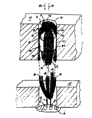
FIG. 1 illustrates a soft-tissue fixation device according to the
invention located in a bone hole;
FIG. 2 shows the soft-tissue fixation device of FIG. 1;
FIG. 3 shows an additional embodiment of a soft-tissue fixation
device;
FIG. 4 shows another embodiment of a soft-tissue fixation
device;
FIG. 5 shows another embodiment of a soft-tissue fixation
device; and
FIG. 6 is an end view of the soft tissue fixation device of FIG. 5.
Referring to FIG. 1, a soft tissue fixation device 10 for securing
soft tissue 12 to bone 14 includes a device body 16 sized to fit within
a bone hole 18. Soft tissue 12 is, e.g., a ligament graft formed from a
CA 02327839 2000-10-06
WO 99/52472 PCT/US99/06750
portion of the patellar tendon, semi-tendonosis or gracilis graft or a
synthetic graft. Bone hole 18 is, e.g., a bone tunnel formed in the
tibia.
5 To replace soft tissue such as a damaged ACL, graft 12 is first
secured to the femur 15 with a securing device 17, e.g., an
endobutton such as described in U.S.S.N 081795,847, filed February
5, 1997, titled GRAFT ATTACHMENT DEVICE AND METHOD OF
ATTACHMENT, incorporated by reference herein, and U.S. Patent
No. 5,306,301, titled GRAFT ATTACHP~AENT DEVICE AND METHOD
OF USING SAME, incorporated by reference herein. The graft is then
secured within bone tunnel 18 in tibia 14 with device 10. Generally,
as is known in the art, the endobutton spans across a hole in the
femur 15 and graft 12 is attached to endobutton 17 with tape 23
looped through openings 25a, 25b in the endobutton. Graft 12 is then
positioned about device body 16, as described below, and device
body 16 is inserted into bone tunnel 18 to reside in a region of
cancellous tissue.
Referring also to FIG. 2, device body 16 is generally disposed
along a longitudinal axis 20. Device body 16 is generally cylindrical in
shape with an outer surface 22 defining longitudinally extending
securing members 32, 34, 36, 38, and channels 24, 26, 28, 30
positioned circumferentially between securing members 32, 34, 36,
38. For example, four channels 24, 26, 28, 30 and four securing
members 32, 34, 36, 38 are equally spaced about the circumference
of device body 16 and extend the entire length, L, of the device body.
Ends 40, 42 of device body 16 are flat and are intersected by
channels 24, 26, 28, 30.
CA 02327839 2000-10-06
WO 99/52472 PCT1US99/06750
6
During use, fixation device 10 is inserted into bone tunnel 18
with soft tissue 12 located within longitudinal channels 24, 26, 28, 30.
As shown in FIG. 1, soft tissue 12 includes four tissue segments 44,
46, 48, 50. Each segment is located in one of the four channels.
However, the number of tissue segments need not equal the number
of channels.
After soft tissue 12 is attached to femur 15, bone tunnel 18 is
drilled, and soft tissue 12 is located within channels 24, 26, 28, 30,
fixation device 10 is positioned within bone tunnel 18 by applying an
axial force to fixation device 10 (along arrow 52) while applying a
tensile load to soft tissue 12 (along arrow 54). Fixation device 10 has
a larger outer diameter than bone tunnel 18 such that there is an
interference fit between securing members 32, 34, 36, 38 and wall 19
of the bone tunnel to secure fixation device 10 to bone tunnel 18. No
rotation of fixation device 10 is required to position fixation device 10
within bone tunnel 18. Additionally, there is no need for a second
member to be inserted to expand the device or wedge the device in
place.
Soft tissue 12 is compressed between the surface of fixation
device 10 defining channels 24, 26, 28, 30 and bone wall 19 to secure
soft tissue 12 within bone tunnel 18. Soft tissue 12 can be tied off
around end 40 of device body 16, e.g., by suturing tissue segments
44, 46, 48, 50 together where the tissue segments exit channels 24,
26, 28, 30. The length, L, of device body 16 is shorter than the length
of bone tunnel 18. This configuration allows fixation device 10 and
soft tissue 12 to reside completely within bone tunnel 18. Tissue
segments 44, 46, 48, 50 can be trimmed after suturing so that they do
not extend beyond an opening 21 of bone tunnel 18.
CA 02327839 2000-10-06
WO 99/52472 PCT/US99/06750
7
Other embodiments are within the scope of the following
claims.
For example, referring to FIG. 3, a soft tissue fixation device
100 has a device body 106 that is generally cylindrical in shape and
disposed along a longitudinal axis 104. Device body 106 includes an
outer surface 126 that defines four channels 108, 110, 112, 114, and
four securing members 116, 118, 120, 122. A proximal end 128 of
device body 106 defines a recess 124 for receiving a driver, not
shown. A set of ribs, e.g., nine evenly spaced ribs 130a-1301, extend
from outer surface 126 into channels 108, 110, 112, 114. The
distance that ribs 130a-130i extend from surface 126 generally
decreases in the direction of arrow 140 from proximal end 128 toward
a distal end 129. For example, ribs 130a-130f project outward 1.28
mm (0.05") from surface 126, rib 130g projects outward 1.21 mm
(0.047") from surface 126, rib 130h projects outward 1.00 mm (0.039")
from surface 126, and rib 130i projects outward 0.82 mm (0.032")
from surface 126. The decrease in distance prevents the ribs 130g,
130h, 1301, which are subject to large forces on insertion, from
breaking.
Securing members 116, 118, 120, 122 each include a set of
grooves 132 that lie along outer surface 126. Grooves 132 form, e.g.,
a set of evenly spaced and uniformly sized wedges 136 oriented
toward distal end 129 to oppose force applied by soft tissue 12 which
would tend to pull fixation device 110 in a distal direction. At a distal
section 134, securing members 116, 118, 120, 122 taper to a smaller
outer diameter. Distal section 134 is tapered to facilitate insertion of
device body 106 into bone tunnel 18.
CA 02327839 2000-10-06
WO 99/52472 PCT/US99/06750
8
A cannulation 138 runs through device body 106 for receiving a
guidewire (not shown). To aid in inserting fixation device 100 into
bone tunnel 18, the guidewire is positioned through bone tunnel 18
and fixation device 100 is passed over the guide-wire and into bone
tunnel 18 with the aid of an insertion tool, not shown, located within
recess 24. No further manipulation of device 100 is required. For
example, neither rotation of the fixation device or insertion of second
member to expand the device or wedge the device in place is
required to position the fixation device within the bone tunnel.
Ribs 130a-1301 engage soft tissue 12 located within channels
108, 110, 112, 114 to aid in securing the soft tissue within the
channels and thus within bone tunnel 18. The ribs have rounded
tissue contacting edges 142 so that the ribs do not damage the soft
tissue.
As discussed above, soft tissue 72 can be tied off around end
128 of device body 106, e.g., by suturing tissue segments 44, 46, 48,
50 together where the tissue segments exit channels 108, 110, 112,
114.
Referring to FIG. 4, a soft tissue fixation device 150 has a
device body 152 that is generally cylindrical in shape and disposed
along a longitudinal axis 154. The device body 152 includes an outer
surface 156 that defines four channels 158, 160, 162, 164, and four
securing members 166, 168, 170, 172. A set of ribs 174a-174e
extend from outer surface 156 into channels 158, 160, 162, 164 for
securing soft tissue within the channels such that the soft tissue is
secured between surface 156 and bone wall 19. Ribs 174a-174e are,
e.g., evenly spaced and uniformly sized or tapered as described
above with reference to FIG. 3. Device body 152 includes an
CA 02327839 2000-10-06
WO 99/52472 PCT/US99/06750
9
enlarged head 176 located at a proximal end 178 of device body 152.
Enlarged head 176 has a radius, R, larger than the radius of bone
tunnel 18. Enlarged head 176 has openings 180, 182, 184, 186 that
are aligned with channels 158, 160, 162, 164, respectively, for
permitting tissue 12 to pass through head 176.
Fixation device 150 functions similarly to fixation device 10.
However, device body 152 is not completely inserted into bone tunnel
18. Enlarged head 176 remains outside bone tunnel 18 and rests
against cortical bone while the remainder of device body 152 is
inserted into bone tunnel 18. Enlarged head 176 provides additional
support for fixation device 150 by resisting forces applied to fixation
device 150 by soft tissue 12 which tend to pull the device into the
bone hole. After insertion, soft tissue 12 can be connected around
enlarged head 176 by, e.g., suturing the segments 44, 46, 48, 50 of
the soft tissue 12 where the segments exit the channels 158, 160,
162, 164 through the openings 180, 182, 184, 186.
Referring to FIGS. 5 and 6, a soft tissue fixation device 60 has
a device body 62 that is generally cylindrical in shape and disposed
along a longitudinal axis 92. Device body 62 includes an outer
surface 64 that defines four channels 66, 68, 70, 72, and four
securing members 74, 76, 78, 80. Multiple deployable projections 86
are extendable from within device body 62 into channels 66, 68, 70,
72. A central bore 88 extends longitudinally through device body 62.
An inner member 90, e.g., a locking screw, can be disposed within
bore 88 to engage projections 86 and deploy projections 86 into
channels 66, 68, 70, 72.
In operation, fixation device 60 functions similarly to soft tissue
fixation device 10, shown in FIGS. 1 and 2. However, projections 86
CA 02327839 2000-10-06
WO 99/52472 PCT/US99/06750
are deployable and, when deployed, aid in securing secure soft tissue
12 within the channels by engaging the soft tissue. Before projections
86 are deployed, they reside within device body 16. After tissue
segments 44, 46, 48, 50, are seated within channels 66, 68, 70, 72,
5 and fixation device 60 is inserted and secured in bone tunnel 18 by
applying an axial force to the fixation device, inner member 44 is
manipulated, e.g., rotated or inserted, such that projections 86 are
engaged. When engaged, projections 86 extend into channels 66,
68, 70, 72 to engage soft tissue 12. Projections 86 have rounded tips
10 94 so that they do not damage soft tissue 12 when extended.
The various illustrated embodiments of the soft tissue fixation
device can be constructed of, e.g., Delrin, acetal, or other non-
bioabsorbable or bioabsorbable materials.
Many of the features shown in the embodiments illustrated
above can be combined. The tapered distal section 134 of soft tissue
fixation device 100 could be combined with the enlarged head 176 of
soft tissue fixation device 150. Grooves 132 can be combined with
deployable projections or with no projections. Other combinations of
the features disclosed are also possible.
