Note: Descriptions are shown in the official language in which they were submitted.
CA 02329054 2007-04-24
WO 99/60017 PCT/EP99/03403
Title of the invention
A novel polypeptide hormone phosphatonin
Field of the invention
The present invention relates to a polypeptide which is involved in the
regulation
of phosphate metabolism. More specifically, the present invention relates to a
novel polypeptide Metastatic-tumor Excreted Phosphaturic-Element (MEPE) or
"phosphatonin". This invention also relates to genes and polynucleotides
encoding
phosphatonin polypeptides, as well as vectors, host cells, antibodies directed
to
phosphatonin polypeptides, and the recombinant methods for producing the same.
Also provided are diagnostic methods for detecting disorders relating to
phosphate
metabolism, and therapeutic methods for treating such disorders. The invention
further relates to screening methods for identifying agonists and antagonists
of
phosphatonin activity.
Several documents are cited throughout the text of this specification;
however, there is no admission
that any document cited is indeed prior art as to the present invention.
Background of the invention
Phosphate plays a central role in many of the basic processes essential to the
cell
and the mineralization of bone. In particular, skeletal mineralization is
dependent
on the regulation of phosphate and calcium in the body and any disturbances in
phosphate-calcium homeostasis can have severe repercussions on the integrity
of
bone. In the kidney, phosphate is lost passively into the glomerular filtrate
and is
actively reabsorbed via a sodium (Na+) dependent phosphate cotransporter. In
the
intestine, phosphate is absorbed from foods. A sodium (Na+) dependent
phosphate cotransporter was found to be expressed in the intestine and
recently
cloned (Hilfiker, PNAS 95(24) (1998), 14564-14569). The liver, skin and kidney
are involved in the conversion of vitamin D3 to its active metabolite,
calcitriol,
CA 02329054 2000-11-16
WO 99/60017 PCT/EP99/03403
which plays an active role in the maintenance of phosphate balance and bone
mineralization.
Vitamin D deficiency causes rickets in children and osteomalacia in adults.
Both
conditions are characterized by failure of calcification of osteoid, which is
the
matrix of bone. There are also several non-dietary conditions which can lead
to
rickets, including X-linked vitamin D resistant hypophosphatemic rickets
(HYP),
hereditary hypercalciuria with hypophosphatemic rickets (HHRH), Dent's disease
including certain types of renal Fanconi syndrome, renal 1 alpha-hydroxylase
deficiency (VDDR I), defects in 1,25-dihydroxy vitamin D3 receptor (end organ
resistance, VDDR II), and oncogenic hypophosphatemic osteomalacia (OHO).
Thus, a number of familial diseases have been characterized that result in
disorders of phosphate uptake, vitamin D metabolism and bone mineralization.
Recently a gene has been cloned and characterized that is defective in
patients
with X-linked hypophosphatemic rickets (PHEX) (Francis, Nat. Genet. 11 (1995),
130-136; Rowe, Hum. Genet. 97 (1996), 345-352; Rowe, Hum. Mol. Genet. 6
(1997), 539-549). The PHEX gene is a type II glycoprotein and a member of a
family (M13), of Zn metalloendopeptidases. PHEX is proposed to function by
processing a factor that plays a role in phosphate homeostasis and skeletal
mineralization (Rowe, Exp. Nephrol. 5 (1997), 355-363; Rowe, Current Opinion
in
Nephrology & Hypertension 7(4) (1998), 367-376). Oncogenic hypophosphatemic
osteomalacia (OHO), has many similarities to HYP with an overlapping
pathophysiology, but different primary defects (Rowe, Exp. Nephrol. 5 (1997),
355-363; Rowe, Current Opinion in Nephrology & Hypertension 7(4) (1998), 367-
376; Drezner in Primer on Metabolic Bone Diseases and Disorders of Mineral
Metabolism (ed. Favus, M.J.) 184-188 (Am. Soc. Bone and Min. Res.,
Kelseyville,
CA, 1990)). Osteomalacia is the adult equivalent of rickets, and a key feature
of
tumour-acquired osteomalacia is softening of the bones. The softened bones
become distorted, resulting in bow-legs and other associated changes
reminiscent
of familial rickets. Low serum phosphate, and abnormal vitamin D metabolism
are
also key features shared with HYP. Tumour acquired osteomalacia is rare, and
the tumours are mainly of mesenchymal origin, although a number of different
tumour types have also been reported (Rowe, Exp. Nephrol. 5 (1997), 355-363;
Francis, Baillieres Clinical Endocrinology and metabolism 11 (1997), 145-163;
2
CA 02329054 2000-11-16
WO 99/60017 PCT/EP99/03403
loakimidis, The J. Rheumatology 21(6) (1994), 1162-1164; Lyles, Ann. Intern.
Med. 93 (1980), 275-278; Rowe, Hum. Genet. 94 (1994), 457-467; Shane, Journal
of Bone and Mineral Research 12 (1997), 1502-1511; Weidner, Cancer 59 (1987),
1442-1442). Surgical removal of the tumour(s) when possible, results in the
disappearance of disease symptoms and bone healing, suggesting the role of a
circulating phosphaturic factor(s) in the pathogenesis of the disease. Also,
hetero-
transplantation of tumours into nude mice (Miyauchi, J. Clin. Endocrinol.
Metab.
67 (1988), 46-53) infusion of saline extracts into rats and dogs (Aschinberg,
J.
Paediatr. 91 (1977), 56-60; Popovtzer, Clin. Res. 29 (1981), 418A (Abstract)),
and
the use of tumour conditioned medium (TCM), of human and animal renal cell
lines all confirm that a circulating phosphaturic factor is secreted by these
tumours.
Although the primary-defect in X-linked rickets is confirmed as a mutated Zn
metalloendopeptidase (PHEX), there is considerable evidence that implicates a
circulating phosphaturic factor(s) (Ecarot, J. Bone Miner. Res. 7 (1992), 215-
220;
Ecarot, J. Bone Miner. Res. 10 (1995), 424-431; Morgan, Arch. Intern. Med. 134
(1974), 549-552; Nesbitt, J. Clin. Invest. 89 (1992), 1453-1459; Nesbitt, J.
Bone.
Miner. Res. 10 (1995), 1327-1333; Nesbitt, Endocrinology 137 (1996), 943-948;
Qiu, Genet. Res., Camb. 62 (1993), 39-43; Lajeunesse, Kidney Int. 50 (1996),
1531-1538; Meyer, J. Bone. Miner. Res. 4(4) (1989), 523-532; Meyer, J. Bone.
Miner. Res. 4 (1989), 493-500). The overlapping pathophysiology of HYP and
OHO raises the intriguing possibility that the tumour-factor may be processed
in
normal subjects by the PHEX gene product. Also, it is likely that proteolytic
processing by PHEX may act by either degrading this undefined phosphaturic
factor(s), or by activating a phosphate-conserving cascade (Carpenter,
Pediatric
Clinics of North America 44 (1997), 443-466; Econs, Am. J. Physiol. 273
(1997),
F489-F498; Glorieux, Arch. Pediatr. 4 (1997), 102s-105s; Grieff, Current
Opinion
in Nephrology & Hypertension 6 (1997), 15-19; Hanna, Current Therapy in
Endocrinology & Metabolism 6 (1997), 533-540; Kumar, Nephrol. Dial.
Transplant.
12 (1997), 11-13; Takeda, Ryoikibetsu Shokogun Shirizu (1997), 656-659). The
cloning and characterization of the tumour-phosphaturic factor is thus
prerequisite
to establishing any links between tumour osteomalacia and familial X-linked
rickets as well as other disorders in the phosphate metabolism.
3
CA 02329054 2000-11-16
WO 99/60017 PCT/EP99/03403
Rowe et al (1996) have reported candidates 56 and 58 kDa protein (s)
responsible
for mediating renal defects in OHO (Rowe, Bone 18, (1996), 159-169). A patient
with OHO was treated by tumor removal and pre- and post-operative antisera
from
the patient were used in a Western blotting identification of tumor
conditioned
media proteins. Neither the tumor cells nor the antisera were ever made
available
to the public, however.
In a review in Exp. Nephrol. 5 (1997), 335-363, Rowe (1997) discusses the
above
diseases and the role of the PHEX gene (previously known as the PEX gene). The
PHEX gene product has been identified as a zinc metalloproteinase. In disease
states such as familial rickets, defective PHEX results in uncleaved
phosphatonin
which would result in down regulation of the sodium dependent phosphate
cotransporter and up regulation of renal mitochondrial 24-hydroxylase.
However,
no purification of phosphatonin was reported by Rowe (1997). Thus, no source
material for phosphatonin was made available to the public. Moreover,
purification,
identification and characterization of phosphatonin has not been possible.
Thus, there is a need for polypeptides that regulate phosphate metabolism,
since
disturbances of such a regulation may be involved in hypo- and
hyperphosphatemic diseases, including osteomalacia, particularly osteoporosis
and renal failure. Furthermore, there is a need for identifying and
characterizing
such polypeptides which may play a role in the detection, prevention and/or
correction of such disorders and may be useful in diagnosing those disorders.
Summary of the invention
The present invention relates to novel phosphatonin polypeptides and the
encoding polynucleotides of phosphatonin. Moreover, the present invention
relates to vectors, host cells, antibodies, and recombinant methods for
producing
the polypeptides and polynucleotides. Also provided are diagnostic methods for
detecting disorders related to the polypeptides, and therapeutic methods for
treating such disorders. The present invention further relates to screening
methods for identifying binding partners of phosphatonin.
4
CA 02329054 2009-08-17
The present invention also relates to an isolated polypeptide which up-
regulates
sodium dependent phosphate co-transport encoded by a polynucleotide selected
from the group consisting of:
(a) polynucleotides encoding at least amino acids 1 to 236 of the
polypeptide comprising the amino acid sequence depicted in SEQ ID NO: 2;
(b) polynucleotides comprising the coding sequence as depicted in SEQ
ID NO: 1 encoding at least amino acids 1 to 236 of said polypeptide;
(c) polynucleotides which hybridize under high stringent conditions
comprising incubation at 42 C in a solution comprising 50% formamide, 5x
SSC, 50 mM sodium phosphate (pH 7.6), 5x Denhardt's, 10% dextran
sulfate and 20 pg/ml denaturated, sheared salmon sperm DNA, followed by
wash in 0.1x SSC at about 65 C with the complement of the polynucleotide
of (a) or (b) encoding a polypeptide which up-regulates sodium dependent
phosphate co-transport;
(d) polynucleotides encoding a polypeptide, the sequence of which has
an identity of 60% or more to the amino acid sequence of the polypeptide
encoded by a polynucleotide of (a) or (b), wherein said polypeptide up-
regulates sodium dependent phosphate co-transport;
(e) polynucleotides encoding a fragment of a polypeptide encoded by
the polynucleotide of any one of (a), (b) and (d), wherein said fragment up-
regulates sodium dependent phosphate co-transport;
(f) polynucleotides encoding an epitope-bearing portion of a
phosphatonin polypeptide comprising amino acid residues from (a) 1 to 40,
(b) 141 to 180, (c) 401 to 429 or (d) any combination of (a) - (c) in SEQ ID
NO: 2; and
(g) polynucleotides of any one of (a), (b), (e) or (f), the nucleotide
sequence of which is degenerate as a result of the degeneracy of the
genetic code.
The present invention also relates to an isolated polynucleotide encoding the
above-mentioned polypeptide.
4a
CA 02329054 2009-08-17
The present invention also relates to a vector containing the above-mentioned
polynucleotide.
The present invention also relates to an isolated host cell (a) genetically
engineered with the above-mentioned polynucleotide, (b) genetically engineered
with the above-mentioned vector or (c) produced by introducing an expression
control sequence into a host cell which comprises a polynucleotide encoding
the
above-mentioned polypeptide.
The present invention also relates to a process for producing the above-
mentioned
polypeptide comprising: culturing the above-mentioned host cell and recovering
said polypeptide from the culture.
The present invention also relates to an antibody that binds specifically to
the
above-mentioned polypeptide.
The present invention also relates to an isolated nucleic acid molecule
comprising
at least 15 contiguous bases in length of nucleotides 1 to 1290 of the
polynucleotide as depicted in SEQ ID NO: 1 or of a complementary strand
thereof.
The present invention also relates to a method for identifying a binding
partner to a
phosphatonin polypeptide comprising:
(a) contacting the above-mentioned polypeptide with a compound to be
screened; and
(b) determining whether the compound effects an activity of the polypeptide,
wherein a binding partner is identified when said activity is different in the
presence of said compound as compared to in the absence thereof.
The present invention also relates to a composition comprising (a) the above-
mentioned polypeptide, (b) the above-mentioned polynucleotide, (c) the above-
mentioned vector, (d) the above-mentioned antibody, (e) the above-mentioned
nucleic acid molecule or (f) any combination of (a) - (e), and further
comprising a
pharmaceutically acceptable excipient, diluent or carrier.
4b
CA 02329054 2009-08-17
The present invention also relates to a composition comprising (a) the above-
mentioned polypeptide, (b) the above-mentioned polynucleotide, (c) the above-
mentioned vector, (d) the above-mentioned antibody, (e) the above-mentioned
nucleic acid molecule or (f) any combination of (a) - (e), and further
comprising
enzyme labels, radio labels or fluorescent labels.
4c
CA 02329054 2000-11-16
WO 99/60017 PCT/EP99/03403
Brief description of the drawings
Figure 1: Figure 1(a) and (b) show respectively chromatograms with low
affinity
and high affinity protein-containing peaks from a concanavalin A
column.
Figure 2: Cation exchange chromatogram of fractions from the concanavalin A
column.
Figure 3: Computer prediction of hydrophilicity and hydrophobicity of
phosphatonin.
Figure 4: Computer prediction of antigenicity of phosphatonin.
Figure 5: Computer prediction of flexibility of phosphatonin.
Figure 6: Computer prediction of surface probability of the secondary
structure of
phosphatonin.
Figure 7: Computer prediction of the secondary structure of phosphatonin.
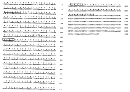
Figure 8: Complete cDNA sequence (SEQ ID NO: 1) and amino acid sequence
(SEQ ID NO: 2) of the largest MEPE clone isolated (pHO11.1). The
five other clone isolated are encompassed by this larger clone and all
clone are in frame with the cloning vehicle pBSCPT SK 11-. Primers
used for PCR are highlighted, and the total number of residues are 430
and 1655 bp respectively. The prokaryotic expression vector pCal-n-
EK contained all in frame residues from MEPE residue V, to the MEPE
stop codon (TAG), at 1291-93 bp. The single polyadenylation sequence
AA{T/U}AAA is double underlined. The region of shared localized
homology with DMA-1, DSSP, and OPN is underlined in wavy line
format (MEPE-motif C-terminus), RGD residues are enclosed in an
ellipsoid), glycosaminoglycan attachment site is boxed (complete line
format), Tyrosine Kinase site is underlined once, and N-glycosylation
motifs are boxed in dotted line format. For a complete list of motifs
including casein kinase 11, protein kinase C etc. please refer to prosite
screen Table 1.
Figure 9: GCG-peptide-structure secondary structure prediction for MEPE. The
primary amino acid backbone is shown as the central line with curves
indicating regions of predicted turn. Hydrophilicity/hydrophobicity
regions are represented as ellipsoids and diamonds respectively and
CA 02329054 2000-11-16
WO 99/60017 PCT/EP99/03403
the RGD motif is indicated. The N-glycosylation sites are represented
as ellipsoids on stalks (C-terminus), and alpha helix by undulating
regions on the primary backbone.
Figure 10: Bar graphs showing phosphate-uptake in the presence of differing
amounts of MEPE: A. 92 ng/ml, B. 300 ng/ml, C. 500 ng/ml, and D.
1000 ng/ml. Choline boxes refer to control Na- independent results with
NaCl replaced with choline chloride. Error bars are SEM, and P values
for the difference between MEPE and control in C and D are < 0.001. In
experiment A (92 ng/ml) P<0.05, and in B (300 ng/ml P, 0.01). N values
for A and B are 4, and for C and D 5 and 6 respectively. Anova followed
by Newman-Keuls Multiple Comparison Test was used.
Figure 11: Dose curve of MEPE administration and phosphate uptake with SEM
error bars.
Figure 12: Sequence similarity analysis using `sim' and Ilanview mathematical
and
software tools (Duret, Comput. Appl. Biosci. 12 (1996), 507-510). In
each computation the gap open penalty was set to 12, and gap
extension penalty 4. Comparison matrix for A was `PAM40', and
BLOSUM62 for B and C respectively (see Duret, Comput. Appi. Biosci.
12 (1996), 507-510; Huang, Comput. Appl. Biosci. 8 (1992), 155-165;
Huang, Comput. Appl. Biosci. (1990) 6, 373-381). The similarity score
threshold was 70% in A, and 40% in B and C respectively. The
highlighted blocks shown on each protein scheme represent sequence
homologies of >80% in A, and > 62% in B and C. Note that in MEPE
versus DSSP (A), there are five homology blocks in DSSP of >80%
sequence similarity to a single motif in MEPE (DSSESSDSGSSSES). A
similar sequence homology is also apparent for DMA-1 and OPN
versus MEPE (B and C) and the MEPE is a feature of all three proteins.
Figure 13: Dot matrix comparison of DSSP versus MEPE using Antheprot
statistical analysis (Deleague, G. Software for protein analysis:
Antheroplot V2.5e. Microsoft group. (7 Passage du Vercours 69-367
Vercors Lyon Cedex 07, 1997)). In (A) a lower stringency comparison
with a window set to 13 is used as screen parameters and in (B) a
wider window of 15 is used. The colors indicate unity matrix scores as
CA 02329054 2000-11-16
WO 99/60017 PCT/EP99/03403
indicated on the diagram. C-terminal residues of MEPE-motif have
>80% sequence homology and the repeat nature of the motif is
illustrated by the striped pattern.
Figure 14: p1BL21 and also p6XL1 recombinant plasmids containing
phosphatonin fusion construct. Lacl: (lac promoter); LIC: (Ligation
independent cloning sequence); EK: Enterokinase cleavage site;
Thrombin (thrombin target sequence); Amp: Ampicillin resistance: Cal
peptide (calmodulin peptide sequence); Phosphatonin (phosphatonin
coding sequence).
Detailed description of the present invention
In view of the need of diagnostic and therapeutic means for the treatment of
diseases related to disorders in the phosphate metabolism in the human body,
the
technical problem of the invention is to provide means and methods for the
modulation of phosphate metabolism which are particularly useful for the
treatment of bone mineral and renal diseases.
The above-defined technical problem is solved by the present invention by
providing the embodiments characterized in the claims. Accordingly, in one
aspect
the present invention relates to an isolated polypeptide having phosphatonin
activity.
Unless otherwise stated, the terms used herein are defined as described in "A
multilingual glossary of biotechnological terms: (IUPAC Recommendations)",
Leuenberger, H.G.W., Nagel, B. and Kolbl, H. eds. (1995), Helvetica Chimica
Acta, CH-4010 Basle, Switzerland, ISBN 3-906 390-13-6. The following
definitions
are provided to facilitate understanding of certain terms used throughout this
specification.
The terms "treatment", "treating" and the like are used herein to generally
mean
obtaining a desired pharmacological and/or physiological effect. The effect
may be
prophylactic in terms of completely or partially preventing a disease or
symptom
thereof and/or may be therapeutic in terms of partially or completely curing a
disease and/or adverse effect attributed to the disease. The term "treatment"
as
CA 02329054 2000-11-16
WO 99/60017 PCT/EP99/03403
used herein covers any treatment of a disease in a mammal, particularly a
human,
and includes: (a) preventing the disease from occurring in a subject which may
be
predisposed to the disease but has not yet been diagnosed as having it; (b)
inhibiting the disease, i.e. arresting its development; or (c) relieving the
disease,
i.e. causing regression of the disease. The present invention is directed
towards
treating patients with medical conditions relating to a disorder of phosphate
metabolism. Accordingly, a treatment of the invention would involve
preventing,
inhibiting or relieving any medical condition related to phosphate metabolism
disorders.
In the present invention, "isolated" refers to material removed from its
original
environment (e.g., the natural environment if it is naturally occurring), and
thus is
altered "by the hand of man" from its natural state. For example, an isolated
polynucleotide could be part of a vector or a composition of matter, or could
be
contained within a cell, and still be "isolated" because that vector,
composition of
matter, or particular cell is not the original environment of the
polynucleotide.
The phosphatonin polypeptide isolated in accordance with the present invention
typically has an approximate molecular weight of 53 to 60 kDa, more preferably
58-60 kDa, as measured on SDS-PAGE, particularly on a 12.5% gel at pH 8.6 in
TRIS-Glycine SDS buffer, see Example 1. An approximate molecular weight of
200 kDa may be measured on bis-tris-SDS-PAGE at pH 7 using a 4-12% gradient
gel with MOPS running buffer. It is possible on such a gel also to see lower
molecular weight bands of 53 to 60 kDa. The polypeptide is generally
glycosylated, and preferably comprises phosphatonin in substantially pure
form.
Surprisingly, it has been found that the phosphatonin is obtainable, following
purification according to the protocol given in Example 1 from Saos-2 cells,
which
are available from the European Collection of Cell Culture under Deposit No.
ECACC 89050205. Accordingly, in a further aspect of the invention, there is
provided use of Saos-2 cells or HTB-96 cells for the production of
phospatonin.
Other transformed or immortalized cell lines may be capable of overexpression
of
phosphatonin, such as transformed osteoblast or bone cell lines.
CA 02329054 2000-11-16
WO 99/60017 PCT/EP99/03403
The present invention also describes the characterization and cloning of a
gene
that is a candidate for the above-described tumour-derived phosphaturic factor
and that is named phosphatonin or MEPE (Metastatic-tumour Excreted
Phosphaturic-Element). To summarize, expression screening of a ?, ZAPII-cDNA
library constructed from mRNA extracted from an OHO tumour using antisera
specific to tumor conditioned media (TCM) phosphaturic-factor was used. The
protein is glycosylated and resolves as two bands on SDS-PAGE electrophoresis
(58-60 kDa), with evidence of possible splicing or post translational
cleavage. The
cloned cDNA codes for a protein of 430 residues (SEQ ID NO: 2) and 1655 bp in
length (SEQ ID NO: 1). The entire 3' end of the gene is present, with part of
the 5'
end missing. The fusion protein containing 10 residues of B-galactosidase is
highly potent at inhibiting Na+ dependent phosphate co-transport in a human
renal
cell line (CL8). Secondary structure prediction confirms that the protein is
highly
hydrophilic with small localized regions of hydrophobicity and no cysteine
residues. A number of helical regions are present, with two distinct N-
glycosylation
motifs at the carboxy-terminus. A key feature is the presence of a cell
attachment
sequence in the same structural context found in osteopontin. Proteolytic-
sites
adjacent to this motif may result in altered receptor specificity for specific
integrins
as found in osteopontin. Screening of the trembi database with MEPE sequence
also demonstrated sequence homology with Dentin phosphoryn (DPP). In
particular there is striking localized residue homology at the C'-terminus of
MEPE
with DPP, dentin-matrix protein-1 (DMA-1) and osteopontin (OPN). This region
of
MEPE contains a recurring series of aspartate and serine residues
(DDSSESSDSGSSSESD), with 80%, 65 % and 62% homology with DSP, DMA-1
and OPN respectively. Moreover, when residue physicochemical character is
considered this homology rises to 93%, suggesting a shared or related
biological-
functionality. It is also of note that this structural motif overlaps a casein
kinase II
phosphorylation motif in MEPE. Skeletal casein kinase II activity is defective
in
rickets, and results in under phosphorylation of osteopontin (Rifas, Calcif.
Tissue
Int. 61 (1997), 256-259). The casein kinase II defect has thus been proposed
to
play a role in the under-mineralization of bone matrix (Rifas, loc. cit.).
Dentin phosphoryn (DPP), is one part of a cleavage product derived from dentin
sialophosphoprotein (DSSP), with the other part known as dentin sialoprotein
CA 02329054 2000-11-16
WO 99/60017 PCT/EP99/03403
(DSP) (MacDougall, J. Biol. Chem. 272 (1997), 835-842). It is of particular
interest
that DSSP, DMA-1, OPN and MEPE are RGD containing phospho-glycoproteins
with distinct structural similarities and major roles in bone-tooth
mineralization
(Linde, Crit. Rev. Oral Biol. Med. 4 (1993), 679-728).
The new OHO tumour-derived phosphaturic factor named phosphatonin or MEPE
described in the present invention, effects bone mineral homeostasis by
regulating
Na+ dependent phosphate co-transport, vitamin D metabolism, and bone
mineralization.
As set out in further detail below, a polynucleotide has been isolated which
encodes polypeptides according to the present invention; see Example 2. The
amino acid and nucleotide sequences of phosphatonin are set out in Figure 8
(SEQ 1D NO: 1 and SEQ ID NO: 2, respectively). Accordingly, the polypeptide of
the present invention comprises the amino acid sequence of Figure 8,
optionally
including mutations or deletions which do not substantially affect the
activity
thereof. Such mutations include substitution of one or more amino acids,
particularly by homologues thereof, as well as additions of one or more amino
acids, especially at the N or C termini. Deletions include deletions from the
N or C
termini. Substitutions by both naturally-occurring and synthetic amino acids
are
possible. Also included are polypeptides modified by chemical modification or
enzymatic modification. Further, fragment peptides, whether chemically
synthesized or produced by a biological method, whether modified or
unmodified,
are included within the scope of this invention.
Accordingly the present invention relates to a phosphatonin polypeptide or an
immunologically and/or biologically active fragment thereof, which comprises
an
amino acid sequence encodable by a polynucleotide selected from the group
consisting of
(a) polynucleotides encoding at least the mature form of the polypeptide
comprising the amino acid sequence depicted in SEQ ID NO: 2 (Figure 8);
(b) polynucleotides comprising the coding sequence as depicted in SEQ ID
NO: 1 (Figure 8) encoding at least the mature form of the polypeptide;
CA 02329054 2000-11-16
WO 99/60017 PCT/EP99/03403
(c) polynucleotides encoding a polypeptide derived from the polypeptide
encoded by a polynucleotide of (a) or (b) by way of substitution, deletion
and/or addition of one or several amino acids of the amino acid sequence
encoded by the polynucleotide of (a) or (b);
(d) polynucleotides comprising the complementary strand which hybridizes
with a polynucleotide of any one of (a) to (c);
(e) polynucleotides encoding a polypeptide the sequence of which has an
identity of 60% or more to the amino acid sequence of the polypeptide
encoded by a polynucleotide of any one of (a) to (d);
(f) polynucleotides encoding a polypeptide capable of regulating phosphate
metabolism comprising a fragment or an epitope-bearing portion of a
polypeptide encoded by a polynucleotide of any one of (a) to (e);
(g) polynucleotides encoding an epitope-bearing portion of a phosphatonin
polypeptide comprising amino acid residues from about 1 to 40, 141 to 180
and/or 401 to 429 in SEQ ID NO: 2 (Figure 8);
(h) polynucleotides comprising at least 15 nucleotides of a polynucleotide of
any one of (a) to (g) and encoding a polypeptide capable of regulating
phosphate metabolism;
(i) polynucleotides encoding a polypeptide capable of regulating phosphate
metabolism comprising the cell and/or glycosaminoglycan attachment motif
and/or the bone mineral motif of a polypeptide encoded by a polynucleotide
of any one of (a) to (h); and
(j) polynucleotides the nucleotide sequence of which is degenerate as a result
of the genetic code to a nucleotide sequence of a polynucleotide of any of
(a) to (i).
As used herein, a phosphatonin "polynucleotide" refers to a molecule having a
nucleic acid sequence contained in SEQ ID NO: 1 or encoding the phosphatonin
polypeptide of the present invention. For example, the phosphatonin
polynucleotide can contain the nucleotide sequence of the full length cDNA
sequence, including the 5' and 3' untranslated sequences, the coding region,
as
well as fragments, epitopes, domains, and variants of the nucleic acid
sequence.
1"
CA 02329054 2000-11-16
WO 99/60017 PCT/EP99/03403
Moreover, as used herein, a phosphatonin polypeptide" refers to a molecule
having the translated amino acid sequence generated from the polynucleotide as
broadly defined.
A phosphatonin "polynucleotide" also includes those polynucleotides capable of
hybridizing, under stringent hybridization conditions, to sequences contained
in
SEQ ID NO: 1 or the complement thereof. "Stringent hybridization conditions"
refers to an overnight incubation at 42 C in a solution comprising 50%
formamide,
5x SSC (750 mM NaCl, 75 mM sodium citrate), 50 mM sodium phosphate (pH
7.6), 5x Denhardt's solution, 10% dextran sulfate, and 20 pg/ml denatured,
sheared salmon sperm DNA, followed by washing the filters in 0.1 x SSC at
about
65 C. Further suitable hybridization conditions are described in the examples.
Also contemplated are nucleic acid molecules that hybridize to the
phosphatonin
polynucleotides at lower stringency hybridization conditions. Changes in the
stringency of hybridization and signal detection are primarily accomplished
through the manipulation of formamide concentration (lower percentages of
formamide result in lowered stringency); salt conditions, or temperature. For
example, lower stringency conditions include an overnight incubation at 37 C
in a
solution comprising 6X SSPE (20X SSPE = 3M NaCl; 0.2M NaH2PO4; 0.02M
EDTA, pH 7.4), 0.5% SDS, 30% formamide, 100 g/ml salmon sperm blocking
DNA; followed by washes at 50 C with 1 X SSPE, 0.1% SDS. In addition, to
achieve even lower stringency, washes performed following stringent
hybridization
can be done at higher salt concentrations (e.g. 5X SSC). Note that variations
in
the above conditions may be accomplished through the inclusion and/or
substitution of alternate blocking reagents used to suppress background in
hybridization experiments. Typical blocking reagents include Denhardt's
reagent,
BLOTTO, heparin, denatured salmon sperm DNA, and commercially available
proprietary formulations. The inclusion of specific blocking reagents may
require
modification of the hybridization conditions described above, due to problems
with
compatibility. Of course, a polynucleotide which hybridizes only to polyA+
sequences (such as any 3' terminal polyA+ tract of a cDNA shown in the
sequence listing), or to a complementary stretch of T (or U) residues, would
not be
included in the definition of "polynucleotide," since such a polynucleotide
would
CA 02329054 2000-11-16
WO 99/60017 PCT/EP99/03403
hybridize to any nucleic acid molecule containing a poly (A) stretch or the
complement thereof (e.g., practically any double-stranded cDNA clone).
The phosphatonin polynucleotide can be composed of any polyribonucleotide or
polydeoxribonucleotide, which may be unmodified RNA or DNA or modified RNA
or DNA. For example, phosphatonin polynucleotides can be composed of single-
and double-stranded DNA, DNA that is a mixture of single- and double-stranded
regions, single- and double-stranded RNA, and RNA that is mixture of single-
and
double-stranded regions, hybrid molecules comprising DNA and RNA that may be
single-stranded or, more typically, double-stranded or a mixture of single-
and
double-stranded regions. In addition, the phosphatonin polynucleotides can be
composed of triple-stranded regions comprising RNA or DNA or both RNA and
DNA. Phosphatonin polynucleotides may also contain one or more modified bases
or DNA or RNA backbones modified for stability or for other reasons.
"Modified"
bases include, for example, tritylated bases and unusual bases such as
inosine. A
variety of modifications can be made to DNA and RNA; thus, "polynucleotide"
embraces chemically, enzymatically, or metabolically modified forms.
Phosphatonin polypeptides can be composed of amino acids joined to each other
by peptide bonds or modified peptide bonds, i.e., peptide isosteres, and may
contain amino acids other than the 20 gene-encoded amino acids. The
phosphatonin polypeptides may be modified by either natural processes, such as
posttranslational processing, or by chemical modification techniques which are
well known in the art. Such modifications are well described in basic texts
and in
more detailed monographs, as well as in a voluminous research literature.
Modifications can occur anywhere in the phosphatonin polypeptide, including
the
peptide backbone, the amino acid side-chains and the amino or carboxyl
termini. It
will be appreciated that the same type of modification may be present in the
same
or varying degrees at several sites in a given phosphatonin polypeptide. Also,
a
given phosphatonin polypeptide may contain many types of modifications.
Phosphatonin polypeptides may be branched, for example, as a result of
ubiquitination, and they may be cyclic, with or without branching. Cyclic,
branched,
and branched cyclic phosphatonin polypeptides may result from posttranslation
natural processes or may be made by synthetic methods. Modifications include
acetylation, acylation, ADP-ribosylation, amidation, covalent attachment of
flavin,
43
CA 02329054 2000-11-16
WO 99/60017 PCT/EP99/03403
covalent attachment of a heme moiety, covalent attachment of a nucleotide or
nucleotide derivative, covalent attachment of a lipid or lipid derivative,
covalent
attachment of phosphatidylinositol, cross-linking, cyclization, disulfide bond
formation, demethylation, formation of covalent cross-links, formation of
cysteine,
formation of pyroglutamate, formulation, gamma-carboxylation, glycosylation,
GPI
anchor formation, hydroxylation, iodination, methylation, myristoylation,
oxidation,
pegylation, proteolytic processing, phosphorylation, prenylation,
racemization,
selenoylation, sulfation, transfer-RNA mediated addition of amino acids to
proteins
such as arginylation, and ubiquitination; see, for instance, PROTEINS -
STRUCTURE AND MOLECULAR PROPERTIES, 2nd Ed., T. E. Creighton, W. H.
Freeman and Company, New York (1993); POST-TRANSLATIONAL COVALENT
MODIFICATION OF PROTEINS, B. C. Johnson, Ed., Academic Press, New York
(1983), pages. 1-12; Seifter, Meth. Enzymol. 182 (1990); 626-646, Rattan, Ann.
NY Acad. Sci. 663 (1992); 48-62. For example, it is possible that phosphatonin
is
expressed as a preproprotein and after processing of the pre-sequence and
optionally pro-sequence is cleaved into two or more fragments which remain
together due to the formation of, for example, hydrogen bonds. The processing
and/or cleavage of the prepro- and even mature form of the phosphatonin
polypeptide may be accompanied by the loss of one or more amino acids at the
cleavage site. It is to be understood that all such forms of the phosphatonin
protein are encompassed by the term "phosphatonin polypeptide", "polypeptide"
or
"protein".
"SEQ ID NO: 1 " refers to a phosphatonin polynucleotide sequence while "SEQ ID
NO:2" refers to a phosphatonin polypeptide sequence.
A phosphatonin polypeptide "having biological activity" refers to polypeptides
exhibiting activity similar, but not necessarily identical to, an activity of
a
phosphatonin polypeptide as measured in a particular biological assay such as
described below, with or without dose dependency. In the case where dose
dependency does exist, it need not be identical to that of the phosphatonin
polypeptide, but rather substantially similar to the dose-dependence in a
given
activity as compared to the phosphatonin polypeptide (i.e., the candidate
polypeptide will exhibit greater activity or not more than about 25-fold less
and,
44
CA 02329054 2000-11-16
WO 99/60017 PCT/EP99/03403
preferably, not more than about ten-fold less activity, and most preferably,
not
more than about three-fold less activity relative to the phosphatonin
polypeptide).
The term "immunologically active" or "immunological activity" refers to
fragments,
analogues and derivatives of the phosphatonin polypeptide of the invention the
essential characteristic immunological- properties of which remain unaffected
in
kind, that is that the polynucleotides of the invention include all nucleotide
sequences encoding proteins or peptides which have at least a part of the
primary
and/or secondary structural conformation for one or more epitopes capable of
reacting specifically with antibodies unique to phosphatonin proteins which
are
encodable by a polynucleotide as set forth above. Preferably, the peptides and
proteins encoded by a polynucleotide of the invention are recognized by an
antibody that specifically reacts with an epitope of the phosphatonin
polypeptide
comprising the amino acid residues of about 20 to 30, 100 to 130, 145 to 160,
300
to 310, 320 to 340 or 380 to 430 of SEQ ID NO: 2 or with an epitope of the
phosphatonin polypeptides described herein below. Residues 380-430
peptides/antibodies are particularly useful for the study of mineralization
processes, residues 145-160 peptides/antibodies for the study of receptor
ligand
interactions (inter gins etc.) and residues 20-30 and 100-130, are of
particular
interest for phosphate regulations studies.
Preferably, the immunologically active phosphatonin peptide fragments,
analogues and derivatives of the phosphatonin polypeptide of the invention are
capable of eliciting an immune response in a mammal, preferably in mouse or
rat.
In a preferred embodiment of the present invention the phosphatonin
polypeptide
is biologically active in that it is capable of regulating or modulating
phosphate
metabolism, preferably it has "phosphatonin activity".
Phosphatonin activity
The term "capable of regulating or modulating phosphate metabolism" as used
herein means that the presence or absence, i.e. the level of the phosphatonin
polypeptide of the invention in a subject modulates Na+-dependent phosphate co-
transport, vitamin D metabolism and/or bone mineralization. Depending on
whether the mentioned activities are up- or down-regulated by the polypeptide
of
CA 02329054 2000-11-16
WO 99/60017 PCT/EP99/03403
the invention, said "capability of regulating or modulating phosphate
metabolism"
is referred to as "phosphatonin activity" and "anti-phosphatonin activity",
respectively.
Phosphatonin activity many be measured by routine assay, particularly as the
ability to down-regulate sodium dependent phosphate co-transport and/or up-
regulate renal 25-hydroxy vitamin D3-24-hydroxylase and/or down-regulate renal
25-hydroxy-D-1 a-hydroxylase. In each case, regulation of the relevant enzyme
activity may be effected directly or indirectly by the phosphatonin; e.g., by
measurement of radioactive Na-dependent uptake of phosphate. These activities
may be assayed using a suitable renal cell line such as CL8 or OK (deposited
at
the European Collection of Cell Cultures under ECACC 91021202). A suitable
assay methodology is found in Rowe et al (1996). Phosphatonin activity may
further be measured by the ability to promote osteoblast-mediated
mineralization
in tissue culture; see, e.g., Santibanez, Br. J Cancer 74 (1996), 418-422;
Stringa,
Bone 16 (1995), 663-670; Aronow, J. Cell Physiol. 143 (1990), 213-221; or as
described in the appended examples.
In a further aspect, the present invention provides a polypeptide comprising a
bioactive fragment of the polypeptide described above. Without intending to be
bound by theory, it is thought that phosphatonin may function as a polyhormone
which may be cleaved in vivo to form one or more fragments at least some of
which possess biological activity such as hormonal activity. In vivo it is
thought
that phosphatonin may be cleaved proteolytically, for example by the PHEX gene
product to produce at least one functional fragment. In a preferred
embodiment,
the polypeptide comprising the bioactive fragment is capable of regulating
phosphate metabolism, for example by possessing phosphatonin activity as
discussed above, or by possessing the opposite of phosphatonin activity as
discussed in further detail below. The bioactive fragment may be an N-
terminal, C-
terminal or internal fragment. The polypeptide comprising the bioactive
fragment
may further comprise additionally amino acid sequence provided that the
activity
of the bioactive fragment is not substantially affected.
CA 02329054 2000-11-16
WO 99/60017 PCT/EP99/03403
Advantageously, the bioactive fragment has a cell attachment motif which
preferably comprises RGD. As discussed in further detail below, this motif may
be
involved in receptor and/or bone mineral matrix interaction. Advantageously,
the
bioactive fragment has a glycosaminoglycan attachment motif, which preferably
comprises SGDG (SEQ ID NO: 3). Attachment of glycosaminoglycan is thought to
permit the fragment to resemble a proteoglycan. Proteoglycans are known to be
involved in bone bioactivity, particularly in cell signaling. These motifs are
discussed in greater detail below.
In one embodiment of the present invention, the polypeptide comprising the
bioactive fragment possesses phosphatonin activity. Without intending to be
bound by theory, such activity is expected in phosphatonin uncleaved by PHEX
metalloproteinase and some bioactive fragments carrying a PHEX
metalloproteinase cleavage site such as the site ADAVDVS (SEQ ID NO: 4)
where cleavage is proposed to occur between residues VD (residues 235 and
236). The bioactive fragment may comprise at least the first 236 residues of
the
amino acid sequence of Figure 8 so that this PHEX metalloproteinase cleavage
site is part of the fragment. Such polypeptides and fragments thereof having
phosphatonin activity will be useful in treating hyperphosphatemic conditions.
Related proteins
Further studies carried out in accordance with the present invention revealed
a
number of distinct similarities between phosphatonin (MEPE), dentin matrix
protein-1 (DMP1), dentin sialo phosphoprotein (DSSP; more specifically the
dentin
phosphoryn C-terminus), bone sialoprotein (BSP) and osteopontin (OPN). In
particular all the aforementioned matrix proteins have RGD motifs, are
glycosylated with unusually high aspartate and serine contents. Casein kinase
11
phosphorylation motifs are a common feature and there are localized regions of
homology shared between each of the proteins. Lanview-sim analyses Swissprot
software (Duret, LALNVIEW: a graphical viewer for pairwise sequence
alignments.
Comput. Biosci. 12 (1996), 507-510) graphically illustrate the regions of high
homology as dot matrix comparisons between phosphatonin and DSSP. The motif
is repeated five times in the dentin phosphoryn (DP) portion of DSSP (Figure
12a),
CA 02329054 2000-11-16
WO 99/60017 PCT/EP99/03403
and this motif has 80% homology to a C-terminal residue in phosphatonin. Based
on physiochemical parameters a 93% homology can be deduced and this
sequence homologue is present in the other bone/dentin molecules described
with
60% to 65% sequence similarity. There is also in the same region extended
sequence homology with a run of residues between DMA-1 and phosphatonin as
is shown in Table 2 and in the sequence comparison below:
408 SSRRRDDSSESSDSGSSSESDG 429 MEPE (SEQ ID NO: 5)
443 SSRSKEDSN-STESKSSSEEDG 463 DMA-1 (SEQ ID NO: 6)
Dentin sialo-phosphoprotein (DSSP) is a large RGD-containing glycoprotein that
in-vivo is cleaved to generate tow proteins known as dentin sialoprotein (DSP)
and
dentin phosphoryn (DP), respectively (MacDougall, J. Biol. Chem. 272 (1997),
853-842). DSP is the N-terminal peptide and DP the C-terminal and both were
originally thought to be derivatives of different genes. A statistical dot-
matrix
comparison of phosphatonin versus DSSP at high and low stringency comparison
is shown in Figure 13. The repeat nature of the "motif-homologue"
(DSSESSDSGSSSES (SEQ ID NO: 7)) in DSSP and its striking homology is
clearly displayed in both graphical presentations. The motif is present only
once in
MEPE at the C-terminus. Moreover, overall low level sequence-similarity to the
C-
terminal portion of DSSP (or the DP component) is clearly displayed. It is
thus
believed that a novel "unique" feature has now been discovered that is likely
to
play a role in bone-mineral interactions in bone-tooth matrix class of
proteins.
In conclusion, all the proteins discussed appear to form integral associations
with
bone mineral or tooth extracellular matrix and the interactions are thought to
be
mediated via integrin/RGD associations. Moreover, the new regional motif (rich
in
serines and aspartate) would be ideal for phosphate calcium interactions. This
therefore supports the hypothesis that the C-terminus of phosphatonin plays a
role
in bone mineral homeostasis, and the N-terminus on renal phosphate regulation.
In summary, the shared features of the proteins comprise:
1. RGD motif in similar structural context.
2. Glycoproteins.
CA 02329054 2000-11-16
WO 99/60017 PCT/EP99/03403
3. Rich in aspartate and serine.
4. Casein kinase and protein kinase motifs.
5. Distinct aspartate-serine rich MEPE motif (repeated in DPP).
6. Large number of phophorylation motif and myristoylation motifs.
7. Evidence of cleavage and/or alternative splicing.
8. All associated with bone or tooth extracellular matrix.
Thus, in a preferred embodiment of the present invention, the phosphatonin
polypeptide comprises the above-described bone mineral motif, preferably the
amino acid sequence of SEQ ID NO: 5 or 7 or an amino acid sequence
corresponding to the same such as those from the mentioned DMP1, DSSP, BSP,
OPN or DMA-1 proteins.
Biloactive fragments
In another embodiment of the present invention, the polypeptide comprising the
bioactive fragment has the reverse of phosphatonin activity and may be
suitable
for treating hypophosphatemic conditions. In this embodiment, the polypeptide
is
directly or indirectly capable of up-regulating sodium dependent phosphate
cotransport and/or down-regulating 25-hydroxy vitamin D3-24-hydroxylase and/or
up-regulating renal 25-hydroxy-D-1 -hydroxylase. The mentioned activities will
also be referred to herein as "anti-phosphatonin" activity. However, use of
the term
"anti-phosphatonin" activity does not exclude the possibility that said
activity is the
one which is predominant of genuine phosphatonin in phosphate metabolism.
These "anti-phosphatonin" activities are also readily measurable using the
methodology of Rowe et al (1996) by assay using a suitable renal cell line
such as
CL8 or OK (deposited at the European Collection of Cell Cultures under ECACC
91021202); see also the methods referred to supra and in the appended
examples. Thus, the phosphatonin polypeptides of the invention can be easily
tested for phosphatonin or "anti-phosphatonin" activity according to the
methods
referred to above or described further herein, e.g., in the appended examples.
Preferably, the fragment is obtainable by proteolytic cleavage of phosphatonin
by
a PHEX metallopeptidase. A PHEX gene has been cloned and found to encode a
zinc metallopeptidase as discussed in Rowe (1997). Again, without intending to
be
13
CA 02329054 2000-11-16
WO 99/60017 PCT/EP99/03403
bound by theory, structurally, bioactive fragments having these activities are
thought to lack at least a part of the N or C terminal portion of the amino
acid
sequence of Figure 8, preferably lacking the C terminal portion up to at least
the
putative PHEX metalloproteinase cleavage site at residues 235/236. This
polypeptide therefore preferably comprises no more than approximately the
first
235 residues of the amino acid sequence of Figure 8.
As is explained in Example 4, the phosphatonin polypeptide of the invention
was
cloned via the use of an expression library, wherein the target cDNA is fused
to a
portion of the P-galactosidase enzyme. In the cDNAs so obtained the N-terminal
methionine was not included. However, it is tempting to predict that genuine
phosphatonin has an N-terminal methionine present in its amino acid sequence.
Therefore, in one embodiment of the phosphatonin polypeptide of the invention
the amino acid sequence of the polypeptide includes the amino acid Met added
to
the N-terminus.
In another embodiment, the polypeptide of the invention can be part of a
fusion
protein. This embodiment will be discussed further below.
The present invention further provides a polynucleotide encoding a
phosphatonin
polypeptide as described herein. Such polynucleotide may be a DNA such as a
cDNA, or an RNA such as mRNA or any other form of nucleic acid including
synthetic or modified derivatives and may encode the polypeptide in a
continuous
sequence or in a number of sequences interrupted by intervening sequences. In
which ever form it is present, the polynucleotide is an isolated
polynucleotide in
that it is removed from its naturally-occurring state. This aspect of the
invention is
based on the cloning of the gene for human phosphatonin. In a preferred
embodiment, the polynucleotide comprises the nucleotide sequence of Figure 8,
optionally including one or more mutations or deletions which do not
substantially
affect the activity of the polypeptide encoded thereby. Such mutations include
those arising from the degeneracy of the genetic code, as well as those giving
rise
to any of the amino acid mutations or deletions discussed above. Accordingly,
by
the employment of techniques routine to those skilled in molecular biology, it
is
2o
CA 02329054 2000-11-16
WO 99/60017 PCT/EP99/03403
possible to use the nucleotide sequence of Figure 8 to generate suitable
polynucleotide sequences which encode polypeptides useful in the present
invention. As mentioned herein before, the present invention also encompasses
phosphatonin polynucleotides, wherein the nucleotide sequence comprises
sequential nucleotide deletions from either the C-terminus or the N-terminus
such
as those described in more detail below.
Extending the Polynucleotide sequence of the Invention
As discussed in Example 4, the phosphatonin polynucleotide obtained by the
expression library may not be full-length at the 5'-end. The polynucleotide
sequences encoding the phosphatonin polypeptides may thus be extended
utilizing partial nucleotide sequence and various methods known in the art to
detect upstream sequences such as promoters and regulatory elements. Gobinda,
(PCR Methods Applic. 2 (1993), 318-322) discloses "restriction-site"
polymerase
chain reaction (PCR) as a direct method which uses universal primers to
retrieve
unknown sequence adjacent to a known locus. First, genomic DNA is amplified in
the presence of primer to a linker sequence and a primer specific to the known
region. The amplified sequences are subjected to a second round of PCR with
the
same linker primer and another specific primer internal to the first one.
Products of
each round of PCR are transcribed with an appropriate RNA polymerase and
sequenced using reverse transcriptase.
Inverse PCR can be used to amplify or extend sequences using divergent primers
based on a known region (Trigiia, Nucleic Acids Res. 16 (1988), 8186). The
primers may be designed using OLIGO 4.06 Primer Analysis Software (1992;
National Biosciences Inc, Plymouth MN), or another appropriate program to be
preferably 22-30 nucleotides in length, to have a GC content of preferably 50%
or
more, and to anneal to the target sequence at temperatures preferably about 68
-
72 C. The method uses several restriction enzymes to generate a suitable
fragment in the known region of a gene. The fragment is then circularized by
intramolecular ligation and used as a PCR template.
Capture PCR (Lagerstrom, PCR Methods Applic. 1 (1991), 111-119) is a method
for PCR amplification of DNA fragments adjacent to a known sequence in, e.g.,
human yeast artificial chromosome DNA. Capture PCR also requires multiple
CA 02329054 2000-11-16
WO 99/60017 PCT/EP99/03403
restriction enzyme digestions and ligations to place an engineered double-
stranded sequence into an unknown portion of the DNA molecule before PCR.
Another method which may be used to retrieve unknown sequences is that of
Parker, (Nucleic Acids Res. 19 (1991), 3055-3060). Additionally, one can use
PCR, nested primers and PromoterFinder libraries to walk in genomic DNA
(PromoterFinderTM Clontech (Palo Alto CA). This process avoids the need to
screen libraries and is useful in finding intron/exon junctions. Preferred
libraries for
screening for full length cDNAs are ones that have been size-selected to
include
larger cDNAs. Also, random primed libraries are preferred in that they will
contain
more sequences which contain the 5' and upstream regions of genes. A randomly
primed library may be particularly useful if an oligo d(T) library does not
yield a
full-length cDNA. Furthermore, direct sequencing of primer extension products
may be employed. Genomic libraries are useful for extension into the 5'
nontranslated regulatory region. Capillary electrophoresis may be used to
analyze
the size or confirm the nucleotide sequence of sequencing or PCR products;
see,
e.g., Sambrook, supra. Systems for rapid sequencing are available from Perkin
Elmer, Beckmann Instruments (Fullerton CA), and other companies.
Computer-assisted identification of phosphatonin polypeptides and their
encoding genes
BLAST2, which stands for Basic Local Alignment Search Tool (Altschul, Nucleic
Acids Res. 25 (1997), 3389-3402; Altschul, J. Mol. Evol. 36 (1993), 290-300;
Altschul, J. Mol. Biol. 215 (1990), 403-410), can be used to search for local
sequence alignments. BLAST produces alignments of both nucleotide and amino
acid sequences to determine sequence similarity. Because of the local nature
of
the alignments, BLAST is especially useful in determining exact matches or in
identifying homologs. The fundamental unit of BLAST algorithm output is the
High-
scoring Segment Pair (HSP). An HSP consists of two sequence fragments of
arbitrary but equal lengths whose alignment is locally maximal and for which
the
alignment score meets or exceeds a threshold or cutoff score set by the user.
The
BLAST approach is to look for HSPs between a query sequence and a database
sequence, to evaluate the statistical significance of any matches found, and
to
report only those matches which satisfy the user-selected threshold of
"A
CA 02329054 2000-11-16
WO 99/60017 PCT/EP99/03403
significance. The parameter E establishes the statistically significant
threshold for
reporting database sequence matches. E is interpreted as the upper bound of
the
expected frequency of chance occurrence of an HSP (or set of HSPs) within the
context of the entire database search. Any database sequence whose match
satisfies E is reported in the program output.
Analogous computer techniques using BLAST (Altschul, 1997, 1993 and 1990,
supra) are used to search for identical or related molecules in nucleotide
databases such as GenBank or EMBL. This analysis is much faster than multiple
membrane-based hybridizations. In addition, the sensitivity of the computer
search
can be modified to determine whether any particular match is categorized as
exact
or homologous. The basis of the search is the product score which is defined
as:
%sequence identity x % maximum BLAST score
100
and it takes into account both the degree of similarity between two sequences
and
the length of the sequence match. For example, with a product score of 40, the
match will be exact within a 1-2% error; and at 70, the match will be exact.
Homologous molecules are usually identified by selecting those which show
product scores between 15 and 40, although lower scores may identify related
molecules.
Examples of the different possible applications of the phosphatonin
polynucleotides and polypeptides according to the invention as well as
molecules
derived from them will be described in detail in the following.
Phosphatonin Polvnucleotides and Polypeptides
The phosphatonin was isolated from a cDNA library constructed from mRNA
extracted from a meningeal phosphaturic-mesenchymal-tumour resected from a
patient suffering from oncogenic hypophosphatemic osteomalacia; see Example
4.
The phosphatonin nucleotide sequence identified as SEQ ID NO:1 was
assembled from partially homologous ("overlapping") sequences obtained from
related DNA clones. The overlapping sequences were assembled into a single
contiguous sequence of high redundancy (usually three to five overlapping
9 -a
CA 02329054 2000-11-16
WO 99/60017 PCT/EP99/03403
sequences at each nucleotide position), resulting in a final sequence
identified as
SEQ ID NO: 1. Therefore, SEQ ID NO: 1 and the translated SEQ ID NO:2 are
sufficiently accurate and otherwise suitable for a variety of uses well known
in the
art and described further below. For instance, SEQ ID NO: 1 is useful for
designing nucleic acid hybridization probes that will detect nucleic acid
sequences
contained in SEQ ID NO: 1. These probes will also hybridize to nucleic acid
molecules in biological samples, thereby enabling a variety of forensic and
diagnostic methods of the invention. Similarly, polypeptides identified from
SEQ ID
NO:2 may be used to generate antibodies which bind specifically to
phosphatonin.
Nevertheless, DNA sequences generated by sequencing reactions can contain
sequencing errors. The errors exist as misidentified nucleotides, or as
insertions
or deletions of nucleotides in the generated DNA sequence. The erroneously
inserted or deleted nucleotides cause frame shifts in the reading frames of
the
predicted amino acid sequence. In these cases, the predicted amino acid
sequence diverges from the actual amino acid sequence, even though the
generated DNA sequence may be greater than 99.9% identical to the actual DNA
sequence (for example, one base insertion or deletion in an open reading frame
of
over 1000 bases).
Accordingly, for those applications requiring precision in the nucleotide
sequence
or the amino acid sequence, the present invention provides not only the
generated
nucleotide sequence identified as SEQ ID NO:1 and the predicted translated
amino acid sequence identified as SEQ ID NO:2, but also means for the cloning
of
the cDNA and genomic DNA corresponding to the nucleotide sequence in SEQ ID
NO:1. The nucleotide sequence of the so obtained phosphatonin clones can
readily be determined by sequencing the clone in accordance with known
methods. The predicted phosphatonin amino acid sequence can then be verified
from such cDNA or genomic clones. Moreover, the amino acid sequence of the
protein encoded by the obtained clones can also be directly determined by
peptide
sequencing or by expressing the protein in a suitable host cell, collecting
the
protein, and determining its sequence and function according to the methods
described herein.
The present invention also relates to the phosphatonin gene corresponding to
SEQ ID NO:1. The phosphatonin gene can be isolated in accordance with known
2A
CA 02329054 2000-11-16
WO 99/60017 PCT/EP99/03403
methods using the sequence information disclosed herein. Such methods include
preparing probes or primers from the disclosed sequence and identifying or
amplifying the phosphatonin gene from appropriate sources of genomic material.
Also provided in the present invention are species homologs of phosphatonin.
Species homologs may be isolated and identified by making suitable probes or
primers from the sequences provided herein and screening a suitable nucleic
acid
source for the desired homologue.
Thus, by the provision of the nucleotide sequence of SEQ ID NO:1 as well as
those encoding the amino acid sequence depicted in SEQ ID NO: 2, it is
possible
to isolate identical or similar nucleic acid molecules which encode
phosphatonin
proteins from other species or organisms, in particular orthologous
phosphatonin
genes from mammals other than human. The term "orthologous" as used herein
means homologous sequences in different species that arose from a. common
ancestor gene during speciation. Orthologous genes may or may not be
responsible for a similar function; see, e.g., the glossary of the "Trends
Guide to
Bioinformatics", Trends Supplement 1998, Elsevier Science.
The phosphatonin polypeptides can be prepared in any suitable manner. Such
polypeptides include isolated naturally occurring polypeptides, recombinantly
produced polypeptides, synthetically produced polypeptides, or polypeptides
produced by a combination of these methods. Means for preparing such
polypeptides are well understood in the art.
Phosphatonin polypeptides are preferably provided in an isolated form, and
preferably are substantially purified. A recombinantly produced version of a
phosphatonin polypeptide, including the secreted polypeptide, can be
substantially
purified by the one-step method described in Smith and Johnson, Gene 67
(1988),
31-40. Phosphatonin polypeptides also can be purified from natural or
recombinant sources using antibodies of the invention raised against the
phosphatonin protein in methods which are well known in the art.
i~J
CA 02329054 2000-11-16
WO 99/60017 PCT/EP99/03403
Polvnucleotide and Polypeptide Variants
"Variant" refers to a polynucleotide or polypeptide differing from the
phosphatonin
polynucleotide or polypeptide, but retaining essential properties thereof such
as
the immunological and preferably biological activity referred to above.
Generally,
variants are overall closely similar, and, in many regions, identical to the
phosphatonin polynucleotide or polypeptide.
Such polynucleotides comprise those which encode fragments, analogues or
derivatives and in particular orthologues of the above-described phosphatonin
proteins and differ, for example, by way of amino acid and/or nucleotide
deletion(s), insertion(s), substitution(s), addition(s) and/or
recombination(s) or any
other modification(s) known in the art either alone or in combination from the
above-described amino acid sequences or their underlying nucleotide
sequence(s). Methods for introducing such modifications in the nucleic acid
molecules according to the invention are well-known to the person skilled in
the
art. All such fragments, analogues and derivatives of the protein of the
invention
are included within the scope of the present invention, as long as the
essential
characteristic immunological and/or biological properties as defined above
remain
unaffected in kind.
The term "variant" means in this context that the nucleotide and their encoded
amino acid sequence, respectively, of these polynucleotides differs from the
sequences of the above-described phosphatonin polynucleotides and
polypeptides in one or more nucleotide positions and are highly homologous to
said nucleic acid molecules. Homology is understood to refer to a sequence
identity of at least 40 %, preferably 50 %, more preferably 60 %, still more
preferably 70 %, particularly an identity of at least 80 %, preferably more
than 90
% and still more preferably more than 95 %. The deviations from the sequences
of
the nucleic acid molecules described above can, for example, be the result of
nucleotide substitution(s), deletion(s), addition(s), insertion(s) and/or
recombination(s); see supra. Homology can further imply that the respective
nucleic acid molecules or encoded proteins are functionally and/or
structurally
equivalent. The nucleic acid molecules that are homologous to the nucleic acid
molecules described above and that are derivatives of said nucleic acid
molecules
are, for example, variations of said nucleic acid molecules which represent
~~O
CA 02329054 2000-11-16
WO 99/60017 PCT/EP99/03403
modifications having the same biological function, in particular encoding
proteins
with the same or substantially the same biological function. They may be
naturally
occurring variations, such as sequences from other mammals, or mutations.
These mutations may occur naturally or may be obtained by mutagenesis
techniques. The allelic variations may be naturally occurring allelic variants
as well
as synthetically produced or genetically engineered variants; see supra.
By a polynucleotide having a nucleotide sequence at least, for example, 95%
"identical" to a reference nucleotide sequence of the present invention, it is
intended that the nucleotide sequence of the polynucleotide is identical to
the
reference sequence except that the polynucleotide sequence may include up to
five point mutations per each 100 nucleotides of the reference nucleotide
sequence encoding the phosphatonin polypeptide. In other words, to obtain a
polynucleotide having a nucleotide sequence at least 95% identical to a
reference
nucleotide sequence, up to 5% of the nucleotides in the reference sequence may
be deleted or substituted with another nucleotide, or a number of nucleotides
up to
5% of the total nucleotides in the reference sequence may be inserted into the
reference sequence. The query sequence may be an entire sequence shown of
SEQ ID NO:1, the ORF (open reading frame), or any fragment specified as
described herein.
As a practical matter, whether any particular nucleic acid molecule or
polypeptide
is at least 40%, 50%, 60%, 70%, 80%, 90%, 95%, 96%, 97%, 98% or 99%
identical to a nucleotide sequence of the presence invention can be determined
conventionally using known computer programs. A preferred method for
determining the best overall match between a query sequence (a sequence of the
present invention) and a subject sequence, also referred to as a global
sequence
alignment, can be determined using the FASTDB computer program based on the
algorithm of Brutlag et al. (Comp. App. Biosci. 6 (1990), 237-245.) In a
sequence
alignment the query and subject sequences are both DNA sequences. An RNA
sequence can be compared by converting U's to T's. The result of said global
sequence alignment is in percent identity. Preferred parameters used in a
FASTDB alignment of DNA sequences to calculate percent identify are:
Matrix=Unitary, k-tuple=4, Mismatch Penalty=1, Joining Penalty=30,
Randomization Group Length=0, Cutoff Score=1, Gap Penalty=5, Gap Size
CA 02329054 2000-11-16
WO 99/60017 PCT/EP99/03403
Penalty 0.05, Window Size=500 or the length of the subject nucleotide
sequence,
whichever is shorter.
If the subject sequence is shorter than the query sequence because of 5' or 3'
deletions, not because of internal deletions, a manual correction must be made
to
the results. This is because the FASTDB program does not account for 5' and 3'
truncations of the subject sequence when calculating percent identity. For
subject
sequences truncated at the 5' or 3' ends, relative to the query sequence, the
percent identity is corrected by calculating the number of bases of the query
sequence that are 5' and 3' of the subject sequence, which are not
matched/aligned, as a percent of the total bases of the query sequence.
Whether
a nucleotide is matched/aligned is determined by results of the FASTDB
sequence
alignment. This percentage is then subtracted from the percent identity,
calculated
by the above FASTDB program using the specified parameters, to arrive at a
final
percent identity score. This corrected score is what is used for the purposes
of the
present invention. Only bases outside the 5' and 3' bases of the subject
sequence,
as displayed by the FASTDB alignment, which are not matched/aligned with the
query sequence, are calculated for the purposes of manually adjusting the
percent
identity score.
For example, a 90 base subject sequence is aligned to a 100 base query
sequence to determine percent identity. The deletions occur at the 5' end of
the
subject sequence and therefore, the FASTDB alignment does not show a
matched/alignment of the first 10 bases at 5' end. The 10 unpaired bases
represent 10% of the sequence (number of bases at the 5' and 3' ends not
matched/total number of bases in the query sequence) so 10% is subtracted from
the percent identity score calculated by the FASTDB program. If the remaining
90
bases were perfectly matched the final percent identity would be 90%. In
another
example, a 9,0 base subject sequence is compared with a 100 base query
sequence. This time the deletions are internal deletions so that there are no
bases
on the 5' or 3' of the subject sequence which are not matched/aligned with the
query. In this case the percent identity calculated by FASTDB is not manually
corrected. Once again, only bases 5' and 3' of the subject sequence which are
not
matched/aligned with the query sequence are manually corrected for. No other
manual corrections are to made for the purposes of the present invention.
CA 02329054 2000-11-16
WO 99/60017 PCT/EP99/03403
By a polypeptide having an amino acid sequence at least, for example, 95%
"identical" to a query amino acid sequence of the present invention, it is
intended
that the amino acid sequence of the subject polypeptide is identical to the
query
sequence except that the subject polypeptide sequence may include up to five
amino acid alterations per each 100 amino acids of the query amino acid
sequence. In other words, to obtain a polypeptide having an amino acid
sequence
at least 95% identical to a query amino acid sequence, up to 5% of the amino,
acid residues in the subject sequence may be inserted, deleted, added or
substituted with another amino acid. These alterations of the reference
sequence
may occur at the amino or carboxy terminal positions of the reference amino
acid
sequence or anywhere between those terminal positions, interspersed either
individually among residues in the reference sequence or in one or more
contiguous groups within the reference sequence.
As a practical matter, whether any particular polypeptide is at least 40%,
50%,
60%, 70%, 80%; 90%, 95%, 96%, 97%, 98% or 99% identical to, for instance, the
amino acid sequences shown in SEQ ID NO: 2 can be determined conventionally
using known computer programs. A preferred method for determining the best
overall match between a query sequence (a sequence of the present invention)
and a subject sequence, also referred to as a global sequence alignment, can
be
determined using the FASTDB computer program based on the algorithm of
Brutlag et al. (Comp. App. Biosci. 6 (1990), 237-245). In a sequence alignment
the
query and subject sequences are either both nucleotide sequences or both amino
acid sequences. The result of said global sequence alignment is in percent
identity. Preferred parameters used in a FASTDB amino acid alignment are:
Matrix=PAMO, k-tuple=2, Mismatch Penalty=1, Joining Penalty=20,
Randomization Group Length=0, Cutoff Score=1, Window Size = sequence
length, Gap Penalty=5, Gap Size Penalty=0.05, Window Size=500 or the length of
the subject amino acid sequence, whichever is shorter.
If the subject sequence is shorter than the query sequence due to N- or C-
terminal
deletions, not because of internal deletions, a manual correction must be made
to
the results. This is because the FASTDB program does not account for N- and C-
terminal truncations of the subject sequence when calculating global percent
identity. For subject sequences truncated at the N- and C-termini, relative to
the
CA 02329054 2000-11-16
WO 99/60017 PCT/EP99/03403
query sequence, the percent identity is corrected by calculating the number of
residues of the query sequence that are N- and C-terminal of the subject
sequence, which are not matched/aligned with a corresponding subject residue,
as a percent of the total bases of the query sequence. Whether a residue is
matched/aligned is determined by results of the FASTDB sequence alignment.
This percentage is then subtracted from the percent identity, calculated by
the
above FASTDB program using the specified parameters, to arrive at a final
percent identity score. This final percent identity score is what is used for
the
purposes of the present invention. Only residues to the N- and C-termini of
the
subject sequence, which are not matched/aligned with the query sequence, are
considered for the purposes of manually adjusting the percent identity score.
That
is, only query residue positions outside the farthest N- and C-terminal
residues of
the subject sequence.
For example, a 90 amino acid residue subject sequence is aligned with a 100
residue query sequence to determine percent identity. The deletion occurs at
the
N-terminus of the subject sequence and therefore, the FASTDB alignment does
not show a matching/alignment of the first 10 residues at the N-terminus. The
10
unpaired residues represent 10% of the sequence (number of residues at the N-
and C-termini not matched/total number of residues in the query sequence) so
10% is subtracted from the percent identity score calculated by the FASTDB
program. If the remaining 90 residues were perfectly matched the final percent
identity would be 90%. In another example, a 90 residue subject sequence is
compared with a 100 residue query sequence. This time the deletions are
internal
deletions so there are no residues at the N- or C-termini of the subject
sequence
which are not matched/aligned with the query. In this case the percent
identity
calculated by FASTDB is not manually corrected. Once again, only residue
positions outside the N- and C-terminal ends of the subject sequence, as
displayed in the FASTDB alignment, which are not matched/aligned with the
query
sequence are manually corrected for. No other manual corrections are to made
for
the purposes of the present invention.
The phosphatonin variants may contain alterations in the coding regions, non-
coding regions, or both. Especially preferred are polynucleotide variants
CA 02329054 2000-11-16
WO 99/60017 PCT/EP99/03403
containing alterations which produce silent substitutions, additions, or
deletions,
but do not alter the properties or activities of the encoded polypeptide.
Nucleotide
variants produced by silent substitutions due to the degeneracy of the genetic
code are preferred. Moreover, variants in which 5-10, 1-5, or 1-2 amino acids
are
substituted, deleted, or added in - any combination are also preferred.
Phosphatonin polynucleotide variants can be produced for a variety of reasons,
e.g., to optimize codon expression for a particular host (change codons in the
human mRNA to those preferred by a bacterial host such as E. coli).
Naturally occurring phosphatonin variants are called "allelic variants," and
refer to
one of several alternate forms of a gene occupying a given locus on a
chromosome of an organism. (Genes II, Lewin, B., ed., John Wiley & Sons, New
York (1985) and updated versions). These allelic variants can vary at either
the
polynucleotide and/or polypeptide level. Alternatively, non-naturally
occurring
variants may be produced by mutagenesis techniques or by direct synthesis.
Using known methods of protein engineering and recombinant DNA technology,
variants may be generated to improve or alter the characteristics of the
phosphatonin polypeptides. For instance, one or more amino acids can be
deleted
from the N-terminus or C-terminus of the protein without substantial loss of
biological function. The authors of Ron, J. Biol. Chem. 268 (1993), 2984-2988,
reported variant KGF proteins having heparin binding activity even after
deleting 3,
8, or 27 amino-terminal amino acid residues. Similarly, Interferon gamma
exhibited up to ten times higher activity after deleting 8-10 amino acid
residues
from the carboxy terminus of this protein. (Dobeli, J. Biotechnology 7 (1988),
199-
216).
Moreover, ample evidence demonstrates that variants often retain a biological
activity similar to that of the naturally occurring protein. For example,
Gayle and
coworkers (J. Biol. Chem. 268 (1993); 22105-22111) conducted extensive
mutational analysis of human cytokine IL-1 a. They used random mutagenesis to
generate over 3,500 individual IL-1a mutants that averaged 2.5 amino acid
changes per variant over the entire length of the molecule. Multiple mutations
were examined at every possible amino acid position. The investigators found
that
"[m]ost of the molecule could be altered with little effect on either [binding
or
biological activity]"; see Abstract. In fact, only 23 unique amino acid
sequences,
34
CA 02329054 2000-11-16
WO 99/60017 PCT/EP99/03403
out of more than 3,500 nucleotide sequences examined, produced a protein that
significantly differed in activity from wild-type.
Furthermore, even if deleting one or more amino acids from the N-terminus or C-
terminus of a polypeptide results in modification or loss of one or more
biological
functions, other biological activities may still be retained. For example, the
ability
of a deletion variant to induce and/or to bind antibodies which recognize the
protein will likely be retained when less than the majority of the residues of
the
protein are removed from the N-terminus or C-terminus. Whether a particular
polypeptide lacking N- or C-terminal residues of a protein retains such
immunogenic activities can readily be determined by routine methods described
herein and otherwise known in the art. Furthermore, using the PESTFIND program
(Rogers, Science 234 (1986), 364-368), PEST sequences (rich in proline,
glutamic
acid, serine, and threonine) can be identified, which are characteristically
present in
unstable proteins. Such sequences may be removed from the phosphatonin
proteins in order to increase the stability and optionally the activity of the
proteins.
Methods for introducing such modifications in the nucleic acid molecules
according to the invention are well-known to the person skilled in the art.
Thus, the invention further includes phosphatonin polypeptide variants which
show
substantial biological activity. Such variants include deletions, insertions,
inversions, repeats, and substitutions selected according to general rules
known in
the art so as have little effect on activity. For example, guidance concerning
how
to make phenotypically silent amino acid substitutions is provided in Bowie,
Science 247 (1990), 1306-1310, wherein the authors indicate that there are two
main strategies for studying the tolerance of an amino acid sequence to
change.
The first strategy exploits the tolerance of amino acid substitutions by
natural
selection during the process of evolution. By comparing amino acid sequences
in
different species, conserved amino acids can be identified. These conserved
amino acids are likely important for protein function. In contrast, the amino
acid
positions where substitutions have been tolerated by natural selection
indicates
that these positions are not critical for protein function. Thus, positions
tolerating
amino acid substitution could be modified while still maintaining biological
activity
of the protein.
CA 02329054 2000-11-16
WO 99/60017 PCT/EP99/03403
The second strategy uses genetic engineering to introduce amino acid changes
at
specific positions of a cloned gene to identify regions critical for protein
function.
For example, site directed mutagenesis or alanine-scanning mutagenesis
(introduction of single alanine mutations at every residue in the molecule)
can be
used. (Cunningham and Wells, Science 244 (1989), 1081-1085) The resulting
mutant molecules can then be tested for biological activity.
As the authors state, these two strategies have revealed that proteins are
surprisingly tolerant of amino acid substitutions. The authors further
indicate which
amino acid changes are likely to be permissive at certain amino acid positions
in
the protein. For example, most buried (within the tertiary structure of the
protein)
amino acid residues require nonpolar side chains, whereas few features of
surface
side chains are generally conserved. Moreover, tolerated conservative amino
acid
substitutions involve replacement of the aliphatic or hydrophobic amino acids
Ala,
Val, Leu and lie; replacement of the hydroxyl residues Ser and Thr;
replacement
of the acidic residues Asp and Glu; replacement of the amide residues Asn and
Gin, replacement of the basic residues Lys, Arg, and His; replacement of the
aromatic residues Phe, Tyr, and Trp, and replacement of the small-sized amino
acids Ala, Ser, Thr, Met, and Gly.
Besides conservative amino acid substitution, variants of phosphatonin include
(i)
substitutions with one or more of the non-conserved amino acid residues, where
the substituted amino acid residues may or may not be one encoded by the
genetic code, or (ii) substitution with one or more of amino acid residues
having a
substituent group, or (iii) fusion of the mature polypeptide with another
compound,
such as a compound to increase the stability and/or solubility of the
polypeptide
(for example, polyethylene glycol), or (iv) fusion of the polypeptide with
additional
amino acids, such as an lgG Fc fusion region peptide, or leader or secretary
sequence, or a sequence facilitating purification. Such variant polypeptides
are
deemed to be within the scope of those skilled in the art from the teachings
herein.
For example, phosphatonin polypeptide variants containing amino acid
substitutions of charged amino acids with other charged or neutral amino acids
may produce proteins with improved characteristics, such as less aggregation.
Aggregation of pharmaceutical formulations both reduces activity and increases
clearance due to the aggregate's immunogenic activity; see, e.g. Pinckard,
Clin.
33
CA 02329054 2000-11-16
WO 99/60017 PCT/EP99/03403
Exp. immunol. 2 (1967), 331-340; Robbins, Diabetes 36 (1987), 838-845;
Cleland,
Crit. Rev. Therapeutic Drug Carrier Systems 10 (1993), 307-377.
Polvnucleotide and Polypeptide Fragments
In the present invention, a "polynucleotide fragment" refers to a short
polynucleotide having a nucleic acid sequence contained in SEQ ID NO:1. The
short nucleotide fragments are preferably at least about 15 nt, and more
preferably at least about 20 nt, still more preferably at least about 30 nt,
and even
more preferably, at least about 40 nt in length. A fragment "at least 20 nt in
length," for example, is intended to include 20 or more contiguous bases from
the
cDNA sequence contained in the nucleotide sequence shown in SEQ ID NO:1.
These nucleotide fragments are useful as diagnostic probes and primers as
discussed herein. Of course, larger fragments (e.g., 50, 150, 500, 600, 1000
nucleotides) are preferred.
Moreover, representative examples of phosphatonin polynucleotide fragments
include, for example, fragments having a sequence from about nucleotide number
1-50, 51-100, 101-150, 151-200, 201-250, 251-300, 301-350, 351-400, 401-450,
451-500, 501-550, 551-600, 651-700, 701-750, 751-800, 800-850, 851-900, 901-
950, 951-1000, 1001-1050, 1051-1100, 1101-1150, 1151-1200, 1201-1250, 1251-
1300 or 1301-1350 of SEQ ID NO:1. In this context "about" includes the
particularly recited ranges, larger or smaller by several (5, 4, 3, 2, or 1)
nucleotides, at either terminus or at both termini. Preferably, these
fragments
encode a polypeptide which has biological activity. More preferably, these
polynucleotides can be used as probes or primers as discussed herein.
In the present invention, a "polypeptide fragment" refers to a short amino
acid
sequence contained in SEQ ID NO:2. Protein fragments may be "free-standing,"
or comprised within a larger polypeptide of which the fragment forms a part or
region, most preferably as a single continuous region. Representative examples
of
polypeptide fragments of the invention, include, for example, fragments from
about
amino acid number 1-20, 21-40, 41-60, 61-80, 81-100, 102-120, 121-140, 141-
160, 161-180, 181-200, 201-220, 221-240, 241-260, 261-280, 281-300, 301-320,
or 321-340, 341-360, 361-380, 381-400 and 401-421 to the end of the coding
region. Moreover, polypeptide fragments can be about 20, 30, 40, 50, 60, 70,
80,
34
CA 02329054 2000-11-16
WO 99/60017 PCTIEP99/03403
90, 100, 110, 120, 130, 140, or 150 amino acids in length. In this context
"about"
includes the particularly recited ranges, larger or smaller by several (5, 4,
3, 2, or
1) amino acids, at either extreme or at both extremes.
Preferred polypeptide fragments include the phosphatonin protein having a
continuous series of deleted residues from the amino or the carboxy terminus,
or
both. For example, any number of amino acids, ranging from 1-60, can be
deleted
from the amino terminus of the phosphatonin polypeptide. Similarly, any number
of amino acids, ranging from 1-30, can be deleted from the carboxy terminus of
the phosphatonin protein. Furthermore, any combination of the above amino and
carboxy terminus deletions are preferred. Similarly, polynucleotide fragments
encoding these phosphatonin polypeptide fragments are also preferred.
Particularly, N-terminal deletions of the phosphatonin polypeptide can be
described by the general formula m-430, where m is an integer from 2 to 416
where m corresponds to the position of the amino acid residue identified in
SEQ
ID NO:2.
Also preferred are phosphatonin polypeptide and polynucleotide fragments
characterized by structural or functional domains. Preferred embodiments of
the
invention include fragments that comprise alpha-helix and alpha-helix forming
regions ("alpha-regions"), beta-sheet and beta-sheet-forming regions ("beta-
regions"), turn and turn-forming regions ("turn-regions"), coil and coil-
forming
regions ("coil-regions"), hydrophilic regions, hydrophobic regions, alpha
amphipathic regions, beta amphipathic regions, flexible regions, surface-
forming
regions, substrate binding region, and high antigenic index regions. As set
out in
the Figures, such preferred regions include Garnier-Robson alpha-regions, beta-
regions, turn-regions, and coil-regions, Chou-Fasman alpha-regions, beta-
regions,
and tum-regions, Kyte-Doolittle hydrophilic regions and hydrophobic regions,
Eisenberg alpha and beta amphipathic regions, Karplus-Schulz flexible regions,
Emini surface-forming regions, and Jameson-Wolf high antigenic index regions.
Polypeptide fragments of SEQ ID NO:2 failing within conserved domains are
specifically contemplated by the present invention and shown in the Figures.
Moreover, polynucleotide fragments encoding these domains are also
contemplated.
CA 02329054 2000-11-16
WO 99/60017 PCT/EP99/03403
Other preferred fragments are biologically active phosphatonin fragments.
Biologically active fragments are those exhibiting activity similar, but not
necessarily identical, to an activity of the phosphatonin polypeptide. The
biological
activity of the fragments may include an improved desired activity, or a
decreased
undesirable activity.
However, many polynucleotide sequences, such as EST sequences, are publicly
available and are accessible through sequence databases. Some of these
sequences may be related to SEQ ID NO:1 and may have been publicly available
prior to conception of the present invention. Preferably, such related
polynucleotides are specifically excluded from the scope of the present
invention.
To list every related sequence would be cumbersome.
Accordingly, preferably excluded from the present invention are one or more
polynucleotides comprising a nucleotide sequence described by the general
formula of a-b, where a is any integer between 1 to 1655 of SEQ ID NO:1, b is
an
integer of 15 to 1655, where both a and b correspond to the positions of
nucleotide residues shown in SEQ ID NO:1, and where the b is greater than or
equal to a +14.
Epitopes & Antibodies
In the present invention, "epitopes" refer to phosphatonin polypeptide
fragments
having antigenic or immunogenic activity in an animal, e.g., a rat, a rabbit,
a
human, a mouse (including a transgenic mouse which carry human
immunoglobulin genes and produce human antibody molecules), and so on. A
preferred embodiment of the present invention relates to a phosphatonin
polypeptide fragment comprising an epitope, as well as the polynucleotide
encoding this fragment. A region of a protein molecule to which an antibody
can
bind is defined as an "antigenic epitope." In contrast, an "immunogenic
epitope" is
defined as a part of a protein that elicits an antibody response; see, for
instance,
Geysen, Proc. Natl. Acad. Sci. USA 81 (1983); 3998-4002. Fragments which
function as epitopes may be produced by any conventional means; see, e.g.,
Houghten, Proc. Natl. Acad. Sci. USA 82 (1985), 5131-5135 further described in
U.S. Patent No. 4,631,211.
CA 02329054 2000-11-16
WO 99/60017 PCT/EP99/03403
In the present invention, antigenic epitopes preferably contain a sequence of
at
least seven, more preferably at least nine, and most preferably between about
15
to about 30 amino acids. Antigenic epitopes are useful to raise antibodies,
including monoclonal antibodies, that specifically bind the epitope; see, for
instance, Wilson, Cell 37 (1984), 767-778; Sutcliffe, Science 219 (1983), 660-
666.)
Similarly, immunogenic epitopes can be used to induce antibodies according to
methods well known in the art; see, for instance, Sutcliffe, supra; Wilson,
supra;
Chow, Proc. Natl. Acad. Sci. USA 82 (1985), 910-914; and Bittle, J. Gen.
Virol. 66
(1985); 2347-2354. A preferred immunogenic epitope includes the soluble
protein.
The immunogenic epitopes may be presented together with a carrier protein,
such
as an albumin, to an animal system (such as rabbit or mouse) or, if it is long
enough (at least about 25 amino acids), without a carrier. However,
immunogenic
epitopes comprising as few as 8 to 10 amino acids have been shown to be
sufficient to raise antibodies capable of binding to, at the very least,
linear
epitopes in a denatured polypeptide (e.g., in Western blotting.)
Using the computer program GCG-Peptide-structure (Rice, Programme Manual
for the EGCG package, Cambridge, CB10 1 RQ England: Hinxton Hall; 1995)
available from the Human Genome Resource Centre
(http://www.hgmr).mrc.ac.uk/homepage.html), SEQ ID NO:2 was found antigenic
at amino acids: regions shown in Figure 4. Thus, these regions could be used
as
epitopes to produce antibodies against the protein encoded by SEQ ID No: 1.
As used herein, the term "antibody" (Ab) or "monoclonal antibody" (Mab) is
meant
to include intact molecules as well as antibody fragments (such as, for
example,
Fab and F(ab')2 fragments) which are capable of specifically binding to
protein.
Fab and F(ab')2 fragments lack the Fc fragment of intact antibody, clear more
rapidly from the circulation, and may have less non-specific tissue binding
than an
intact antibody; see, e.g., Wahl, J. Nucl. Med. 24 (1983), 316-325. Thus,
these
fragments are preferred, as well as the products of a FAB or other
immunoglobulin
expression library. Moreover, antibodies of the present invention include
chimeric,
single chain, humanized antibodies, human antibodies obtainable by or from
phage display, a transgenic mouse carrying human immunoglobulin genes and/or
3T
CA 02329054 2000-11-16
WO 99/60017 PCT/EP99/03403
human chromosomes, isolated immune cells from human body, in vitro or ex vivo
immunization of human immune cells, or any other available methods.
In another embodiment, the present invention relates to a nucleic acid
molecule
which hybridizes with the complementary strand of the phosphatonin
polynucleotide of the invention and which encodes a mutated version of the
protein as defined above which has lost its immunological, preferably
biological
activity. This embodiment may prove useful for, e.g., generating dominant
mutant
alleles of the above-described phosphatonin proteins. Said mutated version is
preferably generated by substitution, deletion and/or addition of 1 to 5 or 5
to 10
amino acid residues in the amino acid sequence of the above-described wild
type
proteins.
Vectors, Host Cells and Protein Production
The present invention also relates to vectors containing the phosphatonin
polynucleotide, host cells, and the production of polypeptides by recombinant
techniques. The vector may be, for. example, a phage, plasmid, viral, or
retroviral
vector. Retroviral vectors may be replication competent or replication
defective. In
the latter case, viral propagation generally will occur only in complementing
host
cells.
Phosphatonin polynucleotides may be joined to a vector containing a selectable
marker for propagation in a host. Generally, a plasmid vector is introduced in
a
precipitate, such as a calcium phosphate precipitate, or in a complex with a
charged lipid. If the vector is a virus, it may be packaged in vitro using an
appropriate packaging cell line and then transduced into host cells.
The phosphatonin polynucleotide insert should be operatively linked to an
appropriate promoter, such as the phage lambda PL promoter, the E. coli lac,
trp,
phoA and tac promoters, the SV40 early and late promoters and promoters of
retroviral LTRs, to name a few. Other suitable promoters will be known to the
skilled artisan. The expression constructs will further contain sites for
transcription
initiation, termination, and, in the transcribed region, a ribosome binding
site for
translation. The coding portion of the transcripts expressed by the constructs
will
preferably include a translation initiating codon at the beginning and a
termination
38
CA 02329054 2000-11-16
WO 99/60017 PCT/EP99/03403
codon (UAA, UGA or UAG) appropriately positioned at the end of the polypeptide
to be translated.
As indicated, the expression vectors will preferably include at least one
selectable
marker. Such markers include dihydrofolate reductase, G418 or neomycin
resistance for eukaryotic cell culture and tetracycline, kanamycin or
ampicillin
resistance genes for culturing in E. coli and other bacteria. Representative
examples of appropriate hosts include, but are not limited to, bacterial
cells, such
as E. coli, Streptomyces and Salmonella typhimurium cells; fungal cells, such
as
yeast cells; insect cells such as Drosophila S2 and Spodoptera Sf9 cells;
animal
cells such as CHO, COS, 293, and Bowes melanoma cells; and plant cells.
Appropriate culture mediums and conditions for the above-described host cells
are
known in the art. Among vectors preferred for use in bacteria include pQE70,
pQE60 and pQE-9, available from QIAGEN, Inc.; pBluescript vectors, Phagescript
vectors, pNH8A, pNH16a, pNH18A, pNH46A, available from Stratagene Cloning
Systems, Inc.; and ptrc99a, pKK223-3, pKK233-3, pDR540, pRIT5 available from
Pharmacia Biotech, Inc. Among preferred eukaryotic vectors are pWLNEO,
pSV2CAT, pOG44, pXTI and pSG available from Stratagene; and pSVK3, pBPV,
pMSG and pSVL available from Pharmacia. Other suitable vectors will be readily
apparent to the skilled artisan.
Furthermore, one could use, e.g., a mammalian cell that already comprises in
its
genome a nucleic acid molecule encoding a phosphatonin polypeptide as
described above, but does not express the same or not in an appropriate manner
due to, e.g., a weak promoter, and introduce into the mammalian cell an
expression control sequence such as a strong promoter in close proximity to
the
endogenous nucleic acid molecule encoding said phosphatonin polypeptide so as
to induce expression of the same.
In this context the term "expression control sequence" denotes a nucleic acid
molecule that can be used to increase the expression of the phosphatonin
polypeptide, due to its integration into the genome of a cell in close
proximity to
the phosphatonin encoding gene. Such regulatory sequences comprise
promoters, enhancers, inactivated silencer intron sequences, 3'UTR and/or
5'UTR
coding regions, protein and/or RNA stabilizing elements, nucleic acid
molecules
encoding a regulatory protein, e.g., a transcription factor, capable of
inducing or
33
CA 02329054 2000-11-16
WO 99/60017 PCT/EP99/03403
triggering the expression of the phosphatonin gene or other gene expression
control elements which are known to activate gene expression and/or increase
the
amount of the gene product. The introduction of said expression control
sequence
leads to increase and/or induction of expression of phosphatonin polypeptides,
resulting in the end in an increased amount of phosphatonin polypeptides in
the
cell. Thus, the present invention is aiming at providing de novo and/or
increased
expression of phosphatonin polypeptides.
Introduction of the construct into the host cell can be effected by calcium
phosphate transfection, DEAE-dextran mediated transfection, cationic lipid-
mediated transfection, electroporation, transduction, infection, or other
methods.
Such methods are described in many standard laboratory manuals, such as
Davis, Basic Methods In Molecular Biology (1986). It is specifically
contemplated
that phosphatonin polypeptides may in fact be expressed by a host cell lacking
a
recombinant vector.
Phosphatonin polypeptides can be recovered and purified from recombinant cell
cultures by well-known methods including ammonium sulfate or ethanol
precipitation, acid extraction, anion or cation exchange chromatography,
phosphocellulose chromatography, hydrophobic interaction chromatography,
affinity chromatography, hydroxylapatite chromatography and lectin
chromatography. Most preferably, high performance liquid chromatography
("HPLC") is employed for purification.
Phosphatonin polypeptides can also be recovered from: products purified from
natural sources, including bodily fluids, tissues and cells, whether directly
isolated
or cultured; products of chemical synthetic procedures; and products produced
by
recombinant techniques from a prokaryotic or eukaryotic host, including, for
example, bacterial, yeast, higher plant, insect, and mammalian cells.
Depending
upon the host employed in a recombinant production procedure, the phosphatonin
polypeptides may be glycosylated or may be non-glycosylated. In addition,
phosphatonin polypeptides may also include an initial (modified) methionine
residue, in some cases as a result of host-mediated processes. Thus, it is
well
known in the art that the N-terminal methionine encoded by the translation
initiation codon generally is removed with high efficiency from any protein
after
translation in all eukaryotic cells. While the N-terminal methionine on most
CA 02329054 2000-11-16
WO 99/60017 PCT/EP99/03403
proteins also is efficiently removed in most prokaryotes, for some proteins,
this
prokaryotic removal process is inefficient, depending on the nature of the
amino
acid to which the N-terminal methionine is covalently linked.
In a particularly preferred embodiment, the present invention relates to a
process
for isolating a phosphatonin polypeptide comprising the steps of:
(a) culturing tumor-conditioned media or osteosarcoma cells to confluence in
serum supplemented media (DMEM Eagles/10%
FCS/glutamine/antimycotic (DMFCS);
(b) incubating the cells on alternate days in serum free media DMEM
Eagles/glutamine/antimycotic antibiotic (DM) up to five hours;
(c) collecting conditioned serum free media from the cells and equilibrating
the
conditioned media to 0.06M sodium phosphate pH 7.2 and 0.5 M NaCl
(PBS) ;
(d) subjecting the media from (c) to an equilibrated column of concanavilin A
sepharose;
(e) washing the column extensively with PBS;
(f) eluting the concanavalin A column with PBS supplemented with 0.5 M a
methyl-D-glucopyranoside;
(g) subjecting the eluted material from (f) to cation exchange chromatography;
and
(h) eluting phosphatonin polypeptide containing fractions with 0.5 M NaCl.
The above-described method is illustrated in Example 1.
Another subject of the invention is a method for the preparation of
phosphatonin
polypeptides which comprises the cultivation of host cells according to the
invention which, due to the presence of a vector or a polynucleotide according
to
the invention or an exogenous expression control sequence, are able to express
such a polypeptide, under conditions which allow expression of the polypeptide
and recovering of the so-produced polypeptide from the culture. It is also to
be
understood that the proteins can be expressed in a cell free system using for
example in vitro translation assays known in the art.
41
CA 02329054 2000-11-16
WO 99/60017 PCT/EP99/03403
Hence, in a still further embodiment, the present invention relates to a
phospatonin polypeptide or an immunologically and/or biologically active
fragment
thereof encoded by the polynucleotide of the invention or produced by a method
of
as described above. Likewise phosphatonin polypeptides are within the scope of
the present invention which are obtainable by proteolytic cleavage of the
above
described phosphatonin polypeptides by a PHEX metallopeptidase.
It will be apparent to those skilled in the art that the protein of the
invention can be
further coupled to other moieties as described above for, e.g., drug targeting
and
imaging applications. Such coupling may be conducted chemically after
expression of the protein to site of attachment or the coupling product may be
engineered into the protein of the invention at the DNA level. The DNAs are
then
expressed in a suitable host system, and the expressed proteins are collected
and
renatured, if necessary.
Regulation of a Phosphate Metabolism
As mentioned hereinbefore, the phosphatonin polypeptide of the present
invention
is capable of regulating phosphate metabolism in different ways. Thus, in one
embodiment, the present invention relates to a phosphatonin polypeptide having
phosphatonin activity in that it has at least one of the following activities:
(a) it is capable of down-regulating sodium dependent phosphate co-transport;
(b) it is capable of up-regulating renal 25-hydroxy vitamin D3-24-hydroxylase;
and/or
(c) it is capable of down-regulating renal 25-hydroxy-D-1-a hydroxylase.
In another embodiment, the present invention relates to a phosphatonin
polypeptide having anti-phosphatonin activity in that it has at least one of
the
following activities:
(a) it is capable of up-regulating sodium dependent phosphate co-transport;
(b) it is capable of down-regulating renal 25-hydroxy vitamin D3-24-
hydroxylase; and/or
(c) it is capable of up-regulating renal 25-hydroxy-D-1-a hydroxylase.
CA 02329054 2000-11-16
WO 99/60017 PCT/EP99/03403
In a particularly preferred embodiment of the present invention, the
phosphatonin
polypeptide comprises a bone mineral motif as described above and positively
regulates bone mineralization.
In a still further embodiment, the present invention relates to phosphatonin
polypeptides which have lost at least one of the above described activities.
Such
polypeptides may be mutant forms of the phosphatonin polypeptide of the
present
invention and can, e.g., be used for studying the effect of mutations in the
phosphatonin encoding gene. In particular, such mutants may prove useful for
the
development of drugs that are capable of compensating a deficiency caused by
the loss of one of the biological activities of the wildtype phosphatonin.
Such
mutant forms of phosphatonin polypeptides may best be studied in the screening
methods described in more detail hereinbelow.
Phosphatonin antibodies
Furthermore, as described above, the provision of the phosphatonin polypeptide
of the present invention enables the production of phosphatonin specific
antibodies. In this respect, hybridoma technology enables production of cell
lines
secreting antibody to essentially any desired substance that produces an
immune
response. RNA encoding the light and heavy chains of the immunoglobulin can
then be obtained from the cytoplasm of the hybridoma. The 5' end portion of
the
mRNA can be used to prepare cDNA to be inserted into an expression vector. The
DNA encoding the antibody or its immunoglobulin chains can subsequently be
expressed in cells, preferably mammalian cells. Depending on the host cell,
renaturation techniques may be required to attain proper conformation of the
antibody. If necessary, point substitutions seeking to optimize binding may be
made in the DNA using conventional cassette mutagenesis or other protein
engineering methodology such as is disclosed herein.
Thus, the present invention also relates to an antibody specifically
recognizing the
phosphatonin polypeptide of the invention.
CA 02329054 2000-11-16
WO 99/60017 PCT/EP99/03403
In a preferred embodiment of the invention, said antibody is a monoclonal
antibody, a polyclonal antibody, a single chain antibody, human or humanized
antibody, primatized, chimerized or fragment thereof that specifically binds
said
peptide or polypeptide also including bispecific antibody, synthetic antibody,
antibody fragment, such as Fab, Fv or scFv fragments etc., or a chemically
modified derivative of any of these. The general methodology for producing
antibodies is well-known and has been described in, for example, Kohler and
Milstein, Nature 256 (1975), 494 and reviewed in J.G.R. Hurrel, ed.,
"Monoclonal
Hybridoma Antibodies: Techniques and Applications", CRC Press Inc., Boco
Raron, FL (1982), as well as that taught by L. T. Mimms et al., Virology 176
(1990), 604-619. Furthermore, antibodies or fragments thereof to the
aforementioned peptides can be obtained by using methods which are described,
e.g., in Harlow and Lane "Antibodies, A Laboratory Manual", CSH Press, Cold
Spring Harbor, 1988.
For the production of antibodies in experimental animals, various hosts
including
goats, rabbits, rats, mice, and others, may be immunized by injection with
polypeptides of the present invention or any fragment or oligopeptide or
derivative
thereof which has immunogenic properties. Techniques for producing and
processing polyclonal antibodies are known in the art and are described in,
among
others, Mayer and Walker, eds., "Immunochemical Methods in Cell and Molecular
Biology", Academic Press, London (1987). Polyclonal antibodies also may be
obtained from an animal, preferably a mammal. Methods for purifying antibodies
are known in the art and comprise, for example, immunoaff inity
chromatography.
Depending on the host species, various adjuvants or immunological carriers may
be used to increase immunological responses. Such adjuvants include, but are
not
limited to, Freund's, complete or incomplete adjuvants, mineral gels such as
aluminium hydroxide, and surface active substances such as lysolecithin,
pluronic
polyols, polyanions, peptides, oil emulsions and dinitrophenol. An example of
a
carrier, to which, for instance, a peptide of the invention may be coupled, is
keyhole limpet hemocyanin (KLH).
The production of chimeric antibodies is described, for example, in
W089/09622.
Methods for the production of humanized antibodies are described in, e.g., EP-
Al
44
CA 02329054 2000-11-16
WO 99/60017 PCT/EP99/03403
0 239 400 and WO90/07861. A further source of antibodies to be utilized in
accordance with the present invention are so-called xenogenic antibodies. The
general principle for the production of xenogenic antibodies such as human
antibodies in mice is described in, e.g., WO 91/10741, WO 94/02602, WO
96/34096 and WO 96/33735.
In a preferred embodiment, the antibody of the invention has an affinity of at
least
about 10'' M, preferably at least about 10'$ M more preferably at least about
10'9
M and most preferably at least about 10'10 M. On the other hand, the
phosphatonin antibody may have a binding affinity of about 105 M'1, preferably
not
higher than 107 M'1 if stimulation of phosphatonin activity is envisaged and
advantageously up to 1010 M'1 or more in case phosphatonin activity should be
suppressed.
Uses of the Phosphatonin Polvnucleotides
The phosphatonin polynucleotides identified herein can be used in numerous
ways as reagents. The following description should be considered exemplary and
utilizes known techniques.
There exists an ongoing need to identify new chromosome markers, since few
chromosome marking reagents, based on actual sequence data (repeat
polymorphisms), are presently available. Phosphatonin related polynucleotides
(genomic and/or cDNA) can be used to carry out restriction analysis as
described
in detail (Rowe, Hum. Genet. 94:5 (1994), 457-467; Benham, Genomics 12
(1992), 368-376; Gillett, Ann. Hum. Genet. 60(3) (1996), 201-211; Rowe,
Nucleic
Acids Res. 22(23) (1994), 5135-5136). In particular, the use of
microsatellites
(Rowe, Hum. Genet. 94:5 (1994), 457-467; Rowe, Nucleic Acids Res. 22(23)
(1994), 5135-5136; Rowe, Hum. Genet. 93 (1994), 291-294; Rowe, Hum. Genet.
91 (1993), 571-575; Rowe, Hum. Genet. 97 (1996), 345-352; Rowe, Hum. Genet.
89 (1992), 539-542), and the isolation of informative markers using
irradiation-
fusion-gene-transfer hybrids and ALU-PCR (Benham, Genomics 12 (1992), 368-
376) will enable the rapid isolation of highly informative methods for the
screening
of phosphatonin and derivative inherited diseases. The above methodologies
have
been particularly successful in the mapping and localization of the PHEX gene
4S
CA 02329054 2000-11-16
WO 99/60017 PCT/EP99/03403
(MEPE is proposed to a PHEX substrate), and extensive mutation analysis has
revealed structural regions and motifs prerequisite for PHEX bio-activity
(Rowe,
Hum. Mol. Genet. 6 (1997), 539-549; Rowe, Exp. Nephrol. 5 (1997), 355-363;
Rowe, Current Opinion in Nephrology & Hypertension 7(4) (1998), 367-376;
Rowe, Clinical and Experimental Nephrology 2(3) (1998), 183-193), these same
approaches can be used for phosphatonin. More recently powerful genome-wide
linkage and screening techniques have been developed that rely on single
nucleotide polymorphisms (SNP's), and the use of a combination of gel-based
sequencing and high-density variation-detection DNA chips (Wang, Science 280
(1998), 1077-1082). Recently SNP data has been made available on the Internet
by the Center for Genome Research at the Whitehead Institute for Biomedical
Research in Cambridge, Massachusetts, USA (Whitehead-MIT) at http://www-
cienome.wi.mit.edu/SNP/human/index.html. This powerful new oligonucleotide-
array based methodology will be the future route for molecular expression
analysis, polymorphism and genotyping, and disease management (Wang,
Science 280 (1998), 1077-1082; Chee, Science 274 (1996), 610-614; Gentalen,
Nucleic Acids Res. 27 (1999), 1485-1491; Hacia, Nucleic Acids Res. 26 (1998),
3865-3866; Lipshutz, Nat. Genet. 21 (1999), 20-24; Fan, Eur. J. Hum. Genet. 6
(1998), 134). Given the sequence information for MEPE in this application the
above new approaches and technology will be used to address the areas
described. The sequence may be mapped to a particular chromosome or to a
specific region of the chromosome using well known techniques. These include
in
situ hybridization to chromosomal spreads, flow-sorted chromosomal
preparations, or artificial chromosome constructions such as yeast artificial
chromosomes, bacterial artificial chromosomes, bacterial P1 constructions or
single chromosome cDNA libraries as reviewed in Price (Blood Rev. 7 (1993),
127-134) and Trask (Trends Genet. 7 (1991), 149-154). The technique of
fluorescent in situ hybridization of chromosome spreads has been described,
among other places, in Verma, (1988) Human Chromosomes: A Manual of Basic
Techniques, Pergamon Press, New York NY. Fluorescent in situ hybridization of
chromosomal preparations and other physical chromosome mapping techniques
may be correlated with additional genetic map data. Extensive mapping data
accessible to the scientific community can be found on the internet at sites
CA 02329054 2000-11-16
WO 99/60017 PCT/EP99/03403
sponsored by the Human-Genome-Mapping-Project United Kingdom (HGMP-RC)
http://www.hamp.mrc.ac.uk/homepage.html, the National Collection of biological
information (NCBI) sponsored by the National Institute of Health USA (NIH),
http://ww.ncbi.nim.nih.gov/, also the Center for Genome Research at the
Whitehead Institute for Biomedical Research in Cambridge, Massachusetts, USA
(Whitehead-MIT) http://www-genome.wi.mit.edu/. Moreover, extensive
microsatellite-maps and related mapping tools covering the entire human genome
can also be accessed via Genethon (French Government sponsored database)
http://www.cienethon.fr/genethon en.html. Seminal maps have also been
published in Science and Nature (see, for example, Dib, Nature 380 (1996), 152-
154), but for up to date data the internet sites should be consulted.
Correlation
between the location of the gene encoding a phosphatonin polypeptide of the
invention on a physical chromosomal map and a specific feature, e.g., a hypo-
or
hyperphosphatemic disease may help to delimit the region of DNA associated
with
this feature. The nucleotide sequences of the subject invention may be used to
detect differences in gene sequences between normal, carrier or affected
individuals. Furthermore, the means and methods described herein can be used
for marker-assisted animal breeding. The nucleotide sequence of the subject
invention may also be used to detect differences in the chromosomal location
due
to translocation, inversion, etc. among normal, carrier or affected
individuals.
In the very least, the phosphatonin polynucleotides can be used as molecular
weight markers on Southern gels, as diagnostic probes for the presence of a
specific mRNA in a particular cell type, as a probe to "subtract-out" known
sequences in the process of discovering novel polynucleotides, for selecting
and
making oligomers for attachment to a "gene chip" or other support, to raise
anti-
DNA antibodies using DNA immunization techniques, and as an antigen to elicit
an immune response.
Uses of Phosphatonin Polypeptides and Antibodies
Phosphatonin polypeptides and antibodies thereto can be used in numerous
ways. The following description should be considered exemplary and utilizes
known techniques.
47
CA 02329054 2000-11-16
WO 99/60017 PCT/EP99/03403
Phosphatonin polypeptides can be used to assay protein levels in a biological
sample using antibody-based techniques. For example, protein expression in
tissues can be studied with classical immunohistological methods; see, e.g.,
Jalkanen, J. Cell. Biol. 101 (1985), 976-985; Jalkanen, J. Cell. Biol. 105
(1987),
3087-3096.) Other antibody based methods useful for detecting protein gene
expression include immunoassays, such as the enzyme linked immunosorbent
assay (ELISA) and the radioimmunoassay (RIA). Suitable antibody assay labels
are known in the art and include enzyme labels, such as, glucose oxidase, and
radioisotopes, such as iodine (1251, 1211), carbon ('4C), sulfur (35S),
tritium (3H),
indium (112In), and technetium (99mTc), and fluorescent labels, such as
fluorescein
and rhodamine, and biotin.
In addition to assaying protein levels in a biological sample, proteins can
also be
detected in vivo by imaging. Antibody labels or markers for in vivo imaging of
protein include those detectable by X-radiography, NMR or ESR. For X-
radiography, suitable labels include radioisotopes such as barium or cesium,
which emit detectable radiation but are not overtly harmful to the subject.
Suitable
markers for NMR and ESR include those with a detectable characteristic spin,
such as deuterium, which may be incorporated into the antibody by labeling of
nutrients for the relevant hybridoma.
A protein-specific antibody or antibody fragment which has been labeled with
an
appropriate detectable imaging moiety, such as a radioisotope (for example,
13'1,
121In, 99mTc), a radio-opaque substance, or a material detectable by nuclear
magnetic resonance, is introduced (for example, parenterally, subcutaneously,
or
intraperitoneally) into the mammal. It will be understood in the art that the
size of
the subject and the imaging system used will determine the quantity of imaging
moiety needed to produce diagnostic images. In the case of a radioisotope
moiety,
for a human subject, the quantity of radioactivity injected will normally
range from
about 5 to 20 millicuries of 99mTc. The labeled antibody or antibody fragment
will
then preferentially accumulate at the location of cells which contain the
specific
protein. In vivo tumor imaging is described in, e.g., Burchiel,
"Immunopharmacokinetics of Radiolabeled Antibodies and Their Fragments",
Chapter 13 in Tumor Imaging: The Radiochemical Detection of Cancer, Burchiel
and Rhodes, eds., Masson Publishing Inc. (1982).
CA 02329054 2000-11-16
WO 99/60017 PCT/EP99/03403
Thus, the invention provides a diagnostic method of a disorder, which involves
(a)
assaying the expression of phosphatonin polypeptide in cells or tissues, or
the
level of phosphatonin or its active fragments or epitopes in the body fluid of
an
individual; (b) comparing the level of gene expression with a standard gene
expression level, whereby an increase or decrease in the assayed phosphatonin
polypeptide gene expression level compared to the standard expression level is
indicative of a disorder.
Moreover, phosphatonin polypeptides can be used to treat disease. For example,
patients can be administered phosphatonin polypeptides in an effort to
increase or
decrease serum phosphate level and/or improve the impaired bone formation (X-
Linked Hyophosphatemic Rickets, Oncogenic Hypophosphatemic Osteomalacia,
Renal Failure, Osteoporosis, Renal Osteodystrophy, and so forth). It can
activate
or inhibit its receptors to up- or down-regulate the expression of sodium
dependent phosphate co-transporters. In addition, the phosphatonin gene
promoter and/or enhancer element can be used in gene therapy applications for
treating phosphate metabolism-specific disorders, particularly X-Linked
Hypophosphatemic Rickets. Also, possibly in bone-mineral loss disorders where
inappropriate gene regulation and/or post-translational modification of MEPE
occurs due to undefined secondary or primary changes (e.g., postmenopausal
women, osteoposis, age related), where supplementation of the hormone (and/or
agonists-antagonists to receptor or hormone) perhaps as an adjunct to hormone
replacement therapy would restore phosphate and bone-mineral balance. A key
feature of MEPE bio-activity and, thus, disease-treatment is the prediction
that N-
terminal sequence regulates renal phosphate uptake, and the C-terminus
(notably
regions associated with the MEPE-motif described earlier) is pre-requisite for
normal bone mineralization and growth.
After renal-transplantation, chronic hyperphosphatemia or in some cases
hypophosphatemia are key features that result in major clinical complications.
For
example, renal transplantation of a normal kidney into a male HYP patient was
reported to result in pathophysiological changes in the normal transplanted
kidney
such that a "rickets-type" renal phosphate leak developed (Morgan, Arch.
Intern.
Med. 134 (1974), 549-552). The clinical use of N-terminal-cleaved processed-
4O
CA 02329054 2000-11-16
WO 99/60017 PCT/EP99/03403
fragments of MEPE could result in effective anti-hypophosphatemic therapy. In
contrast, renal-transplantation cases that result in hyperphosphatemia could
be
treated with whole recombinant MEPE or active derivative peptides modeled on
distinct N-terminal residues. Other diseases that could benefit from treatment
with
MEPE, MEPE derivative peptides, receptor antagonists-agonists (peptides could
be modified to increase potency and specificity of action) include renal
osteodystrophy, renal toxicity, Pagets disease of bone, autosomal-forms of
rickets,
certain forms of renal Fanconi syndrome. Moreover, if receptors are expressed
in
a range of tissues (intestines, etc.) as well as the kidney, then the
potential for
treating patients with end stage renal disease exists (i.e. complete loss of
kidney
function).
Similarly, antibodies directed to phosphatonin polypeptides can also be used
to
treat disease. For example, administration of an antibody directed to a
phosphatonin polypeptide can bind and reduce overproduction of the
polypeptide.
Similarly, administration of an antibody can activate the polypeptide, such as
by
binding to a polypeptide and cleaving it to a different activity form.
At the very least, the phosphatonin polypeptides can be used as molecular
weight
markers on SDS-PAGE gels or on molecular sieve gel filtration columns using
methods well known to those of skill in the art. Phosphatonin polypeptides can
also be used to raise antibodies, which in turn are used to measure protein
expression from a recombinant cell, as a way of assessing transformation of
the
host cell.
Furthermore, phosphatonin polynucleotides and polypeptides can be used in
assays to test for one or more biological activities. If phosphatonin
polynucleotides
and polypeptides do exhibit activity in a particular assay, it is likely that
phosphatonin may be involved in the diseases associated with the biological
activity. Therefore, phosphatonin could be used to treat the associated
disease.
Regulatory sequences of phosphatonin genes
In a further aspect the present invention relates to a regulatory sequence of
a
promoter naturally regulating the expression of a polynucleotide encoding the
phosphatonin polypeptide of the invention described above or of a
polynucleotide
homologous to a polynucleotide of the invention. With methods well known in
the
'o
CA 02329054 2000-11-16
WO 99/60017 PCT/EP99/03403
art it is possible to isolate the regulatory sequences of the promoters that
naturally
regulate the expression of the above-described DNA sequences. For example,
using the above described nucleic acid molecules as probes a genomic library
consisting of human genomic DNA cloned into phage or bacterial vectors can be
screened by a person skilled in the art. Such a library consists e.g. of
genomic
DNA prepared from human blood cells, fractionized in fragments ranging from 5
kb to 50 kb, cloned into the lambda GEM1 1 (Promega) phages. Phages
hybridizing with the probes can be purified. From the purified phages DNA can
be
extracted and sequenced. For example, a human genomic P1 library (Genomic
Systems, Inc.) is screened by a labeled cDNA probe as described in Example 11.
Having isolated the genomic sequences corresponding to the genes encoding the
above-described phosphatonin proteins, it is possible to fuse heterologous DNA
sequences to these promoters or their regulatory sequences via transcriptional
or
translational fusions well known to the person skilled in the art. In order to
identify
the regulatory sequences and specific elements of these phosphatonin genes, 5'-
upstream genomic fragments can be cloned in front of marker genes such as luc,
gfp or the GUS coding region and the resulting chimeric genes can be
transfected
into cells or animals for transient or stable expression. The expression
pattern
observed in the transgenic animals or transfected mammalian cells containing
the
marker gene under the control of the regulatory sequences of the invention can
be
compared with that of the phosphatonin gene described in Example 10 and
reveals the boundaries of the promoter and its regulatory sequences. Usually,
said
regulatory sequence is part of a recombinant DNA molecule, e.g., a vector see
supra. The present invention furthermore relates to host cells transformed
with a
regulatory sequence or a DNA molecule or vector containing the regulatory
sequence of the invention. Said host cell may be a prokaryotic or eukaryotic
cell;
see supra.
Diagnosing disorders of phosphate metabolism
Another object of the present invention concerns the pharmacogenomic selection
of drugs and prodrugs for patients suffering from disorders in phosphate
metabolism (see, e.g., Example 6) and which are possible candidates to drug
therapy. Thus, the findings of the present invention provide the options of
CA 02329054 2000-11-16
WO 99/60017 PCT/EP99/03403
development of new drugs for the pharmacological intervention with the aim of
restituting the function of genetically modified phosphatonin proteins. Also a
gene
therapeutical approach can be envisaged with the aid of the present invention.
Thus, the invention provides a diagnostic method of a disorder, which
involves:
(a) assaying phosphatonin gene expression level in cells or body fluid of an
individual; and
(b) comparing the phosphatonin gene expression level with a standard
phosphatonin gene expression level, whereby an increase or decrease in the
assayed phosphatonin gene expression level compared to the standard
expression level is indicative of disorder in phosphate metabolism, e.g., the
kidney or bone system, or other tissues.
More particularly, the present invention relates to a method of diagnosing a
pathological condition or a susceptibility to a pathological condition in a
subject
related to a disorder of phosphate metabolism comprising:
(a) determining the presence or absence of a mutation in the polynucleotide
encoding phosphatonin; and
(b) diagnosing a pathological condition or a susceptibility to a pathological
condition based on the presence or absence of said mutation.
In another embodiment, the present invention relates to a method of diagnosing
a
pathological condition or a susceptibility to a pathological condition in a
subject
related to a disorder of phosphate metabolism comprising:
(a) determining the presence or amount of expression of a phosphatonin
polypeptide or a mutant form thereof in a biological sample; and
(b) diagnosing a pathological condition or a susceptibility to a pathological
condition based on the presence or amount of expression of the polypeptide.
It is evident that the above-described nucleic acid probes and antibodies of
the
invention are preferably used for the mentioned methods.
The above described diagnosis method can also be employed to determine the
status of said disorders. In connection with the present invention, the term
"pathological condition" include the options that the gene, mRNA, protein or a
CA 02329054 2000-11-16
WO 99/60017 PCT/EP99/03403
transcription control element, e.g. promoter/enhancer sequence may bear a
mutation, deletion or any other modifications which would affect the overall
activity
of the gene when compared to the wild-type normal gene product. Included in
this
term are post-translational modifications of the protein.
In a preferred embodiment of the method of the present invention said status
in
said subject is indicative of a certain form of the disorder in phosphate
metabolism. Furthermore, it can be advantageous that in the method of the
invention said status in said subject is determined in the embryonic status or
in the
newborn status, for example using aminocentesis.
The specific analysis of the status of (potential) disorder of phosphate
metabolism
at the embryonic, newborn or adult stage will provide further insights into,
e.g.,
specific disease states associated with the respective stages. For example, it
is
expected that the etiology of, e.g., X-linked Hypophosphatemic Rickets (XHL)
or
Oncogenic Hypophosphatemic Osteomalacia (OHO) will be elucidated by applying
the methods of the present invention. Upon the basis of this knowledge, new
pharmaceutical active drugs will be developed and tested. The method of the
invention can also be applied to a variety of animals, depending on the
purpose of
the investigation. Thus, in a preferred embodiment, the animal is a mouse.
This
embodiment is particularly useful for basic research to understand more
clearly
the functional interrelationship of different proteins which regulate the
phosphate
metabolism. In a further embodiment the animal is a human. In this embodiment,
preferably diagnostic and therapeutic applications are envisaged.
In a preferred embodiment of the above-described method a further step
comprising treating said newborn with a medicament to abolish or alleviate a
disorder in phosphate metabolism is performed. Early diagnosis of a disorder
in
phosphate metabolism or susceptibility to this disorder is particularly
advantageous and of considerable medical importance. This preferred
embodiment can be used to diagnose the status in, e.g., the coronar villi,
i.e. prior
to the implantation of the embryo. Furthermore, the status can, with the
method of
the present invention, be diagnosed via amniocentesis. The early diagnosis of
disorders in the phosphate uptake and/or reabsorption in accordance with all
CA 02329054 2000-11-16
WO 99/60017 PCT/EP99/03403
applications of the method of the invention allows treatment directly after
birth
before the onset of clinical symptoms.
X-linked rickets patients and tumour osteomalacia patients (prior to tumour
resection, or if resection is not possible), are treated with high doses of
calcitriol or
1,25 dihydroxy vitamin D3 (also known commercially as RocaltroiR and is
available
from Roche; see web site for detailed information on administration
http://www.rochecanada.com/rocaltrol pmI e.html, and oral phosphate
supplements (dibasic sodium phosphate and/or phosphoric acid). Vitamin D
analogs are also occasionally used (e.g., dihydrotachysterol), and urinary
loss of
phosphorus and calcium is reported to be further reduced by the additional use
of
thiazide diuretics such as hydrochlorothiazide and amiloride (Aloe,
Paediatrics 75
(1985), 754-763). For an extensive review of current treatments refer to
(Carpenter, Pediatric Clinics of North America 44 (1997), 443-466). In
children
bones need to be reset by breaking deformed limbs (osteotomy), and the
medications described above result in severe vomiting and diarrhea. Growth
defects associated with familial rickets cannot be satisfactorily addressed
using
current treatments.
Replacing the above medications with phosphatonin and/or phosphatonin-peptide
derivatives would correct the clinical symptoms and normalize the growth
defects
without the unpleasant side effects and surgical osteotomies.
In another preferred embodiment of the above-described methods, said methods
further comprise introducing the functional and expressible phosphatonin gene
into cells of a subject having a disorder or susceptibility to a disorder in
phosphate
metabolism. In this context and as used throughout this specification,
"functional"
phosphatonin gene means a gene wherein the encoded protein having part or all
of the primary structural conformation of the phosphatonin polypeptide
possessing
the biological activity described above. The detection of an expression of a
mutant
form of phosphatonin would allow the conclusion that said expression is
interrelated to the generation or maintenance of a disorder in phosphate
metabolism. Accordingly, one alternative or additional step would be applied
to
54
CA 02329054 2000-11-16
WO 99/60017 PCT/EP99/03403
reduce the expression level to low levels of the mutant phosphatonin or
abolish
the same. This can be done, for example, by at least partial elimination of
the
expression of the mutant gene by biological means, for example, by the use of
ribozymes, antisense nucleic acid molecules or intracellular antibodies
against the
mutant forms of these proteins. Furthermore, pharmaceutical products may be
developed that reduce the expression levels of the corresponding mutant genes.
Binding activity
In a further aspect the present invention relates to a method for identifying
a
binding partner to a phosphatonin polypeptide comprising:
(a) contacting a phosphatonin polypeptide of the invention with a compound to
be screened; and
(b) determining whether the compound effects an activity of the polypeptide.
Phosphatonin polypeptides may be used to screen for proteins that bind to
phosphatonin or for proteins to which phosphatonin binds. The binding of
phosphatonin and the molecule may activate (agonist), increase, inhibit
(antagonist), or decrease activity of the phosphatonin or the molecule bound.
Examples of such molecules include antibodies, oligonucleotides, proteins
(e.g.,
receptors), or small molecules.
Preferably, the molecule is closely related to the natural ligand of
phosphatonin,
e.g., a fragment of the ligand, or a natural substrate, a ligand, a structural
or
functional mimetic; see, e.g., Coligan, Current Protocols in Immunology 1(2)
(1991); Chapter 5. Similarly, the molecule can be closely related to the
natural
receptor to which phosphatonin binds, or at least, a fragment of the receptor
capable of being bound by phosphatonin (e.g., active site). In either case,
the
molecule can be rationally designed using known techniques; see also supra.
Preferably, the screening for these molecules involves producing appropriate
cells
which express phosphatonin, either as a secreted protein or on the cell
membrane. Preferred cells include cells from mammals, yeast, Drosophila, or E.
coli. Cells expressing phosphatonin (or cell membrane containing the expressed
polypeptide) are then preferably contacted with a test compound potentially
CA 02329054 2000-11-16
WO 99/60017 PCT/EP99/03403
containing the molecule to observe binding, stimulation, or inhibition of
activity of
either phosphatonin or the molecule.
The assay may simply test binding of a candidate compound to phosphatonin,
wherein binding is detected by a label, or in an assay involving competition
with a
labeled competitor. Further, the assay may test whether the candidate compound
results in a signal generated by binding to phosphatonin.
Alternatively, the assay can be carried out using cell-free preparations,
polypeptide/molecule affixed to a solid support, chemical libraries, or
natural
product mixtures. The assay may also simply comprise the steps of mixing a
candidate compound with a solution containing phosphatonin, measuring
phosphatonin/molecule activity or binding, and comparing the
phosphatonin/molecule activity or binding to a standard.
Preferably, an ELISA assay can measure phosphatonin level or activity in a
sample (e.g., biological sample) using a monoclonal or polyclonal antibody.
The
antibody can measure phosphatonin level or activity by either binding,
directly or
indirectly, to phosphatonin or by competing with phosphatonin for a substrate.
All of these above assays can be used as diagnostic or prognostic markers. The
molecules discovered using these assays can be used to treat disease or to
bring
about a particular result in a patient (e.g., increase of phosphate level in
the blood)
by activating or inhibiting the phosphatonin/molecule. Moreover, the assays
can
discover agents which may inhibit or enhance the production of phosphatonin
from
suitably manipulated cells or tissues.
Therefore, the invention includes a method of identifying compounds which bind
to
phosphatonin comprising the steps of:
(a) incubating a candidate binding compound with phosphatonin; and
(b) determining if binding has occurred.
Moreover, the invention includes a method of identifying agonists/antagonists
comprising the steps of:
(a) incubating a candidate compound with phosphatonin;
(b) assaying a biological activity as described above, and
(c) determining if a biological activity of phosphatonin has been altered.
5'
CA 02329054 2000-11-16
WO 99/60017 PCT/EP99/03403
As mentioned hereinbefore, the polynucleotides and polypeptides of the present
invention provide a basis for the development of mimetic compounds that may be
inhibitors or activators of phosphatonin or their encoding genes. It will be
appreciated that the present invention also provides cell based screening
methods
that allow a high-throughput-screening (HTS) of compounds that may be
candidates for such inhibitors and activators.
In a further embodiment, the present invention relates to a method of
identifying
and obtaining a drug candidate for therapy of disorders in phosphate
metabolism
comprising the steps of
(a) contacting the polypeptide of the present invention or a cell expressing
said
polypeptide in the presence of components capable of providing a
detectable signal in response to phosphate uptake, with said drug
candidate to be screened under conditions to permit phosphate
metabolism, and
(b) detecting presence or absence of a signal or increase of the signal
generated from phosphate metabolism, wherein the presence or increase
of the signal is indicative for a putative drug.
For example, renal cell line CL8, human primary renal cells, or primary human
osteoblast cells can be used to measure radioactive Na+-dependent phosphate
uptake and/or vitamin D metabolism using methods described by, e.g., Rowe,
1996; supra.
Furthermore, poly A+ RNA or total RNA extracted from cells described in (a),
and
oligonucleotide primers complementary to sequence for phosphate transporter
genes (NPTII etc), renal 24-hydroxylase, a a hydroxylase, PTH, or osteopontin
to
measure expression of these genes using, e.g., the polymerase chain reaction
can be employed.
In addition, the measurement of mineralization of human primary osteoblast
cells
using von kossa stain is feasible. This method comprises, for example,
growing human primary-osteoblasts (obtainable from Clonetics-Biowhitaker) to
confluence using media supplements and conditions recommended by
Clonetics;
CA 02329054 2000-11-16
WO 99/60017 PCT/EP99/03403
- for mineralization experiments supplementing the cells with phosphate donor
p-glycerphosphate, and for controls hydrocortisone-1 1 -hemisuccinate;
supplementing experimental cells with P-glycerphosphate and MEPE 25 ng/ml;
- After 3 weeks in culture and serial changes of media staining the
osteoblasts
for bone mineralization using the Von-Kossa stain as described by Clonetics
(AgNO3; silver salt precipitation).
Furthermore, assays comprising the following measures can be employed:
- Rat perfusion experiments and measuring effects of phosphatonin on renal
phosphate uptake;
determining the expression of a range of relevant genes in human-renal cell
line CL8 and the effects of MEPE supplementation, such as:
= Na+ Phosphate transporters,
= 24 and 1-a hydroxylase,
= Osteopontin and osteocalcin;
co-transfection system in COS cells with MEPE and PHEX;
Bio-assay studies using peptide fragments comprising at least one of the
above described motifs. Hence, another detection method comprises the
measurement of protein kinase C, casein kinase 11, tyrosines kinase or other
signal transduction pathways in cells exposed to phosphatonin and derivative
peptides using contemporary techniques. Furthermore, the methods as
described in the appended examples can be easily adapted to the above-
described screening methods.
The drug candidate may be a single compound or a plurality of compounds. The
term "plurality of compounds" in a method of the invention is to be understood
as
a plurality of substances which may or may not be identical.
Said compound or plurality of compounds may be chemically synthesized or
microbiologically produced and/or comprised in, for example, samples, e.g.,
cell
extracts from, e.g., plants, animals or microorganisms. Furthermore, said
compound(s) may be known in the art but hitherto not known to be capable of
suppressing or activating phosphatonin polypeptides or other components in the
phosphate metabolism. The reaction mixture may be a cell free extract or may
comprise a cell or tissue culture. Suitable set ups for the method of the
invention
CA 02329054 2000-11-16
WO 99/60017 PCT/EP99/03403
are known to the person skilled in the art and are, for example, generally
described in Alberts et al., Molecular Biology of the Cell, third edition
(1994) and in
the appended examples. The plurality of compounds may be, e.g., added to the
reaction mixture, culture medium, injected into a cell or otherwise applied to
the
transgenic animal. The cell or tissue that may be employed in the method of
the
invention preferably is a host cell, mammalian cell or non-human transgenic
animal of the invention described in the embodiments hereinbefore.
If a sample containing a compound or a plurality of compounds is identified in
the
method of the invention, then it is either possible to isolate the compound
from the
original sample identified as containing the compound capable of suppressing
or
activating phosphatonin, or one can further subdivide the original sample, for
example, if it consists of a plurality of different compounds, so as to reduce
the
number of different substances per sample and repeat the method with the
subdivisions of the original sample. Depending on the complexity of the
samples,
the steps described above can be performed several times, preferably until the
sample identified according to the method of the invention only comprises a
limited number of or only one substance(s). Preferably said sample comprises
substances of similar chemical and/or physical properties, and most preferably
said substances are identical.
The compounds which can be tested and identified according to a method of the
invention may be expression libraries, e.g., cDNA expression libraries,
peptides,
proteins, nucleic acids, antibodies, small organic compounds, hormones,
peptidomimetics, PNAs or the like (Milner, Nature Medicine 1 (1995), 879-880;
Hupp, Cell 83 (1995), 237-245; Gibbs, Cell 79 (1994), 193-198 and references
cited supra). Furthermore, genes encoding a putative regulator of phosphatonin
protein and/or which exert their effects up- or downstream the phosphatonin
protein of the invention may be identified using, for example, insertion
mutagenesis using, for example, gene targeting vectors known in the art (see,
e.g., pShooter plasmid series that target expression to the nucleus,
mitochondria,
or cytoplasm pEF/myc/nuc, pCMV/myc/nuc, pEF/myc/mito, pCMV/myc/mito,
pEF/myc/cyto, pCMV/myc/cyto, or pDISPLAY expression vector that targets
CA 02329054 2000-11-16
WO 99/60017 PCT/EP99/03403
recombinant proteins to the surface of mammalian cells. All the vectors are
obtainable from Invitrogen (http://www.invitrogen.com/)).
Determining whether a compound is capable of suppressing or activating
phosphatonin proteins can be done, for example, by monitoring Na+-dependent
phosphate uptake or bone mineralization; see supra. It can further be done by
monitoring the phenotypic characteristics of the cell of the invention
contacted with
the compounds and compare it to that of wild-type cells. In an additional
embodiment, said characteristics may be compared to that of a cell contacted
with
a compound which is either known to be capable or incapable of suppressing or
activating phosphatonin proteins.
Once the described compound has been identified and obtained, it is preferably
provided in a therapeutically acceptable form. Thus, the present invention
also
relates to a method of producing a therapeutic agent comprising the steps of
the
methods of the invention described above; and
(i) synthesizing the compound obtained or identified in step (b) of a method
of
the invention or an analog or derivative thereof in an amount sufficient to
provide said agent in a therapeutically effective amount to a patient; and/or
(ii) combining the compound obtained or identified in step (b) of a method of
the invention or an analog or derivative thereof with a pharmaceutically
acceptable carrier
Methods for the preparation of chemical derivatives and analogues are well
known
to those skilled in the art and are described in, for example, Beilstein,
Handbook of
Organic Chemistry, Springer edition New York Inc., 175 Fifth Avenue, New York,
N.Y. 10010 U.S.A. and Organic Synthesis, Wiley, New York, USA. Furthermore,
said derivatives and analogues can be synthesized and tested for their effects
according to methods known in the art; see also supra and infra.
In summary, the present invention provides methods for identifying compounds
which are capable of modulating phosphate metabolism due to their direct or
indirect activation or phosphatonin. Accordingly compounds identified in
accordance with the method of the present invention to be inhibitors and
CA 02329054 2000-11-16
WO 99/60017 PCT/EP99/03403
activators, respectively, of phosphatonin activity are also within the scope
of the
present invention.
As is evident from the above, the present invention generally relates to
compositions comprising at least one of the aforementioned polynucleotides,
nucleic acid molecules, vectors, proteins, regulatory sequences, recombinant
DNA
molecules, antibodies or compounds. Preferably, said composition comprises
ingredients such as buffers, cryoprotectants etc. which are not naturally
associated with the mentioned components of the invention and render the same
suitable for a particular use.
Advantageously, said composition is for use as a medicament, a diagnostic
means or a kit. Pharmaceutical compositions are described in more detail in
Examples 6 and 7. In particular, bioactive fragments as described above may be
useful as a medicament in the treatment of a disorder of phosphate metabolism
such as X-linked rickets and osteomalacia as well as other diseases of bone
mineral metabolism. There is further provided phosphatonin and PHEX
metallopeptidase as a combined preparation for simultaneous, separate or
sequential use as a medicament. In this way, the PHEX metallopeptidase may be
used to cleave phosphatonin so as to produce active phosphatonin fragments
which may be used for the treatment of disorders of phosphate metabolism as
discussed herein. Whilst all of these diseases are particularly important in
humans, other mammals may also be treated in accordance with the invention.
The present invention has provided for the first time phosphatonin in a
substantially isolated or purified form which is suitably free of
contaminants. Native
phosphatonin and native fragments of phosphatonin, which are free of
contaminants such as SDS and/or other interfering proteins are capable of
regulating phosphate metabolism and of providing active ingredients in
pharmaceutical compositions for the treatment of diseases associated with
disorders of phosphate metabolism.
CA 02329054 2000-11-16
WO 99/60017 PCT/EP99/03403
Hence, the present invention relates to the use of a phosphatonin polypeptide
of
the present invention or a DNA encoding and capable expressing said
polypeptide, the antibody, the activator/agonist, inhibitor/antagonist or
binding
partner of the present invention, for the preparation of a medicament for
treatment
of a disorder of phosphate metabolism.
In particular, the present invention relates to the use of a phosphatonin
polypeptide having phosphatonin activity or a DNA encoding and capable
expressing said polypeptide, the antibody, the activator/agonist or binding
partner
of the invention whose presence in the cell leads to phosphatonin activity,
for the
preparation of a medicament for the treatment of hyperphosphatemia, preferably
for the treatment of renal osteodystrophy, hyperphosphatemia in renal
dialysis/pre-dialysis, secondary hyperparathyrodism or osteitis fibrosa
cystica.
In another embodiment, the present invention relates to the use of a
phosphatonin
polypeptide having anti-phosphatonin activity or a DNA encoding and capable
expressing said polypeptide, the antibody of the invention, the nucleic acid
molecule or the inhibitor/antagonist of the present invention, for the
preparation of
a medicament for the treatment of hypophosphatemia, preferably for the
preparation of a medicament for the treatment of X-linked hypophosphatemic
rickets, hereditary hypophosphatemic rickets with hypercalcuria (HHRH),
hypomineralized bone lesions, stunted growth in juveniles, oncogenic
hypophosphatemic osteomalacia, renal phosphate leakage, renal osteodystrophy,
osteoporosis, vitamin D resistant rickets, end organ resistance, renal Fanconi
syndrome, autosomal rickets, Paget's disease, kidney failure, renal tubular
acidosis, cystic fibrosis or sprue.
In a preferred embodiment of the present invention, the phosphatonin
polypeptide
having anti-phosphatonin activity or a DNA encoding and capable expressing
said
polypeptide, the antibody of the invention, the nucleic acid molecule of the
invention or the inhibitor/antagonist of the invention are used for the
manufacture
of a medicament for the treatment of a bone mineral loss disorder.
CA 02329054 2000-11-16
WO 99/60017 PCT/EP99/03403
In another preferred embodiment, the present invention relates to the use of a
phosphatonin polypeptide and PHEX metallopeptidase for the manufacture of a
combined preparation for simultaneous, separate or sequential use for the
treatment of a disorder of phosphate metabolism.
The above-mentioned uses and methods are described in more detail in Example
6.
In another embodiment, the present invention relates to the use of a
transformed
osteoblast or bone cell line capable of phosphatonin overexpression for the
production and isolation of phosphatonin.
The following examples are put forth so as to provide those skilled in the art
with a
complete disclosure and description of how to carry out various aspects of the
invention and are not intended to limit the scope of what the inventors regard
as
their invention, nor are they intended to represent or imply that the
experiments
below are all of or the only experiments performed. Efforts have been made to
ensure accuracy with respect to numbers used (e.g., amounts, temperatures,
etc.)
but some experimental error and deviation should be accounted for. Unless
indicated otherwise parts are parts by weight, molecular weight is weight
average
molecular weight and temperature is in degrees centigrade.
Example 1: Purification of Phosphatonin from Tumor
A mesenchymal tumor with phosphaturic expression was removed from a patient
and the following samples taken:
A: Sample of pure tumor tissue, size of two large peas, was placed into a 2 ml
vial containing DMEM Eagles/10%FCS/glutamine/antibiotic antimycotic Gibco-
BRL.
B: Sample of sub-dura tumor approximately the same size possibly smaller.
Placed in same media as A.
C: Sample of abnormal dura: tough white material: Placed in same media as A.
D: Sample of tumor fluid.
CA 02329054 2000-11-16
WO 99/60017 PCT/EP99/03403
Processing of Samples:
Day 1:
The samples were each cut into small 0.5 cm cubes using a sterile scalpel.
Half of
each sample was placed into a cryotube and frozen down in N2(1) immediately.
The fluid surrounding the tissue (DMEM/10% FCS etc.), was also collected and
frozen down. The other half of each sample was added to DMEM Eagles/10%
FCS/glutamine/antimycotic antibiotic supplemented with collagenase Al 0.2mg/mI
(15m1), and left at 37 C overnight.
Day 2:
1. After overnight incubation in serum supplemented DMEM, the cells appeared
to be predominantly RBC's and very few adherent cells were observed. The
cells were spun down at room temp and the supernatants collected and
immediately frozen down (15ml).
2. The pellets were then resuspended in 10 ml of DMEM Eagles supplemented
with antibiotic/antimycotic (medium flasks), and then incubated for a further
8h
min.
3. The serum-free supernatants were collected as described in 1 (-10 ml), and
the cells were resuspended in DMEM EAGs with 10% FCS etc., (-15 ml), and
incubation continued. The supernatants were stored at -80 C.
Day 6:
1. After incubation from Day 2, cells were spun down as described for 1 of Day
2.
10% FCS samples were collected and frozen.
2. Pellets were resuspended in serum free DMEM (10 ml), as for Day 2 and this
time left for four hours.
3. Same as for 3 of Day 2.
Day 7:
1. The subdura and tumor culture in particular, had developed innumerable foci
containing clumps of cells which appeared attached to the plastic of the
tissue
culture plates. Underneath these polyp like protuberances was a monolayer of
fibroblast like cells which spread out radially from underneath the tumor like
(04
CA 02329054 2000-11-16
WO 99/60017 PCT/EP99/03403
structures. This layer of cells appeared to act as a matrix to anchor the
polyp
like tumors. None of this was seen in the dura sample, which appeared to lack
cells at this stage, and contained fibrous like matted structures.
2. Cultures were spun down, and the supernatants collected (10% FCS). The
pellets were then placed to one side.
3. The plates were then incubated with 10 ml of trypsin EDTA soln Gibco/BRL
1/10 dilution in PBS for -15 min. Plates were then tapped vigorously and 5 ml
of FCS added.
4. The resuspended cells were then added to the pellets obtained in 2,
resuspended and spun down. The supernatant was discarded.
5. Cells were then plated out in 18 ml of 10 % FCS DMEM Eagles medium with
glutamine and antibiotic antimycotic supplements (large flakes were used.)
6. Finally cells were incubated at 37 C in CO2 atmosphere.
Day 9:
1. Tumor cells and to some extent the subdura cells appeared as innumerable
clumps of cells, and appeared to have the same morphology as the cells prior
to trypsin treatment. Some of the clumps were quite large, and visible to the
naked eye.
2. The serum supplemented media was collected and stored down. Large flasks
were used and 18 ml of media per flask added (DMEM 10% FCS
antimycotic/anti biotic/g l utami ne) .
Day 13:
1. Cells were frozen down (15 ml), and stored in falcons as 10% FCS DMEM
conditioned media.
2. Cells resuspended in serum free DMEM Eags (-1 1 ml) 11.10 am, and left for
6
h at 37 C (CO2 incubator).
3. Cells were then spun down and the supernatants collected (serum free
control
media). 10% FCS DMEM EAG was then added to the remaining cells.
CA 02329054 2007-04-24
WO 99/60017 PCT/EP99/03403
Day 16:
The above process was repeated and Tumor Conditioned Medium (TCM) collected
over several weeks. Alternatively, TCM may be collected from Saos-2 cells
(ECACC
89050205) or U-2 OS cells (ATCC HTB-96).
Purification of Phosphatonin:
Concanavalin A SepharoseTM affinity chromatography:
1. 3m1 of TCM was adjusted with 1 M sodium phosphate pH 7.2 and 5M NaCl to
give
a final concentration of 0.06M Sodium phosphate pH 7.2 and 0.5M NaCl plus
0.01%
sodium azide.
2. Con A SepharoseTM (Pharmacia Code No:17-0440-01), arrived in 20% Ethanol,
and this was first washed with several column volumes of water, and then
equilibrated
in the running buffer. A small C10/10 column (Pharmacia code No: C10/10 id
10mm),
was packed with Con A to a height of 5.5 cm (approx. volume 4.3 to 5.Oml).
Equilibration was carried out at max flow rate of 0.5 ml/min.
3. The sample (adjusted to pH 7.2 sodium phosphate/0.5M NaCl /0.01% sodium
azide), was then added to the column by gravity feed, and reloaded three
times. The
color of the sample enabled visualization of the passage through the column.
Unbound material was then collected and stored for future reference.
4. Waters LC system was then connected and the sample was washed with several
column volumes of loading buffer.
5. After loading and washing, elution was carried out using sodium phosphate
buffer
60 mM pH 7.2/0.5M Nacl/0.5M a-methyl-D-glucopyranoside/0.01 % azide buffer.
See
Figure 1a. A single peak was detected and this was collected.
6. The column was then run to base line approximately 40 ml max, and then left
overnight.
7. After overnight incubation in methyl glycoside buffer, a second peak was
eluted
(see Figure 1b), which peaked at 5 ml.
8. The second peak was collected and dialyzed against 0.05M acetic acid, and
then
lyophilized. Both Concanavalin peaks Al (low affinity), and concanavalin
66
CA 02329054 2000-11-16
WO 99/60017 PCT/EP99/03403
A2 (high affinity), are potent at inhibiting Na+ dependent phosphate co-
transport and vitamin D metabolism in a human renal cell line (CL8). The high
affinity fraction, the human renal cell line (CL8), and the conditions used
for
assay are described in Rowe et al 1996. A further suitable known renal cell
line
for this assay is the OK cell line deposited as ECACC 91021202.
Cation exchange Chromatography using HiTrap SP cation exchange 1 ml column
(Code No 17-1151-01; Pharmacia):
1. The lyophilized protein was then re-dissolved in 0.05M ammonium acetate pH
and the applied to an equilibrated 1 ml HiTrap SP sepharose cation
exchange column.
2. The column was equilibrated prior to sample addition by washing with water,
and then 5 volumes of start buffer (0.02 M ammonium acetate pH 5).
3. Sample was eluted using the following protocol;
Num Time Flow rate %NH4 acetate % NH4 acetate/ 0.5M
min ml /min pH5 NaCl pH 5
1 0.5 100 0
2 15 0.5 25 75
3 20 0.5 0 100
4 25 0.5 0 100
5 35 0.5 100 0
6 50 0.5 100 0
A Single sharp peak was obtained, and the sample was then dialyzed against
0.05M acetic acid and lyophilized; see Figure 2.
After resuspending in 10 mM phosphate buffer pH 7.2 20 pl, aliquots were
resuspended in SDS-PAGE sample buffer (to a final concentration = 125mM
TRIS-HCL pH6; 2.5% glycerol; 0.5% w/v SDS: 5% P-mercaptoethanol; 0.01%
bromophenol blue), boiled (5 mins), cooled and then run on an SDS PAGE gel
12.5% (see chromatogram), and a double band of 55 kD was resolved (see Rowe
et al 1996). Both the Concanavalin A and cation bands also have an aggregated
form. All fractions including the tumor conditioned media were potent at
inhibiting
CA 02329054 2000-11-16
WO 99/60017 PCT/EP99/03403
Na-'* dependent phosphate co-transport in a human renal cell line (1/1000
diln),
and also altered vitamin D metabolism. For a full description of the methods
used
to measure phosphate transport and vitamin D metabolism see Rowe et al 1996.
All purification modalities were carried out on a waters HPLC/FPLC system
programmed by computer-millennium software. The most active fraction was the
concanavalin Al fraction from OHO tumor. Anti pre-operation antisera was used
to screen the immobilized purified fraction. The fraction is also potent at
inhibiting
NaPi, and affects vitamin D metabolism in a human renal cell line (CL8).
Example 2: Screening of tumor conditioned-medium (TCM), and purified
fractions with are/post- operation antisera: plus glycoprotein screen
Pre-operation and post-operation antisera from a patient has been described
previously in Rowe et al 1996. Only pre-operation antisera detected the
purified
fractions and hormone in TCM in which Western and glycoprotein detection of
TCM and purified fractions was achieved using enhanced chemiluminescence.
Protein markers were biotinylated, and tagged with streptavidin peroxidase
conjugate. The arrows show the aggregate and active glycoprotein. Post-
operation antisera and rabbit pre-immune sera did not detect any of the
fractions.
Also, only those tumors secreting phosphaturic factor were positive. Media and
skin controls were negative. A distinct feature of the Con Al, Con A2 and CA1
samples was their potent ability to inhibit NaPi, and alter vitamin D
metabolism in
a human renal cell line (CL8). All the purified fractions have a tendency to
aggregate into a lower mobility form on SDS-PAGE. Also, the purified fractions
and TOM active fractions are heavily glycosylated. The extent of glycosylation
was
confirmed by periodate oxidation of immobilized proteins on PVDF membranes
followed by biotinylation of carbohydrate moieties. These were then screened
with
streptavidin conjugated to horse radish peroxidase and enhanced
chemiluminescence. The active form (inhibits NaPi etc.), is associated with
the 58
to 60 kDa fraction. An additional and powerful way of purifying the protein to
homogeneity is the use of a neutral pH 7 SDS-PAGE system using a 4-12% Bis-
Tris Gel with MOPS running buffer. Pre-caste gels can be purchased from Novex.
CA 02329054 2000-11-16
WO 99/60017 PCT/EP99/03403
Example 3: SDS-PAGE at neutral PH using 4-12% polvacrylamide gradient
and Bis-Tris gel with MOPS running buffer (Nu-PAGE system from NOVEX):
Reduced mobility of hormone
On this system a fraction of the glycosylated hormone has a reduced mobility,
and
runs at -200 kDa. The lower molecular weight form is also visible at 58/60
kDa.
Appearance of the -200kDa protein may be due to the isoelectric point of the
protein (different charge at neutral pH), and the interaction of carbohydrate
moiety
with the gel matrix. Also, increased efficiency of electro-blotting of high
molecular
weight components occurs due to the low % acrylamide (4-12% gradient), at the
top of the gradient gel. Running fractions through this system increases the
purity
and homogeneity of the molecule. A Western blot using this system and
including
the following samples (pre-operation antiserum was used to screen the blots
using
enhanced chemiluminescence detection): 1. protein markers; 2. intracranial
tumor
cell line OHO; 3. cells from sub-dura adjacent to tumor; 4. cells from dura
adjacent
to sub-dura; 5. HTB6 cell line; 6. Saos-2 cell line; 7. defined medium
control; 8.
Skin fibroblast control; 9. Linear sebaceous naevus polyp tumor demonstrated
that
Naevus polyp tumor showed a specific phosphaturic band at -200kDa on SDS-
PAGE Neutral gels.
Example 4: Cloning and Sequencing of Phosphatonin
1. Library construction:
A tumor derived from a patient described in an earlier publication (BD, Rowe
et al.,
1996), was sectioned and mRNA extracted using standard techniques. The mRNA
was copied using reverse transcriptase to generate a cDNA population that was
then subsequently subcloned into a bacteriophage vector ?-ZAP II uni (vector
purchased from Stratagene Ltd., Unit 140, Cambridge Science Park, Milton Road,
Cambridge, CB4 4GF United Kingdom). The cloning was uni-directional and the 5'
end of the gene was adjacent to the T3 promoter and abutted an EcoRl site. The
3' end of the cDNA's abutted an Xho-1 site upstream of a bacterial T7
promoter.
Briefly, resected tumour from patient BD was cut into 1 mm blocks and poly A+
RNA extracted directly using Streptavidin-Magnesphere paramagnetic particle
technology (PolyATractR system Promega). The purified mRNA was then used to
generate a cDNA template using the cDNA synthesis kit from Stratagene. Linker
CA 02329054 2007-04-24
WO 99/60017 PCT/EP99/03403
primers were added to the cDNA to generate a 5' EcoRl compatible cDNA end, and
an Xhol compatible 3' cDNA end, to facilitate forced orientation cloning into
\ZAP II
uni bacteriophage vector. Recombinant bacteriophages were plated out and
amplified on E. coli XL1-Blue mrf'. Total primary clones numbered 800000 with
6%
wild type representation.
2. Screening with pre-operative antisera:
The cDNA bacteriophage library was plated out of NZY agar plates and the R-
galactosidase operon induced using IPTG. Expressed fusion proteins were then
transferred to HybondTM-C membranes (Amersham) and the membranes were then
screened with pre-operation antisera from the patient. The antisera used has
been
described (Rowe et al., 1996). Prior to use the antisera was extensively pre-
absorbed with E. coli lysate, and whole blood to reduce signal to noise.
Rabbit
antisera raised against patient BD-pre-operation serum (Rowe, Bone 18 (1996),
159-
169), was extensively pre-absorbed with normal human serum and E. coli lysate
in
order to remove E. coli antibodies and background human-serum derived
antibodies.
Briefly, five 80 mm diameter nitrocellulose filters were added to whole E.
coli
lysate(Stratagene), and a second set of five filters were soaked with normal
human
serum (10ml). The impregnated filters were each incubated for 10 min at room
temperature in sequence with 250 ml of 1:1000 diluted anti rabbit pre-
operation
antisera in 1% BSA; 20 mM Tris-HCI (pH7.5), 150 mM NaCl (TBS); 0.02 % NaN3.
The preabsorbed pre-operation antisera (pre-Aanti-op) was then used to screen
cDNA library. Bacteriophage \ZAP II uni OHO cDNA-clones were plated out on E.
coli XL1-Blue mrf' and incubated for 3 hours at 37 C. HybondTM N+ filters
preincubated with 10 mM IPTG were then placed on top of the developing plaques
and incubated a further 3 h at 42 C. Filters were then removed and washed with
TBS
supplemented with TweenTM 20 (TBST), and then blocked with 1% BSA in TBS with
0.02% NaN3 overnight at 4 C. Pre-Aanti-op was then added to the blocked
filters and
left for 1 h at room temperature. Subsequent washes of the filters and
incubation with
goat-anti-rabbit alkaline phosphatase conjugate, followed by visualization
using 5-
bromo-4-chloro-3-indolyl phosphate/nitroblue tetrazolium was as described by
Stratagenes picoblueTM
CA 02329054 2000-11-16
WO 99/60017 PCT/EP99/03403
immunoscreening kit. After screening 600,000 clones, nine positives were
selected and purified by secondary and tertiary screening. The bacteriophage
clones were rescued as phagemids using ExAssist helper phage and cloned into
E. coli SOLR cells. ExAssist helper phage and SOLR cells were purchased from
Stratagene Ltd., Suite 140, Cambridge Science Park, Milton Road, Cambridge,
CB4 4GF, United Kingdom.
3. Sequencing clone:
Phagemids were prepared and the DNA sequenced. All nine clones were
sequenced. Positive bacteriophage-plaques were removed from agarose plates
after tertiary screening with a sterile hollow quill. The agarose plugs
containing
the lytic plaques was then added to 0.5 ml of SM buffer supplemented with
0.02%
chloroform, and left at 4 C overnight. Rescue and transformation of
bacteriophage
clones to BSCPT SKIT" phagemids was carried out using ExAssist phage as
described by Stratagene. The host cells for the purified phagemid were E. coli
SOLR cells. Plasmid DNA was then prepared using standard techniques (Rowe,
Nucleic Acids Res. 22 (1994), 5134-5136), and sequenced using ABI fluorescent
automated sequencing and standard vector specific primers. Six of the clones
were overlapping and in frame with the bacterial p-galactosidase promoter to
give
contiguous/overlapping epitopes and expressed proteins with identical
overlapping
DNA sequences. The longest sequenced clone encompassed the cDNA
sequences of the five others and is shown in Figure 8. This sequence (amino
acid/cDNA) is a complete sequence for phosphatonin. There are 430 amino acid
residues cloned (SEQ ID NO: 2) and 1655 bp of DNA sequence (SEQ ID NO: 1).
Secondary structure prediction indicates a highly hydrophilic protein with
glycosylation at the COOH end, and the presence of a cell attachment
tripeptide at
the amino end (RGD), see Figure 8. The protein is also highly antigenic with a
number of major helical domains (Figure 10). Extensive screening of all
available
databases using BLAST has not revealed any statistically relevant homology to
known genes or protein sequences.
CA 02329054 2000-11-16
WO 99/60017 PCT/EP99/03403
4. Purification of recombinant human phosphatonin:
The isolated cDNA clone is represented as rescued phagemids in Bscpt SKIT -
vector (Stratagene vector), and contained within SOLR E. coli host cells. Low
level
fusion protein expression via induction of the R-galactosidase promoter by
IPTG
has been achieved. The phosphatonin clone fusion-product reacts with pre-
operation antisera on western blots. Increased expression and bioactivity of
the
fusion proteins can be achieved by sub-cloning into the pCAL-n-EK vector
(Stratagene vector) (see below). The construct containing human phosphatonin
is
contained in E. coli (BL21 ( DE3) pLysS) cells (purchased from Stratagene).
!PTG
induction of fusion protein is much higher, and essentially pure protein can
be
obtained by calmodulin affinity-chromatography of cell lysates. Recombinant
phosphatonin with fusion-tag binds to the calmodulin resin in the presence of
Cat,_
Phosphatonin fusion protein is then released after washing with EGTA. The
small
microbial fusion-tag is removed by treatment with enterokinase, leaving pure
human phosphatonin.
4a. Subcloning Phosphatonin into pCAL-n-EK vector
The entire deduced cDNA coding sequence (deduced from the largest cDNA
clone pOHO11.1), of phosphatonin (MEPE) was subcloned into the prokaryote
expression vector plasmid pCAL-n-EK (Stratagene vector), and the construct
transformed into E. coli BL21 (DE3) pLysS and E. coli XL1 -Blue mrf'
respectively
(strains obtained from Stratagene). The method of ligation independent cloning
(LIC) was used as described by Stratagene AffinityTM cloning and protein
purification kit (cat No: #214405 and #214407). Two primers were designed from
the phosphatonin sequence 5' and 3' end respectively with additional overhang
linker sequence as follows (bold sequence represents linker):
Forward 5' GACGACGACAAG.GTGAATAAAGAATATAGTATCAGTAA 3'
Linker (SEQ ID NO: 8)
Reverse 5' GGAACAAGACCCGT.CTAGTCACCATCGCTCTCACT 3'
Linker (SEQ ID NO: 9)
CA 02329054 2000-11-16
WO 99/60017 PCT/EP99/03403
PCR amplification of phosphatonin includes DNA sequence coding for the first
valine residue to the stop codon of phosphatonin (see Figure 8), plus linker
sequence. A 5' overhang of the linker sequence is then generated by treating
the
PCR fragment with Pfu polymerase and dATP. Induction of fusion protein is
carried by growing the cells and adding IPTG. The PCR conditions were as
follows. Predenaturation; 95 C 3min, followed by 20 cycles of Denaturation; 95
C
45 sec, Annealing 59 C 60 sec, 72 C 2 min, and then 72"C7 min final extension;
followed by cooling 4 C. A Perkin Elmer 9600 thermocycler was programmed to
carry out the PCR, and the following PCR buffer (PB), was used: 10mM Tris-HCI
pH 8, 50mM KCI, 1 pM primers, 200 gM dNTP's. PB buffer was supplemented
with 2 mM MgCl2. For ligation independent cloning (LIC), the amplified product
was then treated with pfu polymerase and dATP as described by Stratagene, and
then directly annealed to linearized pCAL-n-EK plasmid vector with.
complementary linker overhangs. The construct was then transformed into
competent E. coli XL1-blue mrf' cells, and competent E. coli BL21 (DE3) Clones
were then selected on ampicillin plates, and plasmids prepared and sequenced.
A
summary of the vector and fusion construct is shown in Figure 14. High copy
number plasmid is achieved with E. coli XL1 -blue mrf' host, and high
recombinant
protein expression is obtained with E. coli BL21 (DE3).
4b. Purifying phosphatonin by calmodulin affinity resin
The method as described by Stratagene (cat-214405), can be used. Sequence
upstream from the phosphatonin specific residues will contain a calmodulin
binding sequence. Calmodulin resin is added to the crude cell lysate in the
presence of calcium, and the protein allowed to bind. The slurry is then
washed
with calcium containing buffer, and the phosphatonin fusion protein eluted by
addition of EGTA 2 mM in a Tris buffer (50 mM Tris-HCI pH 8). Removal of the
calmodulin binding protein tag is then accomplished by digestion with site-
specific
protease EK, leaving pure recombinant human phosphatonin. Preferably, the
method may be performed as follows (See table below for buffer compositions):
CA 02329054 2000-11-16
WO 99/60017 PCT/EP99/03403
1. Cells are cultured and induced as described by the Stratagene protocol for
pCAL-n-EK vectors (Cat No: #214405), using BL23 (DE3) E. coli host cells
comprising plasmid p1 BL21; see Figure 14.
2. Protein lysate is also prepared as described by the Stratagene protocol but
using CCBB-II as resuspension buffer (resuspend cell pellet from 500 ml in 10
ml of CCBB-11). It is essential to sonicate in 30 sec pulses followed by 4 min
cooling with ice. Tubes containing cells are kept on ice during sonication.
3. After sonication cells are spun at 10000g and the supernatant decanted.
Most
of the recombinant MEPE remains in the supernatant (protein-lysate).
4. The protein-lysate is then concentrated by using a VIVASCIENCE VIVASPIN
(Cat No: VS1521 called 30,000 MWCO PES) concentrator with a 30000
molecular weight cut off. Approximately 8 ml of supernatant from 500 ml of
cells concentrates down to 3.2 ml (X2.5 conc). Further concentration is not
advisable.
5. For protein-lysate prepared from 190-200 ml of cells (- 1.3 ml of
equivalent
protein-lysate), 1 ml of equilibrated calmodulin resin is then added
(equilibrate
resin as described by Stratagene using CCBB-II buffer).
6. The suspension is rotated overnight at 40 C.
7. The suspension is spun down (- 3000 rpm on eppendorf centrifuge for 2 min),
the supernatant removed and the resin resuspended in 1 ml of CCBB-II buffer.
8. The resin is spun down again and the first wash removed. This is repeated
twice more (total of three washes in CCBB-II).
9. It is then washed once with WB-III; note non of the buffers including the
final
wash buffer contain detergents. The cells used for bio-assay are extremely
sensitive to detergents even in trace amounts. WB-III is the same as CCBB-II
but without protease inhibitors.
10. Non-specific proteins are eluted by washing with buffer EB-I twice (1 ml).
11. MEPE is eluted with EB-II 2-3 times (1 ml).
12. Protein is concentrated using a flowgen 10K microsep concentrator at 4 C.
Generally 3 ml of MEPE eluate can be concentrated down to - 170 pl in 2 hr.
13. After running samples on an SDS-PAGE gel to assess purity and quantity
multiple aliquots are made and frozen at -80 C. Repeated freeze thaw is
avoided. tj
1*
CA 02329054 2000-11-16
WO 99/60017 PCT/EP99/03403
Buffers:
Component CCBB-II WBIII EBI EBII
Tris-Buffer pH 8 50 mm 50 mM 50 mM 50 mM
NaCl 300 mM 300 mM 150 mM 1 M
MgAcetate 1 mm 0 0 0
Imidazole 1 mm 0 0 0
CaC12 2mM 2mM 0 0
Protease Inhibitors w/o EDTA YES No No No
EGTA 0 0 4mM 4 mM
Protease inhibitor tablets were added 1 per 10 ml when used (Boehringer
Mannheim), protease inhibitor w/o EDTA (Cat No: 1836 170). A final elution
with
1 M NaCl, EGTA (4 mM) buffer results in >95% purity of phosphatonin.
Example 5: Structure of phosphatonin
1. Primary structure and motifs:
The primary structure of the protein and the nucleic acid sequence are shown
in
Figure 8. The largest cDNA clone isolated for MEPE was 1655 bp and contained
the entire 3' end of the gene with poly A+ tail and a single polyadenylation
sequence (AA[T/U]AAA) (figure 8). An open reading frame of 430 residues was
found that overlapped and extended the other smaller MEPE cDNA clones
isolated, with a predicted Mr 47.3 kDa and a pI of 7.4. The best fit consensus
start
codon Kozak, Nucleic Acids. Res. 15 (1987), 8125-8148), occurs at 255 bp,
although two other methionines preceded this. It is possible that additional
5'
sequence is missing, and an earlier start codon and or extended 5'
untranslated
sequence needs to be characterized. GCG- secondary structure prediction
indicates that the protein is very hydrophilic with three localized areas of
low
hydrophobicity (figure 9). The protein has giycosylation motifs at residues
382 and
385 (NNST), and residues 383-386 (NSTR). There is also a glycosaminoglycan
attachment site at residues 161-164 (SGDG). The approximate molecular weight
without glycosylation is 54 kDa, and is in close agreement with the purified
glycosylated form of (58-60 kDa). There are a number of phosphorylation site
motifs (see Table 1), and these are predicted to play a role in the biological
activity
of the hormone or fragments thereof.
CA 02329054 2000-11-16
WO 99/60017 PCTIEP99/03403
Table 1
Site (on Figure 8) Motif
Protein Kinase C hos ho lation 8-10 SNK
77-79 TPR
118-120 THR
203-205 TKK
228-230 TAK
311-313 STR
312-314 TRK
319-321 SNR
384-386 STR
403-405 SNR
408-410 SSR
409-411 SRR
Casein Kinase II phosphorylation 8-11 SNKE
139-142 SDFE
177-180 TGPD
194-197 SEAE
199-202 THLD
224-227 TRDE
228-231 TAKE
238-241 SLVE
325-328 TLNE
423-426 SSSE
425-428 SESD
427-430 SDGD
CAMP- & cGMP-dependent protein 405-408 RRFS
kinase hos ho lation
Tyrosine Kinase phosphorylation 40-47 KLHDQEEY
M risto lation 16-21 GLRMSI
143-148 GSGYTD
119-224 GNTIGT
266-271 GSQNAH
291-296 GSSDAA
315-320 GVDHSN
389-394 GMPQGK
Amidation 370-373 HGRK
RGD 152-154 RGD
G cosamino I can Attach. Site 161-165 SGDG
Asu-GI cos lation 382-386 NNST
383-387 NSTR
CA 02329054 2000-11-16
WO 99/60017 PCT/EP99/03403
A key feature of the protein is a cell attachment sequence at residues 152-154
(RGD). The Arg-Gly-Asp sequence plays a role in receptor interactions in
general,
and in fibronectin is essential for cell surface receptor binding to a
specific integrin.
More notable is the presence of this motif in some forms of collagens (bone
matrix
protein), fibrinogen, vitronectin, von Willebrand factor (VWF), snake
disintegrins,
and slime mould discoidins. It is highly probable that this part of the
phosphatonin
is involved in receptor and/or bone mineral matrix interactions. Also these
interactions mediate the following:
1. osteoid mineralization (osteoblasts).
2. Na-dependent phosphate co-transporter gene expression regulation.
3. 24 hydroxylase and/or 1 alpha hydroxylase gene expression regulation
(kidney).
4. bone and dental mineral matrix interactions and regulation of mineral
deposition via nucleation.
The presence of a glycosaminoglycan attachment sequence at residues 161-164
(SGDG), has important implications concerning bone mineral attachment and
interactions. The role of proteoglycans in bone is well documented
particularly in
cell signaling. It is highly probable that this part of the molecule is also
essential
for the above bioactivities (point 1 to 4), and in particular osteoblast
mediated
mineralization of osteoid.
The RGD motif is in a region of predicted turn (Gamier prediction Antheprot),
and
is flanked by two regions of n-sheet (residues 134 to 141 and 172 to 178). The
predicted sheet structure is in turn flanked by two regions of extended (X-
helix (121
to 132 and 196 to 201). The general structural context, predicted turn and
presence of the RGD cell attachment sequence is similar to that found in
osteopontin. The protein also has a number of predicted phosphorylation motifs
for protein kinase C, casein kinase II, tyrosine kinase, and cAMP cGMP-
dependent protein kinase. MEPE was also found to have a large number of N-
myristoylation sites, and these sites appear to be a feature of RGD containing
phospho glycoproteins (osteopontin, vitronectin, collagen, h-integrin binding
protein, denti n-si alophos phop rote in, dentin-matrix-protein-1, bone-
sialoprotein-II
Hry
CA 02329054 2000-11-16
WO 99/60017 PCT/EP99/03403
and fibronectin). There is an unusually high content of aspartate, serine and
glutamate residues (26%), as in osteopontin (37%). Of particular interest is
the
complete absence of cysteine residues in MEPE sequence, indicating that
cysteine-cysteine disulphide bridges do not play a role in the secondary
structure
of this molecule. Sequence homology to dentin phosphoryn (DPP) was found after
screening the trembi database with MEPE. A region at the C-terminus of MEPE
has a sequence of aspartate and serine residues (residues 414-427) that are
almost identical (80% homology), to a recurring motif found in DPP) (figure
26A
and 26B). Physicohemical comparison of the MEPE motif
(DDSSESSDSGSSSESD) with the DSP motif (SDSSDSSDSSSSSDSS),
increases the homology to 93%. The MEPE-motif occurs once at the C-terminus
in MEPE (residues 414 to 427), whereas the DSP homologue is repeated at DSP
residue positions 686 to 699, 636 to 646, and 663 to 677. Moreover, two
related
sequences DSSDSSDSNSSSDS and DSSDSSDSSNSSDS, also with 80%
homology to the MEPE-motif are found in DSP at positions 576 to 589 and 800 to
813 respectively. A similar motif with 60% homology (DDSHQSDESHHSDESD), is
also found in osteopontin (residues 101 to 116), and a casein kinase II
phosphorylation site is contained within the region of homology (figure 12).
Skeletal casein kinase II activity is defective in X-linked rickets (Rifas,
loc. cit.).
Although the osteopontin MEPE-motif is central and not C-terminal, cleavage of
osteopontin in vivo has been reported and this would generate a peptide with
the
MEPE motif placed C-terminal (Smith, J. Biol. Chem. 271 (1996), 28485-28491).
Additional sequence homology to the C-terminal MEPE-motif is also found in
DMA-1 at residues 408 to 429 (SSRRRDDSSESSDSGSSSESDG). A graphical
presentation of the regional sequence homology of the MEPE-motif in DSSP,
DMA-1 and OPN is presented in figure 12 as a `llanview' statistical plot, and
Table 2 presents the sequence similarities in alignment.
Table 2
MEPE versus DSSP
Upper sequence MEPE:
414 DSSESSDSGSSSES 427 (SEQ ID NO: 7)
686 DSSDSSDSSSSSDS 699 (SEQ ID NO: 13)
CA 02329054 2000-11-16
WO 99/60017 PCT/EP99/03403
414 DSSESSDSGSSSES 427 (SEQ ID NO: 7)
633 DSSDSSDSSSSSDS 646 (SEQ ID NO: 13)
413 DDSSESSDSGSSSES 427 (SEQ ID NO: 10)
551 DDSSDSSDSSDSSDS 565 (SEQ ID NO: 14)
414 DSSESSDSGSSSES 427 (SEQ ID NO: 7)
576 DSSDSSDSNSSSDS 589 (SEQ ID NO: 15)
414 DSSESSDSGSSSES 427 (SEQ ID NO: 7)
663 DSSDSSDSSSSSDS 677 (SEQ ID NO: 13)
414 DSSESSDSGSSSES 427 (SEQ ID NO: 7)
752 DSSESSDSSNSSDS 765 (SEQ ID NO: 16)
414 DSSESSDSGSSSES 427 (SEQ ID NO: 7)
800 DSSDSSDSSNSSDS 813 (SEQ ID NO: 17)
MEPE versus Osteopontin:
Upper sequence MEPE
413 DDSSESSDSGSSSESD 428 (SEQ ID NO: 11)
101 DDSHQSDESHHSDESD 116 (SEQ ID NO: 18)
Osteopontin versus DSSP:
Upper sequence osteopontin
106 SDESHHSDESD 116 (SEQ ID NO: 19)
638 SDSSSSSDSSD 648 (SEQ ID NO: 20)
106 SDESHHSDESD 116 (SEQ ID NO: 19)
846 SDSSDSSDSSD 857 (SEQ ID NO: 21)
106 SDESHHSDESD 116 (SEQ ID NO: 19)
857 SDSSDSSDSSN 878 (SEQ ID NO: 22)
MEPE versus DMA-1
MEPE top sequence
408 SSRRRDDSSESSDSGSSSESDG 429 (SEQ ID NO: 12)
443 SSRSKEDSN-STESKSSSEEDG 463 (SEQ ID NO: 23)
VIA
CA 02329054 2000-11-16
WO 99/60017 PCT/EP99/03403
Of interest is the repetitive occurrence of the motif at the C-terminal region
of
DSSP or the dentin-phosphoryn portion. A dot-matrix sequence-comparison of
MEPE against DSSP at high and low stringency is shown in figure 13, and this
illustrates the repetitive occurrence of the aspartate-serine rich MEPE motif
in
DSSP.
DPP is formed by post-translational cleavage of a much larger protein, dentin
sialo-phosphoprotein (DSSP), into two distinct proteins DPP and dentin
sialoprotein (DSP). There is considerable sequence homology of MEPE and
osteopontin to the dentin phosphoryn (DPP), part of dentin siaolo-
phosphoprotein
(DSSP), with no homology to the dentin siaolprotein portion of the molecule
(DSP)
(figure 13). Of note is the close alignment of the RGD motif, casein kinase II
phosphorylation motifs and N-glycosylation sites in both DPP and MEPE (figure
13). Also, all the protein kinase C sites associated with DSSP are clustered
in the
region of overlap with MEPE (dentin phosphoryn portion), with none found in
the
DSP portion of the molecule.
2. Secondary structure:
GCG peptide structure prediction profiles of hydrophobicity/hydrophilicity,
antigenicity, flexibility and cell surface probability are shown in Figures 3
to 6.
These Figures show GCG-peptide structure prediction analysis of the primary
amino acid sequence. Hydrophobicity and hydrophilicity indices are represented
as triangles and ovals respectively. Glycosylation motifs are represented as
circles
on stalks at residues 382-386. Glycosylation symbols can been seen more
clearly
in Figure 6. Protein turn is indicated by the shape of the line representing
primary
amino acid sequence. Regions of a-helix, coil and sheet structure are
indicated by
localized undulations of the line (refer to Figure 7 for more detail).
Computer
predictions were made using GCG-software derived from HGMP resource center
Cambridge (Rice, 1995) Programme Manual for the EGCG package. (Cambridge,
CB10 1RQ, England: Hinxton Hall). A striking feature is the lack of Sistine
residues and the high degree of hydrophilicity, with four minor sites with low
hydrophobic indices (residues 48-53, 59-70, 82-89, and 234-241). The protein
does not have a transmembranous profile as deduced from a secondary structure
CA 02329054 2000-11-16
WO 99/60017 PCT/EP99/03403
prediction using antheorplot software. The protein is also highly antigenic
and
flexible (Figures 4 and 5). The overall secondary structure profile is
indicative of
an extracellular secreted protein, and is in agreement with the proposed
function
of the molecule. Figure 7 shows the helical, sheet structure, turn and coil
regions
of the phosphatonin. This is based on a prediction using Garnier analysis of
the
antheplot v2.5e package. The four lines in each section (top to bottom),
represent
helix, coil, sheet, and turn probability indices of primary amino acid
sequence. The
graph at the bottom presents the same data in block form. Notable is the high
helical content, particularly at the NH2 terminus and also towards the C-
terminus,
which may have a functional context.
Example 6: Medical Uses of Phosphatonin and Phospatonin Fragments
A number of disorders are amenable to treatment using polypeptides according
to
the present invention.
X-linked rickets (hypophosphatemia) (HYP):
X-linked hypophosphatemic rickets is one of the commonest inherited diseases
of
bone mineral metabolism (Rowe, 1997). Phosphatonin bioactive fragments such
as those cleaved by PHEX and the uncleaved hormone will play a major role in
the treatment of the disease. The protein cloned and described herein, is
predicted to interact with its cognate receptor in the kidney and cause an
inhibition
in the expression of a renal Na-dependent phosphate co-transporter (NaPi), and
either directly or indirectly up-regulation of a renal 24 hydroxylase. It is
also
predicted to down regulate expression of renal 1 a hydroxylase
(directly/indirectly).
After cleavage with PHEX or other post-translational modifiers, the peptide
fragments derivative of the hormone are predicted to have the opposite bio-
function (up-regulation of NaPi, down-regulation of 24 hydroxylase, up
regulation
of 1 alpha hydroxylase). The fragment containing the RGD cell attachment
residue
(152-154), is predicted to play a role in the receptor interactions, although
other
peptide derivatives may also mediate receptor ligand interactions for
disparate
bioactivities. Also, phosphatonin derivatives will play an important function
in the
normalization of the hypomineralised bone lesions. This is predicted to occur
by
mediating changes in the osteoblast mediated mineralization of osteoid, and by
CA 02329054 2000-11-16
WO 99/60017 PCT/EP99/03403
correcting the aberrant expression/phosphorylation of bone mineral matrix
proteins (osteopontin/osteocalcin). The RGD cell attachment sequence and also
the glycosaminoglycan attachment motif could be required for the functional
nucleation and crystallization of hydroxyapatite and bone mineral.
Growth impairment is a major feature of HYP, and current treatments are
unsuitable. Treatment by administration of phosphatonin-derived fragments as
opposed to inorganic phosphate and vitamin D supplementation, may correct
this.
Accordingly, among the useful effects of peptide fragments of phosphatonin
are:
1. Correction of hypophosphatemia (NaPi, preferably renal)
2. Normalization of 24-hydroxylase 1 alpha hydroxylase activity (renal).
3. mineralization of bone and bone repair (correction/prevention of rickets).
4. Complete loss of bone pain symptoms.
5. Correction -of stunted growth.
Oncoaenic hypophosphatemic osteomalacia (OHO):
The clinical profile of OHO is similar to HYP. There is a renal phosphate
leak, low
circulating levels of 1,25 dihydroxy vitamin D3 (calcitrioi), elevated
alkaline
phosphatase, bone hypomineralization that in adults is presented as a
generalized
bone softening (osteomalacia) and low serum phosphate. The pathophysiologies
of HYP and OHO clearly overlap. In rickets, the defect is a non functional
PHEX
gene. However, in OHO it is circulating unprocessed phosphatonin. The tumours
are often difficult to find, and can be extremely difficult and dangerous to
resect.
Control of phosphate metabolism and bone mineralization is essential when
removal of tumour is contra-indicated. Administration of PHEX to patients to
cleave hormone is predicted to be dangerous as other circulating hormones and
proteins may also be affected by promiscuous cleavage. Phosphatonin-fragments
could instead be designed that have high receptor affinity and bioactivity,
such
that they would compete effectively with unprocessed tumour-derived
circulating
hormone.
CA 02329054 2000-11-16
WO 99/60017 PCT/EP99/03403
Other rickets or hypophosphatemic conditions:
There are many causes of rickets besides HYP and OHO, the most common
involve abnormalities of vitamin D, but there are causes such as
hypophosphatemia, renal tubular acidosis, use of certain medications, sprue,
cystic fibrosis etc. Use of fragments of phosphatonin, and phosphatonin itself
may
be of use in treating these diseases. Some of the diseases are briefly
discussed
below (diseases resulting in hyperphosphatemia are potentially treatable by
use of
the whole hormone).
= Renal transplants and renal osteodystrophy:
A chronic feature of renal transplantation is the development of a renal
phosphate leak (hypophosphatemia), and abnormal bone mineralization.
Phosphatonin fragments would be effective in treating this without the side-
effects associated with current medications.
Osteodystophy (a combination of bone disorders), is usually caused by
chronic kidney failure (renal disease). Renal failure will result in death,
unless
dialysis is given (end stage renal disease). Therefore, patients with
osteodystrophy are usually on dialysis therapy. This bone disease, which is
also referred to as "renal osteodystrophy", is common in patients on chronic
hemodialysis. Secondary hyperparathyroidism develops in most patients with
chronic renal failure, and is associated with the histologic finding of
osteitis
fibrosa cystica. The disease is characterized by growth failure and severe
bone deformities in children, especially the very young. The pathogenesis of
renal osteodystrophy is related to phosphate retention (hyperphosphatemia),
and its effect on calcium and calcitriol metabolism, in addition to roles
played
by metabolic acidosis, cytokines, and degradation of parathyroid hormone.
Treatment includes restriction of dietary phosphorous intake, phosphate
binders, and use of active metabolites of vitamin D. In this context addition
of
unprocessed hormone would be a powerful means of controlling phosphate
levels, and would lead to bone healing. If receptors for phosphatonin are
expressed in a range of tissues as well as the kidney, then the potential for
treating patients with end stage renal disease exists (i.e. complete loss of
kidney function).
CA 02329054 2000-11-16
WO 99/60017 PCT/EP99/03403
= Osteoporosis/bone mineral loss:
Post-menopausal women are prone to loss of bone mineral with consequent
damage to the integrity of the skeleton. The cause is unknown but is likely to
involve a complex interaction of genetic and environmental factors. Current
research is focussed on refining statistical models to analyze multifactorial
diseases such as osteoporosis.
The use of phosphatonin-derivative fragment(s) would help in the treatment of
this disease by potentially reversing the bone mineral loss. Moreover, the
bioactive peptides could be modified to increase potency and specificity of
action.
= Pagets disease of bone:
Pagets disease occurs due to asynchronous bone re-modeling. Bone
mineralization (mediated by osteoblasts), and bone resorption (mediated by
osteoclasts), are out of step. Excessive osteoclast resorptive activity occurs
(predominantly in the early resorptive phase), and bone marrow is replaced by
fibrous tissue and disorganized trabeculae. Although the cause is unknown,
administration of peptide derivatives of phosphatonin may help in the
treatment of the disease.
= Diseases related to disorders in NaPi in other tissues than kidney:
The sodium dependent phosphate co-transporter (NaPi) is expressed not just
in the kidney but in many other tissues. Three type of NaPi, namely Type I,
II,
and III have been described thus far and all of them are said to be expressed
in the kidney. In tissues other than the kidney, Type III is said to be
expressed
ubiquitously (Murer, Eur. J. Physiol. 433 (1997) 379-389; Kavanaugh, Kidney
Int. 49 (1996) 956-963) and Type I has been confirmed to be expressed in the
liver and brain in addition to the kidney (Hilfiker, PNAS 95 (1998), 14564-
14569). On the other hand, Type II had been believed to be expressed only in
the proximal tubule of the kidney.
W4
CA 02329054 2000-11-16
WO 99/60017 PCT/EP99/03403
Although the proximal tubule of the kidney is known to express all of the
above three types, it is widely accepted that Type II plays the most
significant
role in terms of phosphate reabsorption at this site. This has been
demonstrated by a knockout mouse in which the gene (named Npt2) encoding
Type II NaPi was inactivated. The homozygous mutants (Npt2-/-) exhibited
increased urinary phosphate excretion, hypophosphatemia, elevation in the
serum concentration of 1,25-dihydroxyvitamin D, and other typical symptoms
with hereditary hypophosphatemic rickets with hypercalciuria (HHRH) (Beck,
PNAS 95 (1998), 5372-5377). Since the regulation of phosphate homeostasis
in mammals is largely determined by the kidney, this result is thought to
demonstrate that Type II NaPi plays the most important role in systemic
phosphate homeostasis among all three types. Also, these facts, together with
the result from the CL8 cell line experiment in the examples indicate that the
NaPi that is regulated by Phosphatonin in the kidney is predominantly the
Type II.
One of the major clinical problems with renal failure patients is
hyperphosphatemia. There is a significant clinical value if such excessive
serum phosphate is controlled. Therefore, phosphatonin, its fragments or
derivatives which can downregulate NaPi and reduce serum phosphate level
has a major potential value. In progressive renal failure patients (before so-
called end stage renal disease = ESRD), downregulation of NaPi expressing
in the kidney by phosphatonin will be valuable.
However, once these patients become ESRD and the majority of kidney
function is lost, phosphatonin will eventually lose its action site in the
kidney
because no more phosphate will be excreted from glomeruli. At such a
disease stage, a potential value exists in controlling phosphate absorption
from the diet in the digestive tract. The digestive tract, particularly the
intestine, is the only place where phosphate is taken up from the diet into
the
circulation. Therefore, this will be the next major target to control
phosphate
uptake into the circulation after the kidney function is lost.
CA 02329054 2000-11-16
WO 99/60017 PCT/EP99/03403
A subtype of the Type II NaPi, named Type Ilb was reported to be cloned from
mouse intestine (Hilfiker, PNAS 95 (1998), 14564-14569). Although it is yet to
be known if phosphatonin can effect on the intestinal Type IIb NaPi, it is
reasonably expected that this Type IIb NaPi in the intestine plays a major
role
in the absorption of phosphate from the diet and that phosphatonin may be the
most significant factor for its up- and downregulation.
Example 7: Pharmaceutical Compositions
Pharmaceutical compositions may be formulated comprising a polypeptide
according to the present invention optionally incorporating a pharmaceutically-
acceptable excipient, diluent or carrier. The exact nature and quantities of
the
components of such compositions may be determined empirically and will depend
in part upon the route of administration of the composition. Routes of
administration to patients include oral, buccal, sublingual, topical
(including
ophthalmic), rectal, vaginal, nasal and parenteral (including intravenous,
intraarterial, intramuscular, subcutaneous and intraarticular). In order to
avoid
unwanted proteolysis, a parenteral route is preferred.
Suitable dosages of a molecule of the present invention will vary, depending
upon
factors such as the disease or disorder to be treated, the route of
administration
and the age and weight of the individual to be treated. For instance for
parenteral
administration, a daily dosage of from 0.1 g to 1.5 mg/kg of a molecule of the
invention may be suitable for treating a typical adult. More suitably the dose
might
be 1 g to 150 g. Accordingly, it is envisaged that the active polypeptide
ingredient may be given in a dose range of from 0.01 to 100 mg, typically 0.1
to 10
mg, on a daily basis for an adult human.
Compositions for parenteral administration for example will usually comprise a
solution of the molecule dissolved in an acceptable carrier, preferably an
aqueous
carrier. A variety of aqueous carriers can be used such as water, buffered
water,
0.4% saline, 0.3% glycine etc. Such solutions should advantageously be sterile
and generally free of aggregate and other particulate matter. The compositions
may contain pharmaceutically acceptable buffers to adjust pH, or alter
toxicity, for
example sodium acetate, sodium chloride, potassium chloride, calcium chloride,
sodium lactate, etc. The concentration of molecule in these formulations can
vary
96
CA 02329054 2000-11-16
WO 99/60017 PCT/EP99/03403
widely, for example from less than about 0.5% to as much as 15 or 20% by
weight
and could be selected as appropriate by a skilled person.
Typical pharmaceutical compositions are described in detail in Remington's
Pharmaceutical Science, 15th ed., Mack Publishing Company, Easton,
Pennsylvania (1980). For example, pharmaceutical compositions for injection
could be made up to contain 1 ml sterile buffered water, and 50 mg of
molecule. A
typical composition for infusion could be made up to contain 250 ml of sterile
Ringer's solution, and 150 mg of molecule. Actual methods for preparing
compositions will be known or apparent to those skilled in the art. Approaches
to
formulation and administration of polypeptide pharmaceutical compositions are
well-known to those skilled in this art and are discussed, for example, by P.
Goddard in Advanced Drug Delivery Reviews, 6(1991) 103-131.
Example 8: Further characterization of phosphatonin (MEPE) and its
encoding gene
Clinical profile of patients (BD, ND, EM and DS) with oncogenic osteomalacia:
Patient BD has been described in an earlier publication (Rowe, Bone 18 (1996),
159-169), and a case report for patient ND has also been published (David, J.
Neurosug. 84 (1996), 288-292). Both patients exhibited classical tumour-
osteomalacia, and presented with low serum phosphate and radiological
osteomalacia, and low serum 1,25 vitam D3. Patient BD (44 year old woman), and
patient ND (66 year old woman), exhibited complete remission of symptoms after
removal of tumours from the left nasal cavity (haemangiopericytoma), and the
intracranial space (mesenchymal hemopericytoma like tumour), respectively.
Patient ND had three such operations over a period of twenty years, and
remission occurred after each resection.
Tumour conditioned media:
Tumour samples from both BD, ND and EM were collected immediately after
resection. Samples were then cut into - 1 mm pieces and some frozen in liquid
nitrogen. The remaining pieces of tumour tissue were processed for tissue
culture
as described previously (Rowe, Bone 18 (1996), 159-169). In brief, samples
were
digested with collagenase overnight, and then subjected to alternate cycles of
CA 02329054 2000-11-16
WO 99/60017 PCT/EP99/03403
culture in the presence and absence of serum (DMEM media). With patient ND,
additional samples from, surrounding sub-dura, and dura were also collected
and
treated as described above. Also, control skin fibroblast cultures from
patient BD
were obtained on the same day as tumour resection, and treated in the same way
as the tumour samples. Samples from patient BD were labeled as follows: 1:
tumour conditioned media (TCM-BD); 2: skin conditioned media (SCM-BD).
Samples from patient ND were labeled as follows: 1: Tumour conditioned media
(TCM-ND); 2: sub-dura conditioned media (SDCM-ND); 3: dura conditioned media
(DCM-ND); 4: fluid surrounding intracranial-tumour (FST-ND). All samples were
collected from culture cycles in which cells were grown in serum-free DMEM
media, unless indicated in the text by addition of `serum supplemented' to the
above abbreviations.
Concanavilin A affinity chromatography of TCM:
Concanavilin-A affinity chromatography of tumour conditioned medium (TCM)
from patient ND, performed in accordance with Example 1 resulted in the
isolation
of high and low affinity fractions (HCA, and LCA respectively). Both HCA and
LCA
fractions were eluted with amethyl-D-glucopyranoside (0.5M) elution buffer.
Briefly, partial purification of TCM proteins was carried out by Conacanavilin
A
affinity chromatography using a method described by (Wagner, Gen. Comp.
Endocrinol. 63 (1986), 481-491), with modifications. Concanavilin A Sepharose
(Pharmacia Code No: 17-0440-01, 14 ml), in 20% Ethanol, was first washed with
several column volumes of water, and then equilibrated in running buffer (CRB;
0.06M Sodium phosphate pH 7.2 and 0.5M NaCl). The equilibrated slurry was
then added to a 12 mm X 115 mm Pharmacia screw top column, and three
column volumes of CRB running buffer added at a flow rate of 0.4 ml/min
(FPLC/HPLC millenium Waters chromatography system). Conditioned media
equilibrated in CRB buffer (10 ml), was then added to the column and allowed
to
bind. The column was then washed with several column volumes of CRB loading
buffer, and elutions of bound proteins was then carried out by addition of
sodium
phosphate elution buffer (ERB; 60 mM pH 7.2/ 0.5M NaCl/0.5M (X-methyl-D-
glucopyranoside/0.01 % azide), at a flow rate of 0.2 ml/min (40 ml). High
affinity
proteins were eluted after incubation of the column overnight in ERB buffer
CA 02329054 2000-11-16
WO 99/60017 PCT/EP99/03403
followed by a second passage of ERB buffer at 0.2 ml/min. Elution profiles for
both
high and low concanavilin A-affinity TCM-proteins were identical and produced
a
single symmetrical peak at -1.6 column volumes. Peak LCA represented 1/3 the
total mass of peak HCA, and 1 ug of HCA material was retrieved from 10 ml of
tumour conditioned media (TCM), from patient ND.
SDS-PAGE of TCM and concanavilin A fractions:
Tumour conditioned medium, conditioned media and concanavilin A peaks (HCA
and LCA), were separated by SDS-PAGE and visualized after Sybr-Orange
staining. SDS-polyacrylamide gel electrophoresis was carried out using a Novex
NuPAGETM Electrophoresis system consisting of 4-12% Bis-Tris acrylamide-
gradient gels (pH 6.4), and MOPS-SDS (50 mM 3-[N-morpholino] propane sulfonic
acid; 50 mM Tris-base; 3.5 mM SDS; 1.0 mM EDTA; pH 7.7) running buffer. Runs
were carried out at a constant voltage of 200 for 50 min. Samples were
denatured
at 70 C for 10 minutes in NuPage LDS sample buffer (10% glycerol; 1.7% Tris-
Base; 1.7% Tris-HCI; 2% Lithium Dodecyl Sulfate; dithiothreitol 50 mM; 0.015%
EDTA; 0.075% Serva Blue G250; 0.025% Phenol red; pH7.5 final concentration).
NuPage antioxidant was added to the upper electrophoresis chamber as
recommended by the manufacturers. Following electrophoresis proteins were
stained by incubating the gels in 7.5% acetic acid supplemented with SYPRO-
Orange. Visualization of proteins was achieved after UV illumination using a
Bio-
Rad Fluorlmager gel-imaging system. HCA and LCA fractions stained positive for
two proteins at 56 kDa and 200 kDa respectively and gave identical profiles.
Conditioned media (patient ND), from intracranial-tumour, sub-dura
(immediately
adjacent to tumour in the patient), and dura material contained several major
bands spanning -50-80 kDa. A prominent band was present in all preparations at
66 kDa with a weaker very high molecular weight component at -- 200 kDa
present in tumour and sub-dura. The relative intensity of the - 200 kDa was
highest in the tumour material, and absent in the dura. A diffuse set of bands
at
55-60 kDa was present in tumour and sub-dura but absent in the dura
conditioned
media (patient ND). Conditioned media from skin and media control did not
reveal
any staining for protein. Conditioned media from patient BD and EM gave
similar
profiles except for the absence of the high molecular weight protein at 200
kDa.
CA 02329054 2000-11-16
WO 99/60017 PCT/EP99/03403
Non phosphaturic tumour tissues from patients LA and SL, and also skin
controls
all contained the 66 kDa band and also diffuse staining at 50-60 kDa.
Concanavilin-A affinity peaks HCA and LCA were enriched for the high molecular
weight 200 kDa band and also contained proteins from the 50-66 kDa range.
Conditioned media from bone cell lines HTB96 and SaOs2 gave almost identical
protein profiles to tumour conditioned media from OHO-patient ND. The 200 kDa
band intensity in SaOS2 was reduced relative to TCM from brain tumour (patient
ND), sub-dura (patient ND), and CM from HTB96.
Immuno-blotting and glycoprotein staining of TCM and purified fractions:
For western-blotting, proteins were transferred to PVDF membranes (Amersham),
using submarine electrophoresis. After SDS-PAGE electrophoresis, gels were
equilibrated in transfer buffer: 25 mM Tris-HCI; 0.38 M glycine; 0.2% SDS (TB)
for
1 h at room temperature. PVDF membranes were cut to size, briefly rinsed in
methanol, washed in distilled water, and then equilibrated in TB. The
equilibrated
gel and PVDF membrane were then sandwiched between filters and placed in a
cassette. The cassette was then placed in a Hoeffer system submarine
electroblotter with TB buffer and cooling maintained at 4 C by thermocooler.
Transfer of proteins was then carried out by positioning the PVDF end of the
sandwich towards the anode, and electrophoresis at a constant 0.4 A (45V), for
45
min. Blots were screened with 1/1000 dilution of pre-Anti-op antisera, post-
Anti-
op-antisera, or calmodulin conjugated to alkaline phosphatase using the
methods
described in the Enhanced-Chemiluminescence kit (Amersham; ECL+), or the
calmodulin affinity detection kit (Stratagene) respectively.
Chemiluminescence,
was detected and filmed using the Bio-Rad Fluorlmaging system, and the
calmodulin-affinity binding was visualized using the colourometric system
discussed earlier for clone detection (Stratagene). Biotinylated molecular
weight
markers (Amersham), were used as internal controls to asses transfer and
molecular weight. Streptavidin conjugated to horse radish peroxidase (HRP),
was
added to the secondary antibody (goat-anti-rabbit IgG conjugated to HRP), to
facilitate visualization of the biotinylated-markers via chemiluminescence.
Western blots of phosphaturic tumour-conditioned-media (TCM), from OHO-
patients gave positive chemiluminescent bands when screened with pre-absorbed
CA 02329054 2000-11-16
WO 99/60017 PCT/EP99/03403
pre-operation antisera. Non-phosphaturic tumours, tissue controls from skin
and
media controls were all negative when screened with pre-absorbed pre-operation
antisera. Also, all TCM and conditioned media samples were negative when
screened with post-operation antisera.
Screening of TCM proteins from patient ND, and osteosarcoma cell lines HTB96
and SaOS2 with pre-absorbed pre-operation antiserum revealed two distinct
immuno-positive bands at - 54-57 kDa and - 200 kDa. Patient ND tumour sample
and adjacent sub-dura tissue gave much stronger 54-57 kDa signals relative to
dura brain-sample conditioned-media, and no staining for the 200 kDa band was
found in the dura conditioned-media. Both HCA and LCA concanavilin-A fractions
contained a very strong signal for the 200 kDa band, and a reduced but visible
signal at 54-57 kDa. Cell lines SaOS2 and HTB96 were also positive for the
same
bands, but SaOS2 conditioned media had a reduced signal for the 200 kDa band
relative to TCM and HTB96.
Skin conditioned media (patient ND and BD), and media controls were negative,
as were screenings with post-operation antisera (Rowe, Bone 18 (1996), 159-
169). Recombinant MEPE (rec-MEPE), stained positively with pre-absorbed pre-
operation antisera, and this could be competed out with added rec-MEPE). A
positive band of 54-57 kDa was obtained with Sybr-Orange protein stained, and
pre-absorbed pre-operation antisera screened rec-MEPE. This was the same size
as the 55-57 kDa band (pre-absorbed-pre-operation western screened), found
with patient ND tumour conditioned media, and osteosarcoma cell lines HTB96
and SaOS2. Recombinant-MEPE contains an additional 4.5 kDa CBP-tag at the
N-terminus that decreases mobility and results in an apparent increase in
molecular weight on SDS-PAGE gels. Thus, the equivalent size of tumour derived
protein and rec-MEPE may be due to post-translational modification of tumour
derived MEPE (possibly glycosylation).
TCM western blots from OHO-tumour patients BD and EM contained major pre-
absorbed-pre-operation antisera positive bands at slightly lower molecular
weight
(48-52 kDa), as well as a band co-migrating at 55-57 kDa with rec-MEPE. Other
91
CA 02329054 2000-11-16
WO 99/60017 PCT/EP99/03403
higher molecular weight bands were also seen at 61, 75, 80, and 93 kDa (weaker
signals).
In all samples the major SYBR-Orange stained protein band at 66 kDa was
negative when screened with pre-absorbed pre-operation antisera. Glycoprotein
screening of duplicate blots gave the same results as screening with pre-
operation
antisera and both 54-57 kDa and 200 kDa bands stained positive confirming that
these proteins are glycosylated. Proteins were separated by SDS-PAGE and
blotted onto PVDF membranes as described in methods above. Specific
glycoprotein detection was carried out using an Immuno-Blot kit for
glycoprotein
detection (Bio-Rad), and Amersham biotinylated markers were added as internal
controls. Briefly, after transfer membranes were treated with 10 mM sodium
periodate in sodium acetate/EDTA buffer to oxidise carbohydrate moieties. The
blots were then washed in PBS and incubated with hydrazide in
sodium/acetate/EDTA buffer for 60 minutes at room temperature. Filters were
then
washed three times (10 minutes) with TBS. Subsequent blocking and detection
was carried out as described earlier using the Enhanced chemiluminescence kit
(Amersham), and streptavidin horse radish peroxidase. Primary antibody and
secondary goat anti-rabbit-HRP was not used.
In conclusion pre-absorbed pre-operation antisera specifically detects
proteins
derived from oncogenic hypophosphatemic osteomalacia-TCM. The major
proteins detected fall into two three distinct molecular size ranges 48-52
kDa, 54-
57 kDa, and 200 kDa. All OHO-TCM samples were positive for the 54-57 kDa
protein, and all proteins detected by pre-absorbed-pre-operation antisera
stained
positive when screened for glycoprotein status. Non OHO-tumours control
tissues
and media were negative when screened with pre-absorbed pre-operation
antisera.
Example 9: Expression of MEPE fusion-protein from pCAL-n-EK vector
The entire cDNA coding sequence was subcloned into pCAL-n-EK as described in
Example 4a. Validation of the fusion construct generated by IPTG induction of
the
E. coli host BL21 (DE3), was achieved by screening western blots with pre-
120)
CA 02329054 2000-11-16
WO 99/60017 PCT/EP99/03403
operation antisera, and also with calmodulin conjugated to alkaline
phosphatase
as described above. The fusion protein with microbial CBP-tag (calmodulin
binding peptide of 4.5 kDa), containing calmodulin peptide, enterokinase site,
and
thrombin site was 56 kDa as deduced by SDS-PAGE. This is in approximate
agreement with the expected molecular size (-48 kDa). Purification of protein
was
achieved by calmodulin affinity chromatography as described above.
Preincubation of pre-operation antisera with purified fusion construct
resulted in a
diminution of the 55-57 kDa signal observed on screening TCM western blots,
but
not the 200 kDa band. The failure to completely reduce the 55-57 kDa signal
was
presumed to be due to specific recognition of the highly antigenic
glycosylation
moiety present in the nascent MEPE-protein (TCM), but absent in the microbial
fusion-construct of rec-MEPE. The fusion protein was soluble in aqueous Tris-
buffers and detergents were not required at any stage of the purification
process.
Example 10: Tissue expression (RT/PCR and Northern analysis)
Northern blots containing poly A+ RNA were screened with MEPE cDNA and no
hybridization was detected to stomach, thyroid, spinal cord, lymph node,
trachea,
adrenal gland, bone marrow, heart, brain, lung, liver, skeletal muscle,
kidney, and
pancreas (Clontech MTN-blots I and III). For Northern analysis two blots from
Clontech (MTNTM and MTNTMIII), containing the following poly A+ RNA's: 1;
heart,
2; brain, 3; placenta, 4; lung, 5; liver, 6; skeletal muscle, 7; kidney, 8;
pancreas, 9;
stomach, 10; thyroid, 11; spinal cord, 12; lymph node, 13; trachea, 14;
adrenal
gland, 15; bone marrow, were screened with MEPE cDNA amplified with specific
internal primers (Pho433-111 F and PHO877-111 R). Primer sequences for
Pho433-1 11 F and PH0877-1 11 R are highlighted in figure 8 (nucleotide
positions
433 to 456 (SEQ ID NO: 24) and 877 to 900 (SEQ ID NO: 25), respectively), and
the following PCR conditions were used: predenaturation; 95 C 3 min; followed
by
thirty cycles of denaturation; 95 C 45 sec, annealing; 65 C 30 sec,
polymerization;
72 C 45 sec, and a final extension of 72 C 7 min followed by cooling to 4 C.
PCR-
buffer (PB), was used with a final concentration of 2 mM MgCI2. The 444 bp
amplified MEPE cDNA product was then resolved by submarine agarose
electrophoresis, visualized by ethidium bromide staining, and purified using
glass
beads (Gene-clean II kit; Bio 101 INC). Purified DNA was then radiolabeled
using
CA 02329054 2000-11-16
WO 99/60017 PCT/EP99/03403
a-P32 dCTP (3000 ci/mmol) in conjunction with the MegaPrime labeling-kit from
Amersham. Specific activities of 5 X 10⁹ cpm/pg were routinely obtained.
Hybridization (60 C), and prehybridization (60 C), of blots were carried out
using
published methods (Rowe, Hum. Genet. 97 (1996), 354-352), and stringency
washes were carried out as follows: 1; two washes at room temperature for 30
min
with 2X SSC 0.1%SDS, two washes at 60 C for 30 min in OA X SSC 0.1% SDS.
Filters were then exposed to film for 7 days at -80C and the films developed.
Total
human-RNA from adrenal glands, brain, duodenum, heart, kidney, liver, lung,
skin,
spleen, thymus, thyroid, tonsil, did not amplify using RT/PCR and MEPE
specific
primers, although evidence for low level expression using cDNA template was
found for brain, kidney, liver and pancreas. For this experiment, total RNA
was
extracted from the following human tissues: 1; Thymus, 2; brain, 3; testis, 4;
duodenum, 5; heart, 6; skin, 7; liver, 8; tonsil, 9; spleen, 10; thyroid, 11;
adrenal,
12; lung, 13; kidney, 14; OHO-tumour tissue, 14; Human primary osteoblast.
Total
RNA from Rat primary osteoblast was also obtained. MEPE Internal primers as
described above (Pho433-1 11 F and PHO877-111 R), were used to copy total RNA
using reverse transcriptase-PCR and the Perkin Elmer-Roche RNA PCR kit.
Briefly, 1 ug of total RNA was dissolved in 20 l of 10 mM Tris-HCI (pH 8.3),
50
mM KCI, 5 mM MgC12, 1 mM dNTPs, 1 unit/ l ribonuclease inhibitor, 2.5 unit/ l
MULV reverse transcriptase, 0.75 M down stream primer (PHO877-111 R). The
mixture was then incubated at 37 C for 10 min. Upstream primer (Pho433-1 11
F),
dNTPs, MgC12, and AmpliTaq DNA polymerase, was then added to give final
concentrations of 0.15 M, 200 M, 2 mM, and 2.5 units/100 d respectively in a
total volume of 100 l. PCR was then carried out using a Perkin Elmer
thermocycler (system 9700), set to the following program: predenaturation; 95
C 3
min; followed by thirty five cycles of denaturation; 95 C 45 sec, annealing;
65 C
30 sec, polymerization; 72 C 45 sec, and a final extension of 72 C 7 min
followed
by cooling to 4 C. Amplified products were resolved using agarose-gel
electrophoresis, and verified by southern blotting, and sequencing. Also, a
panel
of normalized cDNA's derived from a range of non-OHO tumours (Breast
carcinoma, lung carcinoma I, colon adenocarcinoma I, lung carcinoma II,
prostatic
adenocarcinoma, colon adenocarcinoma II, ovarian carcinoma, pancreatic
54
CA 02329054 2000-11-16
WO 99/60017 PCT/EP99103403
carcinoma; Clontech human-tumour panel #K1422-1) were all negative to MEPE
PCR, except for very low level expression in one case of colon adenocarcinoma,
ovarian carcinoma, and prostatic carcinoma respectively (detected after
southern
screening of RT/PCR amplified products with radiolabeled MEPE cDNA). In sharp
contrast, RT/PCR using MEPE primers amplified poly A+ RNA, from OHO
tumours, from four separate patients BD, DM, EM, and DS, indicating high
levels
of expression (normalized against glyceraldehyde 3-phosphate dehydrogenase
and transferrin). Poly A+ RNA from non-phosphaturic tumours and control
tissues
from OHO-patients (skin and material adjacent to tumours), CL8 human-renal
cell
line, human primary osteoblast cells (purchased from Clonetics H-OST, see
materials), and poly A+ RNA extracted from a presumed tumour-polyp from a
patient with linear sebaceous naevus syndrome (TCM from polyp did not inhibit
phosphate uptake in human renal cell line CL8), did not amplify using MEPE
specific primers. Using Clontech purchased cDNA's derived from heart, brain,
placenta, lung, liver, skeletal muscle, kidney, and pancreas (human panel I
#K1420-1), as templates for MEPE primer PCR, low level expression was
detected in brain, liver, lung and pancreas. Sequencing of the MEPE-primer
amplified bands revealed complete homology to MEPE cDNA and southern
screening of the amplified bands with MEPE cDNA confirmed the sequencing
results. OHO template poly A+ RNA from all OHO-patients consistently amplified
an expected band of 480 bp and a lower band of 190 bp. The upper band was
confirmed by sequencing and southern autoradiography as completely
homologous to MEPE sequence, and the lower band was confirmed as a MEPE-
derivative by southern analysis. The lower band did not appear in the low
level
expression normal-tissues or non OHO-tumours. This indicates that alternative
splicing may play a role in the tumour derived MEPE. All RT/PCR and PCR
experiments were normalized against G3PDH and transferrin.
In summary high level expression of MEPE (as measured by mRNA levels), was
found only in OHO-tumour samples, and evidence for very low level expression
(possibly ectopic), was found in brain, liver, kidney and three out of eleven
non-
OHO tumours. Eight out of eleven tumours were negative for MEPE mRNA
Air
CA 02329054 2000-11-16
WO 99/60017 PCT/EP99/03403
expression (RT/PCR), and all results were standardized against GA3PDH and
transferrin RT/PCR primers,
Example 11: Southern analysis (Genomic blots)
Genomic blots containing immobilized DNA derived from a family with autosomal
rickets (Rowe, Hum. Genet. 91 (1993), 571-575), and digested with Pstl, EcoRl,
Pvull, and Mspl respectively were screened with radiolabeled MEPE cDNA as
described above. Southern analysis was carried out using genomic digests of
DNA extracted from blood as described previously (Rowe, Hum. Genet. 93 (1994),
291-294). The Pstl blot revealed the presence of an 11 kb band and also a 4 kb
polymorphism in one of the sixteen family members screened. The EcoRl, Pvull,
and Mspl blots were all positive for single bands of 6 kb, 6.5 kb, and 4 kb
respectively, and confirmed the human provenance of the gene. Due to the lack
of
genetic information it was not possible to deduce whether the gene segregated
with the disease in this autosomal rickets family.
Example 12: Phosphate uptake in a human renal cell line CL8: TCM and
MEPE supplementation
Phosphate and glucose uptake experiments were conducted on a human renal
cell line (CL8) as described previously (Rowe, Bone 18 (1996), 159-169). In
brief
cells were cultured in defined medium (DM), to confluency or overnight
incubation
in 24 well flat bottom tissue culture plates (Falcon 3047). The DM was then
replaced with fresh DM supplemented with purified fusion protein or
concanavilin
affinity purified TCM and left overnight at 370 C. Uptake of P32 and C14
methyl-
glucose was then measured (Rowe, Bone 18 (1996), 159-169).
Addition of TCM (1/20 dilution), to human renal cell lines resulted in a
significant
reduction in Na+ dependent phosphate uptake as reported earlier (Rowe, Bone 18
(1996), 159-169). This inhibition was prevented by preincubation of TCM with
pre-
operation and not post operation antisera, also reported earlier (Rowe, Bone
18
(1996), 159-169). Addition of high and low affinity concanavilin-A purified
fractions
(HCA and LCA respectively), at concentrations of 40 ng/ml also resulted in
inhibition of Na+ dependent phosphate uptake (NaPi). In both TCM and
concanavilin-A fractions the inhibition was specific to phosphate uptake, and
did
CA 02329054 2007-04-24
WO 99/60017 PCT/EP99/03403
not affect of Na+ dependent amethyl-D-glucose uptake. In all cases the affects
were dose dependent.
Similar experiments were carried out with MEPE fusion-protein purified by
calmodulin affinity chromatography. Surprisingly, recombinant MEPE did not
inhibit Na+ dependent phosphate co-transport, but increased phosphate uptake
in
a dose dependent manner (see figure 24). A doubling of phosphate uptake was
observed at 1000 ng/ml (p<0.001). These experiments confirm that MEPE fusion
protein specifically increases Na+ dependent phosphate co-transport in a human
renal cell line CL8.
While the present invention has been described with reference to the specific
embodiments it should be understood by those skilled in the art that various
changes may be made and equivalents may be substituted without departing from
the true scope and spirit of the invention: In addition, many modifications
may be
made to adapt to a particular situation, material, composition of matter,
process
step or steps, to the objective, spirit or scope of the present invention. All
such
modifications are intended to be within the scope of the claims appended
hereto.
9,
CA 02329054 2007-04-24
98
SEQUENCE LISTING
<110> University College London
<120> A novel polypeptide hormone phosphatonin
<130> 12976.16
<140> PCT/EP99/03403
<141> 1999-05-18
<160> 25
<170> Patentln Ver. 2.1
<210> 1
<211> 1655
<212> DNA
<213> Homo sapiens
<220>
<221> CDS
<222> (1)..(1290)
<400> 1
gtg aat aaa gaa tat agt atc agt aac aaa gag aat act cac aat ggc 48
Val Asn Lys Glu Tyr Ser Ile Ser Asn Lys Glu Asn Thr His Asn Gly
1 5 10 15
ctg agg atg tca att tat cct aag tca act ggg aat aaa ggg ttt gag 96
Leu Arg Met Ser Ile Tyr Pro Lys Ser Thr Gly Asn Lys Gly Phe Glu
20 25 30
gat gga gat gat get atc agc aaa cta cat gac caa gaa gaa tat ggc 144
Asp Gly Asp Asp Ala Ile Ser Lys Leu His Asp Gln Glu Glu Tyr Gly
35 40 45
gca get ctc atc aga aat aac atg caa cat ata atg ggg cca gtg act 192
Ala Ala Leu Ile Arg Asn Asn Met Gln His Ile Met Gly Pro Val Thr
50 55 60
gcg att aaa ctc ctg ggg gaa gaa aac aaa gag aac aca cct agg aat 240
Ala Ile Lys Leu Leu Gly Glu Glu Asn Lys Glu Asn Thr Pro Arg Asn
65 70 75 80
gtt cta aac ata atc cca gca agt atg aat tat get aaa gca cac tcg 288
Val Leu Asn Ile Ile Pro Ala Ser Met Asn Tyr Ala Lys Ala His Ser
85 90 95
aag gat aaa aag aag cct caa aga gat tcc caa gcc cag aaa agt cca 336
Lys Asp Lys Lys Lys Pro Gln Arg Asp Ser Gln Ala Gln Lys Ser Pro
100 105 110
gta aaa agc aaa agc acc cat cgt att caa cac aac att gac tac cta 384
Val Lys Ser Lys Ser Thr His Arg Ile Gln His Asn Ile Asp Tyr Leu
115 120 125
aaa cat ctc tca aaa gtc aaa aaa atc ccc agt gat ttt gaa ggc agc 432
Lys His Leu Ser Lys Val Lys Lys Ile Pro Ser Asp Phe Glu Gly Ser
130 135 140
CA 02329054 2007-04-24
99
ggt tat aca gat ctt caa gag aga ggg gac aat gat ata tct cct ttc 480
Gly Tyr Thr Asp Leu Gln Glu Arg Gly Asp Asn Asp Ile Ser Pro Phe
145 150 155 160
agt ggg gac ggc caa cct ttt aag gac att cct ggt aaa gga gaa get 528
Ser Gly Asp Gly Gln Pro Phe Lys Asp Ile Pro Gly Lys Gly Glu Ala
165 170 175
act ggt cct gac cta gaa ggc aaa gat att caa aca ggg ttt gca ggc 576
Thr Gly Pro Asp Leu Glu Gly Lys Asp Ile Gln Thr Gly Phe Ala Gly
180 185 190
cca agt gaa get gag agt act cat ctt gac aca aaa aag cca ggt tat 624
Pro Ser Glu Ala Glu Ser Thr His Leu Asp Thr Lys Lys Pro Gly Tyr
195 200 205
aat gag atc cca gag aga gaa gaa aat ggt gga aat acc att gga act 672
Asn Glu Ile Pro Glu Arg Glu Glu Asn Gly Gly Asn Thr Ile Gly Thr
210 215 220
agg gat gaa act gcg aaa gag gca gat get gtt gat gtc agc ctt gta 720
Arg Asp Glu Thr Ala Lys Glu Ala Asp Ala Val Asp Val Ser Leu Val
225 230 235 240
gag ggc agc aac gat atc atg ggt agt acc aat ttt aag gag ctc cct 768
Glu Gly Ser Asn Asp Ile Met Gly Ser Thr Asn Phe Lys Glu Leu Pro
245 250 255
gga aga gaa gga aac aga gtg gat get ggc agc caa aat get cac caa 816
Gly Arg Glu Gly Asn Arg Val Asp Ala Gly Ser Gln Asn Ala His Gln
260 265 270
ggg aag gtt gag ttt cat tac cct cct gca ccc tca aaa gag aaa aga 864
Gly Lys Val Glu Phe His Tyr Pro Pro Ala Pro Ser Lys Glu Lys Arg
275 280 285
aaa gaa ggc agt agt gat gca get gaa agt acc aac tat aat gaa att 912
Lys Glu Gly Ser Ser Asp Ala Ala Glu Ser Thr Asn Tyr Asn Glu Ile
290 295 300
cct aaa aat ggc aaa ggc agt acc aga aag ggt gta gat cat tct aat 960
Pro Lys Asn Gly Lys Gly Ser Thr Arg Lys Gly Val Asp His Ser Asn
305 310 315 320
agg aac caa gca acc tta aat gaa aaa caa agg ttt cct agt aag ggc 1008
Arg Asn Gln Ala Thr Leu Asn Glu Lys Gln Arg Phe Pro Ser Lys Gly
325 330 335
aaa agt cag ggc ctg ccc att cct tct cgt ggt ctt gat aat gaa atc 1056
Lys Ser Gln Gly Leu Pro Ile Pro Ser Arg Gly Leu Asp Asn Glu Ile
340 345 350
aaa aac gaa atg gat tcc ttt aat ggc ccc agt cat gag aat ata ata 1104
Lys Asn Glu Met Asp Ser Phe Asn Gly Pro Ser His Glu Asn Ile Ile
355 360 365
aca cat ggc aga aaa tat cat tat gta ccc cac aga caa aat aat tct 1152
Thr His Gly Arg Lys Tyr His Tyr Val Pro His Arg Gln Asn Asn Ser
370 375 380
aca cgg aat aag ggt atg cca caa ggg aaa ggc tcc tgg ggt aga caa 1200
CA 02329054 2007-04-24
100
Thr Arg Asn Lys Gly Met Pro Gln Gly Lys Gly Ser Trp Gly Arg Gln
385 390 395 400
ccc cat tcc aac agg agg ttt agt tcc cgt aga agg gat gac agt agt 1248
Pro His Ser Asn Arg Arg Phe Ser Ser Arg Arg Arg Asp Asp Ser Ser
405 410 415
gag tca tct gac agt ggc agt tca agt gag agc gat ggt gac 1290
Glu Ser Ser Asp Ser Gly Ser Ser Ser Glu Ser Asp Gly Asp
420 425 430
tagtccacca ggagttccca gcggggtgac agtctgaaga cctcgtcacc tgtgagttga 1350
tgtagaggag agccacctga cagctgacca ggtgaagaga ggatagagtg aagaactgag 1410
tgagccaaga atcctggtct ccttggggga atttttgcta tcttaatagt cacagtataa 1470
aattctatta aaggctataa tgtttttaag caaaaaaaaa tcattacaga tctatgaaat 1530
aggtaacatt tgagtaggtg tcatttaaaa atagttggtg aatgtcacaa atgccttcta 1590
tgttttttac tctgtagaca tgaaaataaa caatatctct cgatgataaa aaaaaaaaaa 1650
aaaaa 1655
<210> 2
<211> 430
<212> PRT
<213> Homo sapiens
<400> 2
Val Asn Lys Glu Tyr Ser Ile Ser Asn Lys Glu Asn Thr His Asn Gly
1 5 10 15
Leu Arg Met Ser Ile Tyr Pro Lys Ser Thr Gly Asn Lys Gly Phe Glu
20 25 30
Asp Gly Asp Asp Ala Ile Ser Lys Leu His Asp Gln Glu Glu Tyr Gly
35 40 45
Ala Ala Leu Ile Arg Asn Asn Met Gln His Ile Met Gly Pro Val Thr
50 55 60
Ala Ile Lys Leu Leu Gly Glu Glu Asn Lys Glu Asn Thr Pro Arg Asn
65 70 75 80
Val Leu Asn Ile Ile Pro Ala Ser Met Asn Tyr Ala Lys Ala His Ser
85 90 95
Lys Asp Lys Lys Lys Pro Gln Arg Asp Ser Gln Ala Gln Lys Ser Pro
100 105 110
Val Lys Ser Lys Ser Thr His Arg Ile Gln His Asn Ile Asp Tyr Leu
115 120 125
Lys His Leu Ser Lys Val Lys Lys Ile Pro Ser Asp Phe Glu Gly Ser
130 135 140
Gly Tyr Thr Asp Leu Gln Glu Arg Gly Asp Asn Asp Ile Ser Pro Phe
145 150 155 160
CA 02329054 2007-04-24
101
Ser Gly Asp Gly Gln Pro Phe Lys Asp Ile Pro Gly Lys Gly Glu Ala
165 170 175
Thr Gly Pro Asp Leu Glu Gly Lys Asp Ile Gln Thr Gly Phe Ala Gly
180 185 190
Pro Ser Glu Ala Glu Ser Thr His Leu Asp Thr Lys Lys Pro Gly Tyr
195 200 205
Asn Glu Ile Pro Glu Arg Glu Glu Asn Gly Gly Asn Thr Ile Gly Thr
210 215 220
Arg Asp Glu Thr Ala Lys Glu Ala Asp Ala Val Asp Val Ser Leu Val
225 230 235 240
Glu Gly Ser Asn Asp Ile Met Gly Ser Thr Asn Phe Lys Glu Leu Pro
245 250 255
Gly Arg Glu Gly Asn Arg Val Asp Ala Gly Ser Gln Asn Ala His Gln
260 265 270
Gly Lys Val Glu Phe His Tyr Pro Pro Ala Pro Ser Lys Glu Lys Arg
275 280 285
Lys Glu Gly Ser Ser Asp Ala Ala Glu Ser Thr Asn Tyr Asn Glu Ile
290 295 300
Pro Lys Asn Gly Lys Gly Ser Thr Arg Lys Gly Val Asp His Ser Asn
305 310 315 320
Arg Asn Gln Ala Thr Leu Asn Glu Lys Gln Arg Phe Pro Ser Lys Gly
325 330 335
Lys Ser Gln Gly Leu Pro Ile Pro Ser Arg Gly Leu Asp Asn Glu Ile
340 345 350
Lys Asn Glu Met Asp Ser Phe Asn Gly Pro Ser His Glu Asn Ile Ile
355 360 365
Thr His Gly Arg Lys Tyr His Tyr Val Pro His Arg Gln Asn Asn Ser
370 375 380
Thr Arg Asn Lys Gly Met Pro Gln Gly Lys Gly Ser Trp Gly Arg Gln
385 390 395 400
Pro His Ser Asn Arg Arg Phe Ser Ser Arg Arg Arg Asp Asp Ser Ser
405 410 415
Glu Ser Ser Asp Ser Gly Ser Ser Ser Glu Ser Asp Gly Asp
420 425 430
<210> 3
<211> 4
<212> PRT
<213> Artificial Sequence
<220>
<223> Description of Artificial Sequence: Artificial
CA 02329054 2007-04-24
102
Sequence
<400> 3
Ser Gly Asp Gly
1
<210> 4
<211> 7
<212> PRT
<213> Artificial Sequence
<220>
<223> Description of Artificial Sequence: Artificial
Sequence
<400> 4
Ala Asp Ala Val Asp Val Ser
1 5
<210> 5
<211> 22
<212> PRT
<213> Artificial Sequence
<220>
<223> Description of Artificial Sequence: Artificial
Sequence
<400> 5
Ser Ser Arg Arg Arg Asp Asp Ser Ser Glu Ser Ser Asp Ser Gly Ser
1 5 10 15
Ser Ser Glu Ser Asp Gly
<210> 6
<211> 21
<212> PRT
<213> Artificial Sequence
<220>
<223> Description of Artificial Sequence: Artificial
Sequence
<400> 6
Ser Ser Arg Ser Lys Glu Asp Ser Asn Ser Thr Glu Ser Lys Ser Ser
1 5 10 15
Ser Glu Glu Asp Gly
<210> 7
<211> 14
CA 02329054 2007-04-24
103
<212> PRT
<213> Artificial Sequence
<220>
<223> Description of Artificial Sequence: Artificial
Sequence
<400> 7
Asp Ser Ser Glu Ser Ser Asp Ser Gly Ser Ser Ser Glu Ser
1 5 10
<210> 8
<211> 38
<212> DNA
<213> Artificial Sequence
<220>
<223> Description of Artificial Sequence: Artificial
Sequence
<400> 8
gacgacgaca aggtgaataa agaatatagt atcagtaa 38
<210> 9
<211> 15
<212> PRT
<213> Artificial Sequence
<220>
<223> Description of Artificial Sequence: Artificial
Sequence
<400> 9
Asp Asp Ser Ser Glu Ser Ser Asp Ser Gly Ser Ser Ser Glu Ser
1 5 10 15
<210> 10
<211> 16
<212> PRT
<213> Artificial Sequence
<220>
<223> Description of Artificial Sequence: Artificial
Sequence
<400> 10
Asp Asp Ser Ser Glu Ser Ser Asp Ser Gly Ser Ser Ser Glu Ser Asp
1 5 10 15
<210> 11
<211> 22
<212> PRT
<213> Artificial Sequence
CA 02329054 2007-04-24
104
<220>
<223> Description of Artificial Sequence: Artificial
Sequence
<400> 11
Ser Ser Arg Arg Arg Asp Asp Ser Ser Glu Ser Ser Asp Ser Gly Ser
1 5 10 15
Ser Ser Glu Ser Asp Gly
<210> 12
<211> 14
<212> PRT
<213> Artificial Sequence
<220>
<223> Description of Artificial Sequence: Artificial
Sequence
<400> 12
Asp Ser Ser Asp Ser Ser Asp Ser Ser Ser Ser Ser Asp Ser
1 5 10
<210> 13
<211> 15
<212> PRT
<213> Artificial Sequence
<220>
<223> Description of Artificial Sequence: Artificial
Sequence
<400> 13
Asp Asp Ser Ser Asp Ser Ser Asp Ser Ser Asp Ser Ser Asp Ser
1 5 10 15
<210> 14
<211> 14
<212> PRT
<213> Artificial Sequence
<220>
<223> Description of Artificial Sequence: Artificial
Sequence
<400> 14
Asp Ser Ser Asp Ser Ser Asp Ser Asn Ser Ser Ser Asp Ser
1 5 10
<210> 15
<211> 14
<212> PRT
CA 02329054 2007-04-24
105
<213> Artificial Sequence
<220>
<223> Description of Artificial Sequence: Artificial
Sequence
<400> 15
Asp Ser Ser Glu Ser Ser Asp Ser Ser Asn Ser Ser Asp Ser
1 5 10
<210> 16
<211> 14
<212> PRT
<213> Artificial Sequence
<220>
<223> Description of Artificial Sequence: Artificial
Sequence
<400> 16
Asp Ser Ser Asp Ser Ser Asp Ser Ser Asn Ser Ser Asp Ser
1 5 10
<210> 17
<211> 16
<212> PRT
<213> Artificial Sequence
<220>
<223> Description of Artificial Sequence: Artificial
Sequence
<400> 17
Asp Asp Ser His Gln Ser Asp Glu Ser His His Ser Asp Glu Ser Asp
1 5 10 15
<210> 18
<211> 11
<212> PRT
<213> Artificial Sequence
<220>
<223> Description of Artificial Sequence: Artificial
Sequence
<400> 18
Ser Asp Glu Ser His His Ser Asp Glu Ser Asp
1 5 10
<210> 19
<211> 11
<212> PRT
<213> Artificial Sequence
CA 02329054 2007-04-24
106
<220>
<223> Description of Artificial Sequence: Artificial
Sequence
<400> 19
Ser Asp Ser Ser Ser Ser Ser Asp Ser Ser Asp
1 5 10
<210> 20
<211> 11
<212> PRT
<213> Artificial Sequence
<220>
<223> Description of Artificial Sequence: Artificial
Sequence
<400> 20
Ser Asp Ser Ser Asp Ser Ser Asp Ser Ser Asp
1 5 10
<210> 21
<211> 11
<212> PRT
<213> Artificial Sequence
<220>
<223> Description of Artificial Sequence: Artificial
Sequence
<400> 21
Ser Asp Ser Ser Asp Ser Ser Asp Ser Ser Asn
1 5 10
<210> 22
<211> 21
<212> PRT
<213> Artificial Sequence
<220>
<223> Description of Artificial Sequence: Artificial
Sequence
<400> 22
Ser Ser Arg Ser Lys Glu Asp Ser Asn Ser Thr Glu Ser Lys Ser Ser
1 5 10 15
Ser Glu Glu Asp Gly
<210> 23
<211> 25
CA 02329054 2007-04-24
107
<212> DNA
<213> Artificial Sequence
<220>
<223> Description of Artificial Sequence: Artificial
Sequence
<400> 23
ggttatacag atcttcaaga gagag 25
<210> 24
<211> 24
<212> DNA
<213> Artificial Sequence
<220>
<223> Description of Artificial Sequence: Artificial
Sequence
<400> 24
gttggtactt tcagctgcat cact 24
<210> 25
<211> 35
<212> DNA
<213> Artificial Sequence
<220>
<223> Description of Artificial Sequence: Artificial
Sequence
<400> 25
ggaacaagac ccgtctagtc accatcgctc tcact 35