Note: Descriptions are shown in the official language in which they were submitted.
CA 02330029 2000-10-26
WO 99/56824 1 PCT/US98/08657
METHOD OF SELECTIVE PHOTOTHERMOLYSIS
FIELD AND BACKGROUND
The present invention relates to dermatological surgery and, more
specifically,
to a method of selective photothermolysis that allows the destruction of
targets, such
as varicose veins, that are too large to be destroyed by presently known
methods
without damaging the surrounding healthy tissue.
Selective photothermolysis is a surgical method, introduced by Anderson and
Parrish in 1983 ("Selective Photothermolysis: Precise Microsurgery by
Selective
Absorption of Pulsed Radiation", Science, Vol. 220, pp. 524-527), for
destroying
certain diseased or unsightly tissue, on or near the skin, with minimal damage
to the
surrounding healthy tissue. The tissue to be destroyed must be characterized
by
significantly greater optical absorption at some wavelength of electromagnetic
radiation than the surrounding tissue. The method consists of irradiating the
target
and the surrounding tissue with pulsed electromagnetic radiation, usually
visible
radiation, that is preferentially absorbed by the target. The energy and
duration of the
pulses is such that the target is heated to between about 70 C and about 80 C,
at
which temperature the proteins of the target coagulate. Because the target
absorbs the
incident radiation much more strongly than the surrounding tissue, the
surrounding
tissue is heated negligibly.
Usually, the radiation source is a laser, for example a flashlamp-pulsed dye
laser. A laser source has the advantage of being inherently monochromatic.
Other
sources include broad band sources used in conjunction with narrow band
filters, as
described, for example, by Gustaffson in Patent No. WO 91/15264. A similar
device,
called the "Photoderm-VL", is manufactured by ESC Medical Systems.
CA 02330029 2000-10-26
WO 99/56824 PCT/US98/08657
2
Suitable targets for selective photothermolysis include birthmarks, port-wine
stains, spider veins, and varicose veins, all of which tend to be much redder
than the
surrounding tissue because of their higher concentration of oxyhemoglobin-
containing
red blood cells. Anderson and Parrish used light of a wavelength of 577
nanometers,
corresponding to the 577 nanometer oxyhemoglobin absorption band. It was
subsequently determined (Tian, Morrison, and Kurban, "585 nm for the Treatment
of
Port-Wine Stains", Plastic and Reconstructive Surgery, vol. 86 no. 6 pp. 1112-
1117)
that 585 nanometers is a more effective wavelength to use.
One constraint on the pulse duration is that the surrounding tissue must not
be
heated to the point that it, too, begins to coagulate. As the target is
heated, heat begins
to diffuse from the target to the cooler surrounding tissue. To keep the
surrounding
tissue from being heated to the point of damage, the pulse length must be kept
on the
order of the target's thermal relaxation time. For relatively small targets,
such as
birthmarks, port-wine stains, and spider veins, typical pulse lengths are on
the order of
hundreds of microseconds. For varicose veins, pulse lengths on the order of
milliseconds should be used.
A complication arises in the treatment of varicose veins by selective
photothermolysis. The normal tissue surrounding varicose veins typically
includes
other blood vessels, notably capillaries, that also absorb the incident
radiation but,
being much smaller than the varicose veins, have much shorter thermal
relaxation
times. Therefore, heat diffusing from these other blood vessels into the
surrounding
tissue tends to heat the surrounding tissue to the point of damage, thereby
causing
scarring.
CA 02330029 2000-10-26
WO 99/56824 PCT/US98/08657
3
There is thus a widely recognized need for, and it would be highly
advantageous to have, a method of selective photothermolysis that is effective
in
removing larger surgical targets, such as varicose veins, without peripheral
damage.
SUMMARY OF THE INVENTION
According to the present invention there is provided a method of selective
photothermolysis of a target within surrounding tissue, comprising the steps
of. (a)
heating the target and the surrounding tissue above normal body temperature;
and (b)
heating the target to between about 70 C and about 80 C.
According to the present invention there is provided a device for selective
photothermolysis of a target within surrounding tissue, comprising: (a) means
for
generating broad-band electromagnetic radiation; and (b) means for generating
at least
one pulse of substantially monochromatic electromagnetic radiation, each of
said at
least one pulse being substantially simultaneous with said broad-band
electromagnetic
radiation.
The method of the present invention is based on the fact that the rate of heat
diffusion from a warm body to a cold body is proportional to the thermal
gradient
between the bodies. Therefore, heating the surrounding tissue to a temperature
higher
than normal body temperature, but not high enough to cause damage, and only
then
heating the target to the point of coagulation, creates an environment in
which the
thermal gradient between the target and the surrounding blood vessels, on the
one
hand, and the other surrounding tissue, on the other hand, is sufficiently
small that the
surrounding tissue is not damaged. In the context of the present invention,
"higher
CA 02330029 2000-10-26
WO 99/56824 PC /US98/08657
4
than normal body temperature" means a temperature of at least about 40 C, but
preferably between about 55 C and about 65 C. Furthermore, the pulse of
monochromatic light used to heat the target may be of lower power and shorter
duration than in the prior art, because the target is heated from a higher
initial
temperature.
The device of the present invention accomplishes this end by heating the
surrounding tissue using broad-band electromagnetic radiation. The scope of
the
present invention includes all effective wavelengths of electromagnetic
radiation, and
effective spectral bands for this purpose include microwave radiation; but the
to preferred spectral band, both for heating the surrounding tissue and for
heating the
target itself, is visible radiation. The preferred device for generating the
broad-band
(white) light is a high intensity lamp such as a xenon arc lamp. The device
includes a
mechanism for pulsing the light from the lamp. This mechanism may include
circuitry for controlling the current supplied to the lamp (e.g., the
mechanism may
operate by turning the lamp on and off); or may include a mechanical shutter.
There are two preferred means for generating the substantially monochromatic
radiation used to heat the target. The first is a laser that operates at the
desired
wavelength, preferably a wavelength between about 570 nanometers and about 610
nanometers. The second is to pass light from the high intensity lamp through a
suitable wavelength selection device, such as a narrow band filter or a
monochromator.
The device of the present invention synchronizes the monochromatic pulses
with the broad-band electromagnetic radiation, by means well-known in the art,
to
CA 02330029 2000-10-26
WO 99/56824 PCT/US98/08657
ensure that the surrounding tissue has been heated sufficiently before the
monochromatic pulse is turned on to heat the target further, and to ensure
that the
target is heated further before the surrounding tissue has a chance to cool
down. In
general terms, this means that, if the broad-band electromagnetic radiation is
pulsed,
5 then each monochromatic pulse is substantially simultaneous with a broad-
band pulse.
As used herein "substantially simultaneous" means that the monochromatic pulse
is
turned on either while the broad-band pulse is on, or substantially
immediately after
the broad-band pulse is turned off.
1o BRIEF DESCRIPTION OF THE DRAWINGS
The invention is herein described, by way of example only, with reference to
the accompanying drawings, wherein:
FIG. I is a schematic diagram of a preferred embodiment of the device of the
present invention in which the source of monochromatic light is a laser;
FIG. 2 shows a pulse schedule for the device of FIG. 1;
FIG. 3 is a schematic diagram of a preferred embodiment of the device of the
present invention in which the source of monochromatic light is the same as
the
source of the broad-band light;
FIG. 4 shows a pulse schedule for the device of FIG. 3;
FIG. 5 shows an alternative embodiment of the device of FIG. 4.
FIG. 6 shows a pulse schedule for the device of FIG. 5.
CA 02330029 2005-06-01
6
DESCRIPTION OF THE PREFERRED EMBODIMENTS
The present invention is a method and device for selective photothermolysis of
relatively large surgical targets. Specifically, the present invention can be
used to remove
varicose veins and similar diseased or unsightly tissue with minimal damage to
the
surrounding healthy tissue.
The principles and operation of a method and device for selective
photothermolysis according to the present invention may be better understood
with
1o reference to the drawings and the accompanying description.
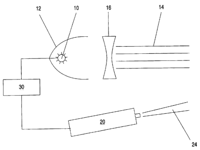
Referring now to the drawings, Figure 1 is a schematic diagram of a preferred
embodiment of the device of the present invention. A high intensity lamp 10
functions as
a source of broad-band (white) light 14. Because lamp 10 emits light in all
directions, a
parabolic reflector 12 and a concave lens 16 are provided to collimate broad-
band light
14, so that substantially all the energy emitted by lamp 10 is directed at the
target and the
surrounding tissue. A laser 20 emits substantially monochromatic light 24,
preferably at
a wavelength of 585 nanometers, also towards the target and the surrounding
tissue. A
control system 30 supplies power to lamp 10 and laser 20, and also turns lamp
10 and
laser 20 on and off in accordance with the pulse schedule shown in Figure 2.
Preferably, lamp 10 is a xenon arc lamp. Preferably, laser 20 is a flashlamp-
pulsed dye laser, for example the ScleroLASER* manufactured by Candela
Corporation
of Wayland, MA.
Figure 2 shows a pulse schedule for the device of Figure 1. The solid line in
Figure 2 represents then duration and intensity of a pulse-band light 14. The
* Trade mark
CA 02330029 2000-10-26
WO 99/56824 PCT/US98/08657
7
dashed line in Figure 2 represents the duration and intensity of a pulse of
monochromatic light 24. Broad-band light 14 is turned on at time T0, and is
kept on
long enough, until time T2, to heat the target and the surrounding tissue to
about 60 C.
As the temperature of the surrounding tissue approaches the desired final
value,
monochromatic light 24 is turned on at time T1, and is kept on until time T3,
long
enough to cause coagulation of the target but not long enough to damage the
surrounding tissue. Preferably, the duration of the monochromatic pulse is
between
about 0.1 milliseconds and about 10 milliseconds.
Figure 3 is a schematic diagram of another preferred embodiment of the device
of the present invention. In this embodiment, lamp 10 serves as the source of
both the
broad-band radiation and the monochromatic radiation that are incident on the
target
and the surrounding tissue. In this embodiment, a mechanical shutter 32 serves
to
alternately block and pass broad-band light 14, thus causing the light
emerging from
the device to be pulsed. A rotating circular filter 34 having two sections, a
white
section 36 and a colored section 38, serves to filter the broad-band pulses
passed by
shutter 32. White section 36 attenuates all wavelengths to substantially the
same
degree, thereby providing a broad-band pulse of the proper intensity and
duration to
heat the target and the surrounding tissue to about 60 C. Colored section 38
attenuates all but a narrow spectral band of light centered on a wavelength of
585
nanometers. Control system 30 synchronizes the movement of shutter 32 and
filter 34
to provide light pulses according to the pulse schedule of Figure 4.
Note that lamp 10 must be much more powerful in the embodiment of Figure
3 than in the embodiment of Figure 1, because in the embodiment of Figure 3,
lamp
CA 02330029 2000-10-26
WO 99/56824 PCT/US98/08657
8
must provide enough spectral power in the vicinity of 585 nanometers to
coagulate
the target. It is for this reason that white section 36 of filter 34 is
required in this
embodiment.
Figure 4 shows a pulse schedule for the device of Figure 3. As in Figure 2, a
5 solid line represents a broad-band pulse and a dashed line represents a
monochromatic
pulse. At time To, with filter 34 positioned so that white section 36 is in
the optical
path of broad-band light 14, shutter 32 is opened, allowing broad-band light
14 to pass
through, and to be attenuated by, white section 36. Filter 34 is rotated,
until, at time
T1, colored section 38 begins to intercept broad-band light 14. At time T2,
all of
1o broad-band light 14 is passing through colored section 38, so that the
light emerging
from the device is substantially monochromatic. At time T3, shutter 32 is
closed,
terminating the monochromatic pulse.
Figure 5 is a schematic diagram of a variant of the device of Figure 3. In the
device of Figure 5, a movable mirror 40 is provided to deflect light passed by
shutter
32 to a fixed mirror 41 and a monochromator 42. The device of Figure 5
generates
pulses according to the pulse schedule of Figure 6, in which, again, the solid
line
represents a broad-band pulse and the dashed line represents a monochromatic
pulse.
At time To, with mirror 40 withdrawn, shutter 32 is opened, allowing broad-
band light
14 to pass through an attenuation filter 44 and thence to the target and the
surrounding
tissue. Like white region 36 of filter 34, attenuation filter 44 attenuates
all
wavelengths to substantially the same degree, to provide a broad-band pulse of
the
proper duration and intensity to heat the target and the surrounding tissue to
about
60 C. At time T1, mirror 40 is moved into place, terminating the broad-band
pulse,
CA 02330029 2000-10-26
WO 99/56824 PCT/US98/08657
9
and, deflecting broad-band light 14 so that it passes, via mirror 41, through
monochromator 42, thereby initiating the monochromatic pulse. Thus, the
monochromatic pulse starts substantially immediately after the termination of
the
broad-band pulse. Monochromator 42 passes on to the target only a narrow
spectral
band of light centered on a wavelength of 585 nanometers. At time T2, shutter
32
closes, terminating the monochromatic pulse.
While the invention has been described with respect to a limited number of
embodiments, it will be appreciated that many variations, modifications, and
other
applications of the invention may be made.