Note: Descriptions are shown in the official language in which they were submitted.
CA 02330100 2000-10-23
WO 99/60402 PCT/SE99/00722
LIGAND BINDING ASSAY AND KIT WITH A SEPARATION ZONE
FOR DISTURBING ANALYTES.
Technical field of the invention
The invention relates to a method for determining an analyte in a sample and
to a
kit for use in the method.
Starting from the prior art, the method of the invention comprises the steps:
i. The sample is applied in a sample application zone (ASZ) on a flow matrix
in
which transport of components present in the sample may take place (transport
flow).
The flow matrix further comprises:
a) optionally an application zone (AR*Z) for a binding reactant (Reactant* =
R*)
which is analytically detectable,
b) a detection zone (DZ) which is located downstream of ASZ and exhibits
another binding reactant (Capturer) firmly anchored to the matrix and in which
a
complex (signal complex) containing the Capturer and the analyte and/or the
Reactant*
is formed in the method.
ii. The flow is allowed to effect the transport of sample components.
iii. The signal complex is detected in the detection zone and the measured
signal is
used for the determination of the analyte.
The invention is primarily directed to the flow matrix which may be of the
same
type as those previously used in, for example, immunochromatography, see
below.
Suitable binding reactants are those which participate in so-called affinity
reactions, especially biospecific affinity reactions, and covalent binding
reactions,
especially exchange reactions between free thiol and reactive disulphide and
other
reactions between soft electrophiles and soft nucleophiles. Common biospecific
affinity
reactions are immunochemical, i.e. between antibody and antigen or hapten.
Other types
of bioaffine reactions are hybridization between complementary nucleic acids
(including
oligonucleotides), reaction between lectin and carbohydrate structure, between
Ig(Fc)-
structure and Ig(Fc)-binding protein, such as protein A or protein G, etc. The
bioaffine
reactions include the reaction between a biomolecule and a synthetically
prepared
ligand/capturer.
For the type of method in question, one talks about non-competitive methods,
for
example sandwich technique, and competitive methods. Sandwich technique
usually
CA 02330100 2000-10-23
WO 99/60402 PCT/SE99/00722
2
means that an analytically detectable complex is formed in which the analyte
binds to
two bioaffine counterparts, one of which is analytically detectable and the
other is
Capturer. In common competitive variants, the analyte and an analytically
detectable
analyte analogue will compete for a limiting amount of bioaffine counterpart.
As
examples of two competitive variants may be mentioned those that use: a)
competition
between analyte and analyte analogue, which is labelled, for a limiting amount
of ligand
in the form of a firmly anchored Capturer, and b) competition between analyte
and
analyte analogue in the form of firmly anchored Capturer for a limiting amount
of
soluble and analytically detectable bioaffine counterpart.
For further information on previously used methodology within the technical
field of the invention it is referred to US-A-4,861,711 (Behringwerke), WO
88/08534
(Unilever), US-A-5,120,643 and 4,740,468 (Abbott), EP-A-284,232 and
US-A-4,855,240 (Becton Dickinson) and WO 96/22532 (Pharmacia AB).
Heteroforms
Compounds which can compete for the binding to a counterpart via one of the
above mentioned binding reactions. Heteroforms may be isoforms of proteins,
e.g.
isoenzymes etc. Within the term heteroforms are included inter alia different
forms of
bioaffine complexes which "resemble" each other by meeting the above
definition.
2 0 Examples are immunocomplexes where the antigen is the same but the
antibody is of
different class/subclass. See further under the title "Analyte" below.
Determination of whether two compounds are heteroforms to each other may be
made in so-called inhibition tests.
Problems to be solved by the invention
The components of a sample that may affect or influence the signal that is to
be
detected in DZ can be divided into two main groups: a) the analyte and b)
components
which directly or indirectly disturb the detection. Directly disturbing
components are
those which interfere with the signal as such, for example fluorescent
components in
3 0 serum in case the complex is to be detected by fluorescence. Examples of
indirectly
disturbing components are heteroforms with regard to Capturer and/or an added
bioaffine reactant R (for example R*). Other indirectly disturbing components,
for
CA 02330100 2000-10-23
WO 99/60402 PCT/SE99/00722
3
example heterophilic antibodies, may be present in the original sample and
interfere
with the formation of the signal complex in DZ. In certain embodiments of the
invention, ligands that are released from the separation zone of the invention
may act
disturbingly (see Example 1).
Problems with disturbing components in samples have often meant that for
analytes that are present in low concentrations, the separation of disturbing
components
and the detection have been performed in different systems.
An example where after ion-exchange separation, analysis has been carried out
either by immunological systems or by on-line measurement of an absorbing
group (460
nm), is in the measurement of carbohydrate deficient transferrins (CDT = CD-
transferrin
= asialo-, monosialo- and disialo-transferrin). When CDT is present at a
relatively high
concentration (10-9 M), both detection alternatives have been possible, but at
lower
concentrations of analyte, immunological measurement is required. The ion-
exchange
chromatography separation is controlled from an advanced and costly equipment,
which
requires specially educated personnel. Also the traditional immunological
tests are
expensive and require well-educated personnel.
The technique for immunological on-line measurement after a chromatographic
separation step has been described by Afeyan et al. (Nature 358 (1992) 603-
604) and
Irth et al. (Anal. Chem. 14 (1995) 355-361). Its difficulties have been
summarized by
2 0 Krull et al. (LC-GC 15(7) (1997) 620-629).
Transport of whole cells into DZ may interfere with the signal from the
detection
complex. It is previously known to use flow matrices where the cells are
captured
mechanically (through filtration) in a denser pre-zone (Oudheusden et al.,
Ann. Clin.
Biochem. 28 (1991) 55-59).
2 5 EP-A-696,735 discloses a chromatographic immunoanalytical system where, in
order to extend the measuring range for the analyte, a predetermined amount of
analyte-
binding antibody has been immobilized in the sample application zone so that a
certain
amount of analyte is retained therein.
EP-A-702,233 discloses a chromatographic immunoanalytical system where, in
3 0 a similar manner to that described in EP-A-696,735, a dilution effect of
the sample is
achieved by capturing a certain amount of analyte before it reacts with
labelled reactant
which is then detected in the detection zone.
CA 02330100 2000-10-23
WO 99/60402 PCT/SE99/00722
4
WO 97/35205 discloses a chromatographic membrane for immunoanalysis
having (i) a zone for the detection of labelled analyte-binding reactant which
has not
bound to the analyte, and (ii) a zone for the detection of the complex between
analyte-
binding reactant and the analyte. The relative amounts of unbound analyte-
binding
reactant and analyte: reactant complex gives a measure of the amount of
analyte in the
sample.
WO 94/06012 discloses an analytical test apparatus having a negative control
zone placed before the analyte detection zone. The negative control zone has
the
function to indicate the presence in the sample of components that affect the
analyte
detection so that it becomes unreliable.
Objects of the invention
A first main object of the invention is to create a simple and rapid method
that
facilitates the determination of an analyte in the presence of disturbing
components. A
particular object is to avoid problems with disturbing components that are
soluble or
suspendable in liquid media of interest.
A second main object of the invention is more rapid and simpler determinations
of individual heteroforms or combinations thereof, especially heteroforms,
that exhibit
peptide, carbohydrate or lipid structures, including various types of
biologically active
2 0 compounds. Among lipids are included steroids and other fat-soluble
substances.
A third main object of the invention is to facilitate the measurement of
analytes
in the concentration range < 10-7 M, particularly < 10-9 M, especially for
samples
containing disturbing heteroforms of the analyte.
A fourth main object of the invention is to simplify the determination of
individual heteroforms or combinations thereof in samples originating from
biological
materials.
A fifth main object of the invention is to provide more rapid and simpler
evaluations of libraries of compounds, for example chemical libraries, such as
combinatorial libraries.
3 0 A subobject of the above mentioned four main objects is to improve the
possibilities of making determinations in field environment (usually semi-
CA 02330100 2000-10-23
WO 99/60402 PCT/SE99/00722
quantitatively) as well as in advanced laboratories (with the possibility of
accurate
quantification).
The invention
5 The above mentioned objects may be achieved with the method mentioned in the
introductory part herein, if the flow matrix contains one or more separation
zones (SZ)
between ASZ and DZ, which should permit at least one component, capable of
influencing the signal from the signal complex in DZ, to be
retarded/separated. This
should take place in SZ by means of the ligand interactions mentioned below,
which can
be reversible or irreversible. The component may be either a disturbing
component or
the analyte. If the component is not an analyte, the retardation means that
the
component (or components) migrates more slowly than the analyte through SZ or
is
bound irreversibly to SZ and thereby is prevented from reaching DZ such that
the
detection of analyte in DZ essentially will not be disturbed by the component
(or
components) in question. Usually, this means that there should be a sufficient
amount of
ligand for substantially all of the disturbing component or components in the
sample to
be affected. "Substantially all" depends on the relative concentrations of the
component(s), but usually means that at least 90 %, preferably at least about
95 %, and
more preferably at least 99 % of the disturbing component(s) are retarded or
captured in
2 0 the separation zone. The component may be the analyte if it is desired to
study the
capability of one or more ligands to bind the analyte. In this case such a
ligand is
immobilized in the separation zone.
The choice of retarding structure/ligand in the separation zone is determined
by
the components that are retarded. The retardation may be based on various more
or less
specific interactions between the ligand structure and the component(s) to be
retarded;
see below under the title "Separation zone". After the passage of SZ, the
analyte will
migrate with the transport flow to the detection zone (DZ), in which a complex
containing the Capturer and the analyte and/or R* are formed.
In those cases where it is intended to retard one or more disturbing
components,
3 0 the formation of signal complexes will take place in the absence thereof.
The detection
of signal complexes in DZ may be taken as a qualitative or quantitative
measure of the
analyte.
CA 02330100 2000-10-23
WO 99/60402 PCT/SE99/00722
6
In those cases where it is intended to retard the analyte, the point of time
for the
formation of a signal complex will be changed, or, if the analyte-ligand
binding in SZ is
irreversible, the formation of a signal complex may be completely inhibited.
The
formation of a signal complex in DZ will be a measure of the capability of the
analyte to
bind to the ligand in SZ.
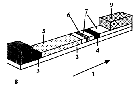
Figures 1-3 illustrate different variants of flow matrices according to the
invention.
Figure 1 is a simple variant having an ASZ, an ARZ, a SZ and a DZ. ARZ and ASZ
are
separated.
Figure 2A differs from the variant in Figure 1 primarily by having five
separation zones
with the same ligand. ARZ and ASZ are separated.
Figure 2B is the same as the variant in Figure 2A except that ARZ and ASZ
coincide.
Figure 3 illustrates the variant of flow matrix of the invention that is used
in Example 1
with three separation zones, two zones (SZI) thereof exhibiting a certain
ligand and one
zone (SZ2) exhibiting another ligand. ASZ and ARZ (= AR*Z) are separated.
A more detailed description of Figure 1 is given under the title "Matrix and
transport flow", and of Figures 2-3 in the introduction to Example 1. The flow
matrices
represented by Figures 1-3 may in principle have any of the geometric
embodiments
below.
Matrix and transport flow
The matrix is of the same type as those previously used in so-called
immunochromatographic determination methods (flow matrix) and defines the room
in
which reactants and sample components are transported. The matrix may thus be
the
internal surface of a single flow channel (for example a capillary), the
internal surface of
a porous matrix having a penetrating system of flow channels (porous matrix)
etc. The
matrix may be in the form of monolith, sheet, column, membrane, separate flow
channel(s), for example of capillary dimensions, or aggregated systems of such
flow
channels etc. They may also be in the form of particles packed in column
cartridges or
3 0 in cut grooves, compressed fibres etc. Another alternative is so-called
nanocolumns for
liquid chromatography, i.e. silicon or quartz plates having channels of about
2 m or
less prepared by micro lithography (see e.g. He, B., et at., Anal. Chem. 1998,
70,
CA 02330100 2000-10-23
WO 99/60402 PCT/SE99/00722
7
3790-3797). The inner surface of the matrix, i.e. the surface of the flow
channels, should
be sufficiently hydrophilic to permit aqueous media (primarily water) to be
transported
through the matrix, either by means of capillary force or by means of applied
pressure or
suction. The smallest inner dimension of the flow channels (for round channels
measured as a diameter) should be sufficiently great to permit transport
through the
matrix of analyte, added reactants, and components that interfere in the
detection zone
and that are to be retarded in SZ. The rule of thumb is that suitable matrices
may be
selected among those with flow channels having a smallest inner dimension in
the range
of 0.1-1000 m, with preference for 0.4-100 m if the matrix has a system of
communicating flow channels. Flow channels having their smallest dimension in
the
upper part of the broad range (up to 1000 m) are primarily of interest for
flows driven
by externally applied pressure/suction.
Suitable matrices are often built up from a polymer, for example
nitrocellulose,
polyester, polyethersulphone, nylon, cellulose nitrate/acetate, cellulose,
regenerated
cellulose. Advantageously, these membranes may be provided with a tight
backside of
e.g. polyester.
The material of the matrix as well as the physical and geometric design of the
flow channels may vary along the flow depending on the intended use of a
certain part
of the matrix [WO 96/22532 (Pharmacia AB); WO 94/15215 (Medix)]. One and the
2 0 same matrix may comprise several transport flows that are parallel or
directed radially
from a common centre, for example in the form of separate channels. In some of
the
most important embodiments, at least the detection zone and the most adjacent
parts of
the matrix should be in such a form that the transport flow into, in and out
of DZ may
take place laterally in the matrix, i.e. at least this part of the matrix is
in the form of a
membrane strip or plate having cut grooves or the like.
Various flow matrices that may be used in the type of tests in question are
described in prior patent publications. See e.g. US-A-4,861,711
(Behringwerke), WO
88/08534 (Unilever), US-A-5,120,643 and US-A-4,740,468 (Abbott), EP-A-284,232
and US-A-4,855,240 (Becton Dickinson); WO 96/22532 (Pharmacia AB).
3 0 The most important embodiment of the invention at the priority date is
based on
liquid transport in a flow matrix which is in the form of e.g. a membrane
strip (see Fig.
CA 02330100 2000-10-23
WO 99/60402 PCT/SE99/00722
8
1). The strip is made up of a matrix that defines a transport flow (1) and is
applied to a
liquid-tight backing (2), suitably of plastic. On the matrix there is an
appplication zone
for sample (3, ASZ) and a detection zone (4, DZ) located downstream thereof.
The
transport flow is in the direction from ASZ towards DZ. Between the sample
application
zone (ASZ) and the detection zone there is a separation zone (5, SZ). In the
transport
flow there may, if required by the particular embodiment, also be application
zones (6)
for additional reactants (R, for example R*, with application zone ARZ, for
example
AR*Z). Between said zones there may be zones (7) the only function of which is
to
transport reactants. The position of an application zone ARZ (AR*Z) is
determined by
the test protocol to be used, and may be upstream or downstream of or coincide
with
ASZ. For the case that ARZ (for example AR*Z) is upstream of ASZ, it may be
advantageous if the addition of liquid in ASZ takes place substantially
simultaneously
as the addition of liquid in the zone ARZ (AR*Z) located upstream thereof. See
our
earlier filed international patent application PCT/SE98/02463 (incorporated by
reference
herein). For certain types of test protocols, ARZ (AR*Z) may coincide with DZ.
In some embodiments it is advantageous if a reactant R, for example R*, is pre-
deposited. This is especially the case if ARZ is located downstream of ASZ and
the test
protocol variant used is simultaneous, i.e. the reactant R and the analyte are
to migrate
into DZ substantially simultaneously.
2 0 In the cases where it is desired to use variants that are sequential in
the sense that
the analyte is to be transported into DZ before the reactant (R), R should be
added after
the sample has passed ARZ if the application zone for reactant (ARZ) is
downstream of
ASZ. Sequential methods may also be achieved if ARZ is upstream of ASZ, in
which
case R optionally may be pre-deposited in ARZ.
In alternative embodiments, reactants (R), for example R*, may migrate into DZ
in separate transport flows from another direction than that of the flow that
transports
the analyte into DZ. See, for example, US-A-4,855,240 (Becton & Dickinson).
In one and the same transport flow there may be several detection zones
intended
for different analytes or different concentration ranges of the same analyte.
For the case
3 0 that the analytes are different, the Capturers in the respective DZ must,
of course, not
exhibit any substantial cross-reactivity against any of the analytes.
CA 02330100 2000-10-23
WO 99/60402 PCT/SE99/00722
9
The transport flow from ASZ through the separation zone (SZ) and further to
the
detection zone (DZ) may be a liquid flow driven by capillary force. When
necessary, the
flow matrix may exhibit a liquid reservoir (8) in the form of a porous matrix
that is
soaked with transport liquid and applied upstream of ASZ and/or a sucking
porous
matrix (9) placed downstream of DZ. The liquid reservoir and the sucking
matrix assist
in maintaining the flow. Liquid flow may also be achieved by means of pressure
or
suction through the matrix. Thus, the pressure may be driven hydrostatically,
for
example by a part of the matrix being designed as a minicolumn placed
vertically and
with its outlet in direct liquid communication with a horizontally located
flow matrix. In
the latter form, the horizontally located part of the matrix may be in the
form of a
strip/membrane. An alternative for transport of analyte, reactants and
disturbing
components may be the application of an electric field across the matrix.
Similar sequences of zones, like that in Figure 1, may also be constructed for
other types of flow matrices, for example capillary tubes and matrices in
which the
transport flow may be in depth.
One or more matrices/transport flows according to the above may be placed
together, for example on a common backing, optionally with a liquid barrier
between
them. Optionally, the flows may have a common ASZ, a common ARZ (AR*Z) etc. As
a rule, DZ is separate for each transport flow.
2 0 In the above mentioned variants, matrices having a separation zone may be
used
to determine one heteroform (analyte). A matrix without separation zone may be
used to
determine all heteroforms of the analyte that may be present in the sample in
an
analogous manner to that for the analyte. By combining these two types of zone
sequences, relative as well as absolute quantities of analyte in the sample
may easily be
2 5 measured.
Separation zone (SZ)
The separation zone exhibits a ligand/structure having binding capability for
one
or more sample components that would have disturbed the detection in DZ. A
3 0 characteristic feature is that the separation is achieved by means of some
type of
specific/selective binding reaction and not because the matrix in SZ provides
a
mechanical obstacle for disturbing components (filtration). Guiding principles
for the
CA 02330100 2000-10-23
WO 99/60402 PCT/SE99/00722
choice of separating/retarding ligand/structure, especially with regard to
specificity,
binding strength (affinity), and kinetics are the same as in affinity
chromatography,
including ion-exchange chromatography, covalent chromatography, and
biospecific
analytical methods in which solid-phase technology is used for capture. With
regard to
5 binding strength (affinity, avidity) and kinetics, the main object of the
presently
preferred variants of the invention is to retard disturbing components in
relation to the
analyte so that detection in DZ may take place without presence of these
components.
Generally, this means that the disturbing components should be retarded as
effectively
as possible or be bound as strongly and quickly as possible in the separation
zone.
10 The ligands that make separation in SZ possible may thus be a) charged
(anionic,
cationic, amphoteric = ion-exchange ligands), amphoteric/amphiphilic,
bioaffine,
chelating, sulphur-containing (primarily thioether for so-called thiophilic
affinity), those
permitting covalent chromatography (reactive disulphide such as pyridyl
disulphide) or
n-it interaction, hydrophobic etc.
In those cases where disturbing components are to be retarded, the rule of
thumb
is that the binding capability of the ligand to one or more disturbing
components should
be stronger than that to the analyte. This applies to the conditions used for
the separation
in SZ. Factors that determine how the separation will succeed are the length
of the
separation zone, ligand density, ligand availability, temperature, flow
velocity, buffer,
2 0 ion-strength, pH, etc.
Among biospecific affinity ligands, primarily so-called immunoligands are
noted, i.e. antibodies and antigen-binding fragments thereof, and antigen and
hapten.
Other examples of affinity ligands are lectin (for example, sialic acid-
binding lectins);
Ig(Fc)-binding protein (such as Protein A and G); nucleic acid, such as oligo-
or
2 5 polynucleotide in single or double-stranded form, analogues of substrates
for enzymes,
enzyme inhibitors, etc. For biospecific affinity ligands, the specificity may
be directed
towards one or more binding sites on the component(s) to be retarded. The
corresponding binding sites should not be available to the same degree on the
analyte
(by which is also intended the case that they do not even exist in non-exposed
form).
3 0 The ligands/structures in question may be anchored to the separation zone,
either
by covalent binding to the matrix, via physical or biospecific adsorption.
Examples of
the latter is the interaction between biotin and streptavidin, between highly
affine
CA 02330100 2000-10-23
WO 99/60402 PCT/SE99/00722
11
antibody and hapten etc. The anchorage to the matrix may take place via a
polymer or
other substituent which in turn carries covalently, physically adsorptively,
or
biospecifically bound ligands that are used in the separation. Another
possibility is
deposition of polymeric particles which exhibit a desired type of ligand. The
particles
may be of hydrophilic or hydrophobic character and to which a compound
exhibiting
the ligand structure has been adsorbed or covalently bound. The technique for
binding a
separating ligand to the matrix SZ may basically be selected in the same way
as
previously known for the Capturer in DZ. See, for example, our earlier filed
international patent applications PCT/SE98/02462, PCT/SE98/02463 and
PCT/SE98/02464 which are hereby incorporated by reference with regard to the
introduction of Capturer into the detection zone. In this connection it may be
mentioned
that there are commercially available membranes which have covalently bound
ligands,
for example DEAE cellulose paper (diethyl aminoethyl) (DE8 1, Whatman
International
Ltd, England).
Detection zone
The Capturer in the detection zone may be selected according to the same rules
as those applying to the ligand in the separation zone, with the proviso that
the binding
capability of the Capturer should be directed towards the analyte and/or
towards an
2 0 analvte-related reactant. It is advantageous to choose highly affine
Capturers with rapid
kinetics for capture of the ligand. It is primarily of interest to use
antibodies or
antigen/hapten for which it is often easy to find highly affine antibodies.
By analyte-related reactant is intended a reactant (R) that is added and when
migrating through DZ may bind to the Capturer in an amount that is related to
the
presence of analyte in the sample. Examples of analyte-related reactants are
R* in the
form of a) labelled analyte analogue in competitive methods that use
competition for a
limiting amount of solid-phase-bound anti-analyte antibody, and b) labelled or
non-
labelled soluble anti-analyte antibody in methods that use
competition/inhibition
between solid-phase-bound analyte analogue and analyte for a limiting amount
of anti-
3 0 analyte antibody in dissolved form.
The Capturer may be anchored to the detection zone by a technique analogous to
that used to bind the ligand to the separation zone.
CA 02330100 2000-10-23
WO 99/60402 PCT/SE99/00722
12
It may be suitable to combine a separation principle in the separation zone
with a
different capturing principle in the detection zone, e.g. ion-exchange
chromatography
for separation and immunochemical adsorption for capture in DZ. In some
situations it
may be practical to use the same principle for retardation and capture in the
two zones
(e.g. two monoclonal antibodies having different specificities, see the
Examples).
Analyte
By analyte is intended the compound or compounds that are determined
quantitatively or qualitatively. Quantitative determination relates to the
measurement of
quantities in absolute as well as relative terms. Qualitative determination of
an analyte
refers to detecting the existence or non-existence of something (yesino test)
or
qualitative properties of a compound, such as capability of affinity-binding
to a certain
ligand.
By relative measurement is intended that the measurement value obtained is a
ratio of the sum of one or more selected heteroforms and the sum of another
combination of heteroforms. An example is the ratio of analyte amount and
total amount
of all heteroforms with regard to a certain counterpart (total amount includes
the amount
of analyte).
The invention is applicable to analytes that may function as a binding
reactant.
2 0 This means that the analyte basically can be any substance for which it is
possible to
provide a Capturer as above. As specific examples may be mentioned
antigen/hapten,
enzyme or antibody or nucleic acid which completely or partly are in single-
stranded
form. The analyte may exhibit amino acid/peptide, carbohydrate or lipid
structure.
Particularly great advantages are obtained for analytes existing together with
heteroforms with regard to binding capability to Capturer and/or an added
reactant R,
for example R*. This applies particularly to the cases where the analyte is in
sample
concentrations which are < 10-7 M, especially < 10-9 M. As examples of this
type of
heteroforms may be mentioned: a) Compounds which differ from each other in
charge,
such as isotransferrins with, for example, CDT as analyte, isohemoglobins
with, for
3 0 example, HbAI c as analyte; b) Compounds which differ from each other in
certain parts
of the basic structure, such as additionally inserted or cleaved (e.g. by
degradation)
amino acids, or partial differences in peptide chains; c) Compounds which
differ from
CA 02330100 2000-10-23
WO 99/60402 PCT/SE99/00722
13
each other due to the fact that different substances/structures have been
added to a basic
structure, for example covalently bonded carbohydrate structures; d)
macromolecules
consisting of two or more subunits which in the macromolecule bind to each
other via
non-covalent bonds, such as bioaffine bonds between receptor and ligand in
receptor-
ligand complexes and between antigen and antibody in immunocomplexes, or via
cystine bridges, for example between the chains of an antibody.
Examples of potential uses/analytes are:
a) The analyte is a heteroform which differs from other heteroforms with
regard to
carbohydrate contents (glycosylation), for example glycoproteins having the
same or a
similar protein part. Variations in this type of heteroforms are known in a
number of
disease conditions such as cancer, inflammation and liver diseases. (Turner G
A, "N-
glycosylation of serum proteins in disease and its investigation using
lectins", Clin.
Chim. Acta 208 (1992) 149-171; and Varki A, "Biological roles of
oligosaccharides: all
of the theories correct", Glycobiology 3(2) (1993) 97-130). Particularly may
be
mentioned the measurement of i) combinations of asialo-, monosialo- and
disialo-
transferrin for which separation may be performed by ion-exchange ligand and
also by
lectin ligand in SZ, and ii) HbAlc which may be separated by means of ion-
exchange or
boronate ligand. Variations in the carbohydrate contents of proteins are also
known in
normal biological changes, for example during the menstrual cycle and for
differences
2 0 in age and sex.
b) The degree of glycosylation of recombinant proteins could be determined by
means of ion-exchange, lectin or boronate ligands in SZ. The analyte will in
this case be
the fraction of a recombinant protein that does not contain a carbohydrate
structure that
binds to the ligand in SZ and therefore migrates most rapidly through SZ.
2 5 c) Recombinant proteins into which a separation handle has been inserted,
for
example a histidine sequence or an IgG-binding sequence, and where total
cleavage of
the handle is important, could be checked after separation in SZ by means of a
metal
chelate ligand or and IgG(Fc)-ligand, respectively. The analyte will in this
case be the
fraction of the recombinant protein from which the histidine sequence or the
Ig(Fc)-
3 0 binding sequence, respectively has been cleaved off.
CA 02330100 2000-10-23
WO 99/60402 PCT/SE99/00722
14
d) Enzymes could be separated into an active and an inactive form by means of
a
ligand in SZ which is a substrate analogue or an inhibitor of the enzyme in
question.
The analyte will be the inactive enzyme.
e) Proteins, peptides or other biomolecules which exert their biological
function by
binding to a specific receptor could be separated by means of a ligand in SZ
which is a
receptor for the biomolecule. The analyte will be the fraction of the
molecules that lack
or have a reduced capability of binding to the receptor.
f) Proteins (e.g. IgE) may in vivo have autoantibodies (IgG, IgA, IgM) bound
thereto. These autoantibodies give rise to, on the one hand, a differing
response in
immunochemical determination of the protein, and, on the other hand, an
altered
turnover rate/function. By using antibodies to the autoantibodies in question
as ligand in
the separation zone, autoantibodies in free and immunocomplex-bound form may
be
separated and the amount of the free form of the protein (= analyte, e.g. of
IgE) may be
calculated.
g) By means of a monoclonal antibody directed against a certain binding site
of a
protein and immobilized to SZ, the presence of heteroforms to the protein
which do not
exhibit the binding site (= analyte) could detected by quantification in DZ.
h) The presence of different substances bound to transport proteins, e.g. a
drug
bound to albumin, could be measured by using suitable ligands in SZ. By the
choice of a
2 0 suitable ligand in SZ, transport proteins with or without bound drug may
be measured in
DZ.
i) IgG and IgA in serum may in certain rheumatic or autoimmune diseases have
an
increased adsorption to different surfaces. By anchoring ligands in the
separation zone
which are capable of binding to IgG and IgA with changed properties, it will
be possible
to measure the proportion of IgG and IgA with unchanged adsorption properties
(=
analyte) in DZ. By having the corresponding autoantigen/hapten as Capturer in
DZ,
specific autoantibodies of IgG or IgA class could be measured with better
sensitivity.
j) Many biologically active compounds (for example, peptides or steroids) are
transported in serum in the form of complexes with binder proteins. By using
antibodies
3 0 against the binder protein as ligand in SZ, the non-complex-bound (free)
form of these
compounds (= analyte) could be determined inimunochemically in the following
detection zone. Examples are triiodothyronine and thyroxine which are
transported
CA 02330100 2000-10-23
WO 99/60402 PCT/SE99/00722
bound to thyroxine-binding globulin (TBG) or thyroxine-binding prealbumin
(TBPA).
Analogously, free forms of estradiol and testosterone which are transported in
bound
form with sexual hormone-binding globulin may be measured.
k) The binding capability of a first compound (= analyte) for a second
compound
5 may be determined with the invention. In this embodiment, one may have the
second
compound as ligand in SZ, and a Capturer with a known binding capability to
the
analyte in DZ. Capture/retardation in SZ will be a measure of the binding
capability of
the analyte and may be measured in DZ.
This embodiment of the invention may be particularly advantageous in the
10 screening of different libraries of compounds with the library members as
ligands in SZ
(chemical libraries, for example).
1) Degradation isoforms of proteins where amino acids have been cleaved off,
can
be determined by the invention. For example, degradation isoforms of creatine
kinase
(CK) are interesting cardiac markers.
Detection in DZ and labelled reactant (R*)
Detection and quantification of signal complexes may be performed by means of
an analytically detectable reactant (Reactant* = R*). For those cases where
the analyte
per se is detectable and is part of a signal complex, detection and
quantification may
2 0 take place without using R*.
R* is usually a biospecific affinity reactant which is labelled with an
analytically
detectable group, such as an enzymatically active group, radioactive group,
fluorescent
group, chromogenic group, hapten, biotin, particles, etc. Analytically
detectable
reactants (R*) also include reactants which per se have binding sites or
properties which
2 5 may be detected analytically when the reactant is part of the signal
complex. Examples
of such binding sites are Ig-class- and Ig-subclass-specific determinants when
the
reactant is an antibody and the antigen-binding part thereof is used to form
the complex
in the detection zone.
Usual forms of analytically labelled reactant are labelled antibody and
labelled
3 0 antigen/hapten. Labelled antibody has its primary use in
A) non-competitive techniques, such as sandwich technique, in which the
captureris
CA 02330100 2000-10-23
WO 99/60402 PCT/SE99/00722
16
a) an antibody which is directed against the same antigen (= analyte) as
the labelled antibody, or
b) an antigen/hapten, or
B) competitive techniques in which competition takes place between an analyte
and a solid phase-bound analyte analogue for a limiting amount of anti-analyte
antibody and the detection of free or occupied sites on the solid phase may be
performed by means of labelled anti-analyte antibody and anti-anti-analyte
antibody, respectively.
Labelled antigen/hapten has its primary use in
A) competitive techniques in which a labelled antigen/hapten is allowed to
compete with an unlabelled antigen/hapten for a limiting amount of antibody
(Capturer), or
B) sandwich-techniques in which antigen/hapten-specific antibody is determined
with anti-antibody as Capturer.
Examples of variants of the invention in which an analytically detectable
reactant (R*) is not utilized are those where the analyte per se is detectable
when it is
part of the complex in DZ. This is illustrated with enzyme as analyte in
combination
with a substrate that gives an analytically detectable product, for example a
substrate
that gives a coloured or fluorescent product that should be insoluble.
2 0 R* may, but need not, exhibit binding capability to the disturbing
components
that are separated in SZ. To the extent that R* has binding capability, the
application
zone thereof should be located downstream of the separation zone (SZ), unless
it is
desired to measure the level of disturbing heteroforms by means of the amount
of R*
binding to SZ.
A particularly useful labelling group is particles which optionally contain
one of
the above mentioned detectable groups, such as fluorophoric group or
chromogenic
group (fluorescent and coloured particles, respectively). Useful particles
often have a
size in the range of 0.001 to 5 .tm, with preference for the range of 0.05 to
5 m. The
particles may be of colloidal dimensions, so-called sol (i.e. usually
spherical and
3 0 monodisperse having a size in the range of 0.001 to 1 m). Especially may
be
mentioned metal particles (for example, gold sol), non-metal particles (for
example,
CA 02330100 2000-10-23
WO 99/60402 PCT/SE99/00722
17
SiO2, carbon, latex and killed erythrocytes and bacteria). Also particles of
non-colloidal
dimensions have been used. These particles have been more or less irregular
and more
or less polydisperse (for example, carbon particles < 1 m; Pharmacia AB, WO
96/22532).
When particles are the label group in the invention, the complex in DZ may
often be detected visually or by optical measuring equipment (e.g. a CCD
camera
coupled to a computer with special software for image analysis or a laser
scanner).
For particles as the label group, it is referred to WO 88/08534 (Unilever);
US-A-5,120,643 (Abbott); EP-A-284,232 (Becton Dickinson) and others.
Samples
The invention is primarily intended for biological samples, for example, blood
(serum, plasma, whole blood), saliva, tear fluid, urine, cerebrospinal fluid,
sweat, etc.
The invention is also applicable to other samples, such as fermentation
solutions,
reaction mixtures, solutions containing a certain protein for which the
binding capability
to a ligand in SZ is to be investigated, etc. See above under the title
"Analytes". It may
be particularly interesting to use the invention for analysis of environmental
samples.
In addition to the method, the invention also relates to an apparatus and a
kit,
respectively, containing the above defined flow matrix.
2 0 The inventions disclosed in the above-mentioned international applications
PCT/SE98/02462, PCT/SE98/02463 and PCT/SE98/02464 may in relevant parts
constitute preferred embodiments of the present invention. All three
applications have
been incorporated by reference.
PATENT EXAMPLES
EXAMPLE 1. TEST STRIP FOR MEASUREMENT OF THE PROPORTION OF
FREE IgE, IgE BOUND TO IgG AND ANTIBODIES TO IgE
3 0 In Figures 2A, 2B and 3 the direction of the transport flow is indicated
by an
arrow (10). In each variant there may at the beginning of the transport flow
be a zone
ASZ (11) for sample, downstream thereof a zone DZ (12), at the end of the
transport
CA 02330100 2000-10-23
WO 99/60402 PCT/SE99/00722
18
flow a sucking part (13), and between each type of zone, parts which only
serve as
transport zones (14).
Figure 2A: The variant according to this figure has five separation zones (SZ)
in which
the ligand may be the same or different or be present in different amounts (15-
19) and
an AR*Z (20) for reagents.
Figure 2B: This is the same sequence of zones as in Figure 2A except that ASZ
(11)
and AR*Z (20) coincide (21). This zone sequence may also be used for the cases
where
the analyte per se is detectable when it is part of a signal complex in DZ. An
AR*Z is
then not necessary.
Figure 3: The sequence of zones according to this figure exhibits two types of
separation zones SZ1 (22, 23) and SZ2 (24), respectively, and separately AR*Z
(25)
downstream of SZ1 (23) and SZ2 (24).
Background: Free IgE and IgE complex-bound to autoantibody (IgA, IgG and IgM)
may be of interest to measure. Above all, however, free IgE should be
quantified
correctly. In the current tests for measurement of IgE, the autoantibodies may
bind to
the same epitopes on IgE as the reagent antibodies (anti-IgE antibody) and
this may then
give rise to falsely too low total IgE levels that vary depending on the
design of the test.
By separating IgG, IgM and IgA before the measurement of IgE, free IgE may be
2 0 detected. The amount of autoantibodies should also be quantified both as
complexes and
as free IgG antibodies directed against IgE.
The most common tests measure free antibodies by methods which use IgE
bound to a solid phase (corresponding to DZ) with which a heavily diluted
serum
sample is allowed to interact. If the serum sample contains anti-IgE antibody,
the latter
is bound to the solid phase forming an immunocomplex. After unbound serum
components have been washed away, anti-IgG antibody that is labelled (R*),
e.g. with
enzyme, is added. Excess of labelled antibody (R*) is removed and the amount
of
enzyme-labelled anti-IgG antibody (R*) bound to the immobilized immunocomplex
is
determined by the addition of a suitable substrate. The sensitivity of these
tests is
3 0 limited by the unspecific binding of IgG to the solid phase. The IgE-
specific part of the
IgG population is generally very small and may be difficult to distinguish
from the
CA 02330100 2000-10-23
WO 99/60402 PCT/SE99/00722
19
amount of unspecifically bound IgG. By capturing IgG to the solid phase and
measuring
the binding of IgE, this limitation may be avoided.
When measuring IgG-complex bound IgE, IgG is captured to a solid phase s
(corresponding to DZ) which supports covalently bound anti-IgG antibody
(Capturer).
By adding labelled anti-IgE antibody (R*), the amount of complex-bound IgE may
be
measured.
The use of an immunoassay technique based on lateral liquid transport in
membranes as described above where the flow first passes through one or more
separation zones (SZ) and then a detection zone (DZ), opens many possibilities
for
simple measurement of IgE-IgG related parameters. If e.g. a sample that
contains a
mixture of free IgE and IgE bound to a human anti-IgE antibody of IgG class
first is
made to pass through a zone containing solid phase-bound anti-human IgG
antibody
(Ligand in SZ) and then a zone containing solid phase-bound anti-IgE antibody
(Capturer in DZ), the sample content of complex between IgE and anti-IgE
antibody of
IgG class will be bound in the separation zone while free IgE passes to the
detection
zone where it is determined by adding labelled anti-IgE antibody (R*) upstream
of the
detection zone (12) but downstream of the separation zones (15-19) for passage
only
through the detection zone (addition in zone 20 in Fig. 2A). By having anti-
IgE
antibody (R*) pass also the separation zone, the amount of IgE-IgG complex
captured in
2 0 the separation zone by binding to anti-human IgG (Ligand) may also be
determined
(ASZ and AR*Z coincide) (addition in zone 21 in Fig. 2B, ASZ common with
AR*Z).
In the separation zone there are, in addition to complex between IgE and anti-
IgE
antibody of IgG class, also free antibodies against IgE. The amount of the
latter may be
determined by having labelled IgE (R* 1) pass through the separation zone.
Labelled IgE
(R* 1) is then added in a separate test to the membrane strip upstream of SZ.
See Figure
2B.
When the amount of IgG is very high in serum, several bands with high
concentrations of anti-IgG must be used as SZ. Both complex-bound and free
anti-IgE
antibodies will then be distributed over several bands due to the total amount
of IgG,
3 0 and the sum of the signal intensities of these bands gives the amount of
antibodies
against IgE.
CA 02330100 2000-10-23
WO 99/60402 PCT/SE99/00722
In the example below, the test principle of artificially prepared complexes of
IgE
and IgG is demonstrated. The complexes have been prepared with monoclonal
antibodies against IgE, and antibodies against mouse-IgG have therefore been
bound to
the separation membrane. In the detection system, antibodies to IgE directed
against
5 other epitopes than the complex-forming antibody have been used. This makes
it
possible to measure the complex equally well as free IgE in the detection
system.
Separation membrane 1 (SZ1): Sheep anti-mouse IgG(Fc) (Ligand 1) was coupled
to
polystyrene aldehyde particles (0.29.tm diameter, IDC, Portland, Oregon,
U.S.A.) by
10 mixing 1.0 mg/ml of antibodies and 20 mg/ml of polystyrene aldehyde
particles in 25
mM phosphate buffer, pH 6.6, at +4 C for 20 hours. The particles were washed
in 20
mM borate buffer, pH 8.6, and were reacted with 15 mg of NaCNBH3 (Sigma-
Aldrich
Chemie, Steinheim, Germany) per 50 mg of particles for 20 hours. The particles
were
then washed in 20 mM borate buffer, pH 8.6, by repeated suspension,
centrifugation and
15 decanting. The particle suspension was diluted in 3% trehalose, 20 mM
borate buffer, to
mg of particles/ml. The diluted suspension was sprayed on strips (20 cm x 3
cm) of
membranes of nitrocellulose (nitrocellulose on polyester, 5 m pore size,
Whatman
International Ltd, England) in two 0.3 cm wide lines which were parallel to
the long
sides of the strips. The spraying equipment (IVEK linear striper, IVEK
Corporation,
2 0 Vermont, U.S.A.) delivered about 50 g of polystyrene particles/cm for
each line. The
membranes were dried at room temperature and then cut to smaller pieces (0.5
cm x 3
cm).
Separation membrane 2 (SZ2): Mouse IgG (Ligand 2) was diluted in 20 mM borate
2S buffer to 3.4 mg of protein/ml. The diluted antibody was sprayed on strips
(20 cm x 4
cm) of membranes of nitrocellulose (the same type as above) in a 0.3 cm wide
line
(spraying equipment as above) with about 6.8 g of antibodies/cm. The
membranes
were dried at room temperature and then cut to smaller pieces (0.5 cm x 1 cm).
3 0 Detection membrane (DZ): Mouse anti-IgE monoclonal antibody (directed
against
domain 4 on IgE, Capturer) was diluted in 20 mM borate buffer to 1.0 mg of
protein/ml.
CA 02330100 2000-10-23
WO 99/60402 PCT/SE99/00722
21
The diluted antibody was sprayed on strips (20 cm x 4 cm) of membranes of
nitrocellulose (the same type as above) in a 0.15 wide line (spraying
equipment as
above) with about I g of antibodies/cm. The membranes were dried at room
temperature and then cut to smaller pieces (0.5 cm x 4 cm) so that the line
with antibody
was parallel with a short side.
Combination membrane: See Figure 3. A piece of separation membrane 1 (0.5 cm x
3
cm, SZ1, 22 and 23, respectively, in Figure 3) were mounted to a piece of
separation
membrane 2 (0.5 cm x 1 cm, SZ2, 24 in Figure 3) and the thus obtained combined
separation membrane was in turn joined to a strip of the detection membrane
(0.5 cm x
4 cm, the line = DZ = 12 in Figure 3) (short side to short side with a gap
between them).
The pieces were kept together on the bottom side by adhesive tape. On the top
side were
placed pieces of nitrocellulose (0.5 cm x 0.3 cm) (A100, 12 m, Schleicher and
Schull,
Dassel, Germany) which somewhat overlapped two adjacent short sides. The
latter
pieces were kept in place by more adhesive tape. A cellulose filter (13 in
Figure 3) (0.5
cm x 2 cm; GB 004, Schleicher and Schull, Dassel, Germany) overlapping the
free short
side of the detection membrane was mounted as a sucking membrane. The sequence
of
zones was ASZ, SZ1, SZ2, DZ.
2 0 Preparation of carbon particle conjugate (R*):
Carbon suspension (stock solution): 2 g of carbon particles (sp 100, Degussa,
Germany) were suspended in 200 ml of 5 mM borate buffer, pH 8.4, and sonicated
(VibraCell 600 W, 1.5 cm probe, Soniced Materials, Danebury, Connecticut,
U.S.A.) in
an ice-bath for 3 x 5 minutes at 100 % amplitude and with 9.9 + 2 seconds
pulse.
2 5 Carbon particle conjugate (R*): 35 p.g/ml of Fab'2 of anti-IgE monoclonal
antibody (directed against domain 3 in IgE) and a suspension of carbon
particles (250
g /ml) were mixed for 3 hours. Bovine serum albumin (BSA) was added to 1 % and
the particles were mixed for another 30 minutes and then washed by means of
centrifugation in I % BSA (0.1 M borate buffer, pH 8.5, 0.05 % NaN3) and
diluted to
3 0 0.8 mg carbon/ml in the wash buffer. The ready carbon particle conjugate
was stored at
+4 C in the wash buffer.
CA 02330100 2000-10-23
WO 99/60402 PCT/SE99/00722
22
Sample material:
Preparation of complex between IgE and IgG: 1 mg of IgE (ND)/ml and 5 mg/ml
of mouse anti-IgE monoclonal antibody (of IgG class and directed against
domain 2)
were reacted in 50 mM phosphate buffer, pH 7.5, for 2.75 hours at room
temperature.
The sample mixture (0.35 ml) was separated on SuperdexT" 200 prep grade, 16/60
(Amersham Pharmacia Biotech AB, Sweden). The separation gave two discernible
complex peaks, one peak corresponded to IgE-IgG and one peak corresponded to
IgG-
IgE-IgG.
Control with 125I-labelled proteins (labelled anti-IgE antibody and labelled
IgE):
Separation membrane I (Ligand = anti-mouse IgG): Mouse anti IgE antibody
(against domain 2 of IgE) and IgE were labelled with 1251 (Chloramine T) to a
labelling
degree of 0.03 for anti-IgE antibody and 1.5 for IgE. The labelled proteins
were diluted
in 6 % BSA (50 mM phosphate buffer, pH 7.5): anti-IgE antibody to about 2.4
g/ml
and IgE to 0.06 g/ml. 1251 anti-IgE antibody (domain 2) was mixed with
unlabelled
anti-IgE antibody (against domain 2) for measuring higher levels of anti-IgE
antibody.
A sucking membrane (0.5 cm x 2 cm, GB004, Schleicher and Schuell, Dassel,
Germany) was attached with tape to one end of a piece of separation membrane 1
(0.5
2 0 cm x 4 cm) with adsorbed sheep anti-mouse IgG(Fc). 10 l of 0.1 M borate
buffer, pH
8.5 (6 % BSA, 0.05 % NaN3), followed by 10 pl of a solution of 1251 -protein
were
applied to the free end of the separation membrane. The lateral flow was then
initiated
by the addition of 4 x 10 l of 0.1 M borate buffer, pH 8.5 (1 % BSA, 0.05 %
NaN3) to
the free end. After all liquid had migrated into the membrane, it was cut to
pieces for
measurement of the radioactivity in the different zones of the sheet
(separation and
transport zones). The measurement was made in a gamma counter, and the
proportion of
1251-protein (labelled anti-IgE antibody and labelled IgE, respectively) that
had been
captured in the different zones was calculated after correction for the amount
of free
radioactive iodine. IgE did not bind any more to the separation zones in which
anti-IgG
3 0 antibody was the ligand than to the intermediate transport zones. More
than 85 % IgE
passed through the membrane. On the other hand, all labelled anti-IgE antibody
was
CA 02330100 2000-10-23
WO 99/60402 PCT/SE99/00722
23
bound to the two separation zones when up to 120 ng of anti-IgE antibody were
added.
When 1000 ng of anti-IgE antibody were added, 200 no, were bound in each anti-
mouse
IgG zone (separation zone) and 500 ng passed. For IgG in human serum this
capacity
maybe sufficient if the serum is diluted 1/100 (about 1000 ng of IgG) and more
anti-
IgG antibody (against human IgG) is used as firmly anchored ligand.
Separation membrane 2 (Ligand = mouse IgG): This membrane was introduced
to bind any anti-mouse IgG antibody that may have been released from the
separation
membrane 1 and which otherwise would be bound to the detection zone resulting
in an
increased background signal (anti-mouse IgG antibody has two Fab parts and may
therefore simultaneously bind to R* and Capturer which both are mouse-IgG).
The
amount of sheep anti-IgG that was released could advantageously be bound with
a
separation zone containing mouse IgG before the detection zone. By means of
this
capturing zone (SZ2) the non-specific binding in the detection zone could be
reduced by
more than 6 times.
Standard protocol for combined separation and immunochemical determination:
l of wash buffer (1 % BSA, 0.9 % NaCl, 1 % Tween 20, 0.1 M borate
buffer, pH 8.4, 0.05 % NaN3) were applied to the edge of the free end (ASZ =
11 in
Figure 3) of the separation membrane 1 on a combination strip according to the
above
2 0 (Sequence SZ1, SZ2, DZ). Then 10 l of IgE standard (IgE, 4-500 kU/1, 0.01-
1.2 g/ml)
and sample (IgE-IgG complex with about I p.g complex/ml and IgG-IgE-IgG
complex
with about 1.3 g complex/ml), respectively, were added. Both sample and
standard
were diluted in 50 mM phosphate buffer, pH 7.5, containing 6 % BSA and 0.05 %
NaN3. A lateral flow was initiated by placing a 0.6 cm x 0.6 cm x 0.3 cm
cellulose
2 5 sponge containing wash buffer, 0.1 M borate buffer, pH 8.4 (1 % BSA, 0.9 %
NaCl, 1
% Tween 20, 0.05 % NaN3) on the free end of the separation part of the strip.
The test
solution migrated through the separation zones (22, 23, 24 in Figure 3) and
the detection
zone (12 in Figure 3) and into the sucking cellulose sponge (13 in Figure 3).
After 7
minutes flow, 10 l of conjugate (R*) of carbon particles and anti-IgE
antibody (0.8 mg
3 0 carbon/ml in 0.1 M borate buffer, pH 8.4 (1 % BSA, 0.05 % NaN3) were added
in the
position between the detection zone and the separation part (25) of the strip.
After
CA 02330100 2008-03-12
WO 99/60402 PCT/SE99/00722
24
another 5 minutes flow, the detection zone was coloured grey to black. The
blackening
was read in a laser scanner (Ultroscan, Amersham Pharmacia Biotech AB,
Uppsala,
Sweden), the peak intensity was calculated and the concentration determined by
reading
against the IgE standard curve. The higher the IgE concentration, the blacker
the signal.
As a comparison, strips having the separation zone I replaced by
nitrocellulose
without ligand (both standard and sample) were evaluated in the same way.
Results:
The standards (IgE) gave the same intensity on the blackening curve in both
measuring systems. The complexes (IgE-IgG and IgG-IgE-IgG) were detected by a
strong black signal in DZ if SZ i was replaced by nitrocellulose without
ligand. If SZ1
contained anti-mouse IgG as ligand, no signal could be detected in DZ for the
complexes. Table 1 illustrates results obtained.
Table I
Sample Separation zone (SZ1)
Immune complex Without ligand Ligand = anti-mouse IgG
IgE-IgG complex 131 kU/1 < 4 kU/1
IgG-IgE-IgG complex 141 kU/1 < 4 kU/1
The separation zone with anti-mouse IgG thus captured up to more than 97 % of
the complexes.
EXAMPLE 2. DETERMINATION METHOD FOR CD-TRANSFERRIN IN PATIENT
SAMPLES
Separation membrane having anion-exchanging properties: A sheet of
nitrocellulose membrane (5 um, nitrocellulose on polyester, Whatman
International Ltd,
England) was placed in a solution of 0.1 % polyethylene imine (PEI, Sigma, St
Louis,
MO, U.S.A.) in ultrapure water (Milli Q, Millipore Corp., Bedford, MA,
U.S.A.). The
solution was shaken for 3 hours and then placed in 0.1 % in Tween 20 for 30
minutes,
CA 02330100 2000-10-23
WO 99/60402 PCT/SE99/00722
air-dried and then stored in a plastic bag at +4 C. The modification degree of
the
membrane was checked with bromophenol blue (pK = 4.1).
The function of modified membranes to interact with charged proteins was
confirmed by transporting 1251-labelled proteins (bovine serum albumin,
tetrasialo- and
5 asialo-transferrin which had been labelled by the Chloramine T method) in a
lateral
liquid flow in strips of the sheet. The protein having the highest pI had the
strongest
tendency to migrate with the liquid flow. If the liquid in different tests
contained an
increasing concentration of NaCl (0-1000 mM), the migration rate was affected
most for
the proteins having the lowest pl. Both these function controls support the
fact that
10 positively charged groups had been introduced in the treatment with
polyethylene imine,
and that these groups can function as ion-exchanging groups towards protein
and NaCl.
Detection membrane: Anti-transferrin monoclonal antibody was coupled to
polystyrene-aldehyde particles (0.29 tm diameter, IDC, Portland, Oregon,
U.S.A.) by
15 mixing 1.3 mg/ml antibody and 22 mg/ml polystyrene-aldehyde particles in 25
mM
phosphate buffer, pH 6.6, at +4 C for 18 hours. The particles were washed in
20 mM
borate buffer, pH 8.4, and were reacted with 5 mg of NaCNBH3 (Sigma-Aldrich
Chemie GmbH, Steinheim, Germany) per 40 mg of particles per ml for 18 hours.
The
particles were washed in 20 mM borate buffer, pH 8.6, and diluted in 20 mM
borate
2 0 buffer containing 6 % trehalose to 14 mg particles/ml. The diluted
suspension was
sprayed on strips (20 cm x 4 cm) of membranes of nitrocellulose (5 m,
nitrocellulose
on polyester backing, Whatman International Ltd, England) in a 1.4 mm wide
line in the
middle of the strip and in parallel with the long side of the strip. The
spraying
equipment was the same as in Example I and now delivered 14 4g of polystyrene
25 particles/cm. The membranes were dried at room temperature and stored in a
plastic bag
at +4 C.
Combination membrane: See Figure 1. The end of a strip of the separation
membrane
(0.5 cm x 3 cm) (= SZ = 5 in Figure 1) was mounted by means of tape on the
underside
3 0 to the end of a strip of the detection membrane that had been shortened by
0.5 cm (0.5
cm x 3.5 cm, the line with antibody = DZ = 4 in Figure 1). The gap between the
ends
CA 02330100 2000-10-23
WO 99/60402 PCT/SE99/00722
26
was bridged with an overlap by a piece of nitrocellulose membrane (0.3 cm x
0.5 cm,
A 100, 12 m, Schleicher and Schuell, Dassel, Germany) which was kept down by
tape.
As sucking membrane (9 in Figure 1), a cellulose filter (0.5 cm x 2 cm, GB
004,
Schleicher and Schuell, Dassel, Germany) was mounted by tape so that it
overlapped the
free end of the strip derived from the detection membrane.
Carbon particle conjugate (R*):
Carbon suspension (stock solution): 2 g of carbon particles (sp 4, Degussa,
Germany) were suspended in 100 ml of 5 mM borate buffer, pH 8.4, and sonicated
with
the same apparatus as in Example I in an ice-bath for 5 minutes at 100 %
amplitude and
5 + 5 seconds pulse.
Carbon-particle conjugate: 100 g/ml of anti-transferrin monoclonal antibody
and carbon suspension (250 g/ml) were mixed for 2 hours. BSA was added to 1 %
and
the particles were mixed for another 30 minutes and then washed by means of
centrifugation in 0.1 M borate buffer, pH 8.5 (containing 1 % BSA and 0.05 %
NaN3)
and diluted to 1.9 mg carbon/ml with wash buffer. The ready carbon particle
conjugate
was stored at +4 C in wash buffer.
Sample materials:
2 0 Tetrasialo-transferrin: Tetrasialo-transferrin was isolated from an iron-
saturated
preparation of human transferrin (mainly tetrasialo-transferrin) by ion-
exchange
chromatography on Mono Q (Amersham Pharmacia Biotech AB, Uppsala, Sweden).
Asialo-transferrin: An iron-saturated preparation of transferrin (Sigma, St
Louis,
MO, USA) was treated with neuramidase (Behringwerke, Marburg, Germany),
whereupon asialo-transferrin was isolated by ion-exchange chromatography on
Mono Q
(Amersham Pharmacia Biotech AB, Uppsala, Sweden).
Isoelectric points (pI): These values were determined for the respective
isoform
preparation and for BSA by isoelectric focusing in Phast System (Amersham
Pharmacia
Biotech AB, Uppsala, Sweden). Asialo-form pI = 5.7, tetrasialo-form pI = 5.3
and
3 0 bovine serum albumin pI = 4.7.
CA 02330100 2000-10-23
WO 99/60402 PCT/SE99/00722
27
Transferrin standard: Asialo-transferrin prepared as above was diluted in 20
mM
BIS-TRIS pH 6.3 containing 0.2 % BSA, 0.1 % Tween 20, 0.1 mM Fe3+-citrate, 1
mM
NaHCO3 and 0.05 % NaN3 to the concentrations 0.07-16.6 pg transferrin/ml and
was
used as standard.
Serum samples: 11 serum samples and 6 serum controls were diluted 1/50 in 20
mM BIS-TRIS pH 6.3 containing 0.1 % bovine gammaglobulin (Sigma, St Louis,
U.S.A.), 0.1 % Tween 20, 0.1 mM Fe3+-citrate, 1 mM NaHCO3, and 0.05 % NaN3.
The serum samples were previously analysed with regard to CDT by means of
CDTect
(Pharmacia & Upjohn Diagnostics AB, Uppsala, Sweden). CDTect measures CD-
transferrin.
Standard protocol for combined separation and immunochemical determination: 2
l of sample (dilution series of transferrin and diluted serum samples,
respectively) were
applied at 1 cm from the edge (ASZ = 3 in Figure 1) of the free end of the
membrane
part with separation zone on a combination strip according to the above. A
lateral liquid
flow was initiated by placing a 0.6 cm x 0.6 cm x 0.3 cm cellulose sponge (8
in Figure
1) soaked with 20 mM BIS-TRIS buffer, pH 6.5, containing 15 mM NaCI and 0.1 %
Tween 20 on the free end of the separation zone. In the separation zone (5 in
Figure 1)
the analyte (CD-transferrin) and its heteroforms (other transferrins) are
attracted by
2 0 positive charges firmly anchored in the zone (Ligand introduced in the PEI
treatment) so
that a heteroform having a greater negative charge (other transferrins) is
attracted more
than a heteroform having a smaller negative charge (CD-transferrin), i.e. CD-
transferrins migrate easier with the liquid flow than trisialo-, tetrasialo-,
pentasialo- etc
transferrin. During its migration through the combination strip/matrix, a
certain
proportion of the total amount of transferrin will therefore be able to bind
to the anti-
transferrin antibody (Capturer) in the detection zone (DZ = 4 in Figure 1).
After 4
minutes flow, 5 l of conjugate (R*) between carbon particles and anti-
transferrin
antibody (1.8 mg carbon/ml in 0.1 M borate buffer, pH 8.4, containing 30 %
trehalose, I
% Tween 20, 1 % BSA, 0.05 % NaN3) were added between the separation zone and
the
3 0 detection zone (in zone (6) in Figure 1 (= AR*Z)). After another 5
minutes, the flow
was stopped and the blackening in the detection zone was read with a laser
scanner
CA 02330100 2008-03-12
WO 99/60402 PCT/SE99/00722
28
(Ultroscan, Amersham Pharmacia Biotech AB, Uppsala, Sweden) and the
concentration
was calculated by reading against measurement values for the dilution series
of asialo-
transferrin. The higher the level of CD-transferrin is in the sample, the
stronger is the
blackening signal.
Table 2: Results
Sample CDTect Invention Sample CDTect Invention
U/L arbitrary U/L arbitrary
units/L units/L
1 5 0.09 10 38 0.87
2 11 0.24 11 40 1.24
3 13 0.22 12 40 1.51
4 17 0.30 13 58 1.71
5 18 0.49 14 78 1.60
6 22 0.44 15 86 2.18
7 22 0.57 16 90 2.85
8 26 0.55 17 110 3.36
9 26 0.64
The measurement values obtained with the method of the invention are
illustrated in Table 2 and showed very good conformity with those obtained
with
CDTect (correlation coefficient 0.971). The invention is considerably faster
and simpler
to perform than CDTect.
EXAMPLE 3. TEST STRIP WITH SAMBUCUS NIGRA LECTIN IN THE
SEPARATION ZONE
Separation membrane: A sheet (4 cm x 12 cm) of cellulose (cellulose filter 54,
Whatman International Ltd, England) was activated with cyan-diethyl-
aminopyridine
(CDAP) (Kohn and Wilchek, Appl. Biochem. Biotechnol. 9 (1984) 285-304). The
activated sheet was placed in a solution of 0.1 mg/ml of Sambucus Nigra lectin
(binds
sialic acid which is in the terminal position of a carbon chain; Vector
Laboratories Inc.,
Burlingame. CA, U.S.A.) in 0.1 M NaHCO3, pH 8.4. The solution was shaken for 2
CA 02330100 2000-10-23
WO 99/60402 PCT/SE99/00722
29
hours, and the sheet was then placed in a) 0.1 M NaHCO3, b) 0.5 M NaCl, c)
distilled
water, d) 0.1 M acetate buffer, pH 4.5, e) 0.1 M NaHCO3, pH 8.4, f) 0.5 M
NaCl, g)
distilled water, h) 0.1 M acetate buffer, pH 4.5, i) 5 mM BIS-TRIS, pH 6.4,
containing
0.1 % Tween 20. Between the different baths, excess liquid was sucked off by
means of
kitchen roll paper. After the wash procedure, the sheet was air-dried and
stored in a
plastic bag at +4 C.
Before the sheet was used, the sheet was mounted to self-adhering plastic (75
m self-adhering polyester film; Gelman Science Inc, Ann Arbor, MI, U.S.A.).
Membranes with detection zone and combination strip: These membranes can be
produced in analogy with Example 2. See also Figure 1. The ligand in SZ is now
lectin.
Carbon-particle conjugate (R*) and 125I-labelled proteins. See Example 2.
Control of separation membrane by means of 1251-labelled proteins: Tetrasialo-
and asialo-transferrin and bovine albumin were labelled with 1251 (Chloramine
T,
labelling degree 0.08-0.13). The labelled proteins were diluted in 10 mM BIS-
TRIS pH
6.4 containing 0.1 % Tween 20, 0.04 mM Fe3+-citrate and 0.05 % NaN3 to about
0.3
g/ml. Additionally, 0.4 mg BSA/ml was added.
A (0.5 cm x 4 cm) strip of the separation membrane and a piece of a sucking
membrane of cellulose (0.5 cm x 2 cm, GB004, Schleicher and Schuell, Dassel,
Germany) were joined by tape on the underside so that their ends overlapped
somewhat.
I l of the solutions of the 1251-labelled proteins were applied at 1 cm from
the free end
of a respective separation membrane. The lateral flow was initiated by placing
a
cellulose sponge (0.6 cm x 0.6 cm x 0.3 cm) on the free end of the separation
membrane. The sponge was soaked with 20 mM TRIS-HCL buffer, pH 7.5, containing
0.5 M NaCl, 1 mM CaC12 with 0.1 % Tween 20. The flow was interrupted by
removing
the cellulose sponge after 2, 4, 6 and 10 minutes, respectively, and the
membranes were
cut 2 and 3 cm from the free end of the separation membrane. The radioactive
3 0 membrane pieces were measured in a gamma counter and the proportion of
added 125I-
CA 02330100 2008-03-12
WO 99/60402 PCT/SE99/00722
protein that had passed 2 and 3 cm was calculated. The values for migration of
1 cm or
more is shown in Table 3.
Table 3. % of totally added 1251-protein that had migrated more than 1 cm in
the
5 separation membrane:
Asialo-transferrin Tetrasialo- BSA
transfemn
PI 5.7 5.3 4.7
% of total % of total % of total
Migration time
min
2 min 54 10 86
4 min 74 10 87
6 min 78 11 91
10 min 91 11 92
Conclusion: It appears from the results that tetrasialo-transferrin is heavily
retarded in
the separation membrane by the Sambucus Nigra lectin, while asialo-transferrin
and
10 BSA are not retarded to the same extent. The results indicate that a
separation
membrane with Sambucus Nigra lectin may be combined with a detection membrane
in
analogy with Example 2 and be used for quantifying CD-transferrin in samples
containing transferrin with a greater content of sialic acid than CD-
transferrin.