Note: Descriptions are shown in the official language in which they were submitted.
CA 02330674 2005-03-02
-1-
METHOD AND APPARATUS) FOR PERFORMING
MINIMALLY INVASIVE SURGICAL PROCEDURES
BACKGROUND OF THE INVENTION
1. FIELD OF THE INVENTION
The present invention relates to a system and
method for performing minimally invasive cardiac
procedures. More particularly, the present invention
relates to a robotic system and surgical instruments
that may be removably attached thereto, wherein said
system aids in performing minimally invasive surgical
procedures.
CA 02330674 2000-10-30
WO 00/51486 PCT/US00/05351
-2-
2. BACKGROUND INFORMATION
Blockage of a coronary artery may deprive the
heart of the blood and oxygen required to sustain life.
The blockage may be removed with medication or by an
angioplasty. For severe blockage a coronary artery
bypass graft (CABG) is performed to bypass the blocked
area of the artery. CABG procedures are typically
performed by splitting the sternum and pulling open the
chest cavity to provide access to the heart. An
incision is made in the artery adjacent to the blocked
area. The internal mammary artery (IMA) is then
severed and attached to the artery at the point of
incision. The IMA bypasses the blocked area of the
artery to again provide a full flow of blood to the
heart. Splitting the sternum and opening the chest
cavity, commonly referred to as `open surgery', can
create a tremendous trauma on the patient.
Additionally, the cracked sternum prolongs the recovery
period of the patient.
There have been attempts to perform CABG
procedures without opening the chest cavity. Minimally
invasive procedures are conducted by inserting surgical
instruments and an endoscope through small incision in
the skin of the patient. Manipulating such instruments
can be awkward, particularly when suturing a graft to
an artery. It has been found that a high level of
dexterity is required to accurately control the
instruments. Additionally, human hands typically have
at least a minimal amount of tremor. The tremor
CA 02330674 2000-10-30
WO 00/51486 PCT/US00/05351
-3-
further increases the difficulty of performing
minimally invasive cardiac procedures.
To perform MIS, the surgeon uses special
instruments. These instruments allow the surgeon to
maneuver inside the patient. One type of instrument
that is used in minimally invasive surgery is forceps,
an instrument having a tip specifically configured to
grasp objects, such as needles. Because forceps and
other instruments designed for minimally invasive
surgery are generally long and rigid, they fail to
provide a surgeon the dexterity and precision necessary
to effectively carry out many procedures in a minimally
invasive fashion. For example, conventional MIS
forceps are not well suited for manipulating a needle
during a minimally invasive procedure, such as during
endoscopy. Therefore, many MIS procedures that might
be performed, have, as of yet, not been accomplished.
In essence, during open surgeries, the tips of the
various instruments may be positioned with six degrees
of freedom. However, by inserting an instrument
through a small aperture, such as one made in a patient
to effectuate a minimally invasive procedure, two
degrees of freedom are lost. It is this loss of
freedom of movement within the surgical site that has
substantially limited the types of MIS procedures that
are performed.
Dexterity is lacking in MIS because the
instruments that are used fail to provide the
additional degrees of freedom that are lost when the
instrument is inserted into a patient. One problem
associated with this lack of dexterity is the inability
CA 02330674 2000-10-30
WO 00/51486 PCT/USOO/05351
-4-
to suture when the instruments are in certain
positions. As a result, surgeries that require a great
deal of suturing within the surgical site are almost
impossible to perform because the surgical instruments
to enable much of this work are not available.
Another problem associated with MIS is the lack of
precision within the surgical site. For procedures
such as the MICABG (Minimally Invasive Coronary Artery
Bypass Graft), extremely small sutures must be emplaced
in various locations proximate the heart. As such,
precise motion of the tool at the tip of a surgical
instrument is necessary. Currently, with hand
positioned instruments, the precision necessary for
such suturing is lacking.
As such, what is needed in the art is a tool and
class of surgical instruments that may be articulated
within the patient such that a surgeon has additional
degrees of freedom available to more dexterously and
precisely position the tool at the tip of the
instrument, as is needed.
Additionally, what is needed in the art is a
method and mechanism that provides simple handle,
instrument and tool changing capabilities so that
various tools may be easily and readily replaced to
enable faster procedures to thus minimize operating
room costs to the patient and to lessen the amount of
time a patient is under anesthesia.
It is to the solution of the aforementioned
problems to which the present invention is directed.
U.S. Patent No. 5,649,956 issued to Jensen et al.
and assigned to SRI International discloses a system
CA 02330674 2000-10-30
WO 00/51486 PCTIUSOO/05351
-5-
for holding a surgical instrument. The system includes
an instrument holder that can hold a surgical
instrument. The instrument can be inserted into a
collar assembly an instrument holder. The instrument
has a pair of pins that are rotated into
circumferential slots of the collar assembly. The
collar assembly further contains a latch which secures
one of the pins within a corresponding slot to prevent
the instrument from being inadvertently detached from
the holder. When installed into the collar assembly
the surgical instrument can be rotated and actuated
through the holder.
The surgical instrument can be detached from the
holder by twisting and then pulling the instrument away
from the collar. These steps may require valuable time
during a surgical procedure. Additionally, it appears
that a cover of the holder must be opened to pull the
instrument out of the collar. Opening the cover
exposes the mechanism that rotates and actuates the
instrument. The exposed mechanism may introduce
contaminants into the operating site. It would be
desirable to provide an instrument and tool driver
which allow an operator to quickly change instruments
without introducing contaminates into the surgical
site.
SUMMARY OF THE INVENTION
One embodiment of the present invention is a
surgical instrument which has an actuator rod that is
coupled to an end effector. The actuator rod is also
CA 02330674 2005-03-02
-6-
detachably connected to a push rod that can move relative to
a handle to actuate the end effector.
Accordingly, in one aspect, the present invention
provides a surgical instrument, comprising: a handle; a push
rod that is coupled to and movable relative to said handle,
said push rod defining a locking cavity therein; an actuator
rod detachably coupled to said push rod, said actuator rod
including a locking barrel that is located within the
locking cavity of said push rod when said actuator rod is
coupled to said push rod; and an end effector that is
coupled to said actuator rod.
In a further aspect, the present invention provides a
surgical instrument, comprising: a handle; a push rod that
can move relative to said handle; an actuator rod that is
coupled to said push rod; an end effector that is coupled to
said actuator rod; and a plunger that is attached to said
push rod and coupled to said handle, said plunger can be
depressed so that said actuator rod can be detached from
said push rod.
In a still further aspect, the present invention
provides a surgical instrument, comprising: a handle having
an inner channel; a push rod coupled to the inner channel of
the handle; an end effector; and ar. actuator rod that is
coupled to the end effector and is detachably coupled to the
push rod without using a tool, wherein said push rod is
movable relative to the handle to actuate the end effector.
In a further aspect, the present invention provides a
tool driver that can actuate a surgical instrument,
CA 02330674 2005-03-02
-6a-
comprising: a housing; a sheath that is detachably connected
to said housing and contains an inner channel that receives
the surgical instrument, said sheath having a wiper that
engages the surgical instrument and a valve that can be
sealed against a valve seat; and an actuator that is coupled
to said housing and which can actuate the surgical
instrument.
In a still further aspect, the present invention
provides a tool driver that can be coupled to a surgical
instrument, comprising: a housing; a sleeve that is coupled
to said housing and can be coupled to the surgical
instrument; an actuator that is coupled to said housing and
can move said sleeve to actuate the surgical instrument; a
bellows that couples said actuator to said sleeve.
In a further aspect, the present invention provides a
tool driver that can be coupled to a surgical instrument,
comprising: a housing; a tube that is coupled to said
housing and the surgical instrument; a first actuator that
can move said sleeve and actuate the surgical instrument; a
second actuator that can rotate said tube and the surgical
instrument; and, a worm gear that couples to said second
actuator to said tube.
In a still further aspect, the present invention
provides a tool driver that can be coupled to a surgical
instrument, comprising: a housing; an actuator that is
attached to said housing and which can actuate the surgical
instrument; a printed circuit boarc. assembly that is coupled
to said housing and said actuator; a wire assembly that is
CA 02330674 2009-05-12
-6b-
connected to said printed circuit board assembly and extends
from said housing, said wire assembly includes an inner jacket
that is within an outer jacket, and a wire that is within said
inner jacket.
In another aspect, the present invention provides a
surgical instrument that can be used to grasp a needle which
has a curved profile, comprising: an end effector that
includes an outer surface which has a curved shape that
conforms to the curved profile of the needle; and, an actuator
element that can actuate said end effector.
BRIEF DESCRIPTION OF THE DRAWINGS
Figure 1 is a perspective view of a minimally invasive
surgical system in accordance with the present invention;
Figure 2 is a schematic of a master of the system;
Figure 3 is a schematic of a slave of the system;
Figure 4 is a schematic of a control system of the
system;
Figure 5 is a schematic showing the instrument within a
coordinate frame;
Figure 6 is a schematic of the instrument moving about a
pivot point;
Figure 7 is an exploded view of an end effector in
accordance with the system of the present invention;
Figure 8 is a side sectional view of a master handle of
CA 02330674 2009-05-12
-6c-
the system in accordance with the present invention;
Figure 8a is a side view of the master handle of the
system in accordance with the present invention;
Figures 9-10A-I are illustrations showing an internal
mammary artery being grafted to a coronary artery;
Figure 11 is a side view of a force feedback tool in
accordance with one aspect of the present invention;
Figure 12 is a perspective view of a robotic arm
including an additional joint;
Figure 13 is a side cross sectional view of an instrument
in accordance with the present invention
CA 02330674 2000-10-30
WO 00/51486 PCT/US00/05351
-7-
wherein said instrument includes irrigation and suction
lines;
Figure 14 is an end sectional view of the
instrument of Figure 13;
Figure 15 is a side view of a rear-loading tool
driver in accordance with the system of the present
invention;
Figure 16 is a plan view of the motor assembly of
the back loading tool driver of Fig. 15;
Figure 17 is a side plan view of an articulable
instrument in accordance with the present invention;
Figure 18 is a side plan view of an articulable
instrument, where the instrument tip is articulated;
Figure 19 is an exploded view of the articulable
portion of the articulable instrument in accordance
with the present invention;
Figure 20 is a plan view of a pivot linkage in
accordance with the articulate portion of the
articulable surgical instrument of the present
invention;
Figure 21 is a perspective view of an articulating
tool driving assembly in accordance with the present
invention;
Figure 22 is a view of a removable tool-tip in
accordance with an articulable instrument of the
present invention;
Figure 23 is a tool-tip receptacle in accordance
with the present invention;
Figure 24 is a cross-sectional view of an
articulable instrument attached to the articulate-
translator of the present invention;
CA 02330674 2000-10-30
WO-00/51486 PCTIUSOO/05351
-8-
Figure 25 is an enlarged cross-sectional view of
the articulate-translator in accordance with the
present invention;
Figure 26 is an end view of the articulate
translator in accordance with the present invention;
Figure 27 is a cross-sectional view of the sterile
section of the articulating tool driving assembly in
accordance with the system of the present invention;
Figure 28 is a cross sectional view of the tool
driver of the articulating tool driving assembly in
accordance with the system of the present invention;
Figure 29 is an schematic of a master of a system
in accordance with the present invention that includes
the articulating tool driving assembly;
Figure 30 is a plan view of a drape for use with
the robotic arm in accordance with the present
invention;
Figure 31 is a plan view of a surgical instrument
having a stapling tool disposed at the end thereof and
wherein the surgical instrument is attached to the
robotic arm in accordance with the present invention;
Figure 32 is a plan view of a surgical instrument
having a cutting blade disposed at the end thereof
wherein the instrument is attached to the robotic arm
in accordance with the present invention;
Figure 33 is a plan view of a surgical instrument
having a coagulating/cutting device disposed at the end
thereof, the instrument attached to a robotic arm in
accordance with the present invention;
Figure 34 is a plan view of a surgical instrument
having a suturing tool disposed at the end thereof and
CA 02330674 2000-10-30
WO 00/51486 PCT/US00/05351
-9-
wherein the surgical instrument is attached to the
robotic arm in accordance with the present invention;
Figure 35 is a plan view of an alternative master-
handle console in accordance the present invention;
Figure 36 is a plan view of an alternative master-
handle console in accordance with the present
invention;
Figure 37 is a partial cut away cross-section of
the master handle console in accordance with the
present invention;
Figure 38 is a partial cut-away plan view of a
handle in accordance with the present invention;
Figure 39 is a perspective view of an alternative
embodiment of a handle in accordance with the present
invention;
Figure 40 is a top plan cross-sectional view of
the handle depicted in Figure 39;
Figure 40A is a top plan view which shows an
interchange mechanism of the handle shown in Fig. 40;
Figure 41 is an alternative embodiment of a handle
in accordance with the present invention;
Figure 42 is an alternative embodiment of a handle
in accordance with the present invention;
Figure 43 is an alternative embodiment of a handle
in accordance with the present invention.
Figure 44 is a perspective view of a tool driver
and surgical instrument coupled to an articulate arm;
Figure 45 is a cross-sectional perspective view of
an embodiment of a surgical instrument coupled to a
tool driver;
CA 02330674 2000-10-30
WO 00/51486 PCT/US00/05351
-10-
Figure 46 is a side cross-sectional view of a
sheath of the tool driver;
Figure 47 is a cross-sectional view similar to
Fig. 46 showing a surgical instrument inserted into the
sheath;
Figure 48 is a cross-sectional view showing a
surgical instrument coupled to a tool holder;
Figure 49 is an enlarged cross-sectional view of
the holder without an instrument;
Figure 50 is a cross-sectional view of an
embodiment of a surgical instrument;
Figure 51 is a cross-sectional view of an actuator
pin assembly;
Figure 52 is a perspective view of a sleeve of the
tool holder;
Figure 53 is an enlarged cross-sectional view
showing a connector assembly of the instrument;
Figure 54 is a cross-sectional view of an
alternate embodiment of a connector assembly;
Figure 55 is a perspective view of an embodiment
of an effector;
Figure 56 is a cross-sectional view of a pin/slot
interface of the tool driver and instrument;
Figure 57 is a perspective view of an embodiment
of a console;
Figure 58 is a cross-sectional view of a grasper
of a handle assembly;
Figure 59 is a cross-sectional view of a swing arm
of the handle assembly;
Figure 60 is a side sectional view of the swing
arm;
CA 02330674 2000-10-30
WO 00/51486 PCTIUSOO/05351
-11-
Figure 61 is a schematic of an articulate arm of
the system.
DETAILED DESCRIPTION
Referring to the drawings more particularly by
reference numbers, Figure 1 shows a system 10 that can
be used to perform minimally invasive surgery. In a
preferred embodiment, the system 10 may be used to
perform a minimally invasive coronary artery bypass
graft, or Endoscopic coronary artery bypass graft (E-
CABG) and other anastomostic procedures. Although a
MI-CABG procedure is shown and described, it is to be
understood that the system may be used for other
surgical procedures. For example, the system can be
used to suture any pair of vessels as well as
cauterizing, cutting, and radiating structures within a
patient.
The system 10 is used to perform a procedure on a
patient 12 that is typically lying on an operating
table 14. Mounted to the operating table 14 is a first
articulate arm 16, a second articulate arm 18 and a
third articulate arm 20. The articulate arms 16-20 are
preferably mounted to the table so that the arms are in
a plane proximate the patient. It is to be appreciated
that the arms may be mounted to a cart or some other
device that places the arms proximate the plane of the
patient as well. Although three articulate arms are
shown and described, it is to be understood that the
system may have any number of arms, such as one or more
arms.
CA 02330674 2000-10-30
WO 00/51486 PCTIUSOO/05351
-12-
The first and second articulate arms 16 and 18
each have a base housing 25 and a robotic arm assembly
26 extending from the base housing 25. Surgical
instruments 22 and 24 are preferably removably coupled
at the end of each robotic arm assembly 26 of the first
and second articulate arms 16, 18. Each of the
instruments 22, 24 may be coupled to a corresponding
robotic arm assembly 26 in a variety of fashions which
will be discussed in further detail hereinbelow.
The third articulate arm 20 additionally comprises
a base housing 25 and a robotic arm assembly 26, and
preferably has an endoscope 28 that is attached to the
robotic arm assembly 26. The base housing 25 and
robotic arm assemblies 26 of each of the articulate
arms 16, 18, and 20 are substantially similar.
However, it is to be appreciated that the configuration
of the third articulate arm 20, may be different as the
purpose of the third articulate arm is to hold and
position the endoscope 28 as opposed to hold and
position a surgical instrument. Additionally, a fourth
arm 29 may be included in the system 10. The fourth
arm 29 may hold an additional instrument 31 for
purposes set out hereinbelow.
The instruments 22, 24 and 29 and endoscope 28 are
inserted through incisions cut into the skin of the
patient 12. The endoscope 28 has a camera 30 that is
coupled to a monitor 32 which displays images of the
internal organs of the patient 12.
Each robotic arm assembly 26 has a base motor 34
which moves the arm assembly 26 in a linear fashion,
relative to the base housing 25, as indicated by arrows
CA 02330674 2000-10-30
WO 00/51486 PCT/USOO/05351
-13-
Q. Each robotic arm assembly 26 also includes a first
rotary motor 36 and a second rotary motor 38. Each of
the robotic arm assemblies 26 also have a pair of
passive joints 40 and 42. The passive joints 40, 42
are preferably disposed orthogonal to each other to
provide pivotal movement of the instrument 22, 24 or
endoscope 28 that is attached to a corresponding
robotic arm assembly 26. The passive joints may be
spring biased in any specific direction, however, they
are not actively motor driven. The joint angle is
controlled to a particular value using a feedback
control loop. The robotic arm assemblies 26 also have
a coupling mechanism 45 to couple the instruments 22
and 24, or endoscope 28 thereto. Additionally, each of
the robotic arm assemblies 26 has a motor driven worm
gear 44 to rotate the instrument 22, 24 or endoscope 28
attached thereto about its longitudinal axis. More
particularly, the motor driven worm gear spins the
instruments or endoscope.
The first, second, and third articulate arms 16,
18, 20 as well as the fourth arm 29 are coupled to a
controller 46 which can control the movement of the
arms. The arms are coupled to the controller 46 via
wiring, cabling, or via a transmitter/receiver system
such that control signals may be passed form the
controller 46 to each of the articulate arms 16, 18,
and 20. It is preferable, to ensure error free
communication between each of the articulate arms 16,
18, 20 and 29 and the controller 46 that each arm 16,
18, 20 and 29 be electrically connected to the
controller, and for the purposes of example, each arm
CA 02330674 2000-10-30
WO 00/51486 PCT/US00/05351
-14-
16, 18, 20 and 29 is electrically connected to the
controller 46 via electrical cabling 47. However, it
is possible to control each of the arms 16, 18, 20 and
29 remotely utilizing well-known remote control systems
as opposed to direct electrical connections. As such
remote control systems are well-known in the art, they
will not be further discussed herein.
The controller 46 is connected to an input device
48 such as a foot pedal, hand controller, or voice
recognition unit. For purposes of example, a foot
controller and voice recognition unit are disclosed
herein. The input device 48 can be operated by a
surgeon to move the location of the endoscope 28 and
view a different portion of the patient by depressing a
corresponding button(s) disposed on the input device
48. Alternatively, the endoscope 28 may be positioned
via voice control. Essentially, a vocabulary of
instructions to move the endoscope, such as up, down,
back, and in may be recognized via a speech recognition
system and the appropriate instructions are sent to the
controller. The speech recognition system may be any
well-known speech recognition software. Additionally,
the controller 46 includes a vocabulary of appropriate
words that may be used with the system 10. Including
such a vocabulary in the controller 46 may be
accomplished through the inclusion of the
aforementioned speech recognition software. To
effectuate the voice recognition a microphone 37 is
included in the system 10. The microphone 37 may be
part of a digital system such that integrity of the
signal is ensure.
CA 02330674 2007-05-01
z
-15-
The controller 46 receives the input signals from
the input device 48 and moves the endoscope 28 and
robotic arm assembly 26 of the third articulate arm 20
in accordance with the input commands of the surgeon.
Each of the robotic arm assemblies 26 may be devices
that are sold by the assignee of the present invention,
Computer Motion, Inc. of Goleta, California, under the
trademark AESOP. The system is also described in U.S.
Patent Number 5,515,478. Although a foot pedal 49 is
shown and described, it is to be understood that the
system may have other input means such as a hand
controller, or a speech recognition interface.
The movement and positioning of instruments 22, 24
attached to the first and second articulate arms 16 and
18 is controlled by a surgeon at a pair of master
handles 50 and 52. Each of the master handles 50, 52
which can be manipulated by the surgeon, has a master-
slave relationship with a corresponding one of the
articulate arms 16, 18 so that movement of a handle 50
or 52 produces a corresponding movement of the surgical
instrument 22, 24 attached to the articulate arm 16,
18. Additionally, a switch 51 may be included in the
system 10. The switch 51 may be used by the surgeon to
allow positioning of the fourth arm 29. This is
accomplished because the position of the switch 51
allows the surgeon to select which of the arms a
specific handle 50 or 52 controls. In this way, a pair
of handles 50 and 52 may be used to control a plurality
of robotic arms. The switch 51 may be connected to a
multiplexer to act as a selector so that output from
CA 02330674 2000-10-30
WO.00/51486 PCT/US00/05351
-16-
the multiplexer is transmitted to the appropriate
robotic arm. Alternatively, the switch may have
several positions and may, therefore, direct its output
to the appropriate input on the controller 46.
The handles 50 and 52 may be mounted to a portable
cabinet 54. A second television monitor 56 may be
placed onto the cabinet 54 and coupled to the endoscope
28 via well-known means so that the surgeon can readily
view the internal organs of the patient 12. The
handles 50 and 52 are also coupled to the controller
46. The controller 46 receives input signals from the
handles 50 and 52, computes a corresponding movement of
the surgical instruments, and provides output signals
to move the robotic arm assemblies 26 and instruments
22, 24. Because the surgeon may control the movement
and orientation of the instruments 22, 24 without
actually holding the ends of the instruments, the
surgeon may use the system 10 of the present invention
both seated or standing. One advantage of the present
system is that a surgeon may perform endoscopic
surgeries in a sitting position. This helps reduce
surgeon fatigue and may improve performance and
outcomes in the operating room, especially during those
procedures that are many hours in length. To
accommodate a seated position, a chair 57 may be
provided with the system.
Alternatively, and as depicted in Figures 35-37,
the handles 50 and 52 may be mounted to a handle stand
900. The handle stand 900 essentially provides for
adjustment of the height and tilt of the handles 50 and
52. The handle stand 900 includes a base 902, a neck
CA 02330674 2000-10-30
WO 00/51486 PCT/US00/05351
-17-
904 and a handle portion 906. The base 902 may be
adjusted so that the handle stand 900 is tilted. A
lever 908 connected to an elongated rod 910 may provide
a means for tilting the handle stand 900. As such, the
stand 900 may be tilted such that a surgeon using the
system 10 can remain comfortable standing or sitting
while manipulating the handles 50 and 52.
Additionally, the handle stand 900 may be
heightened or shortened depending upon the position of
the surgeon (i.e. standing or sitting). This is
accomplished via a telescoping section 912. The
telescoping section 912 includes an upper portion 914
telescopingly housed within a lower portion 916. A
spring biased detent 918 is attached to the upper
portion 914 and a plurality of apertures 920 are
provided in the lower portion 916 such that the detent
918 seats in an associated aperture 920. The upper
portion 914 may be extended by depressing the detent
918 and pulling up on the stand 900. Alternatively,
the stand 900 may be lowered by depressing the detent
and pushing down on the stand 900. The telescoping
section 912 and associated mechanisms serve as a means
to raise and lower the stand 900.
Additionally, and as depicted in Figures 35-37,
the handles 50 and 52 may be attached to the stand 900
via a plurality of rollers 930 and an elongated rod
932. Motion of the rod 932 is transmitted to a
plurality of gears 934 disposed on the stand 900. The
gears 934 may be housed within a housing 936 to protect
them from the environment and to preclude access
thereto. Additionally, potentiometers 938 are utilized
CA 02330674 2000-10-30
WO 00/51486 PCTIUSOO/05351
-18-
to measure the position of the handles 50 and 52
relative to a starting position. This will be
discussed in more detail hereinbelow. It is to be
appreciated that the present invention may be
accomplished either utilizing a cabinet 54 or a stand
900. As the handles 50 and 52 are connected to the
controller 46 in either case.
Each handle has multiple degrees of freedom
provided by the various joints Jml-Jm5 depicted in
Figure 2. Joints Jml and Jm2 allow the handle to
rotate about a pivot point in the cabinet 54 or on the
stand 900. Joint Jm3 allows the surgeon to move the
handle into and out of the cabinet 54 in a linear
manner or in a similar manner on the stand 900. Joint
Jm4 allows the surgeon to rotate the master handle
about a longitudinal axis of the handle. The joint Jm5
allows a surgeon to open and close a gripper.
Each joint Jml-Jm5 has one or more position
sensors which provides feedback signals that correspond
to the relative position of the handle. The position
sensors may be potentiometers, or any other feedback
device such as rotary optical encoders that provides an
electrical signal which corresponds to a change of
position. Additionally, a plurality of position
sensors may be emplaced at each joint to provide
redundancy in the system which can be used to alert a
surgeon of malfunctions or improper positioning of a
corresponding robotic arm assembly 26.
In addition to position sensors, each joint may
include tachometers, accelerometers, and force sensing
load cells, each of which may provide electrical
CA 02330674 2000-10-30
WO 00/51486 PCT/US00/05351
-19-
signals relating to velocity, acceleration and force
being applied at a respective joint. Additionally,
actuators may be included at each joint to reflect
force feed back received at a robotic arm assembly 26.
This may be especially helpful at joint jm5 to indicate
the force encountered inside a patient by the gripper
disposed at the end of one of the tools 22, or 24. As
such, a force reflective element must be included at
the gripper of the instrument 22, 24 to effectuate such
a force reflective feedback loop. Force reflective
elements, such as a piezoelectric element in
combination with a whetstone bridge are well-known in
the art. However, it is not heretofore know to utilize
such force reflection with such a system 10.
As such, a force reflective element must be
included at the gripper of the instrument 22, 24 to
effectuate such a force reflective feedback loop.
Force reflective elements, such as a piezoelectric
element in combination with a whetstone bridge are
well-known in the art. However, it is not heretofore
know to utilize such force reflection with such a
system 10. Additionally, and as depicted in Figure 11,
a specialized tool 300 may be used in conjunction with
the system 10. The tool 300 is attached to an
articulate arm 26 as any other instrument used with the
system. However, the instrument 300 includes force
reflective elements at its tip or distal end 302. As
such, the instrument may be dragged across an artery,
vein or the like and provide feedback to the surgeon as
to the rigidity of the vessel. A lead 303 extends the
length of the instrument and connects to the controller
CA 02330674 2000-10-30
WO 00/51486 PCTIUSOO/05351
-20-
46 to provide electrical signals indicative of the
force encountered at the instrument tip. Such signals
are then processed at the controller and transmitted to
the corresponding handle which provides feedback
indicative of the force. Force reflection and feedback
are well known in the robotics art and as such will not
be further discussed herein. In this fashion, the
surgeon may determine whether there is plaque built up
interior the vessel proximate the area that is palpated
with the device. The force sensing portion 304 is
electrically connected to a corresponding handle 51, 52
through the controller 46 and the switches disclosed
herein earlier.
Figure 3 shows the various degrees of freedom of
each articulate arm 16 and 18. The joints Jsl, Js2 and
Js3 correspond to the axes of movement of the base
motor 34 and rotary motors 36, 38 of the robotic arm
assemblies 26, respectively. The joints Js4 and Js5
correspond to the passive joints 40 and 42 of the arms
26. The joint Js6 may be a motor which rotates the
surgical instruments about the longitudinal axis of the
instrument. The joint Js7 may be a pair of fingers
that can open and close. The instruments 22 and 24
move about a pivot point P located at the incision of
the patient.
Joint Js2(s) is a joint that is included directly
after Joints Js2 and Js3 to provide for additional
positionability of the arm 26, and more particularly an
instruments disposed at the end thereof.
Joint Js2(a) is disposed orthogonal to both joints
Js2 and Js3. Essentially, joint Js2(a) allows the arm
CA 02330674 2000-10-30
WO 00/51486 PCTIUSOO/05351
-21-
26 to be offset an angle, theta, from the plane formed
by segments 36 and 38. As such, the controller must
account for this offset which is measured by a
potentiometer or optical encoder emplaced at the joint
Js2(a) and is depicted in figure 12.
Figure 12 shows a robotic arm including the
additional joint, Js2(a). This joint is not motor
drive, however the displacement of this joint from the
plane formed by segments 36 and 38 must be accounted
for to ensure proper functioning of the robotic arm.
As such, and as disclosed hereinbelow, the coordinate
transforms necessary to provide for movement of
surgical instruments disposed at the end of the arm 26
must include transformation at this joint. Coordinate
frame transforms are well known in the robotic art and
as such, they will not be further discussed herein. It
is the inclusion of the additional joint itself that is
unobvious over the prior art. More particularly, the
inclusion of the additional joint provides additional
maneuverability of the robotic arm making it easier to
position for use with a patient.
The joints Js4 and Js5 correspond to the passive
joints 40 and 42 of the arms 26. The joint Js6 may be
a motor which rotates the surgical instruments about
the longitudinal axis of the instrument. The joint Js7
may be a pair of fingers that can open and close. The
instruments 22 and 24 move about a pivot point P
located at the incision of the patient.
Figure 4 shows a schematic of a control system
that translates a movement of a master handle into a
corresponding movement of a surgical instrument. In
CA 02330674 2000-10-30
WO 00/51486 PCTIUSOO/05351
-22-
accordance with the control system shown in Fig. 4, the
controller 46 computes output signals for the
articulate arms so that the surgical instrument moves
in conjunction with the movement of the handle. Each
handle may have an input button 58 which enables the
instrument to move with the handle. When the input
button 58 is depressed the surgical instrument follows
the movement of the handle. When the button 58 is
released the instrument does not track the movement of
the handle. In this manner the surgeon can adjust or
"ratchet" the position of the handle without creating a
corresponding undesirable movement of the instrument.
The "ratchet" feature allows the surgeon to
continuously move the handles to more desirable
positions without altering the positions of the arms.
Additionally, because the handles are constrained by a
pivot point the ratchet feature allows the surgeon to
move the instruments beyond the dimensional limitations
of the handles. Although an input button 58 is shown
and described, it is to be understood that the surgical
instrument may be activated by other means such as
voice recognition. Using the voice recognition would
require a specifically vocabulary such as " AWAKE" and
" SLEEP" or some other two words having opposing
meanings. Voice recognition is well known in general,
and it is the specific use of voice recognition in this
system 10 that has substantial novelty and utility.
The input button may alternatively be latched so
that movement of the corresponding instrument toggles
between active and inactive each time the button is
depressed by the surgeon.
CA 02330674 2000-10-30
WO 00/51486 PCTIUSOO/05351
-23-
When the surgeon moves a handle, the position
sensors provide feedback signals Ml-M5 that correspond
to the movement of the joints Jml-Jm5, respectively.
The controller 46 computes the difference between the
new handle position and the original handle position in
computation block 60 to generate incremental position
values -Ml--M5.
The incremental position values _Ml- _M5 are
multiplied by scale factors S1-S5, respectively in
block 62. The scale factors are typically set at less
than one so that the movement of the instrument is less
than the movement of the handle. In this manner the
surgeon can produce very fine movements of the
instruments with relatively coarse movements of the
handles. The scale factors S1-S5 are variable so that
the surgeon can vary the resolution of instrument
movement. Each scale factor is preferably individually
variable so that the surgeon can more finely control
the instrument in certain directions. By way of
example, by setting one of the scale factors at zero
the surgeon can prevent the instrument from moving in
one direction. This may be advantageous if the surgeon
does not want the surgical instrument to contact an
organ or certain tissue located in a certain direction
relative to the patient. Although scale factors
smaller than a unit one are described, it is to be
understood that a scale factor may be greater than one.
For example, it may be desirable to spin the instrument
at a greater rate than a corresponding spin of the
handle.
CA 02330674 2000-10-30
WO 00/51486 PCTIUSOO/05351
-24-
The controller 46 adds the incremental values Ml-
_M5 to the initial joint angles Mjl-Mj5 in adder
element 64 to provide values Mrl-MrS. The controller
46 then computes desired slave vector calculations in
computation block 66 in accordance with the following
equations.
Rdx = Mr3 = sin (Mr2) = cos (Mrl) +Px
Rdy = Mr3=sin(Mr2)=sin(Mrl)+Py
Rdz = Mr3=cos(Mr2)+Pz
Sdr = Mr4
Sdg = Mr5
where;
Rdx,y,z = the new desired position of the end effector
of the instrument.
Sdr = the angular rotation of the instrument about the
instrument longitudinal axis.
Sdg = the amount of movement of the instrument fingers.
Px,y,z = the position of the pivot point P.
The controller 46 then computes the movement of the
robotic arm 26 in computational block 68 in accordance
with the following equations.
Jsdl=Rdz
Jsd3 = 7L - cos-' Rdx2 + Rdy2 - L12 - L22
2L1 = L2
Jsd2=tan-' (Rdy/Rdx) +A for Jsd3-<O
Jsd2=tan-'(Rdy/Rdx)-A for Jsd3>0
A = cos-1 Rdx2 + Rdy2 + L12 - L22
2 = Ll Rdx2 + Rdy2
Jsd6=Mr4
Jsd7=Mr5
CA 02330674 2000-10-30
WO 00/51486 PCT/USOO/05351
-25-
where;
Jsdl = the movement of the linear motor.
Jsd2 = the movement of the first rotary motor.
Jsd3 = the movement of the second rotary motor.
Jsd6 = the movement of the rotational motor.
Jsd7 = the movement of the gripper.
L1 = the length of the linkage arm between the first
rotary motor and the second rotary motor.
L2 = the length of the linkage arm between the second
rotary motor and the passive joints.
The controller provides output signals to the motors to
move the arm and instrument in the desired location in
block 70. This process is repeated for each movement
of the handle.
The master handle will have a different spatial
position relative to the surgical instrument if the
surgeon releases, or toggles, the input button and
moves the handle. When the input button 58 is
initially depressed, the controller 46 computes initial
joint angles Mjl-Mj5 in computational block 72 with the
following equations.
Mj 1 = tan- (ty/tx)
Mj2 = tan-1(d/tz)
Mj3 = D
Mj4 = Js6
Mj5 = Js7
d = tx2 + ty2
tx Rsx - Px _ Rsy - Py _ Rsz - P z
D ty D tz
D
D = )(Rsx_Px)2 + (Rsy-Py)2 + (Rsz-Pz)2
CA 02330674 2000-10-30
WO 00/51486 PCT/USOO/05351
-26-
The forward kinematic values are computed in block 74
with the following equations.
Rsx=L1=cos (Js2) +L2=cos (Js2+Js3 )
Rsy=Li=sin (Js2) +L2=sin (Js2+Js3 )
Rsz=Jl
The joint angles Mj are provided to adder 64. The
pivot points Px, Py and Pz are computed in
computational block 76 as follows. The pivot point is
calculated by initially determining the original
position of the intersection of the end effector and
the instrument PO, and the unit vector Uo which has the
same orientation as the instrument. The position P(x,
y, z) values can be derived from various position
sensors of the robotic arm. Referring to Figure 5 the
instrument is within a first coordinate frame (x, y, z)
which has the angles q4 and q5. The unit vector Uo is
computed by the transformation matrix:
COS 05 0 - sin 05 0
Uo = - sin 04 sin 05 COS 04 - sin 04 COS 05 0
COS 04 sin 05 sin 04 cos 04 - 1
After each movement of the end effector an angular
movement of the instrument DQ is computed by taking the
arcsin of the cross-product of the first and second
unit vectors Uo and U1 of the instrument in accordance
with the following line equations Lo and L1.
AO=aresin(ITI)
T=UoooUl
CA 02330674 2000-10-30
WO 00/51486 PCT/US00/05351
-27-
where;
T = a vector which is a cross-product of unit vectors
Uo and U1.
The unit vector of the new instrument position Ui is
again determined using the position sensors and the
transformation matrix described above. If the angle
06 is greater than a threshold value, then a new pivot
point is calculated and Uo is set to Ui. As shown in
Figure 6, the first and second instrument orientations
can be defined by the line equations Lo and L1:
Lo:
xo = Mx0 = Zo + Cxo
yo = Myo = Zo + Cyo
L1:
xi = Mxi = Z1 + Cxl
yl = Myl = Zl + Cyl
where;
Zo = a Z coordinate along the line Lo relative to the
z axis of the first coordinate system.
Z1 = a Z coordinate along the line Li relative to the
z axis of the first coordinate system.
Mxo = a slope of the line Lo as a function of Zo.
Myo = a slope of the line Lo as a function of Zo.
Mxi = a slope of the line Li as a function of Z1.
Myl = a slope of the line L1 as a function of Zi.
CA 02330674 2000-10-30
WO 00/51486 PCT/USOO/05351
-28-
Cxo = a constant which represents the intersection of
the line Lo and the x axis of the first coordinate
system.
Cyo = a constant which represents the intersection of
the line Lo and the y axis of the first coordinate
system.
Cxl = a constant which represents the intersection of
the L1 and the x axis of the first coordinate system.
Cyl = a constant which represents the intersection of
the line L1 and the y axis of the first coordinate
system.
The slopes are computed using the following algorithms:
Mxo = Uxo/Uzo
Myo = Uyo/Uzo
Mxl = Uxl/Uzl
Myl = Uyl/Uzl
Cx0 = Pox - Mxl=Poz
Cyo = Poy - Myl=Poz
Cxl = Plx - Mxl=Plz
Cyl = Ply - Myl=Plz
where;
Uo(x, y and z) = the unit vectors of the instrument in
the first position within the first coordinate system.
U1(x, y and z) = the unit vectors of the instrument in
the second position within the first coordinate system.
CA 02330674 2000-10-30
WO 00/51486 PCT/US00/05351
-29-
Po(x, y and z) = the coordinates of the intersection of
the end effector and the instrument in the first
position within the first coordinate system.
Pl(x, y and z) = the coordinates of the intersection of
the end effector and the instrument in the second
position within the first coordinate system.
To find an approximate pivot point location, the
pivot points of the instrument in the first orientation
Lo (pivot point Ro) and in the second orientation L1
(pivot point R1) are determined, and the distance half
way between the two points Ro and R1 is computed and
stored as the pivot point Rave of the instrument. The
pivot point Rave is determined by using the cross-
product vector T.
To find the points Ro and R1 the following
equalities are set to define a line with the same
orientation as the vector T that passes through both Lo
and L1.
tx = Tx/Tz
ty = Ty/Tz
where;
tx = the slope of a line defined by vector T relative
to the Z-x plane of the first coordinate system.
ty = the slope of a line defined by vector T relative
to the Z-y plane of the first coordinate system.
Tx = the x component of the vector T.
Ty = the y component of the vector T.
Tz = the z component of the vector T.
CA 02330674 2000-10-30
WO 00/51486 PCT/US00/05351
-30-
Picking two points to determine the slopes Tx, Ty and
Tz (eg. Tx = xl-xo, Ty = yl-yo and Tz = zl-zO) and
substituting the line equations Lo and L1, provides a
solution for the point coordinates for Ro (xo, yo, zo)
and R1 (x1, yl, zl) as follows.
zo = ((Mx I - tx ) z 1 + Cx 1 - Cxo ) l (Mxo - tx )
z 1 = ((Cy 1 - Cyo ) ( Mxo - tx ) - (Cx 1 - Cxo) ( Myo - ty )) l
((Myo - ty ) ( Mx 1 - tx ) - (MY 1 - ty ) ( Mxo - tx ) )
yo = Myo >- zo + Cyo
Y1 = My1 >-z1 + Cyl
xo = Mxo >-zo + Cxo
x1 = Mx1>-z1 +Cx1
The average distance between the pivot points Ro and R1
is computed with the following equation and stored as
the pivot point of the instrument.
Rave = ((x 1 + xo)/ 2, (y1 + yo ) / 2, (z1 +zo2)
The pivot point can be continually updated with the
above described algorithm routine. Any movement of the
pivot point can be compared to a threshold value and a
warning signal can be issued or the robotic system can
become disengaged if the pivot point moves beyond a set
limit. The comparison with a set limit may be useful
in determining whether the patient is being moved, or
the instrument is being manipulated outside of the
patient, situations which may result in injury to the
patient or the occupants of the operating room.
CA 02330674 2007-05-01
-31-
While substantial real time movement of the
robotic arms is provided, it may be appreciated that
pre-planned movements may be incorporated into the
present system 10. This is most advantageous with
regard to movement of the endoscope. Any type of
movement may be stored in am associated memory of the
controller so that a surgeon may define his own
favorite movements and then actuate such movement by
pressing a button or via voice control. Because the
movement is taught in the present application, no
further disclosure of this concept is required.
To provide feedback to the surgeon, the system 10
may include a voice feedback unit. As such, it the
robotic arms suffer any malfunction, the voice feedback
may supply a message that such error has occurred.
Additionally, messages regarding instrument location,
time-in-use, as well as a host of other data may be
supplied to the surgeon through the voice feedback
unit. If such a condition occurs that requires a
message, the system has a set of messages stored in an
associated memory, such message may be encoded and
saved in the memory. A speech synthesis unit 89, as
depicted in Fig. 1 can then vocalize the message to the
surgeon. In this fashion, a surgeon can maintain sight
of the operative environment as opposed to looking for
messages displayed on a video screen or the like.
Speech synthesis is well known, although its inclusion
in a master-slave robotic system for minimally invasive
surgery is heretofore unknown and present novel and
unobvious advantages.
CA 02330674 2000-10-30
WO 00/51486 PCT/US00/05351
-32-
To provide feedback to the surgeon the fingers of
the instruments may have pressure sensors that sense
the reacting force provided by the object being grasped
by the end effector. Referring to Fig. 4, the
controller 46 receives the pressure sensor signals Fs
and generates corresponding signals Cm in block 78 that
are provided to an actuator located within the handle.
The actuator provides a corresponding pressure on the
handle which is transmitted to the surgeon's hand. The
pressure feedback allows the surgeon to sense the
pressure being applied by the instrument. As an
alternate embodiment, the handle may be coupled to the
end effector fingers by a mechanical cable that
directly transfers the grasping force of the fingers to
the hands of the surgeon.
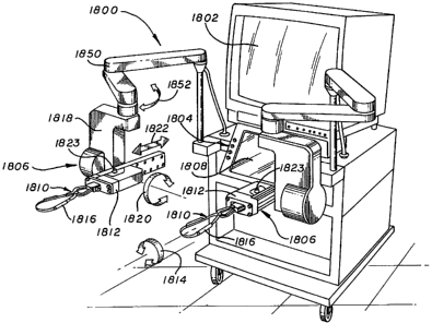
Figure 7 shows a preferred embodiment of an end
effector 80 that may be used in the present invention.
The end effector 80 includes a surgical instrument 82,
such as those disclosed hereinabove 22, 24, that is
coupled to a front loading tool driver 84. The end
effector 80 is mounted to one of the robotic arm
assemblies 26 by coupling mechanism 45. The coupling
mechanism 45 includes a collar 85 that removably
attaches to a holder 86. The holder 86 includes a worm
gear 87 that is driven by a motor in the robotic arm
assembly 26 to rotate the collar 85 and in turn rotate
the instrument 82 about its longitudinal axis. The
holder 86 includes a shaft 88 that seats into a slot in
the robotic arm assembly 26. The shaft 88 may be
turned by the motor in the arm assembly, which then
rotates the worm gear 87 thus rotating the collar 86
CA 02330674 2000-10-30
WO 00/51486 PCTIUSOO/05351
-33-
and the instrument 82. A tightening tool 89 may be
employed to tighten and loosen the collar about the
instrument 82. Such a tool operates like a chuck key,
to tighten and loosen the collar 86.
The surgical instrument 82 has a first finger 90
that is pivotally connected to a second finger 91. The
fingers 90, 91 can be manipulated to hold objects such
as tissue or a suturing needle. The inner surface of
the fingers may have a texture to increase the friction
and grasping ability of the instrument 82. The first
finger 90 is coupled to a rod 92 that extends through a
center channel 94 of the instrument 82. The instrument
82 may have an outer sleeve 96 which cooperates with a
spring biased ball quick disconnect fastener 98. The
quick disconnect 98 allows instruments other than the
finger grasper to be coupled to front loading tool
driver 84. For example, the instrument 82 may be
decoupled from the quick disconnect 98 and replaced by
a cutting tool, a suturing tool, a stapling tool
adapted for use in this system, such as the stapling
apparatus disclosed in U.S. Patent No. 5,499,990 or
5,389,103 assigned to Karlsruhe, a cutting blade, or
other surgical tools used in minimally invasive
surgery. The quick disconnect 98 allows the surgical
instruments to be interchanged without having to re-
sterilize the front loading tool driver 84 each time an
instrument is plugged into the tool driver 84. The
operation of the front loading tool driver 84 shall be
discussed in further detail hereinbelow.
The quick disconnect 98 has a slot 100 that
receives a pin 102 of the front loading tool driver 84.
CA 02330674 2000-10-30
WO 00/51486 PCT/US00/05351
-34-
The pin 102 locks the quick disconnect 98 to the front
loading tool driver 100. The pin 102 can be released
by depressing a spring biased lever 104. The quick
disconnect 98 has a piston 106 that is attached to the
tool rod 92 and in abutment with an output piston 108
of a load cell 110 located within the front loading
tool driver 84.
The load cell 110 is mounted to a lead screw nut
112. The lead screw nut 112 is coupled to a lead screw
114 that extends from a gear box 116. The gear box 116
is driven by a reversible motor 118 that is coupled to
an encoder 120. The entire end effector 80 is rotated
by the motor driven worm gear 87.
In operation, the motor 118 of the front loading
tool driver 84 receives input commands from the
controller 46 via electrical wiring, or a
transmitter/receiver system and activates, accordingly.
The motor 118 rotates the lead screw 114 which moves
the lead screw nut 112 and load cell 110 in a linear
manner. Movement of the load cell 110 drives the
coupler piston 106 and tool rod 92, which rotate the
first finger 88. The load cell 110 senses the
counteractive force being applied to the fingers and
provides a corresponding feedback signal to the
controller 46.
The tool 82 and any tool included in the system
may include an irrigation line 83 and a suction line
85. Each of the irrigation line 83 and suction line 85
extend down the center channel 94 and may be enclosed
within a separate housing 89 disposed interior the tool
82. This is depicted in Figures 13 and 14. The
CA 02330674 2000-10-30
WO 00/51486 PCTIUSOO/05351
-35-
irrigation line 83 is connected to a water source or
saline source and may be used to irrigate the surgical
site or to remove tissue from the instrument 82.
Irrigation systems are generally well known. It is not
heretofore known, though, to include an irrigation line
83 into an endoscopic instrument for use with a robotic
system 10.
Additionally, a suction line 85 may be enclosed
within the housing 89 disposed interior the instrument
82. Suction is generally needed to remove blood, or
other fluids from the surgical site. Again, it is not
heretofore known to include a suction line 85 in an
endoscopic instrument for use with a robotic system 10.
As such, the inclusion of either an irrigation line or
a suction line present advances in the art that are
novel and as of yet unknown.
Each of the suction and irrigation lines run to
well-known suction and irrigation systems which are
well known in the art. The activation of irrigation or
suction is generally accomplished through the use of a
foot controller or hand controller. However, it must
be appreciated that the activation of such devices may
be integrated into the present system by including a
button at the surgeon input device or the cabinet.
Alternatively, the suction and irrigation may be voice
activated and as such, additional vocabulary must be
included in the voice recognition system of the present
invention. More particularly, the voice recognition
system should recognize the commands "suction" and
"irrigate".
CA 02330674 2000-10-30
WO 00/51486 PCT/US00/05351
-36-
The front loading tool driver 84 may be covered
with a sterile drape 124 so that the tool driver 84
does not have to be sterilized after each surgical
procedure. Additionally, the robotic arm assembly 26
is preferably covered with a sterile drape 125 so that
it does not have to be sterilized either. The drapes
124, 125 serve substantially as a means for enclosing
the front loading tool driver 84 and robotic arm
assembly 26. The drape 125 used to enclose the robotic
arm assembly 26 is depicted in further detail in Figure
26. The drape 125 has a substantially open end 300
wherein the robotic arm assembly 26 may be emplaced
into the drape 125. The drape 125 additionally
includes a substantially tapered enclosed end 302 that
effectively separates the arm assembly 26 from the
operating room environment. A washer 304 having a
small aperture 306 formed therethrough allows an
instrument to be coupled to the arm assembly 26 via the
coupling mechanism 45. The washer 304 reinforces the
drape 125 to ensure that the drape 125 does not tear as
the arm assembly 26 moves about. Essentially, the
instrument cannot be enclosed in the drape 125 because
it is to be inserted into the patient 12. The drape
125 also includes a plurality of tape 308 having,
adhesive 310 disposed thereon. At least one piece of
tape 308 is opposedly arranged the other pieces of tape
308 to effectuate the closing of the drape 125 about
the arm assembly 26.
Figures 8 and 8a show a preferred embodiment of a
master handle assembly 130. The master handle assembly
130 includes a master handle 132 that is coupled to an
CA 02330674 2000-10-30
WO-00/51486 PCT/USOO/05351
-37-
arm 134. The master handle 132 may be coupled to the
arm 134 by a pin 136 that is inserted into a
corresponding slot 138 in the handle 132. The handle
132 has a control button 140 that can be depressed by
the surgeon. The control button 140 is coupled to a
switch 142 by a shaft 144. The control button 140
corresponds to the input button 58 shown in Fig. 4, and
activates the movement of the end effector.
The master handle 132 has a first gripper 146 that
is pivotally connected to a second stationary gripper
148. Rotation of the first gripper 146 creates a
corresponding linear movement of a handle shaft 150.
The handle shaft 150 moves a gripper shaft 152 that is
coupled a load cell 154 by a bearing 156. The load
cell 154 senses the amount of pressure being applied
thereto and provides an input signal to the controller
46. The controller 46 then provides an output signal
to move the fingers of the end effector.
The load cell 154 is mounted to a lead screw nut
158 that is coupled to a lead screw 160. The lead
screw 160 extends from a reduction box 162 that is
coupled to a motor 164 which has an encoder 166. The
controller 46 of the system receives the feedback
signal of the load cell 110 in the end effector and
provides a corresponding command signal to the motor to
move the lead screw 160 and apply a pressure on the
gripper so that the surgeon receives feedback relating
to the force being applied by the end effector. In
this manner the surgeon has a "feel" for operating the
end effector.
CA 02330674 2000-10-30
WO 00/51486 PCT/US00/05351
-38-
The handle is attached to a swivel housing 168
that rotates about bearing 170. The swivel housing 168
is coupled to a position sensor 172 by a gear assembly
174. The position sensor 172 may be a potentiometer
which provides feedback signals to the controller 46
that correspond to the relative position of the handle.
Additionally, an optical encoder may be employed for
this purpose. Alternatively, both a potentiometer and
an optical encoder may be used to provide redundancy in
the system. The swivel movement is translated to a
corresponding spin of the end effector by the
controller and robotic arm assembly. This same type of
assembly is employed in the stand 900.
The arm 134 may be coupled to a linear bearing 176
and corresponding position sensor 178 which allow and
sense linear movement of the handle. The linear
movement of the handle is translated into a
corresponding linear movement of the end effector by
the controller and robotic arm assembly. The arm can
pivot about bearings 180, and be sensed by position
sensor 182 located in a stand 184. The stand 184 can
rotate about bearing 186 which has a corresponding
position sensor 188. The arm rotation is translated
into corresponding pivot movement of the end effector
by the controller and robotic arm assembly.
A human hand will have a natural tremor typically
resonating between 6-12 hertz. To eliminate tracking
movement of the surgical instruments with the hand
tremor, the system may have a filter that filters out
any movement of the handles that occurs within the
tremor frequency bandwidth. Referring to Figure 4, the
CA 02330674 2000-10-30
WO 00/51486 PCT/US00/05351
-39-
filter 184 may filter analog signals provided by the
potentiometers in a frequency range between 6-12 hertz.
Alternatively, an optical encoder and digital filter
may be used for this purpose.
As shown in Figures 9 and lOA-J, the system is
preferably used to perform a cardiac procedure such as
a coronary artery bypass graft (CABG). The procedure
is performed by initially cutting three incisions in
the patient and inserting the surgical instruments 22
and 24, and the endoscope 26 through the incisions.
One of the surgical instruments 22 holds a suturing
needle and accompanying thread when inserted into the
chest cavity of the patient. If the artery is to be
grafted with a secondary vessel, such as a saphenous
vein, the other surgical instrument 24 may hold the
vein while the end effector of the instrument is
inserted into the patient.
The internal mammary artery (IMA) may be severed
and moved by one of the instruments to a graft location
of the coronary artery. The coronary artery is severed
to create an opening in the artery wall of a size that
corresponds to the diameter of the IMA. The
incision(s) may be performed by a cutting tool that is
coupled to one of the end effectors and remotely
manipulated through a master handle. The arteries are
clamped to prevent a blood flow from the severed
mammary and coronary arteries. The surgeon manipulates
the handle to move the IMA adjacent to the opening of
the coronary artery. Although grafting of the IMA is
shown and described, it is to be understood that
CA 02330674 2000-10-30
WO 00/51486 PCT/US00/05351
-40-
another vessel such as a severed saphaneous vein may be
grafted to bypass a blockage in the coronary artery.
Referring to Figs. 1OA-J, the surgeon moves the
handle to manipulate the instrument into driving the
needle through the IMA and the coronary artery. The
surgeon then moves the surgical instrument to grab and
pull the needle through the coronary and graft artery
as shown in Fig. 10B. As shown in Fig. 10C, the
surgical instruments are then manipulated to tie a
suture at the heel of the graft artery. The needle can
then be removed from the chest cavity. As shown in
Figs. 10D-F, a new needle and thread can be inserted
into the chest cavity to suture the toe of the graft
artery to the coronary artery. As shown in Fig. 10H-J,
new needles can be inserted and the surgeon manipulates
the handles to create running sutures from the heel to
the toe, and from the toe to the heel. The scaled
motion of the surgical instrument allows the surgeon to
accurately move the sutures about the chest cavity.
Although a specific graft sequence has been shown and
described, it is to be understood that the arteries can
be grafted with other techniques. In general the
system of the present invention may be used to perform
any minimally invasive anastomostic procedure.
Additionally, it may be advantageous to utilize a
fourth robotic arm to hold a stabilizer 75. The
stabilizer may be a tube or wire or some other medical
device that may be emplaced within an artery, vein or
similar structure to stabilize such structure. Using
the switch 51 to interengage the fourth robotic arm,
with a handle 50 or 52 a surgeon may position the
CA 02330674 2000-10-30
WO 00/51486 PCT/US00/05351
-41-
stabilizer 75 into the vessel. This eases the task of
placing a stitch through the vessel as the stabilizer
75 maintains the position of the vessel. Once the
stabilizer 75 has been placed, the surgeon then flips
the switch or like mechanism to activate the robotic
arm that had been disconnected to allow for movement of
the fourth robotic arm. The stabilizer 75 should be
substantially rigid and hold its shape. Additionally,
the stabilizer should be formed form a material that is
steralizable. Such material are well known in the
medical arts. However, this application and
configuration is heretofore unknown.
As disclosed hereinabove, the system may include a
front loading tool driver 84 which receives control
signals from the controller 46 in response to movement
of a master handle 50 or 52 and drives the tool
disposed at the end of a surgical instrument.
Alternatively, a back loading tool driver 200 may be
incorporated into the system 10 of the present
invention, as depicted in Figures 15 and 16. The back
loading tool driver 200 cooperates with a back loadable
surgical instrument 202. The incorporation of such a
back loading tool driver 200 and instrument 202
expedites tool changing during procedures, as tools may
be withdrawn from the tool driver 200 and replaced with
other tools in a very simple fashion.
The back loading tool driver 200 is attached to a
robotic arm assembly 26 via a collar and holder as
disclosed hereinabove. The back loading tool driver
includes a sheath 204 having a proximal end 206 and a
distal end 208. The sheath 204 may be formed of
CA 02330674 2000-10-30
WO 00/51486 PCT/USOO/05351
-42-
plastic or some other well-known material that is used
in the construction of surgical instruments. The
sheath 204 is essentially a hollow tube that fits
through the collar 85 and is tightened in place by the
tightening tool that is described in more detail
hereinabove.
The back loadable surgical instrument 202 has a
tool end 210 and a connecting end 212. A surgical tool
214, such as a grasper or some other tool that may be
driven by a push/pull rod or cable system, or a
surgical tool that does not require such a rod or
cable, such as a coagulator, or harmonic scalpel is
disposed at the tool end 210 of the instrument 202.
A housing 216 is disposed at the connecting end
212 of the instrument 202. The housing has a lever 218
disposed interiorly the housing 216. The lever 218 has
a pivot point 220 that is established by utilizing a
pin passing through an associated aperture 222 in the
lever. The pin may be attached to the interior wall
224 of the housing. A push/pull cable or rod 226, that
extends the length of the instrument 202 is attached to
the lever 218, such that movement of the lever 218
about the pivot point 220 results in a linear movement
of the cable or rod 226. Essentially the cable or rod
226 servers as a means 227 for actuating the tool 214
at the tool end 210 of the instrument 202. The cable or
rod 226 may be attached to the lever via a connection
pin as well. The lever 218 has a C-shape, wherein the
ends of the lever 218 protrude through two apertures
228, 230 in the housing 216. The apertures 228, 230
CA 02330674 2000-10-30
WO 00/51486 PCT/US00/05351
-43-
are preferably surrounded by O-rings 232 the purpose of
which shall be described in more detail hereinbelow.
The tool end 210 of the back loadable surgical
instrument 202 is emplaced in the hollow tube of the
back loading tool driver 200. The tool 202 may be
pushed through the tool driver until the tool end 210
extends beyond the sheath 204. The 0-rings 232 seat in
associated apertures 234, 236 in a housing 238 of the
tool driver 200. The housing additionally has an
aperture 240 centrally formed therethrough, the
aperture being coaxial with the interior of the hollow
tube. In this fashion, the surgical instrument 202 may
be inserted into. and through the tool driver 200. Each
of the O-rings 232 snugly seats in its associated
aperture in the housing 238 of the tool driver 200.
The housing 238 additionally includes a motor
assembly 242 which is depicted in Figure 16. The motor
assembly 242 is attached to the housing 238 and is held
firmly in place therein. The motor assembly generally
includes a motor 244 attached to a reducer 246. The
motor drives a leaf 248 attached at the end thereof.
The leaf 248 engages the ends of the lever 218 such
that rotational movement of the motor results in the
movement of the lever 218 about the pivot point 220.
This in turn results in the lateral movement of the
means 227 for actuating the tool 214 at the tool end
210 of the instrument 202. The motor moves in response
to movements at a control handle. Additionally, force
sensors 248, 250 may be attached at the ends of the
leaf 248. As such, a force feedback system may be
incorporated to sense the amount of force necessary to
CA 02330674 2000-10-30
WO 00/51486 PCT/US00/05351
-44-
actuate the tool 214 at the tool end 210 of the
instrument 202. Alternatively, the motor 244 may have
a force feedback device 252 attached thereto, which can
be used in a similar fashion.
One advantage of utilizing the back loading tool
driver 200 is that the sheath 204 always remains in the
patient 12. As such, the tools do not have to be
realigned, nor does the robotic arm assembly 26 when
replacing or exchanging tools. The sheath 204 retains
its position relative to the patient 12 whether or not
a toll is placed therethrough.
The system 10 of the present invention may
additionally be supplied with one or two additional
degrees of freedom at the tip of an instrument. For
the purposes of example, two additional degrees of
freedom will be disclosed; however it is to be
appreciated that only one degree of freedom may be
included as well. To provide the additional degrees of
freedom, and as depicted in Figures 17-20, an
articulable surgical instrument 300 may be incorporated
into the present. The instrument 300 may be coupled to
the arm assembly 26 via a collar and holder as
disclosed hereinabove. In order to articulate the tip
of the articulable instrument 300 an articulating tool
driver 500 must be employed. The articulating tool
driver 500 shall be described in more detail
hereinbelow. The master must have an additional two
degrees of freedom added thereto to proved the controls
for the articulation at the tip of the instrument 300.
Figure 29 depicts an alternative master schematic that
includes the two additional degrees of freedom. As
CA 02330674 2000-10-30
WO 00/51486 PCT/US00/05351
-45-
disclosed hereinbelow, the two additional degrees of
freedom are mapped to the articulable portion of the
instrument 300. The two additional axes at the master
are referred to as Jm6 and Jm7.
By incorporating the articulable instrument 300
and the articulating tool driver 500 and the additional
degrees of freedom at the master, difficult maneuvers
may be carried out in an easier fashion.
With reference to figs. 17-20, the articulable
instrument 300 generally includes an elongated rod 302,
a sheath 304, and a tool 306. The tool can be a
grasper, a cutting blade, a retractor, a stitching
device, or some other well-known tool used in minimally
invasive surgical procedures. Figures 27-30 show
various tools that may be emplaced at the distal end of
the articulable surgical instrument 300.
The instrument 300 includes an articulable portion
301 having a proximal portion 308, a pivot linkage 310
and a distal portion 212 each of which will be
discussed in more detail hereinbelow. Additionally,
the instrument 300 includes means 311 for articulating
the articulable portion 301 of the instrument 300 with
respect to the elongated rod 302. The inclusion of the
articulable portion 301 provides two additional degrees
of freedom at the instrument tip. It must also be
appreciated that although the articulable portion 301
is described as including a proximal portion, a pivot
linkage and a distal portion, there may be provided a
plurality of intermediate portions each mounted to each
other via a corresponding pivot linkage.
CA 02330674 2000-10-30
WO 00/51486 PCTIUSOO/05351
-46-
Disposed between and mounted to each of the
respective proximal portion and distal portion and any
intervening intermediate portions are pivot linkages
310. The pivot linkage 310 interengages with the
proximal and distal portions of the articulable portion
to provide articulation at the instrument tip.
Essentially, the cooperation of the proximal portion,
pivot linkage and distal portion serves as a universal
joint.
The elongated rod 302 is preferably hollow and
formed of stainless steel or plastic or some other
well-know material that is steralizable. Because the
rod 302 is hollow, it encompasses and defines an
interior 314. The elongated rod 302 additionally has a
proximal end 316 and a distal end 318. The distal end
318 of the elongated rod 302 should not be confused
with the distal portion 312 of the articulable portion
301 of the instrument 300.
The proximal portion 308 of the articulable
portion 301 may be integrally formed with the elongated
rod 302 or it may be attached thereto vie welding, glue
or some other means well-known to the skilled artisan.
It is preferable that the proximal portion 308 be
integrally formed with the elongated rod 302 to ensure
sufficient stability and durability of the instrument
300. The proximal portion 308 of the articulable
portion 301 comprises two fingers 320, 322 each of
which have an aperture 324, 326 formed therethrough.
A pivot linkage 310 is mounted to the proximal
portion 308 via a plurality of pins 328 that each pass
through an associated aperture in an adjoining finger.
CA 02330674 2000-10-30
WO 00/51486 PCT/USOO/05351
-47-
The pivot linkage 310 is a generally flat disk 330
having a central aperture 332 passing therethrough and
four apertures 334, 336, 338, 340 evenly spaced at the
periphery of the disk 330. Additionally pins 328 are
attached to and extend from the edge 342. The pins 328
seat in the apertures of the associated fingers to
provide the articulability of the instrument 300. Five
leads 350, 352, 354, 356, 358 extend interiorly the
hollow shaft. On lead 350 extends down the center and
passes through the center aperture 332 in the pivot
linkage 310. Two 352, 354 of the five leads extend
down the hollow interior of the instrument and are
attached to the pivot linkage such that linear tension
on one of the leads results in rotational movement of
the pivot portion 301. These two leads 352, 354 attach
to the pivot linkage at two of the apertures formed.
therethrough. Additionally, they attach at those
apertures that are adjacent to the pins that pass
through the fingers of the proximal portion 308 of the
articulable portion 301 of the instrument 300. The
other two leads 356, 358 pass through the two other
apertures in the pivot linkage and attach at the distal
end of the articulable portion 301. Movement of these
two leads results in movement of the articulable
portion 301 that is orthogonal to the movement when the
two other leads 352, 354 are moved.
To articulate the instrument as a part of the
present system, and as depicted in Figures 21-28, there
is provided an articulating mechanism 400. The
articulating mechanism 400 generally comprises the
CA 02330674 2000-10-30
WO 00/51486 PCTIUSOO/05351
-48-
articulating tool driver 500, a sterile coupler 600, a
translator 700 and the articulable tool 300.
The translator is attached to the proximal end 316
of the instrument 300. The instrument 300 may
additionally have a removable tool 420 as shown in
Figs. 22-23. The removable tool 420 may be any tool,
such as a cutter 422 that is attached to an elongated
rod or cable 424. At the end of the rod 246 there is
disposed a flat section 428 with an aperture 430 formed
therethrough. The flat section 428 seats into a
channel 432 disposed at the end of a second cable or
rod 434 that travels down the elongated shaft of the
instrument 300. The second cable 434 has a channel 432
formed in the end thereof such that the flat section
428 seats in the channel 432. At least one spring
biased detent 436 seats in the aperture 430 disposed
through the flat section 428. This connects the tool
420 to the rest of the instrument 300. As such, tools
may be exchanged at the tip of the instrument without
having to remove the instrument from the system 10
every time a new tool is required.
The tool 300 is attached to the translator 700 and
essentially is integrally formed therewith. The
articulating mechanism 400 is attached to the robotic
arm assembly 26 via the collar 85 as is disclosed
hereinabove. The collar 85 fits about the shaft 302 of
the instrument 300.
The translator 700 has a proximal end 702 and a
distal end 704. The distal end 704 of the translator
700 has a cross sectional shape that is substantially
similar to the cross sectional shape of the elongated
CA 02330674 2000-10-30
WO- 00/51486 PCT/US00/05351
-49-
rod 302 of the instrument 300. Additionally, the
translator 700 has a hollow interior 706. The center
rod 350 extends through the hollow interior 706 of the
translator 700 and emerges at the proximal end 702
thereof. Two of the leads 352, 354 terminate
interiorly the translator at two shoulders 708, 710
that are attached to a first hollow tube 712 through
which the center lead 350 extends. The first hollow
tube 712 may be formed of some strong durable material
such as stainless steel, steel, hard plastic or the
like.
The first hollow tube 712 is mounted to a bearing
714 such that it may be rotated. Rotation of the first
hollow tube 712 results in the linear motion of the
leads 352, 254 and the articulation of the articulable
portion 301 of the instrument 300 in one plane of
motion.
A second hollow tube 716 has a pair of shoulders
718, 719 extending therefrom. Two leads 356, 358
attach to one each of the shoulders 718, 719. The
hollow tube 716 is disposed within a bearing assembly
720 such that it may be rotated. Again, rotation of
the second hollow tube 716 results in linear movement
of the leads 356, 358 which articulates the articulable
portion 301 of the instrument 300 in a plane orthogonal
the plane of motion established through the rotation of
the first hollow tube. It is to be appreciated that
the second hollow tube 714 radially surrounds the first
hollow tube 712. The translator 700 additionally
includes a quick disconnect 722 comprising a pin 724
disposed at the end of a spring biased lever 726 which
CA 02330674 2000-10-30
WO 00/51486 PCT/US00/05351
-50-
provides removable attachment of the translator 700 to
the sterile coupler 600. Both of the hollow tubes 712
and 716 may have notches 750 formed therein at their
ends. The notches serve as a means 752 for
interconnecting each of the tubes to the sterile
coupler 600 which will be discussed in further detail
hereinbelow.
The translator 700 is removably attached to the
sterile coupler 600 via the quick disconnect 722.
Because the articulable tool driver 500 is not easily
sterilized, it is advantageous to include a sterile
coupler 600 so that instruments may be exchanged
without having to sterilize the articulable tool driver
500. Additionally, the coupler 600 provides a means by
which the translator 700 may be attached to the tool
driver 500 while the tool driver is enclosed in a drape
125 such as that depicted in figure 26. The translator
600 has a housing 610 . Preferably the housing and the
components of the coupler 600 are formed of some easily
steralizable mater such as stainless steel, plastics or
other well-known sterilizable materials. The housing
610 has a substantially hollow interior 612 and open
ends 614 and 616. Two hollow tubes 618 and 620 are
rotatively disposed within the housing 610. To
effectuate the rotation of each of the tubes 618 and
620, bearings 622 and 624 are disposed about each of
the tubes. Each of the tubes has notches 626 formed in
the ends thereof so effectuate the attachment of the
translator 700 to the coupler 600 at one end. And to
effectuate the attachment of the coupler 600 to the
articulable tool driver 500 at the other end thereof.
CA 02330674 2000-10-30
WO 00/51486 PCTIUSOO/05351
-51-
The pin 724 on the translator may seat in a notch
628 to attach the translator 700 to the coupler 600.
Additionally, the coupler 600 may include a pin 630
attached to a spring biased pivot 632 to effectuate
attachment of the coupler to the driver 500. The
coupler 600 additionally includes a center section 634
that slidably receives the end 351 of the center cable
or rod 350. The end 351 may include a tip with a
circumferential groove 353 disposed thereabout. The
tip seats in a recess 636 formed in the center section
634 and is removably locked in place by at least one
spring biased detent 638. A tip 640, which is
substantially similar to the tip containing the
circumferential groove 353 is disposed adjacent the
recess 636 and serves to attach the cable center cable
350 to the articulable tool driver 500, which will be
discussed in further detail hereinbelow.
The center section 634 is intended to laterally
slide within the innermost tube 618. To effectuate
such a sliding motion, a linear bearing may be disposed
about the center section interiorly of the innermost
tube. Alternatively, the center section 634 may be
formed of a bearing material that provides smooth
sliding within the innermost tube 618.
The coupler 600 is removably attached to the
articulable tool driver 500. It is intended that the
articulable tool driver be enclosed by a drape 125.
The articulable tool driver 500 includes a
substantially hollow housing 502 having a closed first
end 504 and a substantially open second end 504.
Securely disposed interiorly the housing 502 is a
CA 02330674 2000-10-30
WO 00/51486 PCT/US00/05351
-52-
gripper motor 506, and a pair of wrist motors 508 and
510. Each of the motors are in electrical connection
with the controller 46. Alternatively, the motors may
receive signals from the controller via a
transmitter/receiver system where such systems are well
known. It is the application of such a
transmitter/receiver system to the present invention
that is new. The gripper motor 506 is attached to a
load nut 510 that surrounds a load screw 512. The
motor 506 receives the control signals and turns in
response thereto. The load nut 510 turns which
laterally moves the load screw 512. The load screw 512
is attached to a load cell 514 which may be employed to
measure the force required to laterally move the cable
350 which is attached vie the coupler 600 to the
gripper motor 506. This may be used in a force
feedback system that may be incorporated in the system
of the present invention. A rod 516 having a
channel 518 formed at the end thereof is attached to
the load cell 514. As such, the rod 516 moves in a
linear fashion. The tip 640 of the coupler 600 seats
in the channel 518 and is removably held in place by at
least one spring biased detent or some other similar
attachment mechanism 520. Therefore, if a surgeon at a
master handle actuates the grippers, the gripper motor
506 turns, thus laterally moving the rod 516, and in
turn the center cable 350 which opens and closes the
grippers at the tool accordingly. Of course, the
action at the tool will depend upon the type of tool
disposed thereat. For example, if a stapling tool is
CA 02330674 2000-10-30
WO 00/51486 PCT/USOO/05351
-53-
disposed at the end of the surgical instrument 300 then
a stapling action would take place.
If a master handle 50 or 52 is turned about axes
J6 or J7 then one of the two wrist motors 510, 508
corresponding to the required motion turns. Each of
the motors 508, 510 are attached to a corresponding
gear 522, 524. Each of the gears 522, 524 engage a
corresponding slotted section 530, 532 of an associated
hollow tube 526, 528 to turn the associated tube
radially about its longitudinal axis. Each of the
tubes 526, 528 include notched ends 534, 536 to engage
the notched ends of corresponding hollow tubes of the
coupler 600. It is to be appreciated that each of the
hollow tubes 526, 528, 618 and 620 are all coaxial.
Additionally, bearings may be emplaced intermediate
each of the tubes 526 and 528 to provide easy
independent rotatability of the individual tubes.
When the tubes 526, 528 are rotated, they rotate
the tubes in the coupler which rotates the tubes in the
translator. This results in the articulation at the
tip of the surgical instrument 300. More particularly,
this results in the articulation of the articulable
portion of the surgical instrument 300. Additionally,
whether the front loading tool driver, the back loading
tool driver, or the articulable tool driver are
employed, surgical instruments may be easily exchanged.
As such, a cutting blade 800 may be exchanged for
a grasper, and a grasper may be exchanged for a stapler
810. Essentially, such a system simplifies the
performance of minimally invasive surgical procedures
where the procedures include the step of changing one
CA 02330674 2000-10-30
WO 00/51486 PCTIUSOO/05351
-54-
tool for another. And because the system allows
articulation at the tip of certain instruments, the
articulation mechanism may be used to articulate such
stapling, or cutting instruments that incorporate the
articulable portion as disclosed hereinabove.
Additionally, the instrument may not be an
articulable instrument, but the articulating mechanism
can be used to control other functions, such as
stapling. Figure 31 depicts a stapling instrument 810
attached to the robotic arm assembly via the collar 85
and holder 86. The lead that is generally use for the
grasping tool, may be used to effectuate the stapling
mechanism. Endoscopic staplers are generally well
known in the art, however, it is heretofore to known to
use a stapler that is attached to a robotic arm as is
disclosed herein.
Additionally, a cutting blade, such as that
depicted in Figure 32 may be employed in the system of
the present invention. The cutting blade 800 is
attached to the robotic arm assembly 26 via the collar
85 and holder 86. The cutting blade does not require a
lead such as that required by the grasper or the
stapler; however, the cutting tool, may be articulated
via the articulating mechanism that has been disclosed
hereinabove.
A cauterizer or coagulator may additionally be
attached to the robotic arm assembly 26 via the collar
85 and holder. Cauterizers and coagulators are well
known and the cauterizing tool may be attached at the
end of an articulable instrument as disclosed
hereinabove. By using a variety of tools in
CA 02330674 2000-10-30
WO 00/51486 PCT/US00/05351
-55-
predetermined sequences, various procedures may be
carried out. It is generally preferable to be able to
change instruments because many procedures require
such.
As disclosed hereinabove, the handles 50 and 52
allow a surgeon to control the movement of the tools
attached to the robotic arms. As such, the
configuration of the handles 50 and 52 should provide
great ease of use for a surgeon. Figures 38-43 depict
various handle configurations. Additionally, the
handles 50 and 52 may be selected by a surgeon from a
plurality of handles 960 that are available for use by
the surgeon.
A proximally open handle 962 has a proximal end
963 and a distal end 965. The handle 962 has first
finger portion 964 and a second finger portion 966
pivotally attached at the distal end 965 of the handle
962. A joint 968 disposed intermediate the finger
portion 964 and 966 provides linear motion of an
elongated rod 970 which is used to actuate the tool tip
of an instrument attached to the robotic arm. This
handle may serve as one or both of the two handles 50
and 52 of the system.
A distally open handle 972 has a proximal end 973
and a distal end 975. The handle 972 has first finger
portion 974 and a second finger portion 976 pivotally
attached at the proximal end 973 of the handle 972. A
joint 978 disposed intermediate the finger portion 964
and 966 provides linear motion of an elongated rod 980
which is used to actuate the tool tip of an instrument
CA 02330674 2000-10-30
WO 00/51486 PCT/US00/05351
-56-
attached to the robotic arm. This handle may serve as
one or both of the two handles 50 and 52 of the system.
Such handles 962 and 972 may be interchanged
through the inclusion of an interchange mechanism 984.
The interchange mechanism 984 includes a biased detent
latch 986 that engages an aperture in the elongated rod
932 such that the handle may be attached or removed
from the rod 932.
Other handle configurations are depicted in
Figures 41-43. And more particularly, each of the
handles 1000, 1100, and 1200 have a pair of fingerseats
1020. The major difference between each of the handles
1000, 1100, and 1200 is the orientation of the
fingerseats to a pivot point on the handle. The
fingerseats may be parallel, or perpendicular to the
axis S of the pivot point of the handle. Each of these
configurations may be included as an attachable handle.
As such, a surgeon may exchange handles throughout a
procedure depending upon the task to be accomplished.
A surgeon may prefer one handle for a set of tasks and
another handle for a different set of tasks. As such,
the surgeon may exchange handles during the performance
of a surgical procedure to enable such tasks.
While certain exemplary embodiments have been
described and shown in the accompanying drawings, it is
to be understood that such embodiments are merely
illustrative of and not restrictive on the broad
invention, and that this invention not be limited.to
the specific constructions and arrangements shown and
described, since various other modifications may occur
to those ordinarily skilled in the art.
CA 02330674 2000-10-30
WO 00/51486 PCT/US00/05351
-57-
Figure 44 shows an embodiment of a surgical
instrument 1600 that is coupled to a tool driver 1602.
The instrument 1600 may have an end effector 1604 that
is actuated by the tool driver 1602. The tool driver
1602 can be connected to a controller (not shown) by a
wire assembly 1606. Additionally, the tool driver 1602
can be coupled to an articulate arm 1608. The
articulate arm 1608 can both move the tool driver 1602
and spin the instrument 1600.
As shown in Figure 45, the tool driver 1602 may
include a sheath 1610 that is attached to a tool
housing 1612. The sheath 1610 may include a sleeve
portion 1614 that extends from a collar 1616. The
collar 1616 may have internal threads (not shown) that
are screwed onto corresponding threads (not shown) of
the housing 1612 so that the sheath can be detached
from the housing 1612. The sheath 1610 may be
constructed from a material that is both electrically
non-conductive and transparent to x-rays.
The electrically non-conductive material may
prevent electrical current from flowing to the patient
from the surgical instrument. By way of example, if
the instrument is an electro-cautery device the non-
conductive sheath may prevent an electrical short
through the sheath. As an alternate embodiment, the
sheath 1610 may be constructed from a metal material
that has an outer layer of non-conductive material.
Providing a sheath 1610 that is transparent to x-rays
allows x-ray images of the patient to be taken without
interference from the sheath.
CA 02330674 2000-10-30
WO 00/51486 PCT/US00/05351
-58-
The sheath 1610 is typically inserted into an
incision of a patient. The sheath 1610 and incision
define a pivot point for the instrument 1600. The
articulate arm 1608 may include passive joints which
provide additional degrees of freedom for the arm (not
shown). The sheath 1610 provides a structure that may
hold the arm 1608 in place when a new instrument 1600
is being coupled to the tool driver 1602.
Figure 46 shows an embodiment of the sheath collar
1616. The collar 1616 may include a valve assembly
1620 that can move between an open position and a
closed position. A gas is typically introduced into
the patient when performing a procedure with the
surgical instrument 1600. The valve 1620 prevents gas
from escaping the patient and flowing through a sheath
inner channel 1622 when the instrument 1600 is pulled
out of the sheath 1610.
The valve assembly 1620 may include a valve 1624
that controls fluid communication between a valve port
1626 and the inner channel 1622. The valve 1624 may be
coupled to a torsion spring 1628 that biases the valve
1624 into the closed position. In the closed position
the valve 1624 may cooperate with a valve seat 1630 to
prevent gas from flowing through the inner channel 1622
and into the port 1626. The valve 1624 may have a
radial portion 1632 and an annular flat portion 1634
that presses against the seat 1630.
As shown in Figure 47, the instrument 1600 can be
inserted through the port 1626 and into the inner
channel 1622. The port 1626 may have a tapered surface
1636 to guide the instrument 1600 through the valve
CA 02330674 2000-10-30
WO 00/51486 PCT/USOO/05351
-59-
1624. Insertion of the instrument 1600 into the sheath
1610 pushes the valve 1624 into the open position. The
seat 1630 may have a wiper 1638 that presses against
the outside surface of the instrument 1600 and prevents
gas from escaping the patient through the valve
assembly 1620. The wiper 1638 may have an inner
diameter that is approximately equal to the outer
diameter of the surgical instrument 1600 so that there
is a slip fit between the wiper 1638 and the instrument
1600. The valve 1624 may not make contact with the
wiper 1638 even in the closed position.
As shown in Figures 48 and 49, the tool driver
1602 may include a tube 1640 that is coupled to the
surgical instrument 1600. The tube 1640 may have gear
teeth 1642 that are coupled to a mating gear 1644 of
the articulate arm 1608 (also shown in Fig. 45). The
mating gear 1644 can be rotated by a motor (not shown)
in the arm 1608. Rotation of the gear 1644 rotates the
worm gear 1642 and the tube 1640. Rotation of the tube
1640 rotates the instrument 1600 within the patient.
The driver 1602 may include an inner sleeve 1646
that is coupled to an actuator pin 1647 of the
instrument 1600. The sleeve 1646 is attached to a
plate 1648. The plate 1648 is coupled to a linear
actuator 1650. The actuator 1650 can move the plate
1646 along the longitudinal axis of the tube 1640 as
indicated by the arrow. Movement of the plate 1648
translates the sleeve 1646 and pin 1647 to actuate the
end effector 1604 of the instrument 1600. The inner
sleeve 1646 is coupled to the plate 1648 so that the
sleeve 1646 can spin within the instrument 1600.
CA 02330674 2000-10-30
WO 00/51486 PCT/US00/05351
-60-
The tool driver 1602 may include a bellows 1652
that couples the linear actuator 1650 to the plate
1648. The bellows 1652 seals the interface so that the
actuator of the driver 1602 does not become
contaminated. In one embodiment, the bellows 1652 is
constructed from a stainless steel material which is
particularly suitable for reuse when the tool driver
1604 is sterilized under elevated pressures and
temperatures.
Figure 50 shows an embodiment of a surgical
instrument 1600 that can be coupled to the tool driver
1602. The instrument 1600 may have a push rod 1654
that extends through the inner channel 1656 of a handle
1658. The push rod 1654 may be coupled to a detachable
actuator rod 1660. The actuator rod 1660 extends
through an instrument sleeve 1662 and terminates at the
end effector 1604.
The actuator pin 1647 may be pressed into the push
rod 1654. The pin 1647 is free to move along a slot
1666 of the handle 1658. As shown in Figure 51 the
actuator pin 1647 may include a pair of caps 1668 that
snap onto a barbed pin 1670. The caps 1668 secure the
pin 1670 to the push rod 1654.
As shown in Figure 52, when the instrument 1600 is
installed into the driver 1602 the pin assembly 1664
engages a corresponding groove 1672 of the sleeve 1646.
Movement of the sleeve 1646 by the linear actuator 1650
translates the pin 1647 and the attached actuator rod
1660 to provide a controlled movement of the end
effector 1604.
CA 02330674 2000-10-30
WO 00/51486 PCT1US00/05351
-61-
As shown in Figure 53, the actuator rod 1660 may
have a proximal extension 1674 that engages a clamp
portion 1676 of the push rod 1654. The extension 1674
and clamp 1676 allow the actuator rod 1654 and
corresponding end effector to be detachably connected
to the handle 1658. The clamp portion 1676 may include
four barbed tips 1678 that essentially enclose a
locking cavity 1680. The locking cavity 1680 may
receive a locking barrel 1682 located at the end of the
extension 1674.
The extension 1674 can be inserted through an
opening 1684 of the handle 1658. The locking barrel
1682 engages chamfered surfaces 1686 of the barbed tips
1678 to deflect the tips 1678 in an outward direction
and allow the barrel 1682 to be inserted into the
locking cavity 1680. The locking cavity 1680 has a
profile which prevents the barrel 1682 from becoming
detached from the push rod 1654. A collar 1688 can be
screwed onto a threaded portion 1690 to capture a
collar of the instrument sleeve 1662 and secure the
sleeve 1662 to the handle 1658.
The actuator rod 1660 can be removed by moving the
push rod 1654 until the chamfered surfaces 1686 of the
tips 1678 engage an annular lip 1692 of the handle
1658. The annular lip 1692 deflects the tips 1678 so
that the actuator rod 1660 can be separated from the
push rod 1654. The collar 1688 is typically initially
removed to decouple the instrument sleeve 1662 from the
handle 1654. The clamp arrangement allows different
end effectors to be attached to the same handle 1654
CA 02330674 2000-10-30
WO 00/51486 PCT/US00/05351
-62-
during a surgical procedure without having to decouple
the entire instrument 1600 from the tool driver 1602.
Figure 54 shows an alternate embodiment wherein
the actuator rod 1660' is screwed into a push rod
1654'. The instrument sleeve 1662' may be connected to
the handle 1658' by a set screw 1693.
Referring again to Fig. 50, the push rod 1654 may
be attached to a plunger 1694 by a pin 1696. The
plunger 1694 allows an operator to manually move the.
push rod 1654 into the annular lip 1692 shown in Fig.
53 so that the end effector 1604 can be replaced with
another unit. The instrument 1600 may include a return
spring 1698 that biases the plunger 1694 into a
proximal position. The spring 1698 can also insure
that the end effector 1604 is always closed or open,
whatever is desired, when the end effector 1604 is
removed from the patient.
The instrument 1600 may include a detachable
electrode 1700 that is attached to the push rod 1654.
The electrode 1700 can be attached to an electrical
power supply (not shown). The push rod 1654, actuator
rod 1660 and electrode 1700 can all be constructed from
an electrically conductive material which provides an
electrical path between the power supply and a
cauterizing element(s) on the tip of a cauterizing type
instrument. The handle 1658 and plunger 1694 may be
constructed from an expensive disposable non-conductive
plastic material.
Figure 55 shows an embodiment of an end effector
1710 which can be used to grasp a needle 1712. The end
effector 1710 may have a stationary finger 1714 and a
CA 02330674 2000-10-30
WO 00/51486 PCT/US00/05351
-63-
clamp finger 1716 that is moved by the actuator rod
1660. The stationary finger 1714 may have an outer
surface 1718 which has a radius of curvature that
conforms to the shape of the needle 1712. The end
effector 1710 can be manipulated so that the needle
1712 extends along the outer surface 1718. The needle
1712 is held in place by moving the clamp finger 1716
to a closed position. The outer surface 1718 is
preferably oriented to be essentially perpendicular to
the longitudinal axis of the push rod 1660 so that the
needle 1712 can be moved in any direction in space.
Each finger 1714 and 1716 may have a tip 1720 can be
used to grasp tissue.
Referring to Figures 48 and 56, show an interface
which secures the handle 1658 to the sleeve 1662 and
tube 1640 of the driver 1602. The tube 1640 may have a
plurality of spring biased ball detents 1730 which can
slide along corresponding alignment grooves 1732 of the
handle 1658. The grooves 1732 may each have transverse
portions 1734 which receive the detents 1730 when the
handle 1658 is turned in a counter-clockwise direction.
The detents 1730 and grooves 1732 align the instrument
1600 with the driver 1602 so that the actuator pin 1647
is aligned with the grooves of the sleeve 1646.
Referring to Fig. 48 the tool driver 1602 may
include a force sensor 1740 which senses the force
exerted onto the end effector. The force sensor 1740
can be coupled to a controller (not shown) which can
utilize the feedback as part of an algorithm to control
the instrument.
CA 02330674 2000-10-30
WO 00/51486 PCT/US00/05351
-64-
The mechanical advantage may vary for different
surgical instruments. For example, the force ratio
between the handle and tip of a hand-held tweezer may
be 2/5, while the ratio for a needle grasper may be
5/1. It may be desirable to provide the following
variable force transformation algorithm to allow an
operator to vary the force ratio between the handle
held by the hand of the surgeon and the tip of the
instrument. This provides the surgeon with a more
realistic "feel" of an instrument that is normally held
by the surgeon. The force algorithm may be as follows.
Ft = R=(Fh-K=o)
where;
Ft = force at the instrument tip.
R = variable force ratio.
Fh = force applied by the opeartor on the handle.
K = spring constant for instrument jaw.
6 = angle of instrument jaw.
The ratio R may be defined by;
R F Ftm
K 6 where;
hm
Ftm = nominal maximum tip force.
Fhm = nominal maximum handle force.
Each instrument coupled to the tool driver may
have a corresponding R value that is utilized by the
CA 02330674 2000-10-30
WO 00/51486 PCT/US00/05351
-65-
controller to provide a tip force which corresponds to
a handle force that is similar to the ratio of a
corresponding instrument that is normally held in the
hand of a surgeon.
By way of example, if the instrument coupled to
the driver corresponds to a tweezer, an appropriate R
value can be loaded into the controller so that two
pounds of force exerted onto the handle will translate
into 5 pounds of force at the tip of the end effector.
Likewise, 5 pounds of forces sensed by the force sensor
will translate into 2 pounds of feedback to the handle.
The R value can be entered through an input device such
as a keypad, voice recognition system, etc., or
automatically called up from an electronically stored
look-up table when the instrument 1600 is coupled to
the driver 1602.
Referring to Fig. 49, the driver 1602 may include
a printed circuit board assembly 1750 that is connected
to the linear actuator 1650 and the force sensor 1740.
The printed circuit board assembly 1750 may be coupled
to the controller by a wire assembly 1752. The wire
assembly 1752 is introduced to the driver 1602 through
a connector port 1754.
The wire assembly 1752 may include an outer jacket
1756 that encloses an inner jacket 1758. The inner
jacket 1758 may enclose electrical wires 1760. The
inner jacket 1758 may be constructed from a non-porous
material such as TEFLON. The outer jacket 1756 may be
constructed from a flexible material such as silicon.
The outer jacket 1758 may extend through an inner
channel 1762 of a sleeve 1764 that is screwed into the
CA 02330674 2000-10-30
WO 00/51486 PCTIUSOO/05351
-66-
driver housing 1612. The sleeve 1764 may be sealed
against the housing by an O-ring 1766. The assembly
1752 may further include a sealant 1768 that seals the
inner jacket 1758 to the outer jacket 1756 within the
sleeve 1764. The dual jackets and seals insure that
moisture does not enter the tool driver through the
wire assembly when the driver is sterilized.
The printed circuit board assembly 1750 may
include a temperature sensor 1770. By way of example,
the tool driver 1602 may be sterilized after each
surgical procedure under elevated temperatures and
pressures. During sterilization the sensor 1770 may be
connected to an instrument (not shown) through the wire
assembly 1752. The instrument may provide a readout of
the temperature within the tool driver 1602 to allow an
operator to determine whether the driver temperature
exceeds a threshold value that may damage the driver.
Alternatively, or in addition to, the instrument may
provide an indicator, such as the illumination of
light, when the temperature exceeds a threshold value.
Figure 57 shows an embodiment of a console 1800
for the system. The console 1800 may include a video
monitor 1802, an input device 1804 and a pair of handle
assemblies 1806. The handles 1806 can be manipulated
to control the surgical instrument (not shown). The
input device 1804 may include a touchpad screen 1808
which displays a menu(s), commands and other
information which allow an operator to vary different
operating parameters of the systems by pressing the
screen 1808.
CA 02330674 2000-10-30
WO 00/51486 PCTIUSOO/05351
-67-
By way of example, the touchpad 1808 may allow the
operator to vary the force ratio value R, or a scale
factor that correlates the amount of spacial movement
between the handles and the instrument. The operator
may also select between a pull type surgical instrument
and a push type instrument. A push type instrument may
require a distal movement of the actuator rod to close
the end effector. A pull type instrument may require
an opposite proximal movement of the actuator rod to
close the end effector. Selecting push or pull insures
that the tool driver will properly actuate the
instrument. The operator may also control the maximum
jaw angle of the instrument and a jaw locking option
wherein the instrument jaw remains locked even when the
operator releases the handle.
.Each handle assembly 1806 may include a grasper
1810 that can spin relative to a handle 1812 as
indicated by arrow 1814. Rotation of the grasper 1810
can be translated into a corresponding spinning
movement of the instrument about the instrument
longitudinal axis. The grasper 1810 may contain a pair
of pressure plates 1816 that can be depressed by the
operator. The depression of the plates 1816 can cause
a corresponding movement of the actuator rod and the
end effector of the instrument.
The handle 1812 can be pivoted and translated
relative to a swing arm 1818 as indicated by the arrows
1820 and 1822, respectively. Pivoting the handle 1812
may provide a corresponding rotational movement of the
instrument within the patient. Likewise, translational
movement of the handle 1812 may induce a movement of
CA 02330674 2000-10-30
WO 00/51486 PCT/US00/05351
-68-
the instrument into and out of the patient. The handle
1812 may have a release button 1823 which can be
depressed to decouple the handle assembly 1806 from the
instrument, such that movement of the assembly 1806
will not create a corresponding movement of the
instrument.
Figure 58 shows an embodiment of the grasper 1810.
The pressure plates 1816 can be pivotally connected
together by a pin 1824 and bushing 1826. The plates
1816 are also connected to a push ring 1828 by a pair
of rocker arms 1830. The rocker arms 1830 are
pivotally connected to a grasper housing 1832 by pins
1834. The push ring 1828 is connected to a push rod
1836.
The push rod 1836 is connected to an
actuator/sensor assembly 1838 that can translate the
rod 1836 and ring 1828 as indicated by the arrow 1840.
The actuator/sensor assembly 1838 can either induce or
sense the translational movement of the rod 1836.
Movement of the rod 1836 can either induce, or be
responsive to, deflection of the plates 1816. The
plates 1816, ring 1828, rod 1836 and actuator/sensor
assembly 1838 allow the system to both sense the force
being applied by the operator and apply a feedback
force to the operator.
The push rod 1836 may be coaxial aligned with a
connector rod 1839 that is used to couple the grasper
1810 to the handle 1812. The connector rod 1839 is
locked in place by a connecting pin 1842 that sits
within a corresponding groove 1844 of the rod 1840.
The pin 1842 is attached to a threaded collar 1846 and
CA 02330674 2000-10-30
WO 00/51486 PCT/USOO/05351
-69-
a handle 1848. The handle 1848 can be rotated to move
the pin 1842 into engagement with the connector rod
1836 to "frictionally" lock the rod 1836 to the grasper
1810. The grasper 1810 may also have a spring biased
ball detent 1849 which mates with a corresponding
feature of the rod 1840 to properly orient the grasper
1810 with the handle 1812.
Referring to Fig. 57, the swing arm 1818 can pivot
about a forearm 1850 as indicated by arrow 1852.
Pivotal movement of the swing arm 1818 may induce a
corresponding movement of the instrument. The forearm
may be adjusted to a desirable position by the
operator.
Figure 59 shows an embodiment of the swing arm
1818. The swing arm handle 1812 may include a sliding
bearing assembly 1860 which allows an operator to slide
the handle 1812 relative to the arm 1818. The handle
1812 may have cables (not shown) that are coupled to a
roller 1862. The roller 1862 rotates in conjunction
with any linear displacement of the handle 1812.
Rotation of the roller 1862 can be detected by a
rotational sensor 1864 that is connected to the
controller of the system. The sensor 1864 can provide
output signals that are processed to induce a
corresponding movement of the instrument.
Translation of the handle 1812 may move the center
of gravity of the assembly 1806. The assembly 1806 may
include a counterweight assembly 1866 that counteracts
the movement of the handle 1812 so that the handle
assembly 1806 is mechanically balanced.
CA 02330674 2000-10-30
WO 00/51486 PCT/USOO/05351
-70-
The counterweight assembly 1866 may include a
counterweight 1868 that is coupled to a translator
1870. The translator 1870 causes the counterweight
1868 to move in a direction opposite from the movement
of the handle 1812 to counteract the shifting weight of
the handle 1812.
The counterweight 1868 may be connected to a
roller 1872 that is coupled to a variable torque
assembly 1874. The variable torque assembly may
include an actuator 1876 that is connected to a linkage
mechanism 1878. The linkage mechanism 1878 includes a
finger plate 1880 that exerts a force on the roller
1872 and the counterweight 1868.
As shown in Figure 60 the roller 1872 can move
along the finger plate 1880. Movement of the roller
1872 varies the effective lever arm and corresponding
torque exerted by the counterweight to counteract an
opposite movement of the handle 1812. The output of
the actuator 1876 can be varied to change the force
exerted by the plate 1880 and torque applied by the
counterweight 1868.
Figure 61 shows a schematic of an articulate arm
that corresponds to the arm shown in Fig. 3. When
performing a cardiac surgical procedure it may be
desirable to insert a surgical instrument into a
patient and then move the tip of the instrument up
toward the patient's sternum. The active actuator Js4
will allow the instrument to move in such a manner.
The articulate arm can be located within nine
different cartesian coordinate systems designated by
subscripts 0, 1, 2, 3, 4, 5, 6, 7 and t. The zero
CA 02330674 2000-10-30
WO 00/51486 PCT/USOO/05351
-71-
coordinate system is a fixed world system. The first
coordinate system has an origin at the center of the
first rotary motor Js2. The origin can move along a z
direction a distance dl. The x and y unit vectors of
the first coordinate system do not vary regardless of
the position of the first linkage arm L1.
The second coordinate system has an origin at the
center of the first rotary motor Js2 but has x and y
unit vectors that rotate with rotation of the first
linkage arm L1. The third, fourth, fifth, sixth and
seventh coordinate systems correspond to the centers of
joints Js3, Js4, Js5 and Js6 and Js7, respectively.
Joints Js3, Js4 and Js7 may all be active. Joints Js5
and Js6 may be passive.
The tip of the instrument may be located within a
tip coordinate system Xt, Yt and Zt. A movement of the
tip in a desired direction may be translated back into
the world coordinate system X0, Y0 and Z0 utilizing the
following forward transformation matrices. The
1
transformation matrix To transforms world coordinates
T2
into the coordinates of the first coordinate system, 1
transforms the first coordinates into the second
coordinate system and so forth and so on.
1 0 0 0
0 1 0 0
TO =
0 0 1 dl
0 0 0 1
CA 02330674 2000-10-30
WO 00/51486 PCT/USOO/05351
-72-
cos e2 - sin 02 0 0
T2 sin e2 cos e2 0 0
1 0 0 1 0
0 0 0 1
cos O3 - sin e3 0 L1
T3 - sin e3 cos e3 0 0
2 0 0 1 0
0 0 0 1
- cos e4 - sin e4 0 L2
4 0 0 -1 0
T3 =
sin e4 cos 94 0 0
0 0 0 1
1 0 0 L4
t 0 0 1 0
T4 =
0 -1 0 0
0 0 0 1
0 0 1 0
Z,5 - sin 05 cos e5 0 0
t - cos e5 sin e6 0 0
0 0 0 1
- sin e6 - COS 06 0 0
6 0 0 -1 0
T5 =
cos e6 - sin 06 0 0
0 0 0 1
CA 02330674 2000-10-30
WO 00/51486 PCT/US00/05351
-73-
cos 07 - sin 07 0 L3
7 0 0 1 0
T6 =
- sin 07 - cos 07 0 0
0 0 0 1
The inverse of the above listed transformation
matrices can be used to provide the following equations
which define the amount of movement for each actuator
for a desired movement of the instrument tip.
While certain exemplary embodiments have been
described and shown in the accompanying drawings, it is
to be understood that such embodiments are merely
illustrative of and not restrictive on the broad
invention, and that this invention not be limited to
the specific constructions and arrangements shown and
described, since various other modifications may occur
to those ordinarily skilled in the art.