Note: Descriptions are shown in the official language in which they were submitted.
CA 02330746 2004-10-21
ANTERIOR SEGMENT CORONARY RESTORATION APPARATUS
Background of the Invention
Field of the Invention
This invention relates generally to surgical apparatus for addressing ischemic
cardiomyopathy, and more specifically to apparatus for restoring the
architecture and
normal function of a mammalian heart.
Discussion of the Prior Art
The function of a heart in an animal is primarily to deliver life-supporting
oxygenated blood to tissue throughout the body. This function is accomplished
in four
l0 stages, each relating to a particular chamber of the heart. Initially
deoxygenated blood is
received in the right auricle of the heart. This deoxygenated blood is pumped
by the right
ventricle of the heart to the lungs where the blood is oxygenated. The
oxygenated blood is
initially received in the left auricle of the heart and ultimately pumped by
the left ventricle
of the heart throughout the body. It can be seen that the left ventricular
chamber of the
heart is of particular importance in this process as it is relied upon to pump
the oxygenated
blood initially through a mitral valve into and ultimately throughout the
entire vascular
system.
A certain percentage of the blood in the left ventricle is pumped during each
stroke
of the heart. This pumped percentage, commonly referred to as the ejection
fraction, is
2 0 normally about sixty percent. It can be seen that in a heart having a left
CA 02330746 2000-11-O1
WO 99/56655 PCT/US99/02079
ventricular volume such as seventy milliliters, an ejection fraction of sixty
percent
would deliver approximately 42 milliliters of blood into the aorta.
Realizing that the heart is part of the body tissue, and the heart muscle also
requires oxygenated blood, it can be appreciated that the normal function of
the
heart is greatly upset by clotting or closure of the coronary arteries. When
the
coronary arteries are blocked, an associate portion of the heart muscle
becomes
oxygen-starved and begins to die. This is clinically referred to as a heart
attack.
Ischemic cardiomyopathy typically occurs as the rest of the heart dilates in
an
attempt to maintain the heart's output to the body.
to As the ischemic cardiomyopathy progresses, the various structures of the
heart are progressively involved including the sternum, the apex and the
antero
lateral wall of the left ventricle. Within a particular wall, the blood
starvation
begins at the inside of the wall and progresses to the outside of the wall. It
can be
seen that addressing ischemic cardiomyopathy shortly after the heart attack
can limit
the detrimental effects to certain elements of the heart structure, as well as
the inner
most thicknesses of the walls defining those structures.
As a heart muscle is denied blood nourishment support, its ability to
participate, let alone aid, in the cardiac pumping function, is greatly
diminished and
typically nil. Such muscle is commonly referred to as akinetic, meaning it
does not
2 0 move. In some cases the wall will form elastic scar tissue which tends to
balloon in
response to the pumping action. This muscle tissue is not only akinetic, in
that it
does not contribute to the pumping function, but it is in fact dyskinetic, in
that it
detracts from the pumping function.
Perhaps the most notable symptom of ischemic cardiomyopathy is the
reduction in the ejection fraction which may diminish, for example, from a
normal
sixty percent to only twenty percent. This results clinically in fatigue, and
inability
to do stressful activities, that require an increase in output of blood from
the heart.
CA 02330746 2000-11-O1
WO 99/56655 PCT/US99/02079
The normal response of the heart to a reduction in ejection fraction is to
increase the
size of the ventricle so that the reduced percentage continues to deliver the
same
amount of oxygenated blood to the body. By way of example, the volume of the
left
ventricle may double in size. Furthermore, a dilated heart will tend to change
its
architecture from the normal conical or apical shape, to a generally spherical
shape.
The output of blood at rest is kept normal, but the capacity to increase
output of
blood during stress (i.e., exercise, walking) is reduced. Of course, this
change in
architecture has a dramatic effect on wall thickness, radius, and stress on
the heart
wall. In particular, it will be noted that absent the normal conical shape,
the
twisting motion at the apex, which can account for as much as one half of the
pumping action, is lost. As a consequence, the more spherical architecture
must rely
almost totally on the lateral squeezing action to pump blood. This lateral
squeezing
action is inefficient and very different from the more efficient twisting
action of the
heart. The change in architecture of the heart will also typically change the
structure
and ability of the mitral valve to perform its function in the pumping
process.
Valvular insufficiency can also occur due to dilatation.
Although the dilated heart may be capable of sustaining life, it is
significantly
stressed and rapidly approaches a stage where it can no longer pump blood
effectively. In this stage, commonly referred to as congestive heart failure,
the heart
becomes distended and is generally incapable of pumping blood returning from
the
lungs. This further results in lung congestive and fatigue. Congestive heart
failure
is a major cause of death and disability in the United States where
approximately
400,000 cases occur annually.
Following coronary occlusion, successful acute reprefusion by thrombolsys,
(clot dissolution) percutaneous angioplasty, or urgent surgery can decrease
early
mortality by reducing arrhythmias and cardiogenic shock. It is also known that
addressing ischemic cardiomyopathy in the acute phase, for example with
3
CA 02330746 2000-11-O1
WO 99/56655 PCT/US99/02079
reperfusion, may salvage the epicardial surface. Although the myocardium may
be
rendered akinetic, at least it is not dyskinetic. Post-infraction surgical
vascularation
can be directed at remote viable muscle to reduce ischemia. However, it does
not
address the anatomical consequences of the akinetic region of the heart that
is
scarred. Despite these techniques for monitoring ischemia, cardiac dilation
and
subsequent heart failure continue to occur in approximately fifty percent of
post-
infraction patients discharged from the hospital.
Various surgical approaches have been taken primarily to reduce the
ventricular volume. This is also intended to increase the ejection fraction of
the
heart. In accordance with one procedure, viable muscle is removed from the
heart
in an attempt to merely reduce its volume. This procedure, which is typically
accomplished on a beating heart, has been used for hearts that have not
experienced
coronary disease, but nevertheless, have dilated due to leaking heart valves.
Other
attempts have been made to remove the scarred portion of the heart and to
close the
resulting incision. This has also had the effect of reducing the ventricular
volume.
In a further procedure, a round, circular patch has been proposed for
placement typically in the lateral ventricular wall. Unfortunately, providing
the
patch with a circular shape has allowed the dilated heart to remain somewhat
enlarged with a thin and over-stressed wall section. The exact placement of
the
patch has been visually determined using only a visual indication where the
typically
white scar tissue meets the typically red normal tissue. Location of the patch
has
been facilitated in a further procedure where a continuous suture has been
placed
around the ventricular wall to define a neck for receiving the patch. The neck
has
been formed in the white scar tissue rather than the soft viable muscle. This
procedure has relied on cardioplegia methods to stop the beating of the heart
and to
aid in suture placement..
4
CA 02330746 2000-11-O1
WO 99/56655 PCT/US99/02079
These surgical procedures have been met with some success as the ejection
fraction has been increased, for example, from twenty-four percent to forty-
two
percent. However, despite this level of success, little attention has been
paid to
myocardial protection, the potential for monitoring the writhing action
associated
with apical structure, or the preferred structure for the patch. Failure to
protect the
heart during restoration of the segment has increased hospital mortality,
morbidity,
and irreversibly damaged some normal muscle needed to maintain the heart's
output.
Summary of the Invention
The procedure of the present invention is preferably performed on a beating
heart. This is believed to greatly improve the myocardial protection during
the
restoration process. The procedure further benefits from the beating of the
heart by
providing a palpable indication of preferred patch placement. As opposed to
prior
procedures, the primary intent is to exclude, not only the budging dyskinetic
segments, but also the non-contracting akinetic segments of the heart which do
not
contribute to the pumping action. As a result, akinetic segments, despite a
normal
visual appearance, can be included for removal in this procedure. The process
may
include an endoventriclar Fontan suture, but the stitch will typically be
placed in
normal tissue with palpable guidance rather than in scar tissue and only a
visual
determination.
A non-circular, anatomically-shaped, typically oval patch is proposed and
may be formed of a sheet material such as mammalian fixed pericardium. The
patch
may include a continuous ring which separates the body of the material from a
hemostatic rim or flange which facilitates bleeding control. The patch is
fixed to the
Fontan neck preferably using pledgeted, interrupted sutures to secure patch
5
CA 02330746 2000-11-O1
WO 99/56655 PCT/US99/02079
placement and avoid distortion. Closure of the excluded ventricle over the
hemostatic patch avoids dead space and provides security against patch leaks
and
resulting expansion.
These and other features and advantages of the invention will become more
apparent with a description of preferred embodiments and reference to the
associated drawings.
Description of the Drawings
Fig. 1 is a perspective view of the abdominal cavity of a human body
showing the heart in cross section;
Fig. 2 is a front plan view of the heart showing coronary arteries which feed
the septum, apex and lateral wall of the myocardium;
Fig. 3 is a axial cross section view of the ventricular portions of the heart
illustrating a dilated, generally spherical left ventricle;
Fig. 4 is an anterior elevation view of the heart with an incision into the
left
ventricle through dyskinetic scar tissue;
Fig. 5 is an anterior elevation view similar to Fig. 4 where the incision is
made in marbled akinetic tissue;
Fig. 6 is an anterior elevation view similar to Fig. 5 illustrating the
incision
made in normal-looking akinetic tissue;
Fig. 7 is a axial cross section view of the left ventricle showing the
surgeon's
hand palpating the mycardium to define an imaginary circumferential line of
separation between viable and akinetic tissue;
Fig. 8 is a axial cross section view similar to Fig. 7 illustrating the
palpating
heart and a preferred zone of placement for a patch associated with the
present
invention;
6
CA 02330746 2000-11-O1
WO 99/56655 PCT/US99/02079
Fig. 9 is an anterior elevation view similar to Fig. 4 and illustrating
placement of a Fontan stitch in the ventricular wall;
Fig. I O is an axial cross section view taken along lines 10-10 of Fig. 9 and
illustrating a Fontan neck created by the Fontan stitch;
Fig. 11 is a side elevation view of the opening illustrated in Fig. 9 with the
Fontan suture tightened to facilitate the natural oval formation of the
opening;
Fig. 12A is a plan view of sheet material included in one embodiment of the
patch associated with the present invention;
Fig. 12B is a cross section view taken along lines 12B-12B of Fig. 12A and
illustrating the sheet material in a concave configuration;
Fig. 13 is a top plan view of a ring associated with the patch of the present
invention;
Fig. 14 is a circumferential cross section taken along lines 14-14 of Fig. 13;
Fig. 15 is a top plan view showing the sheet material and ring combined to
form one embodiment of the patch of the present invention;
Fig. 16 is a cross section view of the patch taken along lines 16-16 of Fig.
15;
Fig. 17 is a cross section view similar to Fig. 12B and illustrating the sheet
material in a convex configuration;
Fig. 18 is a cross section view similar to Fig. 16 and illustrating the ring
disposed on a concave surface of the sheet material;
Fig. 19 is a cross section view similar to Fig. 18 and illustrating the ring
sandwiched between two pieces of the sheet material;
Fig. 20 is a cross section view similar to Fig. 19 and illustrating the ring
sandwiched between two pieces of material, but having only a single layer in
the
center of the patch;
7
CA 02330746 2000-11-O1
WO 99/56655 PCT/US99/02079
Fig. 21 is an anterior elevation view similar to Fig. 11 and illustrating the
placement of pledgeted, interrupted sutures engaging the patch in a remote
location;
Fig. 22A is an axial cross section view of the left ventricle illustrating the
patch being moved along the interrupted sutures from the remote location to
the
Fontan neck;
Fig. 22B is a perspective view similar to Fig. 21 and illustrating an
alternative method for placement of interrupted sutures;
Fig. 23 is an axial cross section view similar to Fig. 22 and illustrating the
patch in its final disposition against the Fontan neck, and further
illustrating use of
the hemostatic rim to control bleeding;
Fig. 24 is an axial cross section view of the ventricular portion of the
heart,
with the patch mounted in place, the ventricle wall restored to its apical
configuration, and the lateral ventricular wall closed in overlapping
relationship with
the septum wall next to the patch.
Description of Preferred Embodiments and
Best Mode of the Invention
Abdominal portions of the human body are illustrated in Figure 1 and
2 0 designated by the reference numeral 10. The body 10 is merely
representative of
any mammalian body having a heart 12 which pumps blood containing nutrients
and
oxygen, to vitalize tissue in all areas of the body 10. Other organs of
particular
importance to this blood circulation process include the lungs 14 and 16, and
the
vasculature of the body 10 including arteries which carry blood away from the
heart
12 and veins which return blood to the heart 12.
The heart 12 typically includes four chambers, a right auricle 18, a right
ventricle 21, a left auricle 23 and a left ventricle 25. In general, the
auricles 18 and
23 are receiving chambers which the ventricles 21 and 25 are pumping chambers.
8
CA 02330746 2000-11-O1
WO 99/56655 PCT/US99/02079
Each of these chambers 18-25 is associated with a respective function of the
heart
12. For example, it is the purpose of the right auricle 18 to receive the
deoxygenated blood returning in the veins of the body 10, such as the femoral
vein
27. From the right auricle 18, the deoxygenated blood passes into the right
ventricle
21 from which it is pumped through a pulmonary artery 30 to the lungs 14 and
16.
Within the lungs 14 and 16, the deoxygenated blood is reoxygenated and
returned to the left auricle 23 of the heart 12 through a pulmonary vein 32.
From
this chamber, the oxygenated blood passes through a mitral valve 27 into the
left
ventricle 25. With each beat of the heart 12, the left ventricle 25 contracts
pumping
the oxygenated blood into the arteries of the body, such as the femoral artery
36.
The shape of the normal heart 12 is of particular interest as it dramatically
affects the way that the blood is pumped. It will be noted, for example, that
the left
ventricle 25, which is the primary pumping chamber, is somewhat elliptical,
conical
or apical in shape in that it is longer than it is wide and descends from a
base 35 with
a decreasing cross-sectional circumference, to a point or apex 37. The left
ventricle
is further defined by a lateral ventricle wall 38, and a septum 41 which
extends
between the auricles 18, 23 and the ventricles 21, 25.
The pumping of the blood from the left ventricle 25 is accomplished by two
types of motion. One of these motions is a simple squeezing motion which
occurs
20 between the lateral wall 38 and the septum 41 as illustrated by the arrows
43 and 45,
respectively. The squeezing motion occurs as a result of a thickening of the
muscle
fibers in the myocardium. This compresses the blood in the ventricle chamber
25
and ejects it into the body 10. The thickening changes between diastole (when
the
heart is contracting) and systole (when the heart is ejecting). This is seen
easily by
2 5 echocardiogram, and can be routinely measured.
The other type of motion is a twisting or writhing motion which begins at the
apex 37 and rises toward the base 35, as shown by the arrow 47. The rising
9
CA 02330746 2000-11-O1
WO 99/56655 PCT/US99/02079
writhing motion occurs because the heart muscle fibers run in a circular or
spiral
direction around the heart 12. When these fibers constrict, they cause the
heart to
twist initially at the small area of the apex 37, but progressively and
ultimately to the
wide area of the base 35. These squeezing and twisting motions are equally
important as they are each responsible for moving approximately one-half of
the
blood pumped.
The amount of blood pumped from the left ventricle 2S divided by the
amount of blood available to be pumped is referred to as the ejection fraction
of the
heart 12. Generally, the higher the ejection fraction the more healthy the
heart. A
1o normal heart, for example, may have a total volume of one hundred
milliliters and an
ejection fraction of sixty percent. Under these circumstances, 60 milliliters
of blood
are pumped with each beat of the heart 12. It is this volume of blood in the
normal
heart of this example, that is pumped with each beat to provide nutrients
including
oxygen to the muscles and other tissues of the body 10.
The muscles of the body, of course, include the heart muscle or myocardium
which defines the various chambers 18-2S of the heart l2. This heart muscle
also
requires the nutrients and oxygen of the blood in order to remain viable. With
reference to Figure 2, it can be seen that the anterior or front side of the
heart 12
receives oxygenated blood through a common artery SO which bifurcates into a
septal artery branch S2, which is directed toward the septum 41, and an
anterior
descending artery S4 which is directed toward the apex 37 and the lateral
ventricle
wall 38.
When a blockage occurs in one of these coronary arteries, that portion of the
heart muscle which is fed by the blocked artery no longer receives the oxygen
needed to remain viable. These blockages typically occur in the common artery
SO
and in the septal artery branch S2. When the common artery is involved, the
septum
41, apex 37 and lateral wall 38 all become ischemic or oxygen deprived. When
only
CA 02330746 2000-11-O1
WO 99156655 PCT/US99/02079
the septal artery branch 52 is involved, the ischemic symptoms are limited
primarily
to the septum 41 and the apex 37. In this latter case, the septum 41 is almost
always
affected, the apex 31 is usually affected, and the lateral wall 38 is
sometimes
affected.
As the ischemia progresses through its various stages, the affected
myocardium dies losing its ability to contribute to the pumping action of the
heart.
The ischemic muscle is no longer capable of contracting so it cannot
contribute to
either squeezing or the twisting motion required to pump blood. This non-
contracting tissue is said to be akinetic. In severe cases the akinetic
tissue, which is
not capable of contracting, is in fact elastic so that blood pressure tends to
develop a
bulge or expansion of the chamber. This is particularly detrimental as the
limited
pumping action available, as the heart 12 loses even more of its energy to
pumping
the bulge instead of the blood.
The body's reaction to ischemic infraction is of particular interest. The body
10 seems to realize that with a reduced pumping capacity, the ejection
fraction of
the heart is automatically reduced. For example, the ejection fraction may
drop
from a normal sixty percent to perhaps twenty percent. Realizing that the body
still
requires the same volume of blood for oxygen and nutrition, the body causes
its
heart to dilate or enlarge in size so that the smaller ejection fraction pumps
about the
2 0 same amount of blood. As noted, a normal heart with a blood capacity of
seventy
milliliters and an ejection fraction of sixty percent would pump approximately
42
milliliters per beat. The body seems to appreciate that this same volume per
beat
can be maintained by an ejection fraction of only thirty-percent if the
ventricle 25
enlarges to a capacity of 140 milliliters. This increase in volume, commonly
referred
to as "remodeling" not only changes the volume of the left ventricle 25, but
also its
shape. The heart 12 becomes greatly enlarged and the left ventricle 25 becomes
more spherical in shape losing its apex 37 as illustrated in Figure 3. In this
view, the
11
CA 02330746 2000-11-O1
WO 99/56655 PCTlUS99/02079
stippled area of cross section shows the ischemic or infracted region of the
myocardium.
On the level of the muscle fibers, it has been noted that dilation of the
heart
causes the fibers to reorient themselves so that they are directed away from
the inner
heart chamber containing the blood. As a consequence, the fibers are poorly
oriented to accomplish even the squeezing action as the lines of force become
less
perpendicular to the heart wall. It will be noted that this change in fiber
orientation
occurs as the heart dilates and moves from its normal oliptical shape to its
dilated
spherical shape. The spherical shape further reduces pumping efficiency since
the
fibers which normally encircle the apex to facilitate writhing are changed to
a more
flattened formation as a result of these spherical configurations. The
resulting
orientation of these fibers produce lines of force which are also directed
laterally of
the ventricle chamber 25. Thus, the dilation and resulting spherical
configuration
greatly reduces contraction efficiency.
Although the remodeling of the heart 12 by the body 10 helps in maintaining
the blood flow, it places the heart wall under considerable stress which
eventually
can result in congestive heart failure. While myocardial ischemia or
infarction is the
primary cause of death and disability in this country, congestive heart
failure is
certainly the secondary cause with over 400,000 cases reported annually. It is
this
post-infarction congestive heart failure which is a primary focus of the
present
invention.
As noted, successful acute reprefusion by thrombolysis, percutaneous
angioplasty, or urgent surgery can decrease early mortality by reducing
arrhythmia
and cariogenic shock. These procedures applied in the early stages of ischemia
can
also aid in salvaging the epicardia surface of the myocardium and thereby
prevent
akinetic tissue from becoming dyskinetic. Notwithstanding these known methods
of
12
CA 02330746 2000-11-O1
WO 99/56655 PCT/US99/02079
intervention, cardiac dilation and subsequent congestive heart failure occur
in
approximately fifty percent of the post-infraction patients.
The procedure of the present invention addresses the effects myocardial
infraction using a cardioprotective approach to restore the geometry of the
left
ventricle. This is not a "remodeling" procedure automatically produced by the
body
10, nor a "reconstructive" procedure which leaves the heart with other than a
normal geometry. Rather, this is a procedure which attempts to "restore" the
normal geometry, and particularly the apical configuration of the left
ventricle 25.
The procedure reduces the volume of the left ventricle 25, but also increases
the
percentage of the ventricle wall which is viable. This greatly increases the
ejection
fraction of the heart and significantly reduces heart stress.
With a primary purpose of reducing the left ventricle volume, the intent of
the procedure initially is to remove that portion of the wall which is not
capable of
contracting. This, of course, includes the scarred dyskinetic segments, which
are
easy to visualize, but may also include akinetic segments, which do not
contract
despite their normal appearances.
An incision 61 is cut into the myocardial wall of the dilated heart 12 as
illustrated in Figure 4. If the surrounding tissue is dyskinetic, it will
typically be
formed entirely of thin, elastic scar tissue. It is the elasticity of this
scar tissue
which causes the detrimental ballooning or bulging effects previous discussed.
In some cases, the tissue surrounding the incision 61 will be somewhat
marbled as illustrated in Figure 5 with patches of both scar tissue 63 and
viable red
tissue 65. This marbled tissue is often characterized by trabeculae 67 which
form
ridges along the inner surface or endothelium of the wall. In spite of the
presence of
some viable tissue 65, these marbled walls of the heart 12 may nevertheless be
akinetic.
13
CA 02330746 2000-11-O1
WO 99/56655 PCT/US99/02079
With reference to Figure 6, it is apparent that the akinetic portion of the
myocardium may even appear to be viable with an absence of white scar tissue
and
the presence of a full red color. Nevertheless, these portions are akinetic
and offer
no positive effect to the pumping process.
Given these factors, it is apparent that a determination as to where the
akinetic portions begin and end cannot be a visual determination as relied on
by the
prior art. Although the visual appearance may be of some value in this
determination, ultimately, one must palpate the tissue as illustrated in
Figure 7.
Note that this emphasizes the importance of performing the restorative surgery
on a
beating heart. By palpating the myocardial wall, one can feel where the
contractions
of the lateral ventricular wall 38 and the septum 41 begin and end. Without
regard
for color or other properties visually distinguishable, the palpating will
usually
indicate viable tissue on one side of an imaginary circumferential line 70,
with
akinetic and dyskinetic tissue on the other side of the imaginary line 70. As
described in greater detail below, a patch 72 will ultimately be positioned
relative to
this imaginary circumferential line 70 not only to reduce the volume of the
left
ventricle 25 but also to define that reduced volume with a larger percentage
of
viable heart muscle.
After the preferred location of the patch 72 has been determined relative to
the circumferential line 70, a continuous Fontan stitch 74 can be placed in
proximity
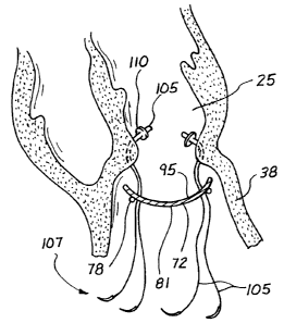
to the line 70 as illustrated in Figure 9. This stitch 74 produces an annular
protrusion 76 which forms a neck 78 relative to the imaginary line 70. This
neck 78
initially may have a round circular configuration as illustrated in Figure 9.
However,
as the suture 74 is tightened, the musculature of the myocardium will form a
natural
2 5 oval shape as illustrated in Figure 11. It is this oval-shaped neck 78,
formed by the
Fontan stitch 74, which in its natural ovoid shape is particularly adapted to
receive
the patch 72 of the present invention.
14
CA 02330746 2004-10-21
Providing the patch 72 with a configuration complimentary to the ovoid shape
of
the Fontan stitch 74 is believed to be of particular importance and advantage
to the present
invention. In the past, patches of a round, circular form were used. This form
maintained
the dilatation of stretch fibers in their more inefficient transverse
orientation. As a result,
the fiber contraction continued to be very inefficient. Providing the patch 72
with an oval
configuration restores the apex 37 or elliptical form of the heart 12. On a
muscle fiber
level, the fibers are directed back to a more normal, meaning generally
perpendicular,
orientation with respect to the heart wall 38. This reorients the lines of
contraction force to
greatly increase the contraction efficiency.
1 o Construction of various embodiments of the patch 72 are discussed with
reference
to Figures 12A-20. In the plan view of Figure 12A, a sheet material 81 is
illustrated to
have the shape of an ellipse with a major axis 83 between 30 and 50
millimeters and a
minor axis 85 between 20 and 30 millimeters. It is contemplated that the sheet
material 81
can be provided in two sizes, such as 20x30 millimeters and 30x40 millimeters.
The sheet material 81 may be formed, for example, from DacronTM
(HemashieldTM), or polytetrafluroethylene (GortexTM). However in a preferred
embodiment, the sheet material 81 is formed of autologous pericardium, or some
other
fixed mammalium tissue such as bovine or porcine pericardium. Importantly, the
sheet
material 81 is preferably sized and configured with a shape similar to that of
the Fontan
2 0 neck 78 as illustrated in Figure 11. As noted, this shape is non-circular
and preferably
oval.
The sheet material 81 can have a generally flat planar configuration, or can
be
shaped as a section of a sphere. The spherical shape can be achieved as
illustrated in Figure
12B by fixing the pericardium while it is stretched over a spherical die to
form a concave
2 5 surface 90.
CA 02330746 2000-11-O1
WO 99/56655 PCT/US99/02079
In addition to the sheet material 81, the patch 72 also preferably includes a
ring 87 which will typically have a toroidal configuration with a
circumferential
cross section that is circular, as shown in Figure 13. The ring will typically
be
formed of a plastic graph material that can also be made of curled autogenous
tissue
such as fascia or pericardium. In general, the ring 87 can be formed from any
biocompatible material having a degree of flexibility suitable to prevent
interference
with the normal contractions of the heart 12.
The circumferential cross section view of Figure 14 illustrates that the ring
87 may be enclosed in a tubular sheath 90 which may be formed from woven
l0 Dacron, and incorporated to promote tissue ingrowth to the patch 72.
The ring 87 will generally have a non-circular shape which may be similar to
but smaller than the shape of the material 81. Providing the ring 87 with a
shape
similar to the material 81 will enable the ring 87 to be attached to the
material 81 as
illustrated in Figures 15 and 16 with a body 91 of the patch disposed within
the ring
87, and a circumferential rim or flange 93 disposed outwardly of the ring 87.
The
rim 93 will preferably have a constant width around its circumference. This
width
will typically be in a range between 5 and 8 millimeters.
Many variations on the patch 72 will be apparent from the foregoing
discussion. For example, as illustrated in Figure 17, the sheet material 81
can be
2 0 provided with a convex surface 95 facing the left ventricle 25 rather than
the
concave surface illustrated in Figure 13. As illustrated in claim 18, the ring
87 can
be disposed on either the interior or exterior side of the material 8 I .
The ring 87 can be attached to the material 81 by adhesive or by stitches 97
passing over the ring 87 and through the material 81. Alternatively, the ring
87 can
be sandwiched between two pieces of the sheet material. In this case, a second
piece of the sheet material 99 can be positioned on the side of the ring 87
opposite
to the sheet material 81. Appropriate sutures extending around the ring 87 and
16
CA 02330746 2000-11-O1
WO 99/56655 PCT/US99/02079
through the materials 81 and 99 will sandwich the ring and maintain it in the
preferred position. The second piece of material 99 can be formed as a circle
with
an inner diameter 100 less than that of the ring 87, and a outer diameter 102
generally equal to that of the material 81.
It will be appreciated that many variations on these preferred embodiments
of the patch 82 will be apparent, each having a generally non-circular sheet
material,
such as the material 81, and perhaps a somewhat flexible toroid or oval ring
87.
In a preferred method for placing the patch 72, interrupted sutures 105 can
be threaded through the Fontan neck 78 as illustrated in Figure 21. Where the
to tissue is soft, the sutures 105 can be looped through pledgets 110 on the
interior
side of the neck 78 with the free ends of the sutures 1 OS extending through
the
exterior side of the neck 78. These free ends, emanating from progressive
positions
around the circumferential neck 78, are passed in complementary positions
through
the body of the patch 72 which is initially positioned remotely of the neck 78
as
illustrated in Figure 21. Since the Fontan stitch 74 may be applied to normal
(although akinetic) tissue, the pledgets 110 are preferred to insure that the
sutures
105 are well anchored in the neck 78.
Another method for placement of the interrupted patch suture is illustrated in
Figure 22B. In this view, which is similar to Figure S l, interrupted sutures
1 1 I are
2o directed through the entire ventricular wall 38 and exit the wall 38 in
proximity to
the protrusion 76 which forms the Fontan neck 78. These sutures 11 1 can also
be
anchored in a pledged strip I 13 disposed on the outer surface of the heart 12
to
further enhance the anchoring of these sutures 1 I 1.
When all of the interrupted sutures 105 have been placed around the
2 5 circumference of the neck 87, the patch 72 can be moved from its remote
location
along the sutures 105 and into proximity with the oval neck 78. This step is
illustrated in Figure 22 where the patch 72 is embodied with the concave
surface 90
17
CA 02330746 2000-11-O1
WO 99/56655 PCT/US99/02079
facing the neck 78 and with the ring 87 disposed outwardly of the material 81.
After the patch 17 has been moved into an abutting relationship with the neck
78,
the interrupted sutures 105 fan be tied as illustrated in Figure 23.
Having closed the left ventricular cavity 25 with the patch 72, one may
proceed to address any bleeding which may have resulted from placement of the
Fontan stitch 74 or the sutures 105. Such bleeding is illustrated by the
reference
numeral 112 in Figure 23. This bleeding I 12 will typically occur in close
proximity
to the neck 78 and beneath the region covered by the rim or flange 93
associated
with the material 81 of the patch 72. This bleeding can normally be stopped by
merely placing a suture through the ventricular wall 38 and the rim 93 at the
point of
bleeding. A pledget 114 can be used to tie the suture 1 12 with the rim 93
closely
held against the bleeding wall 38. This reinforcing stitch, acting in
combination with
the rim 93 of the patch 72, will usually stop any bleeding associated with the
sutures.
With the patch 72 suitably placed, the operative site can be closed by joining
the myocardial walls in a vest-over-pants relationship as illustrated in
Figure 24.
Care should be taken not to distort the right ventricle 21 by folding the
septum over
the wall 4I ventricuiar wall 38. Alternatively, the lateral wall 38 can be
disposed
interiorly of the septum wall 41 so a majority of the force on the patch 72 is
diverted
to the lateral wall 38. These walls 38 and 41 can be overlapped in close
proximity
to the patch 72 in order to avoid creating any cavity between the patch 72 and
the
walls 38, 41. When air evacuation is confirmed by transesophageal echo, the
patient
can be weaned of~bypass usually with minimal, if any, inotropic support.
Decanulasation and closure is routine.
Figure 24 is positioned in proximity to Figure 3 in order to illustrate the
dramatic difference between the pre-operative dilated heart of Figure 3 and
the post-
operative apical heart of Figure 24. For comparison it will again be noted
that the
18
CA 02330746 2000-11-O1
WO 99/56655 PCTNS99/02079
dilated heart of Figure 3 might typically have a left ventricular volume of
140
milliliters which might produce a blood flow of 42 milliliters with an
ejection
fraction of 30%. Comparing this with the postoperative heart of Figure 24, it
can be
seen initially that the ventricular volume is reduced for example to 90
milliliters.
The percentage of viable heart wall as opposed to akinetic heart wall is
greatly
increased thereby providing an increase in the ejection fraction, for example
from
thirty percent to forty-five percent. This combination results in a pumped
blood
volume of about 40 milliliters with each beat of the heart 12.
These structural changes are somewhat quantitative in consideration. But a
further advantage, qualitative in nature, is also associated with the present
procedure. It will be noted that this restorative procedure provides the heart
12
with a more natural apical configuration which facilitates the writhing action
discussed with reference to the arrow 47 in Figure 1. Thus, not only is the
normal
size of the heart achieved, but the restoration procedure also achieves a
normal heart
operation. In combination, the patch 72 and the resulting procedure
significantly
reduce the long term effects of myocardial ischemia and overcome many of the
causes associated with congestive heart failure.
It may be found that muscle function will be restored to some remote areas
following the altered ventricular architecture. Although not fully understood,
it is
believed that this restoration procedure improves remote segmental myocardial
contractility by reducing the wall tension and stress in the myocardium due to
a
reduction in ventricular volume. The stress equation states that --
Stress = P x R
2h
where
P is blood pressure;
3 0 R is radius of the heart wall; and
19
CA 02330746 2000-11-O1
WO 99/56655 PCT/US99/02079
h is wall thickness.
Reducing the ventricular volume decreases the radius, increases the thickness,
and
thereby reduces wall stress. This improves the myocardial oxygen supply/demand
relationship, but may also revive the contractibility of otherwise normal but
previously stressed myocardium. At the very least, the reduced stress on the
heart
12 is relieved along with any potential for congestive heart failure.
A further advantages of this procedure relates to the incision 61 in the left
ventricle 25 which also provides access to the mural valve 34. Replacing this
mitral
value 34 through the left ventricle 25 is much simpler than the present infra-
aortic
replacement procedure. Coronary artery bypass grafts also can be more easily
accommodated intraoperatively. As a result, all of these repairs can be
undertaken
with greater simplicity and reduced time. While blood cardioplegia may be
advantageously used for revascularization and valvular procedures, it would
appear
that the restorative procedure is best accomplished with continuous profusion
of the
beating open heart for cardiac protection.
Placement of patch 70 can be further enhanced by providing in the patch kit
a plurality of sizing disks which can be individually held in proximity to the
Fontan
neck in order to determine appropriate patch size. The disks might have a
generally
planar configuration, and of course, would vary in size. Each disk might have
a
centrally located handle extending from the planar disk for ease of use. The
patch
72 could be removably mounted on a holder also including a disk, on which the
patch is mounted, and an elongate handle extending from the disk to facilitate
placement.
As further support for the restoration procedure, a special suture needle is
contemplated which has a proximal end and a distal end. The proximal end is
generally straight and accounts for more than half of the length of the
needle. The
CA 02330746 2000-11-O1
WO 99/56655 PCT/US99/02079
distal end is curved along a relatively large radius facilitating initial
penetration of
the thick heart wall. With this configuration, the needle can be easily
introduced
through the thick myocardium, but then pulled along a generally straight path
as it is
removed interiorly of the ventricle.
The goal of this procedure is to restore the heart 12 to its normal size,
shape
and function. This includes restoring the conical apex of the heart in order
to
achieve the writhing pumping action. The nonfunctioning segmental ventricular
myocardium is excluded and replaced with a patch so that the only akinetic
wall of
the ventricle is that defined by the small patch area. Not only is visual
assessment
l0 enhanced, but more importantly, palpation affords the surgeon the ability
to
carefully and accurately determine the circumferential line of separation
between the
contracting and noncontracting muscle. This determination is achieved although
the
muscle may have normal color and may not contain either circular or trabecular
scar
tissue.
It is believed that cardioplegia arrest may be deleterious to ventricular
function in the open ventricle because of nonuniform flow distribution. By
avoiding
this cardioplegia arrest and operating on a beating heart, aortic cross
clamping as
well as the use of inter-aortic balloons and ventricular assist devices can be
avoided.
Patch placement can be intraoperatively adjusted guided by echo or radio
nucleotide
2 0 data. Placement of the patch is further simplified by creation of the
Fontan neck 78
and use of interrupted felt or pericardial pledgeted sutures 1 O5. The
circumferential
rim 93 associated with the patch 72 facilitates bleeding control without
distortion of
the patch 72. Finally, using a vest-over-pants closure of the excluded
ventricle
obliterates dead space and provides security against patch leaks and resultant
expansion.
Within these wide objectives and parameters, there will be variations on the
structure of the patch and the methods of restoration. Although the non-
circular
21
CA 02330746 2000-11-O1
WO 99/56655 PCT/US99/02079
configuration of the sheet material and ring are believed to be critical, the
shape of
the patch 72 may vary widely to provide the best anatomical fit with the
natural
shape of the ventricle 25. The sheet material 81 may be composed of a variety
of
materials, both natural and artificial. These materials may be woven or
nonwoven to
achieve a desired structure for the sheet material 81. The ring 87 may
similarly be
formed from a variety of materials and provided with a variety of shapes in
order to
add structure to the patch 72 without interfering with the normal contractions
of the
heart 12. Variations of the steps of the associated restoration method might
include
mounting the patch with a convex surface facing the ventricular cavity, use of
tissue
adhesives are also contemplated for attaching sealing and otherwise fixing the
patch
72 to the Fontan neck 78.
Given these wide variations, which are all within the scope of this concept,
one is cautioned not to restrict the invention to the embodiments which have
been
specifically disclosed and illustrated, but rather encouraged to determine the
scope
of the invention only with reference to the following claims.
22