Note: Descriptions are shown in the official language in which they were submitted.
CA 02331323 2000-11-07
WO 99/58174 PG'f/US99/09979
CIRCULATORY SUPPORT SYSTEM AND METHOD OF USE
FOR ISOLATED SEGMENTAL PERFUSION
FIELD OF THE INVENTION
The present invention relates generally to circulatory support systems and
cardiopulmonary bypass systems. More particularly, it relates to a circulatory
support system
and method of use for isolating organ systems for separate closed loop
perfusion.
BACKGROUND OF THE INVENTION
Circulatory support systems are used in many different medical settings to
supplement
or to replace the pumping function of a patient's heart. Applications of
circulatory support
systems and methods include, inter alia, augmenting cardiac output in patients
with a failing
heart, resuscitating victims of severe trauma or injury, and supporting a
patient's circulatory
functions during surgery.
One particular type of circulatory support system, known as a cardiopulmonary
bypass (CPB) system, is used to temporarily replace the functions of the heart
and the lungs
by supplying a flow of oxygenated blood to the patient's circulatory system.
The CPB system
drains deoxygenated blood from the patient's venous system, passes it through
a blood
oxygenator, and pumps the oxygenated blood back into the patient's arterial
system. CPB
systems may be configured for direct cannulation of the inferior and superior
vena cava or the
right atrium and the aorta, or they may be configured for peripheral
cannulation through the
femoral vein or jugular vein and the femoral artery. The cardiopulmonary
bypass system
allows the patient's heart to be temporarily stopped, for example by
cardioplegic arrest,
hypothermic arrest or fibrillation, for performing a variety of cardiothoracic
surgical
procedures.
Previous CPB systems have generally been configured to provide a single
circulatory
loop for supplying the entire body with oxygenated blood from a single CPB
pump. Thus, all
CA 02331323 2000-11-07
WO 99/58174 PCTNS99/09979
2
organ systems of the body receive oxygenated blood at the same pressure and
temperature
and with the same blood composition. This single-loop configuration has
significant
limitations in many medical circumstances. It has been found, for instance,
that the optimal
perfusion temperature for organ preservation during prolonged circulatory
support is different
for different organs of the body. Likewise, different chemical compositions of
the blood are
beneficial for preservation of different organ systems. For optimal
preservation of all the
organ systems within the body, it would be desirable to be able to selectively
perfuse
different organ systems with different perfusates, which have been optimized
for each of the
organ systems.
US 5308320, 5383854, 5820593 and 5879316 by Peter Safar, S. William Stezoski
and
Miroslav Klain, describe a cardiopulmonary bypass system capable of segmenting
a patient's
aorta and for selectively perfusing the different segments of the aorta with
perfusates of
different temperatures or chemical compositions. Other US patents that address
the concept
of selective aortic perfusion include commonly owned, copending patent
applications;
08/909,293, filed 8/11/97; 08/909,380, filed 8/11/97, and 09/152,589 filed
8/11/98 by Safar et
al.; and US 5738649 and commonly owned copending patent application 09/060,412
filed
4/14/98 by John A. Macoviak; and US 5827237 and 5833671 by John A. Macoviak
and
Michael Ross and commonly owned copending patent application 08/665,635, filed
6/17/96;
filed 6/18/96, by John A. Macoviak and Michael Ross; and 60/067,945, filed
12/8/97, by
Bresnahan et al. These patent applications and all other patents referred to
herein are hereby
incorporated by reference in their entirety. The balloon catheter of Safar et
al. may be
introduced into the patient's aorta from a peripheral entry point, such as the
femoral artery or
the subclavian artery, or it may be introduced by a direct puncture in the
patient's aorta
during open chest surgery.
The previously described system, however, does not isolate the segments of the
circulatory system from one another on the venous side of the circulatory
system because the
blood from each of the segments mingles together. Thus, any organ preserving
temperature
gradients, chemicals or therapeutic agents introduced into one of the segments
will eventually
mix with and be diluted into the entire systemic blood supply. In many
circumstances it
CA 02331323 2000-11-07
WO 99/58174 PCT/US99/09979
3
would be desirable to at least partially segment blood flow on the venous side
of the
circulatory system. For example, when administering anesthesia to a patient
during surgery, it
may be desirable to limit the flow of the anesthetic to the cerebral
circulation only and to
avoid dilution of the anesthetic in the systemic blood supply, and even to
recirculate the
anesthetic to the cerebral circulation. As another example, when administering
a therapeutic
agent that is very costly or which has systemic, central or specific organ
toxicity or other
undesirable effects, it may be desirable to limit the flow of the therapeutic
agent to the target
organs as much as possible without it entering the systemic blood supply such
as gene
therapy, viral vectors protein plasmids and angiogenic genes. As a third
example, when
performing segmented selective perfusion combined with hypothermic organ
preservation, it
would be desirable to isolate the segments of the circulatory system on the
venous side to
allow more precise and efficient temperature control within each circulatory
loop. It would be
desirable, therefore, to provide a circulatory support system or
cardiopulmonary bypass
system that allows segmentation of the circulatory system on the venous side,
as well as on
the arterial side, for isolated closed loop circulatory support of separate
organ systems. Such a
closed loop circulatory support system may be used to supply the entire body
with blood or
other fluids through a plurality of isolated circulatory loops when the heart
is not pumping.
Alternatively, the closed loop circulatory support system may be used to
create a single
circulatory loop for supplying a single segment or organ system of the body
with blood or
other fluids while the beating heart supplies blood to the remainder of the
body.
A plethora of known and newly discovered organ preserving chemicals and
therapeutic agents are suitable for use with the circulatory support system of
the present
invention. Among these are natural and artificial blood substitutes or oxygen
Garners, such as
free hemoglobin, PERFLUBRON, and perfluorocarbons, and hemoglobin modifiers,
such as
RSR-13 (Altos Therapeutics), that increase oxygen delivery from blood to
tissues. Also
among these are neuroprotective agents, which have been the subject of
intensive research in
recent years. Promising neuroprotective agents include Na+ blockers, glutamate
inhibitors,
nitric oxide inhibitors and radical scavengers. A thorough treatment of this
subject can be
found in the book Neuroprotective Agents, published by the New York Academy of
Sciences.
CA 02331323 2000-11-07
WO 99/58174 PGTNS99/09979
4
Possible therapeutic agents include, inter alia, thrombolytic agents, such as
tPA,
streptokinase and urokinase as well as gene therapy including angiogenic
genes.
SUMMARY OF THE INVENTION
The circulatory support system of the present invention generally includes one
or
more venous cannulae for draining blood from the venous side of the patient's
circulatory
system, one or more arterial cannulae for perfusing the arterial side of the
patient's
circulatory system, and one or more blood circulation pumps connected between
the venous
cannulae and the arterial cannulae. The arterial cannulae and the venous
cannulae of the
circulatory support system may take one of several possible configurations.
The circulatory
support system is configured to segment a patient's circulatory system into
one or more
isolated circulatory loops. The circulatory loops may be isolated from one
another and/or
from the remainder of the patient's circulatory system on the venous side, as
well as on the
arterial side, for isolated closed loop circulatory support of separate organ
systems. The
circulatory support system of the present invention is suitable for use in
minimally-invasive
cardiac surgery, using thoracoscopic, port-access or minithoracotomy
techniques, or for
standard open-chest cardiac surgery.
Also disclosed is a method for circulatory support and for cardiopulmonary
bypass
using differential perfusion and/or isolated segmental perfusion of the
circulatory system.
According to the method, a patient's circulatory system is segmented into two
or more
regions that are perfused with perfusate at different temperatures andlor
different chemical
compositions and/or different flow rates and/or different pressures. The
regions may be
isolated from one another and/or from the remainder of the patient's
circulatory system on the
venous side, as well as on the arterial side, for isolated closed loop
circulatory support of
separate organ systems.
In one variant of the method, a cerebral loop, a cardiac loop and a corporeal
loop are
created. A first fluid, preferably containing oxygenated blood, is circulated
to the cerebral
loop at a relatively low temperature of approximately 32° C or lower
for deep protective
CA 02331323 2000-11-07
WO 99/58174 PCT/US99/09979
hypothermia of the brain. Neuroprotective agents may be added to the first
fluid to enhance
the protection. A second fluid, which may include a cardioplegic agent, is
circulated to the
cardiac loop at a moderate temperature between 32° C and 37° C
for mild hypothermia of the
heart to protect the myocardium, while avoiding arrhythmias that can be caused
by deep
hypothermia. A third fluid, preferably containing oxygenated blood, is
circulated to the
corporeal loop at approximately 37° C for normothermic support of the
remainder of the
body. The venous side of the circulatory system may likewise be divided three
ways so that
the cerebral loop, cardiac loop and corporeal loop which are at least
partially isolated from
one another. Alternatively, the venous side of the circulatory system may be
divided two
ways so that the cardiac loop combines with either the cerebral loop or
corporeal loop on the
venous side, or the flow from all three loops may be allowed to commingle on
the venous
side of the circulatory system.
The use of differential perfusion according to this method provides several
other
clinical advantages in addition to those discussed above. The use of differing
degrees of
hypothermia allows optimal protection of the brain and of the heart during
cardiopulmonary
support, while decreasing the likelihood of complications. This method reduces
the thermal
mass of the tissue that must be cooled and rewarmed during the procedure. In
addition,
normothermic corporeal circulation provides a large reservoir of stored
thermal energy for
assisting in rewarming the heart and the brain at the end of the procedure.
Both of these
factors will result in decreasing the procedure time for surgery requiring
cardiopulmonary
bypass.
Still other clinical advantages exist with a closed loop circulatory system of
the
present invention. By isolating the cerebral, myocardial and corporeal
circulation on the
venous side (outputs) as well as the arterial side (inputs), isolated
measurements in the aortic
arch, aortic root, and corporeal circulation can be monitored in relation to
the superior vena
cava, right atrium and inferior vena cava respectively. This relationship will
enable the
clinician to determine oxygen saturation in the cerebral loop and in the
corporeal loop to
better manage the patient during the surgical procedure.
CA 02331323 2000-11-07
WO 99/58174 PCT/US99/09979
6
BRIEF DESCRIPTION OF THE DRAWINGS
FIG 1 illustrates a side view of an aortic catheter according to the present
invention
with a catheter shaft configured for retrograde deployment via femoral artery
access.
FIG 2 is a magnified lateral cross section of the aortic catheter of FIG 1
taken along
line 2-2 showing the multi-lumen arrangement of the catheter shaft.
FIG 3 illustrates a side view of a superior vena cava cannula according to the
present
invention with a tubular shaft configured for introduction into a patient's
venous system
through the jugular vein or other peripheral artery.
FIG 4 is a magnified lateral cross sectional of the superior vena cava cannula
of FIG 3
taken along line 4-4 in FIG 3.
FIG S illustrates a side view of an inferior vena cava cannula according to
the present
invention with a tubular shaft configured for introduction into a patient's
venous system
through the femoral vein or other peripheral artery.
FIG 6 is a magnified lateral cross sectional of the inferior vena cava cannula
of FIG S
taken along line 6-6 in FIG 5.
FIG 7 is a schematic illustration depicting a first embodiment of the present
invention
configured for selective, isolated, dual-loop perfusion of a patient's
circulatory system.
FIG 8 is a cutaway close-up view of the cannula placement as shown in FIG 7
with a
portion of the patient's heart cut away to better show the descending aorta.
FIG 9 illustrates a side view of an aortic catheter according to the present
invention
with a catheter shaft configured for retrograde deployment via femoral artery
access.
FIG 10 is a magnified lateral cross section of the aortic catheter of FIG 9
taken along
line 10-10 showing the mufti-lumen arrangement of the catheter shaft.
FIG 11 illustrates a side view of a dual lumen venous drainage cannula of the
present
invention configured for introduction through the patient's inferior vena cava
via the femoral
vein or other suitable venous access point in the lower extremities.
FIG 12 is a magnified lateral cross section of the venous drainage cannula
taken along
line 12-12 of FIG 11.
FIG 13 is a magnified lateral cross section of the venous drainage cannula
taken along
line 13-13 of FIG 11.
FIG 14 is a schematic illustration depicting a second embodiment of the
present
invention configured for selective, isolated, dual-loop perfusion of a
patient's circulatory
system.
CA 02331323 2000-11-07
WO 99/58174 PCT/US99/09979
7
FIG 15 is a cutaway close-up view of the cannula placement as shown in FIG14
with
a portion of the patient's heart cut away to better show the descending aorta.
FIG 16 illustrates a side view of an aortic catheter according to the present
invention
with a coaxial catheter shaft configured for retrograde deployment via femoral
artery access.
FIG 17 is a magnified lateral cross section of the aortic catheter of FIG 16
taken along
line 17-17 showing the mufti-lumen coaxial arrangement of the catheter shaft.
FIG 18 is a magnified lateral cross-section of the aortic catheter of FIG 16
taken along
line 18-18 showing the mufti-lumen arrangement of the catheter shaft.
FIG 19 illustrates a side view of a coaxial dual lumen venous drainage cannula
of the
present invention configured for introduction through the patient's inferior
vena cava via the
femoral vein or other suitable venous access point in the lower extremities.
FIG 20 is a magnified lateral cross section of the coaxial dual lumen venous
drainage
cannula taken along line 20-20 of FIG 19.
FIG .21 is a magnified lateral cross section of the coaxial dual lumen venous
drainage
cannula taken along line 2I-21 of FIG 19.
FIG 22 illustrates a third embodiment of the support system of the present
invention
configured for selective, isolated, dual-loop perfusion of a patient's
circulatory system.
FIG 23 is a cutaway close-up view of the cannula placement of FIG 22 with a
portion
of the patient's heart cut away to better show the descending aorta.
FIG 24 illustrates an aortic arch perfusion cannula of the present invention
configured
for introduction into the aortic arch through peripheral arterial access in
one of the upper
extremities, such as the left or right subclavian artery, axillary artery or
brachial artery.
FIG 25 is a magnified lateral cross section of the aortic arch perfusion
cannula of FIG
24 taken along line 25-25 of FIG 24 showing the mufti-lumen arrangement of the
catheter
shaft.
FIG 26 illustrates a corporeal perfusion cannula of the present invention
configured
for introduction into the descending aorta through a peripheral arterial
access in one of the
Iower extremities, such as the femoral artery.
FIG 27 is a magnified lateral cross section of the corporeal perfusion cannula
taken
along line 27-27 of FIG 26 showing the mufti-lumen arrangement of the catheter
shaft.
FIG 28 illustrates a fourth embodiment of the support system of the present
invention
configured for selective, isolated, dual-loop perfusion of a patient's
circulatory system.
CA 02331323 2000-11-07
WO 99/581?4 PC'f/US99/099?9
8
FIG 29 illustrates a side view of a dual-balloon, selective, central arterial
perfusion
cannula configured for antegrade introduction into the patient's aortic arch
via a direct
puncture or incision in the ascending aorta.
FIG 30 is a magnified lateral cross section of the aortic catheter of FIG 29
taken along
line 30-30 in FIG 29 illustrating the mufti-lumen arrangement of the aortic
catheter.
FIG 31 illustrates a side view of the central superior vena cava cannula of
the present
invention configured for introduction into the patient's superior vena cava
via an incision in
the right atrium.
FIG 32 is a magnified lateral cross-section of the central superior vena cava
cannula
taken along line 32-32 of FIG 31.
FIG 33 illustrates a side view of the central inferior vena cava cannula of
the present
invention configured for introduction into the patient's inferior vena cava
through the same or
another incision in the right atrium.
FIG 34 is a magnified lateral cross-section of the central superior vena cava
cannula
taken along line 33-33 of FIG 33.
FIG 35 is a schematic diagram of a fifth embodiment of the circulatory support
system of the present invention configured for selective, isolated, dual loop
perfusion of a
patient's circulatory system.
FIG 36 is a side view of an aortic perfusion shunt apparatus configured for
insertion
into a patient's aorta via a peripheral artery such as the femoral artery.
FIG 37 is a distal end view of the expandable shunt conduit of the aortic
perfusion
shunt apparatus of FIG 36 taken along line 37-37.
FIG 38 shows a schematic diagram of a sixth embodiment of the circulatory
support
system of the present invention configured for selective, closed-loop
perfusion of a patient's
cerebral circulation and upper extremities, while the beating heart supplies
the viscera and
lower extremities with blood.
FIG 39 shows a schematic diagram of a seventh embodiment of the circulatory
support system of the present invention configured for selective, closed-loop
perfusion of a
patient's renal system, while the beating heart supplies the remainder of the
circulatory
system with blood.
CA 02331323 2000-11-07
WO 99/58174 PCT/US99/09979
9
DETAILED DESCRIPTION OF THE INVENTION
The circulatory support system of the present invention generally comprises
one or
more arterial cannulae to enable the segmented perfusion of the patient's
circulatory system.
On the arterial side, one or more venous cannulae for enable segmented
draining of the
patient's circulatory system on the venous side, and one or more blood
circulation pumps
connect between the venous cannulae and the arterial cannulae. Preferably, the
circulatory
support system will also include one or more blood oxygenators and one or more
heat
exchangers for conditioning the patient's blood. The circulatory support
system is configured
to segment a patient's circulatory system into one or more isolated
circulatory loops. The
circulatory loops are isolated from one another and/or from the remainder of
the patient's
circulatory system on the venous side, as well as on the arterial side, for
isolated closed loop
circulatory support of separate organ systems.
FIGS 1 through 7 illustrate a first embodiment of the present invention. FIG 1
illustrates a side view of the aortic catheter 100 according to the present
invention with a
catheter shaft 102 configured for retrograde deployment via femoral artery
access. In order to
facilitate placement of the aortic catheter 100 and to improve the stability
of the catheter 100
in the proper position in the patient's aorta, a distal region 144 of the
catheter shaft 102 may
be preshaped with a curve to match the internal curvature of the patient's
aortic arch. The
curved distal region 144 represents a J-shaped curve of approximately 180
degrees of arc with
a radius of curvature of approximately 2 to 4 cm to match the typical
curvature of the aortic
arch in an adult human patient. In addition, the distal end 106 of the
catheter may be skewed
slightly up out of the plane of the curve to accommodate the forward
angulation of the
patient's ascending aorta. Additionally, the catheter shaft 102 may be
reinforced, particularly
in the curved distal region 144, for example with braided or coiled wire, to
further improve
the stability of the catheter 100 in the proper position in the patient's
aorta.
Illustrated in FIG 2, is a magnified lateral cross section of the aortic
catheter 100 of
FIG 1 taken along line 2-2 showing the multi-lumen arrangement of the catheter
shaft 102.
The catheter shaft 102 has six lumens: a corporeal perfusion lumen 108, an
arch perfusion
lumen 110, a first balloon inflation lumen 112, a second balloon inflation
lumen 114, a guide
wire and cardioplegia lumen 116 and a root pressure lumen 118.
CA 02331323 2000-11-07
WO 99/58174 PCTNS99/09979
Referring to FIG 1 the elongated catheter shaft 102 is preferably formed of a
flexible
thermoplastic material, a thermoplastic elastomer or a thermoset elastomer.
The catheter
shaft 102 may be fabricated separately by known extrusion methods and joined
together end-
to-end, for example by heat welding or by adhesive bonding. Alternatively, the
catheter shaft
102 may be fabricated by dipping or by composite construction techniques and
joined
together or the entire catheter shaft 102 may be fabricated integrally.
Suitable materials for
the elongated catheter shaft 102 include, but are not limited to,
polyvinylchloride,
polyurethane, polyethylene, polypropylene, polyamides (nylons), polyesters,
silicone, latex,
and alloys or copolymers thereof, as well as braided, coiled or counterwound
wire or filament
reinforced composites.
An upstream occlusion member 120 is mounted on the catheter shaft 102 near the
distal end 106 of the catheter 100. The upstream occlusion member 120 in this
embodiment is
in the form of an expandable, inflatable balloon bonded to the catheter shaft
102 by heat
welding or with an adhesive. Alternatively, the upstream occlusion member 120
may be in
the form of a selectively deployable external catheter valve. For a discussion
of other
suitable upstream occlusion members as well as the materiai components
thereof, reference is
made to commonly owned US 5827237, and 5833671 which have previously been
incorporated by reference herein in their entirety and commonly owned
copending patent
application 09/205,753 filed 12/4/98, which is herein incorporated by
reference. These
occlusion members discussed therein are suitable for all embodiments discussed
herein in any
combination. Suitable materials for the upstream occlusion member 120 include
flexible
polymers and elastomers, which include, but are not limited to,
polyvinylchloride,
polyurethane, polyethylene, polypropylene, polyamides (nylons), polyesters,
Latex, silicone,
and alloys, copolymers and reinforced composites thereof. In addition, the
outer surface of
the upstream occlusion member 120 may include a friction increasing coating or
texture to
increase friction with the aortic wall when deployed. The upstream occlusion
member 120
has a deflated state, in which the diameter of the occlusion member 120 is
preferably not
much larger than the diameter of the catheter shaft 102, and an inflated
state, in which the
occlusion member 120 expands to a diameter sufficient to occlude blood flow in
the
ascending aorta of the patient. For use in adult human patients, the
inflatable balloon
upstream occlusion member 120 preferably has an inflated outer diameter of
approximately
1.5 cm to 5.0 cm. Preferably, the inflatable occlusion member 120 has an
inflated length that
is not significantly longer than its inflated diameter, or, more preferably,
is shorter than its
CA 02331323 2000-11-07
WO 99/58174 PCTNS99/09979
11
inflated diameter. This shortened inflated profile allows the upstream
occlusion member 120
to be easily placed within the ascending aorta between the coronary arteries
and the
brachiocephalic artery without any danger of inadvertently occluding either.
A downstream occlusion member 122 is mounted on the catheter shaft 102 at a
position proximal to and spaced apart from the upstream occlusion member 120.
The
downstream anchoring member may be made of the same materials as the upstream
anchoring member of different materials and of the same size or a different
size. For a
complete discussion on the potential sizes arid characteristics of the
downstream occlusion
member, reference is made to commonly owned copending patent application
09/205,753
filed 12/4/98 which has previously been incorporated by reference. The
downstream
anchoring members discussed therein are suitable for all embodiments discussed
herein in
any combination. The distance between the upstream occlusion member 120 and
the
downstream occlusion member 122 is preferably between 3 and 20 cm, more
preferably
between 8 and 15 cm, and is chosen so that when the aortic catheter 100 is
deployed and the
upstream occlusion member 120 is positioned within the ascending aorta between
the
coronary arteries and the brachiocephalic artery, the downstream occlusion
member 122 will
be positioned in the descending aorta downstream of the left subclavian
artery. The
downstream occlusion member 122 in this embodiment is in the form of an
expandable,
inflatable balloon bonded to the catheter shaft 102 by heat welding or with an
adhesive. The
downstream occlusion member 122 is more elongated, than the upstream occlusion
member
120. Suitable materials for the inflatable balloon downstream anchoring member
122 include
flexible polymers and elastomers, which include, but are not limited to,
polyvinylchloride,
polyurethane, polyethylene, polypropylene, polyamides (nylons), polyesters,
latex, silicone,
and alloys, copolymers and reinforced composites thereof. In addition, the
outer surface of
the downstream anchoring member 122 may include a friction increasing coating
or texture to
increase friction with the aortic wall when deployed. Alternatively, the
downstream
occlusion member 122 may be in the form of a selectively deployable valve.
The inflatable downstream occlusion member 122 has a deflated state, in which
the
diameter of the occlusion member 122 is preferably not much larger than the
diameter of the
catheter shaft 102, and an inflated state, in which the occlusion member 122
expands to a
diameter sufficient to occlude blood flow in the descending aorta of the
patient. For use in
adult human patients, the downstream occlusion member 122 preferably has an
inflated outer
CA 02331323 2000-11-07
WO 99/58174 PCTNS99/09979
12
diameter of approximately 1.5 cm to 5.0 cm and a length of approximately 1.0
cm to 7.5 cm.
The more elongated the occlusion member 122 the greater the anchoring friction
against the
wail of the descending aorta when the downstream occlusion member 122 is
inflated in order
to prevent migration of the aortic catheter 100 due to pressure gradients
within the aorta
during perfusion.
The corporeal perfusion lumen 108 extends through the catheter shaft 102 from
the
proximal end 104 to one or more corporeal perfusion ports 124 on the exterior
of the catheter
shaft 102 proximal of the downstream occlusion member 122. The arch perfusion
lumen 110
extends through the catheter shaft 102 from the proximal end 104 to one or
more arch
perfusion ports 126 on the exterior of the catheter shaft 102 between the
upstream occlusion
member 120 and the downstream occlusion member 122. The first inflation lumen
112
extends through the catheter shaft 102 from the proximal end 104 to a first
balloon inflation
port 132 residing in the interior of the downstream occlusion member 122. The
second
balloon inflation lumen 114 extends through the catheter shaft 102 from the
proximal end 104
to balloon inflation port 130 residing in the interior of the upstream
occlusion member 120.
Alternatively, a common balloon inflation lumen can serve to simultaneously
inflate and
deflate both the upstream occlusion member 120 and the downstream occlusion
member 122.
When a common inflation lumen is implemented an arch monitoring lumen (not
shown) may
be incorporated having an arch monitoring port residing between the upstream
occlusion
member 120 and the downstream occlusion member 122 to monitor the pressure in
the aortic
arch.
The root pressure lumen 118 extends through the catheter shaft 102 from the
proximal
end 104 to a root pressure port 128 near the distal end 106 of the catheter
shaft 102 to monitor
pressure in the aortic root. The guide wire and cardioplegia lumen 116 extends
from the
proximal end 104 of the catheter shaft 102 to a guide wire/cardioplegia port
136 at the distal
end 106 of the catheter shaft 102, distal to the upstream occlusion member
120. Preferably,
the distal end 106 of the catheter shaft 102 is smoothly tapered or rounded
for easy
introduction and to avoid trauma or injury to the aortic wall during insertion
or withdrawal of
the aortic catheter 100.
Preferably, the aortic catheter 100 includes one or more markers, which may
include
radiopaque markers and/or sonoreflective markers, to enhance imaging of the
aortic catheter
CA 02331323 2000-11-07
WO 99/58174 PCT/US99/09979
13
100 using fluoroscopy or ultrasound, such as transesophageal echocardiography
(TEE). In
this illustrative embodiment, the aortic catheter 100 includes a distal
radiopaque marker 138
positioned near the distal end 106 of the catheter shaft 102, an intermediate
radiopaque
marker 140 positioned near the proximal edge of the upstream occlusion member
120, and a
proximal radiopaque marker 142 positioned near the distal edge of the
downstream anchoring
member 122. Each of the radiopaque markers 138, 140, 142 may be made of a ring
of dense
radiopaque metal, such as gold, platinum, tantalum, tungsten or alloys
thereof, or a ring of a
polymer or adhesive material heavily loaded with a radiopaque filler material.
The proximal end 104 of the catheter shaft 102 is connected to a manifold 150
with
fittings for each of the catheter lumens. The corporeal perfusion lumen 108 is
connected to a
Y-fitting 162 that has a barb connector 152 for connection to a perfusion pump
or the like and
a luer connector 154, which may be used for monitoring perfusion pressure, for
withdrawing
fluid samples or for injecting medications or other fluids. Likewise, the arch
perfusion lumen
110 is connected to a Y-fitting 164 that has a barb connector 156 for
connection to a
perfusion pump and a luer connector 158. The balloon inflation lumens 112 and
114 are
connected to luer connectors 160 and 166 respectively or other fittings
suitable for connection
to a syringe or balloon inflation device. The guide wire and cardioplegia
lumen 116 is
connected to a three-way Y-fitting 170 that has a barb connector 172 for
connection to a
cardioplegia infusion pump, a luer connector 174 and a guide wire port 176
with a Touhy-
Borst adapter or other hemostasis valve. The root pressure lumen 118 is
connected to a luer
connector 168 or other fitting suitable for connection to a pressure monitor.
FIG 3 illustrates a side view of a superior vena cava cannula 399 according to
the
present invention with a tubular shaft 398 configured for introduction into a
patient's venous
system through the jugular vein or other peripheral artery. FIG 4 is a
magnified lateral cross
sectional of the superior vena cava cannula 399 of FIG 3 taken along line 4-4
in FIG 3.
Referring now to FIGS 3 and 4 collectively, the superior vena cava cannula 399
has a
tubular shaft 398 that includes a venous drainage lumen 397 and a balloon
inflation lumen
396. The tubular shaft 398 preferably has a length of approximately 15 cm to
60 cm and a
diameter of approximately 10 to 32 French (3.3 mm to 10.7 mm diameter). An
occlusion
balloon 395 or other expandable occlusion member is mounted on the tubular
shaft 398 near
the distal end of the cannula 399. The occlusion balloon 395 or other
expandable occlusion
CA 02331323 2000-11-07
WO 99/58174 PCT/US99/09979
14
member preferably has an expanded diameter of approximately 5 mm to 40 mm. The
venous
drainage lumen 397 extends through the tubular shaft 398 from a venous
drainage fitting 394
to one or more venous drainage ports 393 on the tubular shaft 398 proximal to
the occlusion
balloon 395. The venous drainage fitting has a luer connector 373 which may be
used for
monitoring pressure, temperature, chemical compositions and for withdrawing
fluid samples
or for injecting medications or other fluids, a barb connector 372 or other
suitable fitting for
being connected to a CPB machine and a guide wire entry connector 392 in the
form of a
Thouy-Borst fitting or other suitable hemostasis valve for creating a fluid
tight seal when
using a guide wire. When a guide wire is used the venous drainage lumen 397
serves as an
additional guide wire lumen capable of receiving a guide wire 301 which is
guided to a guide
wire port 374 on the end of the tubular shaft 398 distal to the occlusion
balloon 395.
Alternatively, a separate lumen may be provided leading to a port distal to
the occlusion
balloon 395 wherein a separate monitoring device may be slidably or integrally
disposed to
give monitoring information inside or outside the cannula 399; and inside the
superior vena
cava. The balloon inflation lumen 396 extends through the tubular shaft 398
from a balloon
inflation fitting 391 on the proximal end of the cannula 399 to one or more
balloon inflation
ports 390 within the occlusion member 395.
FIG 5 illustrates a side view of an inferior vena cava cannula 589 according
to the
present invention with a tubular shaft 588 configured for introduction into a
patient's venous
system through the femoral or other peripheral artery. FIG 6 is a magnified
lateral cross
sectional of the inferior vena cava cannula 589 of FIG 5 taken along line 6-6
in FIG 5.
Referring collectively to FIGS S and 6, the inferior vena cava cannula 589 is
configured for introduction into the patient's inferior vena cava via the
femoral vein or other
suitable venous access point in the lower extremities. The inferior vena cava
cannula 589 has
a tubular shaft 588 that includes a venous drainage lumen 587 and a balloon
inflation lumen
586. The tubular shaft 588 preferably has a length of approximately 15 cm to
90 cm and a
diameter of approximately 10 to 32 French (3.3 mm to 10.7 mm diameter). An
occlusion
balloon 585 or other expandable occlusion member is mounted on the tubular
shaft 588 near
the distal end of the cannula 589. The occlusion balloon 585 or other
expandable occlusion
member preferably has an expanded diameter of approximately 5 mm to 40 mm. The
venous
drainage lumen 587 extends through the tubular shaft 588 from a venous
drainage fitting 584
on the proximal end of the cannula shaft 588 to one or more venous drainage
ports 583 on the
CA 02331323 2000-11-07
WO 99/5$174 PCT/US99/09979
1S
tubular shaft 588 proximal to the occlusion balloon 585. The venous drainage
fitting 584 has
a luer connector 563 which may be used for monitoring pressure, temperature,
chemical
compositions and for withdrawing fluid samples or for injecting medications or
other fluids, a
barb connector S62 or other suitable fitting for being connected to a CPB
machine and a
guide wire entry connector 582 in the form of a Thouy-Borst fitting or other
suitable
hemostasis valve for creating a fluid tight seal when using a guide wire. When
a guide wire
is used the venous drainage lumen 587 serves as an additional guide wire lumen
configured
for receiving a guide wire 501 which is guided to a guide wire port 564 on the
end of the
tubular shaft 588 distal to the occlusion balloon 585. Alternatively, a
separate lumen may be
provided leading to a port distal to the occlusion balloon S85 wherein a
separate monitoring
device integral or nonintegral is slideably disposed to give monitoring
information inside or
outside the cannula 589, and inside the inferior vena cava. The balloon
inflation lumen 586
extends through the tubular shaft 588 from a balloon inflation fitting 581 on
the proximal end
of the cannula 589 to one or more balloon inflation ports 580 within the
occlusion balloon
585.
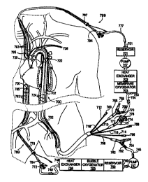
FIG 7 is a schematic illustration depicting a first embodiment of the present
invention
configured for selective, isolated, dual-loop perfusion of a patient's
circulatory system. The
circulatory support system has a cerebral loop for perfusion of the patient's
cerebral
circulation and upper extremities and a separate corporeal loop for perfusion
of the patient's
viscera and lower extremities. Optionally, the patient's coronary circulation
may be included
in the cerebral loop or the corporeal loop or a third, isolated coronary loop
may be created. In
this embodiment of the circulatory support system, arterial cannulation is
provided by a dual-
balloon, selective arterial perfusion cannula 700, and venous cannulation is
provided by a
superior vena cava cannula 799 and a separate inferior vena cava cannula 788.
FIG 8 is a
cutaway close-up view of the cannula placement as shown in FIG 7 with a
portion of the
patient's heart cut away to better show the descending aorta.
Referring now to FIGS 7 and 8, the cerebral closed loop circulation is created
by
having venous drainage port 793 proximal to the occlusion balloon 795 in fluid
communication with the venous drainage lumen 797. Connected to the venous
drainage
lumen 797 of the superior vena cava cannula 799 is a venous drainage fitting
794 which is
connected to inflow tubing 777 in fluid communication with inflow port 751 of
a first blood
circulation pump 750. After the blood is conditioned it is pumped through
outflow port 753
CA 02331323 2000-11-07
WO 99/58174 PCT/US99/09979
16
which is coupled to outflow tubing 754 in fluid communication with barb
connector 756
which is coupled to the arch perfusion lumen 710 of the arterial cannula 700.
The first blood
circulation pump 750 may be a peristaltic roller pump, a centrifugal blood
pump or other
suitable blood circulation pump. For illustrative purposes a membrane
oxygenator system is
provided for the cerebral circulation and a bubble oxygenator is provided for
the corporeal
circulation. It is understood by those skilled in the art that either
oxygenator may be
employed. In addition, a system may use two bubble oxygenators, two membrane
oxygenators or a membrane oxygenator and a bubble oxygenator or any
combination thereof.
This illustrative embodiment and all others contained herein may be configured
with any
combination as so stated.
The cerebral loop of the circulatory support system includes a venous drainage
cannula 799, which drains to a venous blood reservoir 701, the blood is pumped
to a heat
exchanger 702 and membrane oxygenator 703 in series with the first blood
circulation pump.
Optionally, vacuum assist (not shown) may be used to enhance venous drainage
through the
superior vena cava cannula 799. Venous blood from the head and upper
extremities enters
the patient's superior vena cava and is drained out through the venous
drainage lumen 797 of
the superior vena cava cannula 799. The blood is oxygenated, cooled and
recirculated by the
first blood circulation pump 757 to the head and upper extremities through the
arch perfusion
lumen 710 and out the arch perfusion ports 726 within the arterial cannula
700.
The corporeal loop of the circulatory support system includes a venous
drainage
cannula 789, which drains into a combined heat exchange bubble oxygenator to
an arterial
reservoir where it is pumped to arterial cannula 700. The venous drainage
lumen 787 is fluid
communication with drainage port 783 proximal to the occlusion balloon 785 in
fluid
communication with the venous drainage lumen 787. Alternatively there can be a
venous
drainage port 730 distal as well as proximal to the occlusion balloon 785.
Connected to the
venous drainage lumen 787 of the inferior vena cava cannula 789 has a venous
drainage
fitting 784 connected to corporeal inflow tubing 749 in fluid communication
with inflow port
748 of the second blood circulation pump 747. After the blood is conditioned
it is pumped
through outflow port 746 which is coupled to outflow tubing 745 in fluid
communication
with barb connector 752 which is coupled to the corporeal perfusion lumen 708
of the arterial
cannula 700. The second blood circulation pump 747 may be a peristaltic roller
pump, a
centrifugal blood pump or other suitable blood circulation pump. The corporeal
loop of the
CA 02331323 2000-11-07
WO 99158174 PCT/US99/09979
17
circulatory support system includes a venous blood reservoir 706, a blood
oxygenator 705
and heat exchanger 704 in series with the second blood circulation pump.
Optionally, vacuum
assist (not shown) may be used to enhance venous drainage through the inferior
vena cava
cannula 789. Venous blood from the viscera and lower extremities enters the
patient's inferior
vena cava and is drained out through the venous drainage lumen 787 of the
inferior vena cava
cannula 789. The blood is oxygenated, cooled and recirculated by the second
blood
circulation pump 747 to the viscera and lower extremities through the
corporeal perfusion
lumen 708 and out the corporeal perfusion ports 724 of the arterial cannula
700.
Optionally, either the superior vena cava cannula 799 or the inferior vena
cava
cannula 789 may be made without the occlusion balloon or with additional
drainage ports
distal to the balloon so that the cannula drains the patient's right atrium
and the coronary
sinus as part of the cerebral Ioop or the corporeal loop, respectively.
Alternatively, either the
superior vena cava cannula 799 or the inferior vena cava cannula 789 can be
made with a
separate, second drainage lumen connected to drainage ports positioned distal
to the balloon
for draining the patient's right atrium and the coronary sinus. A separate
coronary perfusion
loop can be created by connecting the second drainage lumen to the inflow of a
third blood
circulation pump and connecting the outflow of the pump to the cardioplegia
lumen of the
arterial cannula 700. The third blood circulation pump may be a peristaltic
roller pump, a
centrifugal blood pump or other suitable blood circulation pump. Preferably,
the coronary
loop also includes a venous blood reservoir, a blood oxygenator and heat
exchanger in series
with the third blood circulation pump.
As another alternative, the coronary circulation can be isolated by using a
coronary
sinus catheter for retrograde administration of cardioplegia into the
patient's coronary arteries.
This would eliminate the need for the occlusion balloon on either the superior
vena cava
cannula 799 or the inferior vena cava cannula 789 and the patient's right
atrium could be
drained as part of the cerebral loop or the corporeal loop. For example, a
superior vena cava
cannula 799 without an occlusion balloon (not shown) or with the balloon
deflated (not
shown) could be inserted into the superior vena cava and the right atrium via
the jugular vein.
An inferior vena cava cannula 789 would be inserted into the inferior vena
cava via the
femoral vein and the occlusion balloon 785 inflated to isolate the corporeal
loop. A coronary
sinus catheter can be inserted collaterally with the superior vena cava
cannula 799 via the
jugular vein to isolate the coronary circulation on the venous side and for
antegrade or
CA 02331323 2000-11-07
WO 99/58174 PGT/US99/09979
18
retrograde flow of blood, cardioplegia or other fluids. Suitable coronary
sinus catheter for
retrograde administration of cardioplegia can be found in U.S. patents
5,738,652; 5,722,963;
5,720,726; 5,662,607; 5,653,690; 5,643,231; 5,620,418; 5,617,854; 5,597,377;
5,558,644;
5,549,581; 5,533,957; 5,505,698; 5,488,960; 5,487,730; 5,466,216; 5,423,772;
5,423,745;
5,401,244; 5,395,331; 5,385,548; 5,385,540; 5,324,260; 5,197,952; 5,024,668;
5,021,045;
4,943,277; 4,927,412; 4,753,637; 4,648,384; 4,459,977, which are hereby
incorporated by
reference in their entirety.
To complete the closed loop circulation system an arterial perfusion cannula
700 is
provided. The dual-balloon, selective arterial perfusion cannula 700 is
configured for
retrograde introduction into the patient's aorta via a peripheral arterial
access point, such as
the femoral artery. The dual-balloon, selective arterial perfusion cannula 700
has a tubular
shaft 702 that includes a corporeal perfusion lumen 708, an arch perfusion
lumen 710, a guide
wire cardioplegia lumen 7I6, two balloon inflation lumens 712 and 714 and, a
root pressure
lumen 718. An upstream occlusion balloon 720 or other expandable occlusion
member is
mounted on the tubular shaft 702 so that it is positioned in the ascending
aorta between the
coronary arteries and the right brachiocephalic artery. A downstream occlusion
balloon 722
or other expandable occlusion member is mounted on the tubular shaft 702 so
that it is
positioned in the descending aorta downstream of the left subclavian artery.
The corporeal
perfusion lumen 708 extends through the tubular shaft 702 from a corporeal
barb connector
752 to one or more corporeal perfusion ports 724 on the tubular shaft 702
proximal to the
downstream occlusion balloon 722. The arch perfusion lumen 710 extends through
the
tubular shaft 702 from an arch barb connector 756 to one or more arch
perfusion ports 726 on
the tubular shaft 702 between the upstream occlusion balloon 720 and the
downstream
occlusion balloon 722. The guide wire cardioplegia lumen 716 extends through
the tubular
shaft 702 from a barb connector 772 to one or more cardioplegia ports 736 on
the tubular
shaft distal to the upstream occlusion balloon 720. The root pressure lumen
718 extends
through the tubular shaft 702 from a pressure fitting 768 to a root pressure
port 728 on the
tubular shaft 702 distal to the upstream occlusion balloon 720. A first
balloon inflation lumen
712 extends through the tubular shaft 702 a balloon inflation fitting 760 a
balloon inflation
port 732 within the downstream occlusion balloon 722. A second balloon
inflation lumen
714 extends through the tubular shaft 702 to a balloon inflation fitting 766
to a balloon
inflation port 730 within the upstream occlusion balloon 720.
CA 02331323 2000-11-07
WO 9915$174 PCT/US99/09979
19
FIGS 9 through 15 illustrate a second embodiment of the circulatory support
system of the
present invention, which is also configured for selective, isolated, dual-loop
perfusion of a
patient's circulatory system. The circulatory support system has a cerebral
loop for perfusion
of the patient's cerebral circulation and upper extremities and a separate
corporeal loop for
perfusion of the patient's viscera and lower extremities. As in the previously
described
embodiment, the patient's coronary circulation may optionally be included in
the cerebral
loop or the corporeal loop or a third, isolated coronary loop may be created.
In this
embodiment of the circulatory support system, arterial cannulation is provided
by a dual-
balloon, selective arterial perfusion cannula 900 similar to the one
previously described in
connection with FIG 1 and venous cannulation is provided by a dual-lumen
venous drainage
cannula 1199.
FIG 9 illustrates a side view of the aortic catheter 900 according to the
present
invention with a catheter shaft 902 configured for retrograde deployment via
femoral artery
access. In order to facilitate placement of the aortic catheter 900 and to
imnrore the stability
of the catheter 900 in the proper position in the patient's aorta, a distal
region 944 of the
catheter shaft 902 may be preshaped with a curve to match the internal
curvature of the
patient's aortic arch. The curved distal region 944 represents a J-shaped
curve of
approximately 180 degrees of arc with a radius of curvature of approximately 2
to 4 cm to
match the typical curvature of the aortic arch in an adult human patient. In
addition, the distal
end 906 of the catheter may be skewed slightly up out of the plane of the
curve to
accommodate the forward angulation of the patient's ascending aorta.
Additionally, the
catheter shaft 902 may be reinforced, particularly in the curved distal region
944, for example
with braided or coiled wire, to further improve the stability of the catheter
900 in the proper
position in the patient's aorta.
Illustrated in FiG 10, is a magnified lateral cross section of the aortic
catheter 900 of
FIG 9 taken along line 10-10 showing the mufti-lumen arrangement of the
catheter shaft 902.
The catheter shaft 902 has six lumens: a corporeal perfusion lumen 908, an
arch perfusion
lumen 910, a common balloon inflation lumen 912, an arch monitoring lumen 914,
a guide
wire and cardioplegia lumen 916 and a root pressure lumen 918.
Referring to FIG 9 the elongated catheter shaft 902 is preferably formed of a
flexible
thermoplastic material, a thermoplastic elastomer or a thermoset elastomer.
The catheter
CA 02331323 2000-11-07
WO 99/58174 PCT/US99/09979
shaft 902 may be fabricated separately by known extrusion methods and joined
together end-
to-end, for example by heat welding or by adhesive bonding. Alternatively, the
catheter shaft
902 may be fabricated by dipping or by composite construction techniques and
joined
together or the entire catheter shaft 902 may be fabricated integrally.
Suitable materials for
the elongated catheter shaft 902 include, but are not limited to,
polyvinylchloride,
polyurethane, polyethylene, polypropylene, polyamides (nylons), polyesters,
silicone, latex,
and alloys or copolymers thereof, as well as braided, coiled or counterwound
wire or filament
reinforced composites.
An upstream occlusion member 920 is mounted on the catheter shaft 902 near the
distal end 906 of the catheter 900. The upstream occlusion member 920 in this
embodiment is
in the form of an expandable, inflatable balloon bonded to the catheter shaft
902 by heat
welding or with an adhesive. Suitable materials for the upstream occlusion
member 920
include flexible polymers and elastomers, which include, but are not limited
to,
polyvinylchloride, polyurethane, polyethylene, polypropylene, polyamides
(nylons),
polyesters, latex, silicone, and alloys, copolymers and reinforced composites
thereof. In
addition, the outer surface of the upstream occlusion member 920 may include a
friction
increasing coating or texture to increase friction with the aortic wall when
deployed. The
upstream occlusion member 920 has a deflated state, in which the diameter of
the occlusion
member 920 is preferably not much larger than the diameter of the catheter
shaft 902, and an
inflated state, in which the occlusion member 920 expands to a diameter
sufficient to occlude
blood flow in the ascending aorta of the patient. For use in adult human
patients, the
inflatable balloon upstream occlusion member 920 preferably has an inflated
outer diameter
of approximately 1.5 cm to 5.0 cm. Preferably, the inflatable occlusion member
920 has an
inflated length that is not significantly longer than its inflated diameter,
or, more preferably,
is shorter than its inflated diameter. This shortened inflated profile allows
the upstream
occlusion member 920 to be easily placed within the ascending aorta between
the coronary
arteries and the brachiocephalic artery without any danger of inadvertently
occluding either.
A downstream occlusion member 922 is mounted on the catheter shaft 902 at a
position proximal to and spaced apart from the upstream occlusion member 920.
The
distance between the upstream occlusion member 920 and the downstream
occlusion member
922 is preferably between 3 and 20 cm, more preferably between 8 and 15 cm,
and is chosen
so that when the aortic catheter 900 is deployed and the upstream occlusion
member 920 is
CA 02331323 2000-11-07
WO 99/58174 PCT/US99/09979
21
positioned within the ascending aorta between the coronary arteries and the
brachiocephalic
artery, the downstream anchoring member 922 will be positioned in the
descending aorta
downstream of the left subclavian artery. The downstream occlusion member 922
in this
embodiment is in the form of an expandable, inflatable balloon bonded to the
catheter shaft
902 by heat welding or with an adhesive. The downstream occlusion member 922
is may be
larger, that is to say, more elongated, than the upstream occlusion member 920
of the same
size or smaller. Suitable materials for the inflatable balloon downstream
anchoring member
922 include flexible polymers and elastomers, which include, but are not
limited to,
polyvinylchloride, polyurethane, polyethylene, polypropylene, polyamides
(nylons),
polyesters, latex, silicone, and alloys, copolymers and reinforced composites
thereof. In
addition, the outer surface of the downstream anchoring member 922 may include
a friction
increasing coating or texture to increase friction with the aortic wall when
deployed.
The inflatable downstream occlusion member 922 has a deflated state, in which
the
diameter of the occlusion member 922 is preferably not much larger than the
diameter of the
catheter shaft 902, and an inflated state, in which the occlusion member 922
expands to a
diameter sufficient to substantially prohibit blood flow in the descending
aorta of the patient.
For use in adult human patients, the downstream occlusion member 922
preferably has an
inflated outer diameter of approximately 1.0 cm to 5.0 cm and a length of
approximately 1.0
cm to 7.5 cm. The more elongated the occlusion member 922 the greater the
anchoring
friction against the wall of the descending aorta when the downstream
occlusion member 922
is inflated in order to prevent migration of the aortic catheter 900 due to
pressure gradients
within the aorta during perfusion.
The corporeal perfusion lumen 908 extends through the catheter shaft 902 from
the
proximal end 904 to one or more corporeal perfusion ports 924 on the exterior
of the catheter
shaft 902 proximal of the downstream occlusion member 922. Alternatively, to
simplify
catheter design and to reduce overall catheter diameter a separate
contralateral, or co-lateral
peripheral access arterial cannula may be used to access either the same
femoral artery or the
other femoral artery. The arch perfusion lumen 910 extends through the
catheter shaft 902
from the proximal end 904 to one or more arch perfusion ports 926 on the
exterior of the
catheter shaft 902 between the upstream occlusion member 920 and the
downstream
occlusion member 922. A common balloon inflation lumen 912 extends through the
catheter
shaft 902 from the proximal end 904 to balloon inflation ports 932 and 930
which reside in
CA 02331323 2000-11-07
WO 99/58174 PCT/US99/09979
22
the interior of downstream occlusion balloon 922 and the upstream occlusion
balloon 920
respectively. Alternatively, separate inflation lumens can be implemented to
separately
inflate the downstream occlusion member 922 and the upstream occlusion member
920.
The arch monitoring lumen 914 extends through the catheter shag 902 from the
proximal end 904 to an arch monitoring port 934 proximal to the upstream
occlusion member
920 to monitor pressure in the aortic root. The root pressure lumen 918
extends through the
catheter shaft 902 from the proximal end 904 to a root pressure port 928 near
the distal end
906 of the catheter shaft 902 to monitor piessure in the aortic root. The
guide wire and
cardioplegia lumen 916 extends from the proximal end 904 of the catheter shaft
902 to a
guide wire/cardioplegia port 936 at the distal end 906 of the catheter shaft
902, distal to the
upstream occlusion member 920. Preferably, the distal end 906 of the catheter
shaft 902 is
smoothly tapered or rounded for easy introduction and to avoid trauma or
injury to the aortic
wall during insertion or withdrawal of the aortic catheter 900.
Preferably, the aortic catheter 900 includes one or more markers, which may
include
radiopaque markers and/or sonoreflective markers, to enhance imaging of the
aortic catheter
900 using fluoroscopy or ultrasound, such as transesophageal echocardiography
(TEE). In
this illustrative embodiment, the aortic catheter 900 includes a distal
radiopaque marker 938
positioned near the distal end 906 of the catheter shaft 902, an intermediate
radiopaque
marker 940 positioned near the proximal edge of the upstream occlusion member
920, and a
proximal radiopaque marker 942 positioned near the distal edge of the
downstream anchoring
member 922. Each of the radiopaque markers 938, 940, 942 may be made of a ring
of dense
radiopaque metal, such as gold, platinum, tantalum, tungsten or alloys
thereof, or a ring of a
polymer or adhesive material heavily loaded with a radiopaque filler material.
The proximal end 904 of the catheter shaft 902 is connected to a manifold 950
with
fittings for each of the catheter lumens. The corporeal perfusion lumen 908 is
connected to a
Y-fitting 962 that has a barb connector 952 for connection to a perfusion pump
or the like and
a luer connector 954, which may be used for monitoring perfusion pressure,
temperature,
chemical compositions and for withdrawing fluid samples or for injecting
medications or
other fluids. Likewise, the arch perfusion lumen 910 is connected to a Y-
fitting 964 that has a
barb connector 956 for connection to a perfusion pump and a luer connector 958
which may
be used for monitoring arch perfusion pressure, temperature, chemical
compositions and for
CA 02331323 2000-11-07
WO 99/58174 PCTNS99/09979
23
withdrawing fluid samples or for injecting medications or other fluids. The
common balloon
inflation lumen 912 is connected to a stopcock or luer connector 960 or other
fitting suitable
for connection to a syringe or balloon inflation device. In addition the
inflation lumen 912
may be attached to a pressure monitoring device to give visible and or tactile
feedback
concerning the balloon inflation pressure. The guide wire and cardioplegia
lumen 916 is
connected to a three-way Y-fitting 970 that has a barb connector 972 for
connection to a
cardioplegia infusion pump, a luer connector 974 capable of monitoring root
perfusion
pressure, temperature and chemical compositions and a guide wire port 976 with
a Touhy-
Borst adapter or other hemostasis valve. The root pressure lumen 918 is
connected to a luer
connector 968 or other suitable fitting capable of monitoring arch perfusion
pressure,
temperature and chemical compositions or for withdrawing fluid samples. The
arch
monitoring lumen 914 is connected to a luer connector 966 or other suitable
fitting capable of
monitoring arch perfusion pressure, temperature, and chemical compositions or
for
withdrawing fluid samples. Alternatively, sensors may be placed on the
catheter shaft or
inside the catheter shaft to measure chemical compositions in the aortic arch.
FIG 11 illustrates a side view of a dual lumen venous drainage cannula 1199 of
the
present invention configured for introduction through the patient's inferior
vena cava via the
femoral vein or other suitable venous access point in the lower extremities.
Alternatively, the
dual lumen venous drainage cannula 1199 may be configured for introduction
though the
patient's superior vena cava via the jugular vein or other suitable venous
access point in the
neck or upper extremities. The elongated tubular shaft 1198 may be fabricated
separately by
known extrusion methods and joined together end-to-end, for example by heat
welding or by
adhesive bonding. Alternatively, the elongated tubular shaft 1198 may be
fabricated by
dipping or by composite construction techniques and joined together or the
entire tubular
shaft i 198 may be fabricated integrally. Suitable materials for the elongated
tubular shaft
1198 include, but are not limited to, polyvinylchloride, polyurethane,
polyethylene,
polypropylene, polyamides (nylons), polyesters, silicone, latex, and alloys or
copolymers
thereof, as well as braided, coiled or counterwound wire or filament
reinforced composites.
FIG 12 is a magnified lateral cross section of the venous drainage cannula
1199 taken
along line 12-I2 of FIG 11. FIG 13 is a magnified lateral cross section of the
venous
drainage cannula 1199 taken along line 13-13 of FIG 11. Collectively FIGS 11
through 13
illustrate the mufti-lumen arrangement of the dual-lumen venous drainage
cannula 1199
CA 02331323 2000-11-07
WO 99/58174 PCTNS99/09979
24
having an elongated tubular shaft 1198 which includes a first venous drainage
lumen 1188; a
second venous drainage lumen 1189; a first balloon inflation lumen 1191, and a
second
balloon inflation lumen 1194. Alternatively, the dual-lumen venous drainage
cannula 1199
may have a common balloon inflation lumen capable of simultaneously inflating
both
occlusion balloons. The tubular shaft 1198 preferably has a length of
approximately 15 cm to
90 cm and a diameter of approximately 10 to 32 French (3.3 mm to 10.7 mm
diameter).
The dual-lumen venous drainage cannula 1199 includes a first occlusion balloon
1197
or other expandable occlusion member mounted on the tubular shaft 1198, which
is
positioned within the patient's superior vena cava when in the operative
position, and a
second occlusion balloon 1196 or other expandable occlusion member, mounted on
the
tubular shaft 1198, which is positioned within the patient's inferior vena
cava when in the
operative position. Suitable materials for the first occlusion member 1197 and
the second
occlusion member 1 I96 include flexible polymers and elastomers, which
include, but are not
limited to, polyvinylchloride, polyurethane, polyethylene, polypropylene,
polyamides
(nylons), polyesters, latex, silicone, and alloys, copolymers and reinforced
composites
thereof. The occlusion balloons 1196 and 1197 preferably have an expanded
diameter of
approximately 5 mm to 40 mm. When the dual-lumen venous drainage cannuia 1199
is
configured for femoral artery introduction, the first occlusion balloon 1197
is mounted near
the distal end 1195 of the tubular shaft 1198 and the second occlusion balloon
1196 is
mounted somewhat proximal to the first balloon 1197, as shown. Alternatively,
for jugular
vein introduction, these positions are reversed.
A first balloon inflation lumen 1191 is connected to a stopcock l I90 that
extends
through the tubular shaft 1198 to a balloon inflation port 1192 within the
first occlusion
balloon 1197. The second balloon inflation lumen 1194, is connected to a
stopcock 1193, that
extends through the tubular shaft 1198 to a balloon inflation port 1123 within
the second
occlusion balloon 1196. Alternatively, a common balloon inflation lumen may be
implemented and a superior vena cava monitoring lumen may be implemented to
monitor
pressure, temperature and chemical composition in the superior vena cava.
The first venous drainage lumen 1188 extends from a venous drainage fitting
1187
through the tubular shaft 1198, to one or more superior vena cava drainage
ports 1195 on the
tubular shaft 1 I98 distal to the first occlusion balloon 1197. In addition,
venous drainage
CA 02331323 2000-11-07
WO 99/58174 PGT/US99/09979
ports 1182 which are distal to the second occlusion balloon 1196 are also in
fluid
communication with the first venous drainage lumen 1188. Alternatively, the
venous
drainage ports 1182 may be in fluid communication with the second venous
drainage lumen
I 189. The second venous drainage lumen 1189 extends from a venous drainage
fitting 1181
through the tubular shaft 1198, to one or more inferior vena cava drainage
ports 1173 on the
tubular shaft 1198 proximal to the second occlusion balloon 1196. Preferably,
the distal
portion of the tubular shaft 1198 is smoothly tapered or rounded for easy
introduction and to
avoid trauma or injury to the vena cava during insertion or withdrawal of the
venous cannuia
1199.
Preferably, the venous cannula includes one or more markers, which may include
radiopaque markers and/or sonoreflective markers, to enhance imaging of the
venous cannula
1199 using fluoroscopy or ultrasound, such as transesophageal echocardiography
(TEE). In
this illustrative embodiment, the venous drainage cannula 1199 includes a
distal radiopaque
marker 1178 positioned near the distal end 1195 of the tubular shaft 1 I98, an
intermediate
radiopaque marker 1177 positioned near the drainage ports 1182, and a proximal
radiopaque
marker 1176 positioned near the distal edge of the second occlusion member
1196. Each of
the radiopaque markers 1178, 1177, 1176 may be made of a ring of dense
radiopaque metal,
such as gold, platinum, tantalum, tungsten or alloys thereof, or a ring of a
polymer or
adhesive material heavily loaded with a radiopaque filler material.
The proximal end 1183 of the venous drainage cannula 1199 is connected to a
manifold 1125 with fittings for each of the catheter lumens. The first venous
drainage lumen
1188 is coupled to a three-way fitting 1187 that has a barb connector 1186 for
connection to
an external CPB machine, a luer connector 1185 capable of monitoring superior
vena cava
pressure, temperature and chemical compositions and a guide wire port 1184
with a Touhy-
Borst adapter or other hemostasis valve on the proximal end of the cannula
1183. The second
venous drainage lumen 1189 is coupled to a Y-fitting 1181 having a barb
connector 1180, or
other suitable fitting capable of being coupled to a CPB machine and a luer
fitting 1179
capable of monitoring inferior vena cava pressure, temperature and chemical
compositions.
A first inflation lumen 1 I91 is coupled to a stopcock 1190, or other suitable
fitting capable of
being attached to an inflation mechanism and a second inflation lumen 1194 is
coupled to a
stopcock 1193, or other suitable fitting capable of being attached to an
inflation mechanism.
In addition, each inflation lumen may have an individual pressure-monitoring
device
CA 02331323 2000-11-07
WO 99/58174 PCT/US99/09979
26
proximal or distal to the stopcock to provide visible and tactile feedback
concerning the
balloon inflation pressures. Alternatively, a common inflation lumen may be
implemented.
FIG 14 illustrates the second embodiment of the closed loop circulatory system
of the
present invention. FIG 15 is a cutaway close-up view of the cannula placement
as shown in
FIG 14 with a portion of the patient's heart cut away to better show the
descending aorta. The
cerebral loop of the circulatory support system is created by having venous
drainage ports
1495 and 1482 in fluid communication with the superior vena cava drainage
lumen 1488.
Coupled to the superior vena cava drainage lumen 1488 is a fitting 1487 having
a barb
connector 1486 coupled to tubing 1449 in fluid communication with an inflow
port 1448 of a
first blood circulation pump 1447. The blood is conditioned and pumped through
the outflow
port 1446 of the first blood circulation pump 1447 to the arch perfusion lumen
1410 of the
arterial cannula 1400. The first blood circulation pump 1447 may be a
peristaltic roller pump,
a centrifugal blood pump or other suitable blood circulation pump. Preferably,
the cerebral
loop of the circulatory support system will also include a venous blood
reservoir 1401, a
blood oxygenator 1403 and heat exchanger 1402 in series with the first blood
circulation
pump 1447. Optionally, vacuum assist may be used to enhance venous drainage
through the
first venous drainage lumen 1488 of the dual-lumen venous drainage cannula
1499. Venous
blood from the head and upper extremities enters the patient's superior vena
cava and is
drained out through the first venous drainage lumen 1488 of the dual-lumen
venous drainage
cannula 1499 as the first occlusion balloon 1497 prevents blood from traveling
into the right
atrium from the superior vena cava. The blood is oxygenated, cooled and
recirculated by the
first blood circulation pump 1447 to the head and upper extremities through
the arch
perfusion lumen 1410 of the arterial cannula 1400.
The corporeal loop of the circulatory support system is created by having a
venous
drainage port 1478 in fluid communication with inferior vena cava drainage
lumen 1489. A
second Coupled to the second venous drainage lumen 1489 is a fitting 1481
having a barb
connector 1480 coupled to tubing 1477 in fluid communication with an inflow
port 1451 of a
second blood circulation pump 1455. After the blood is conditioned it is
pumped through
outflow port 1457 in fluid communication with tubing 1459 which is coupled to
a barb
connector 1452 in fluid communication the corporeal lumen 1408 of the aortic
catheter 1400.
The second blood circulation pump may be a peristaltic roller pump, a
centrifugal blood
pump or other suitable blood circulation pump. Preferably, the corporeal loop
of the
CA 02331323 2000-11-07
WO 99/58174 PCT/US99/09979
27
circulatory support system will also include a venous blood reservoir 1404, a
blood
oxygenator 1406 and heat exchanger1405 in series with the second blood
circulation pump
1455. Optionally, vacuum assist may be used to enhance venous drainage through
the second
venous drainage lumen of the dual-lumen venous drainage cannula 1400. Venous
blood from
the viscera and lower extremities enters the patient's inferior vena cava and
is drained out
through the second venous drainage lumen 1489 of the dual-lumen venous
drainage cannula
1499. The blood is oxygenated, cooled and recirculated by the second blood
circulation pump
1455 to the viscera and lower extremities through the corporeal perfusion
lumen 1408 of the
arterial catheter 1400.
Optionally, the dual-lumen venous drainage cannula 1499 may be made without
either
the first occlusion balloon or the second occlusion balloon or one of the
balloons may be
partially deflated or completely deflated when operating in this mode since
isolation of the
patient's right atrium and the coronary sinus is unnecessary. Alternatively,
the dual-lumen
venous drainage cannula 1499 may be provided with a third venous drainage
lumen within
the tubular shaft connected to the drainage ports 1482 between the first and
second balloons
for draining the patient's right atrium and the coronary sinus. A separate
coronary perfusion
loop can be created by connecting the third venous drainage lumen to the
inflow of a third
blood circulation pump and connecting the outflow of the pump to the
cardioplegia lumen of
the arterial cannula 1400. The third blood circulation pump may be a
peristaltic roller pump, a
centrifugal blood pump or other suitable blood circulation pump. Preferably,
the coronary
loop also includes a venous blood reservoir, a blood oxygenator and heat
exchanger in series
with the third blood circulation pump.
As another alternative, the coronary circulation can be isolated by inserting
a coronary
sinus catheter via the jugular vein to isolate the coronary circulation on the
venous side and
for antegrade or retrograde flow of blood, cardioplegia or other fluids into
the patient's
coronary arteries. The first occlusion balloon 1495 could be eliminated from
the dual-lumen
venous drainage cannula 1499 or left uninflated so that the patient's right
atrium will be
drained as part of the cerebral loop.
FIGS 16 through 23 collectively illustrate a third embodiment of the
circulatory
support system of the present invention, which is also configured for
selective, isolated, dual-
loop perfusion of a patient's circulatory system. The circulatory support
system has a cerebral
CA 02331323 2000-11-07
WO 99/58174 PCT/US99109979
28
loop for perfusion of the patient's cerebral circulation and upper extremities
and a separate
corporeal loop for perfusion of the patient's viscera and lower extremities.
As in the
previously described embodiment, the patient's coronary circulation may
optionally be
included in the cerebral loop or the corporeal loop or a third, isolated
coronary loop may be
created through the use of a separate coronary sinus catheter or through a
separate pump. In
this third embodiment of the circulatory support system, arterial cannulation
is provided by a
coaxial dual-balloon, selective arterial perfusion cannula 1600 and venous
cannulation is
provided by a coaxial dual-lumen venous drainage cannula 1799.
FIG 16 illustrates a side view of the aortic catheter 1600 according to the
present
invention with a coaxial catheter shaft 1602 configured for retrograde
deployment via femoral
artery access. Alternatively, a separate contralateral or colateral arterial
cannula may be
provided to provide perfusion to the corporeal body through separate
cannulation of the a
second peripheral artery. In order to facilitate placement of the aortic
catheter 1600 and to
improve the stability of the catheter 1600 in the proper position in the
patient's aorta, a distal
region 1644 of the catheter shaft 1602 may be preshaped with a curve to match
the internal
curvature of the patient's aortic arch. The curved distal region 1644
represents a J-shaped
curve of approximately 180 degrees of arc with a radius of curvature of
approximately 2 to 4
cm to match the typical curvature of the aortic arch in an adult human
patient. In addition, the
distal end 1606 of the catheter may be skewed slightly up out of the plane of
the curve to
accommodate the forward angulation of the patient's ascending aorta.
Additionally, the
catheter shaft 1602 may be reinforced, particularly in the curved distal
region 1644, for
example with braided or coiled wire, to further improve the stability of the
catheter 900 in the
proper position in the patient's aorta.
Illustrated in FIG 17, is a magnified lateral cross section of the aortic
catheter 1600 of
FIG 16 taken along line 17-17 showing the mufti-lumen coaxial arrangement of
the catheter
shaft 1602. The catheter shaft 1602 has six lumens: a corporeal perfusion
lumen 1608; an
arch perfusion lumen 1610; a common balloon inflation lumen 1612; an arch
monitoring
lumen 1614; a guide wire and cardioplegia lumen 1616 and a root pressure lumen
1618.
FIG 18 is a magnified lateral cross-section of the aortic catheter i 600 of
FIG 16 taken
along line 18-18 showing the mufti-lumen arrangement of the catheter shaft
1602. Shown in
FIG 18, five of the six lumens continue distally through the catheter shaft
1602: the arch
CA 02331323 2000-11-07
WO 99/58174 PCT/US99/09979
29
perfusion lumen 1610; the common balloon inflation lumen 1612; the arch
monitoring lumen
1614; the guide wire and cardioplegia lumen 1616 and the root pressure lumen
1618. The
corporeal perfusion lumen terminates at a position distal to the corporeal
perfusion ports
1624.
Refernng to FIGS 16 through 18 the elongated catheter is comprised of an inner
tubular shaft and an outer tubular shaft configured in a coaxial relationship
such that an
annular space is created therebetween. The annular space between the tubular
shafts 1603
and 1606 defines the corporeal lumen 1608. The tubular shafts 1603 and 1606
are preferably
formed of a flexible thermoplastic material, a thermoplastic elastomer or a
thermoset
elastomer. The coaxial catheter shaft 1602 may be fabricated separately by
known extrusion
methods and joined together end-to-end, for example by heat welding or by
adhesive
bonding. Alternatively, the coaxial catheter shaft 1602 may be fabricated by
dipping or by
composite construction techniques and joined together or the entire catheter
shaft 1602 may
be fabricated integrally. Suitable materials for the elongated catheter shaft
1602 include, but
are not limited to, polyvinylchloride, polyurethane, polyethylene,
polypropylene, polyamides
(nylons), polyesters, silicone, latex, and alloys or copolymers thereof, as
well as braided,
coiled or counterwound wire or filament reinforced composites.
An upstream occlusion member 1620 is mounted on the inner tubular shaft 1603
near
the distal end 1606 of the catheter 1600. The upstream occlusion member 1620
in this
embodiment is in the form of an expandable, inflatable balloon bonded to the
catheter shaft
1602 by heat welding or with an adhesive. Suitable materials for the upstream
occlusion
member 1620 include flexible polymers and elastomers, which include, but are
not limited to,
polyvinylchloride, polyurethane, polyethylene, polypropylene, polyamides
(nylons),
polyesters, latex, silicone, and alloys, copolymers and reinforced composites
thereof. In
addition, the outer surface of the upstream occlusion member 1620 may include
a friction
increasing coating or texture to increase friction with the aortic wall when
deployed. The
upstream occlusion member 1620 has a deflated state, in which the diameter of
the occlusion
member 1620 is preferably not much larger than the diameter of the catheter
shaft 1602, and
an inflated state, in which the occlusion member 1620 expands to a diameter
sufficient to
occlude blood flow in the ascending aorta of the patient. For use in adult
human patients, the
inflatable balloon upstream occlusion member 1620 preferably has an inflated
outer diameter
of approximately 1.5 cm to 5.0 cm. Preferably, the inflatable occlusion member
1620 has an
CA 02331323 2000-11-07
WO 99/58174 PCTIUS99/09979
inflated length that is not significantly longer than its inflated diameter,
or, more preferably,
is shorter than its inflated diameter. This shortened inflated profile allows
the upstream
occlusion member 1620 to be easily placed within the ascending aorta between
the coronary
arteries and the brachiocephalic artery without any danger of inadvertently
occluding either.
A downstream occlusion member 1622 is mounted on the catheter shaft 1602 at a
position proximal to and spaced apart from the upstream occlusion member 1620.
The
distance between the upstream occlusion member 1620 and the downstream
occlusion
member 1622 is preferably between 3 and 20 cm, more preferably between 8 and
15 cm, and
is chosen so that when the aortic catheter 1600 is deployed and the upstream
occlusion
member 1620 is positioned within the ascending aorta between the coronary
arteries and the
brachiocephalic artery, the downstream anchoring member 1622 will be
positioned in the
descending aorta downstream of the left subclavian artery. The downstream
occlusion
member 1622 in this embodiment is in the foam of an expandable, inflatable
balloon bonded
to the catheter shaft 1602 by heat welding or with an adhesive. Suitable
materials for the
inflatable balloon downstream anchoring member 1622 include flexible polymers
and
elastomers, which include, but are not limited to, polyvinylchloride,
polyurethane,
polyethylene, polypropylene, polyamides (nylons), polyesters, latex, silicone,
and alloys,
copolymers and reinforced composites thereof. In addition, the outer surface
of the
downstream anchoring member 1622 may include a friction increasing coating or
texture to
increase friction with the aortic wall when deployed.
The inflatable downstream occlusion member 1622 has a deflated state, in which
the
diameter of the occlusion member 1622 is preferably not much larger than the
diameter of the
catheter shaft 1602, and an inflated state, in which the occlusion member 1622
expands to a
diameter capable of regulating blood flow in the descending aorta of the
patient. Therefore,
to gain desired results the downstream occlusion member may be completely
inflated, or
partially inflated. For use in adult human patients, the downstream occlusion
member 1622
preferably has an inflated outer diameter of approximately 1.0 cm to 5.0 cm
and a length of
approximately 1.0 cm to 7.5 cm.
The corporeal perfusion lumen 1608 extends through the catheter shaft 1602
from the
proximal end 1604 to one or more corporeal perfusion ports 1624 on the
exterior of the
catheter shaft 1602 proximal of the downstream occlusion member 1622. The arch
perfusion
CA 02331323 2000-11-07
WO 99/58174 PCTNS99/09979
31
lumen 1610 extends through the catheter shaft 1602 from the proximal end 1604
to one or
more arch perfusion ports 1626 on the exterior of the catheter shaft 1602
between the
upstream occlusion member 1620 and the downstream occlusion member 1622. A
common
balloon inflation lumen 1612 extends through the catheter shaft 1602 from the
proximal end
1604 to balloon inflation ports 1632 and 1630 which reside in the interior of
downstream
occlusion balloon 1622 and the upstream occlusion balloon 1620 respectively.
Alternatively,
separate inflation lumens can be implemented to separately inflate the
downstream occlusion
member 1622 and the upstream occlusion member 1620.
The arch monitoring lumen 1614 extends through the catheter shaft 1602 from
the
proximal end 1604 to an arch monitoring port 1634 proximal to the upstream
occlusion
member 1620 to monitor pressure in the aortic arch though the lumen 1614 or by
providing a
separate sensor slidably disposed in the lumen 1614. The root pressure lumen
1618 extends
through the catheter shaft 1602 from the proximal end 1604 to a root pressure
port 1628 near
the distal end 1606 of the catheter shaft 1602 to monitor pressure in the
aortic root through
the lumen 1618 or through a separate sensor slidably disposed in the lumen
1618. The guide .
wire and cardioplegia lumen 1616 extends from the proximal end 904 of the
catheter shaft
1602 to a guide wire/cardiopiegia port 1636 at the distal end 1606 of the
catheter shaft 1602,
distal to the upstream occlusion member 1620. Preferably, the distal end 1606
of the catheter
shaft 1602 is smoothly tapered or rounded for easy introduction and to avoid
trauma or injury
to the aortic wall during insertion or withdrawal of the aortic catheter 1600.
Preferably, the aortic catheter 1600 includes one or more markers, which may
include
radiopaque markers and/or sonoreflective markers, to enhance imaging of the
aortic catheter
900 using fluoroscopy or ultrasound, such as transesophageal echocardiography
(TEE). In
this illustrative embodiment, the aortic catheter 1600 includes a distal
radiopaque marker
1638 positioned near the distal end 1606 of the catheter shaft 1602, an
intermediate
radiopaque marker 1640 positioned near the proximal edge of the upstream
occlusion
member 1620, and a proximal radiopaque marker 1642 positioned near the distal
edge of the
downstream anchoring member 1622. Each of the radiopaque markers 1638, 1640,
1642 may
be made of a ring of dense radiopaque metal, such as gold, platinum, tantalum,
tungsten or
alloys thereof, or a ring of a polymer or adhesive material heavily loaded
with a radiopaque
filler material.
CA 02331323 2000-11-07
WO 99/58174 PCTNS99/09979
32
The proximal end 1604 of the catheter shaft 1602 is connected to a manifold
1650
with fittings for each of the catheter lumens. The corporeal perfusion lumen
1608 is
connected to a Y-fitting 1662 that has a barb connector 1652 for connection to
a perfusion
pump or the like and a luer connector 1654, which may be used for monitoring
perfusion
pressure, temperature, chemical compositions and for withdrawing fluid samples
or for
injecting medications or other fluids. Likewise, the arch perfusion lumen 1610
is connected
to a Y-fitting 1664 that has a barb connector 1656 for connection to a
perfusion pump and a
luer connector 1658 which may be used for monitoring arch perfusion pressure,
temperature,
chemical compositions and for withdrawing fluid samples or for injecting
medications or
other fluids. The common balloon inflation lumen 1612 is connected to a
stopcock or luer
connector 1660 or other fitting suitable for connection to a syringe or
balloon inflation
device. In addition the inflation lumen may have a pressure monitoring balloon
proximal or
distal to the stopcock or luer fitting to give visible and tactile feedback
concerning the
balloon inflation pressure. The guide wire and cardioplegia lumen 1616 is
connected to a
three-way Y-fitting 1670 that has a barb connector 1672 for connection to a
cardioplegia
infusion pump, a luer connector 1674 capable of monitoring root perfusion
pressure,
temperature and chemical compositions and a guide wire port 1676 with a Touhy-
Borst
adapter or other hemostasis valve. The root pressure lumen 1618 is connected
to a luer
connector 1668 or other fitting suitable capable of monitoring arch perfusion
pressure,
temperature and chemical compositions or for withdrawing fluid samples. The
arch
monitoring lumen 1614 is connected to a luer connector 1666 or other fitting
suitable capable
of monitoring arch perfusion pressure, temperature, chemical compositions or
for
withdrawing fluid samples.
FIG 19 illustrates a side view of a coaxial dual lumen venous drainage cannula
1999
of the present invention configured for introduction through the patient's
inferior vena cava
via the femoral vein or other suitable venous access point in the lower
extremities.
Alternatively, the coaxial dual lumen venous drainage cannula 1999 may be conf
gored for
introduction though the patient's superior vena cava via the jugular vein or
other suitable
venous access point in the neck or upper extremities. The elongated coaxial
tubular shaft
1998 may be fabricated separately by known extrusion methods and joined
together end-to-
end, for example by heat welding or by adhesive bonding. Alternatively, the
elongated
coaxial tubular shaft 1998 may be fabricated by dipping or by composite
construction
techniques and joined together or the entire elongated coaxial tubular shaft
1998 may be
CA 02331323 2000-11-07
WO 99/58174 PCT/US99/09979
33
fabricated integrally. Suitable materials for the elongated coaxial tubular
shaft 1998 include,
but are not limited to, polyvinylchloride, polyurethane, polyethylene,
polypropylene,
polyamides (nylons), polyesters, silicone, latex, and alloys or copolymers
thereof, as well as
braided, coiled or counterwound wire or filament reinforced composites.
FIG 20 is a magnified lateral cross section of the coaxial dual lumen venous
drainage
cannula 1999 taken along line 20-20 of FIG 19. FIG 21 is a magnified lateral
cross section of
the coaxial dual lumen venous drainage cannula 1999 taken along line 21-21 of
FIG 19.
Collectively FIGS 19 through 21 illustrate the mufti-lumen arrangement wherein
the inner
tubular shaft 1915 and an outer tubular shaft 1917 are configured in a coaxial
relationship
such that an annular space is created therebetween, which defines the
corporeal venous
drainage lumen 1989. The venous coaxial mufti-lumen drainage cannula 1900 is
further
comprised of a cerebral drainage lumen 1988, which is defined by the internal
diameter of the
inner tubular shaft 1915, a first balloon inflation lumen 1991, and a second
balloon inflation
lumen 1994. The tubular shaft 1998 preferably has a length of approximately 15
cm to 90 cm
and a diameter of approximately 10 to 32 French (3.3 mm to 10.7 mm diameter).
The dual-lumen venous drainage cannula 1999 includes a first occlusion balloon
1997
or other expandable occlusion member, mounted on the tubular shaft 1998, which
is
positioned within the patient's superior vena cava when in the operative
position, and a
second occlusion balloon 1996 or other expandable occlusion member, mounted on
the
tubular shaft 1998, which is positioned within the patient's inferior vena
cava when in the
operative position to create a segmentation of venous blood flow in the
superior and inferior
vena cava. Suitable materials for the first occlusion member 1997 and the
second occlusion
member 1996 include flexible polymers and elastomers, which include, but are
not limited to,
polyvinylchloride, polyurethane, polyethylene, polypropylene, polyamides
(nylons),
polyesters, latex, silicone, and alloys, copolymers and reinforced composites
thereof. The
occlusion balloons 1996 and 1997 preferably have an expanded diameter of
approximately 5
mm to 40 mm. When the coaxial dual-lumen venous drainage cannula 1999 is
configured for
femoral artery introduction, the first occlusion balloon 1997 is mounted near
the distal end
1995 of the inner tubular shaft 1915 and the second occlusion balloon 1996 is
mounted
somewhat proximal to the first balloon 1997, on the outer tubular shaft 1917.
Alternatively,
for jugular vein introduction, the positions of the occlusion balloons are
reversed.
CA 02331323 2000-11-07
WO 99/58174 PCT/US99/09979
34
A first balloon inflation lumen 1991 is connected to a stopcock 1990 that
extends
through the tubular shaft 1998 to a balloon inflation port 1992 within the
first occlusion
balloon 1997. The second balloon inflation lumen 1994, is connected to a
stopcock 1993, that
extends through the tubular shaft 1998 to a balloon inflation port 1923 within
the second
occlusion balloon 1996.
The cerebral venous drainage lumen 1988 extends from a proximal venous
drainage
fitting 1987 in fluid communication with an external CPB machine through the
tubular shaft
1998, to one or more superior vena cava drainage ports 1995 on the tubular
shaft 1998 distal
to the first occlusion balloon 1997. In addition, venous drainage ports 1982
which are
proximal to the first occlusion balloon 1997 are also in fluid communication
with the first
venous drainage lumen 1988. Alternatively, the venous drainage ports 1982 may
be in fluid
communication with the corporeal venous drainage lumen 1989. Alternatively, a
separate
lumen may be provided to completely isolate the myocardial circulation. The
corporeal
venous drainage lumen 1989 extends from a proximal venous drainage fitting
1981 in fluid
communication with an external CPB machine through the tubular shaft 1998, to
one or more
inferior vena cava drainage ports 1978 on the tubular shaft 1998 proximal to
the second
occlusion balloon 1996. Preferably, the distal portion of the tubular shaft
1998 is smoothly
tapered or rounded for easy introduction and to avoid trauma or injury to vena
cava during
insertion or withdrawal of the coaxial mufti-lumen venous cannula 1999.
Preferably, the coaxial mufti-lumen venous cannula 1999 includes one or more
markers, which may include radiopaque markers andlor sonoreflective markers,
to enhance
imaging of the venous cannula 1999 using fluoroscopy or ultrasound, such as
transesophageal
echocardiography (TEE). In this illustrative embodiment, the multilumen
coaxial venous
drainage cannula 1999 includes a distal radiopaque marker 1908 positioned near
the distal
end of the tubular shaft 1998, an intermediate radiopaque marker 1977
positioned near the
drainage ports 1982, and a proximal radiopaque marker 1976 positioned near the
distal edge
of the second occlusion member 1996. Each of the radiopaque markers 1908,
1977, 1976 may
be made of a ring of dense radiopaque metal, such as gold, platinum, tantalum,
tungsten or
alloys thereof, or a ring of a polymer or adhesive material heavily loaded
with a radiopaque
filler material.
CA 02331323 2000-11-07
WO 99/58174 PCT/US99/09979
The proximal end 1983 of the coaxial multi-lumen venous drainage cannula 1999
is
capable of receiving the inner tubular member and creating a fluid tight seal
through the
Touhy-Borst adapter 1931 or other suitable hemostasis valve capable of
receiving a second
catheter instrument. The cerebral venous drainage lumen 1988 is coupled to a Y-
fitting 1987
that has a barb connector 1986 for connection to an external CPB machine, a
luer connector
1985 capable of monitoring superior vena cava pressure, temperature and
chemical
compositions. The corporeal venous drainage lumen 1989 is coupled to a three-
way fitting
1981 having a barb connector 1980, or other suitable fitting capable of being
coupled to a
CPB machine, a luer fitting 1979 capable of monitoring inferior vena cava
pressure,
temperature and chemical compositions and a guide wire port 1984 with a Touhy-
Borst
adapter 193 I or other hemostasis valve. A first inflation lumen 1991 is
coupled to a stopcock
1990, or other suitable fitting capable of being attached to an inflation
mechanism and a
second inflation lumen 1994 is coupled to a stopcock 1993, or other suitable
fitting capable of
being attached to an inflation mechanism. In addition, each inflation lumen
may have an
individual pressure-monitoring device proximal or distal to the stopcock to
provide visible
and tactile feedback concerning the balloon inflation pressures.
FIG 22 illustrates a third embodiment of the support system of the present
invention
conf gored for selective, isolated, dual-loop perfusion of a patient's
circulatory system. The
circulatory support system has a cerebral loop for perfusion of the patient's
cerebral
circulation and upper extremities and a separate corporeal loop for perfusion
of the patient's
viscera and lower extremities. As in the previously described embodiments, the
patient's
coronary circulation may optionally be included in the cerebral loop or the
corporeal loop or a
third, isolated coronary loop may be created. In this embodiment of the
circulatory support
system, arterial cannulation is provided by a dual-balloon, coaxial selective
arterial perfusion
cannula 2200 and venous cannulation is provided by a dual-lumen, coaxial
venous drainage
cannula 2299. A cutaway close-up view of the cannula placement is shown in FIG
23 with a
portion of the patient's heart cut away to better show the descending aorta.
The dual-lumen, coaxial venous drainage cannula 2299 may be configured for
introduction though the patient's inferior vena cava via the femoral vein or
other suitable
venous access point in the lower extremities, as shown, or, alternatively, it
may configured
for introduction through the patient's superior vena cava via the jugular vein
or other suitable
venous access point in the neck or upper extremities. The dual-lumen coaxial
venous
CA 02331323 2000-11-07
WO 99/58174 PCT/US99/09979
36
drainage cannula 2299 has an inner tubular shaft 2215 that includes a fi_~t
venous drainage
lumen 2288 for draining venous blood from the patient's superior vena cava and
an outer,
coaxial tubular shaft 2217 that includes a second, coaxial venous drainage
lumen 2289 for
draining venous blood from the patient's inferior vena cava. In addition, the
inner tubular
shaft 2215 includes a first balloon inflation lumen 2291 and the outer tubular
shaft 2217
includes a second balloon inflation lumen 2294 for inflating the balloons to
enable the
segmentation of the vena cava to isolate the cerebra, corporeal and myocardial
circulation.
The inner and outer tubular shafts 2215 and 2217 preferably have a length of
approximately
15 cm to 90 cm and the outer tubular shaft 2217 preferably has a diameter of
approximately
to 32 French (3.3 mm to 10.7 mm diameter). A first occlusion balloon 2297 or
other
expandable occlusion member mounted near the distal end of the inner tubular
shaft 2215 and
a second occlusion balloon 2296 or other expandable occlusion member is
mounted near the
distal end of the outer tubular shaft 2217. The occlusion balloons 2297 and
2296 or other
expandable occlusion members preferably have an expanded diameter of
approximately 5
mm to 40 mm. The inner tubular shaft 2215 is slidable within a hemostasis seal
2231 at the
proximal end of the outer tubular shaft 2217. This allows adjustment of the
distance between
the first occlusion balloon 2297 and second occlusion balloon 2296 so that the
first occlusion
balloon 2297 can be positioned within the patient's superior vena cava, and
the second
occlusion balloon 2296 can be positioned within the patient's inferior vena
cava. Preferably,
the hemostasis seal includes a Touhy-Borst fitting or other compression seal
that allows the
user to selectively lock the relative position of the inner 2215 and outer
2217 tubular shafts.
Optionally, a sliding hemostasis seal may be used at the distal end of the
outer tubular shaft to
seal the annular space between the inner and outer tubular shafts.
The superior vena cava drainage lumen 2288 extends through the inner tubular
shaft
2215 from a first venous drainage fitting 2287 on the proximal end of the
inner tubular shaft
2215 to one or more superior vena cava drainage ports 2295 on the inner
tubular shaft 2215
distal to the first occlusion balloon 2297. The superior vena cava drainage
lumen 2288 may
also connect to a distal guidewire port on the end of the inner tubular shaft
2215 distal to the
first occlusion balloon 2297. The second venous drainage lumen 2289 extends
through the
tubular shaft 2298 within the annular space from a second venous drainage
fitting 2281 on the
proximal end of the outer tubular shaft 2217 to one or more inferior vena cava
drainage ports
2278 on the outer tubular shaft proximal to the second occlusion balloon 2296.
In addition,
CA 02331323 2000-11-07
WO 99/58174 PCT/US99/09979
37
extra drainage can also be accomplished through an annular opening 2273, and
extra venous
drainage ports 2282 distal to the second occlusion balloon 2296.
The cerebral loop of the circulatory support system is created by connecting
the
superior vena cava venous drainage lumen 2288 of the inner tubular shaft 2215
to the inflow
2248 of a first blood circulation pump 2247 using suitable blood flow tubing
2249, then
connecting the outflow 2246 of the first blood circulation pump 2247 to the
arch perfusion
lumen 2210 of the arterial cannula 2200. The first blood circulation pump 2247
may be a
peristaltic roller pump, a centrifugal blood pump or other suitable blood
circulation pump.
Preferably, the cerebral loop of the circulatory support system will also
include a venous
blood reservoir 2201, a blood oxygenator 2203 and heat exchanger 2202 in
series with the
first blood circulation pump 2247. Optionally, vacuum assist may be used to
enhance venous
drainage through the first venous drainage lumen 2288 of the inner tubular
shaft 2215.
Venous blood from the head and upper extremities is partitioned into the
superior vena cava
lumen 2288 by the first occlusion balloon 2297 and is drained out through the
superior vena
cava venous drainage lumen 2288 of the inner tubular shaft 2215. The blood is
oxygenated,
cooled and recirculated by the first blood circulation pump 2247 to the head
and upper
extremities through the arch perfusion lumen 2210 of the arterial cannula
2200.
The corporeal loop of the circulatory support system is created by connecting
the
inferior vena cava venous drainage lumen 2289 of the outer tubular shaft 2217
to the inflow
2251 of a second blood circulation pump 2255 using suitable blood flow tubing
2277, then
connecting the outflow of the second blood circulation pump 2255 to the
corporeal perfusion
lumen 2208 of the arterial cannula 2200. The second blood circulation pump
2255 may be a
peristaltic roller pump, a centrifugal blood pump or other suitable blood
circulation pump.
Preferably, the corporeal loop of the circulatory support system will also
include a venous
blood reservoir 2204, a blood oxygenator 2206 and heat exchanger 2205 in
series with the
second blood circulation pump 2255. Optionally, vacuum assist may be used to
enhance
venous drainage through the inferior vena cava venous drainage lumen 2289 of
the tubular
shaft 2298. Venous blood from the viscera and lower extremities enters the
patient's inferior
vena cava and is partitioned into the inferior vena cava venous drainage lumen
2289 by the
second occlusion balloon 2296 and is drained out through the inferior vena
cava venous
drainage lumen 2289. The blood is oxygenated, cooled and recirculated by the
second blood
CA 02331323 2000-11-07
WO 99/58174 PGT/US99/09979
38
circulation pump 2255 to the viscera and lower extremities through the
corporeal perfusion
lumen 2208 of the arterial cannula 2200.
The dual-lumen, coaxial venous drainage cannula 2299 also includes one or more
drainage ports 2282 connected with the first venous drainage lumen 2288 on the
inner tubular
shaft 221 S between the first and second balloons 2297 and 2296 for draining
the patient's
right atrium and the coronary sinus as part of the cerebral loop.
Alternatively, the patient's
right atrium and the coronary sinus may be drained into the inferior vena cava
venous
drainage lumen 2289 through the annular space 2273 between the inner 2215 and
outer 2217
tubular shafts as part of the corporeal loop. Optionally, the dual-lumen,
coaxial venous
drainage cannula 2299 may be made without either the first occlusion balloon
2297 or the
second occlusion balloon 2296 or one of the balloons may be deflated when
isolation of the
patient's right atrium and the coronary sinus is not needed. Alternatively,
the dual-lumen,
coaxial venous drainage cannula 2299 may be provided with a third venous
drainage lumen
within the inner or outer tubular shaft connected to drainage ports between
the first 2297 and
second 2296 balloons for draining the patient's right atrium and the coronary
sinus. A
separate coronary perfusion loop can be created by connecting the third venous
drainage
lumen to the inflow of a third blood circulation pump and connecting the
outflow of the pump
to the cardioplegia lumen 2216 of the arterial cannula 2200. The third blood
circulation pump
may be a peristaltic roller pump, a centrifugal blood pump or other suitable
blood circulation
pump. Preferably, the coronary loop also includes a venous blood reservoir, a
blood
oxygenator and heat exchanger in series with the third blood circulation pump.
FIGS 24 though 29 collectively illustrate a fourth embodiment of the
circulatory
support system of the present invention configured for selective, isolated,
dual-loop perfusion
of a patient's circulatory system. The circulatory support system has a
cerebral loop for
perfusion of the patient's cexebral circulation and upper extremities and a
separate corporeal
loop for perfusion of the patient's viscera and lower extremities. Optionally,
the patient's
coronary circulation may be included in the cerebral loop or the corporeal
loop or a third,
isolated coronary loop may be created. In this illustrative embodiment,
arterial cannulation is
provided by a low profile peripheral arterial cannulation subsystem that
includes an aortic
arch perfusion cannula 2400 and a separate corporeal perfusion cannula 2401.
Venous
cannulation may be provided by any of previously described venous cannulae,
for illustrative
purposes, a superior vena cava cannula 399 as described in FIGS 3 and 4 is
used in
CA 02331323 2000-11-07
WO 99/58174 PCTNS99/09979
39
conjunction with a separate inferior vena cava cannula 589 which was also
fully described in
FIGS S and 6, there descriptions are incorporated by reference herein. The use
of a low
profile peripheral arterial cannulation subsystem with separate superior and
inferior vena cava
cannulae allows easier cannulation of patients with smaller peripheral
arteries, such as
pediatric patients and smaller adults, particularly women. The low profile
peripheral arterial
cannulation subsystem also allows percutaneous cannulation, without an
arterial cutdown, in
adult patients with normal sized peripheral arteries.
FIG 24 illustrates an aortic arch perfusion cannula of the present invention
configured
for introduction into the aortic arch through peripheral arterial access in
one of the upper
extremities, such as the left or right subclavian artery, axillary artery or
brachial artery.
Alternatively, a two catheter arterial system may also be accomplished by
cannulating both
femoral arteries in a contralateral approach, or by cannulating the same
femoral artery with
the second arterial cannula in a collateral approach. The aortic arch
perfusion cannula 2400
has a tubular shaft 2402 preferably having a length of approximately 15 cm to
90 cm and a
diameter of approximately 10 to 32 French (3.3 mm to 10.7 mm diameter). In
order to
facilitate placement of the aortic arch catheter 2400 and to improve the
stability of the
catheter 2400 in the proper position in the patient's aorta, a distal region
2444 of the catheter
shaft 2402 may be preshaped with a curve to match the internal curvature of
the patient's
aortic arch. The curved distal region 2444 represents an S-shaped curve to
match the typical
curvature of the aortic arch in an adult human patient. In addition, the
distal end 2406 of the
catheter may be skewed slightly up out of the plane of the curve to
accommodate the forward
angulation of the patient's ascending aorta. Additionally, the catheter shaft
2402 may be
reinforced, particularly in the curved distal region 2444, for example with
braided or coiled
wire, to further improve the stability of the catheter 2400 in the proper
position in the
patient's aorta.
Illustrated in FIG 25, is a magnified lateral cross section of the aortic arch
perfusion
cannula 2400 of FIG 24 taken along line 25-25 of FIG 24 showing the multi-
lumen
arrangement of the catheter shaft 2402. The cannula shaft 2402 has four lumens
including, an
arch perfusion lumen 2410, a balloon inflation lumen 2412, a cardioplegia
lumen 2416 and, a
root pressure lumen 2418.
CA 02331323 2000-11-07
WO 99/58174 PCTNS99/09979
Referring collectively to FIGS 24 and 25, an occlusion balloon .'420 or other
expandable occlusion member is mounted near the distal end 2406 of the tubular
shaft 2402
so that it will be positioned in the ascending aorta between the coronary
arteries and the right
brachiocephalic artery, when the balloon 2420 is deployed. The arch perfusion
lumen 2410
extends through the tubular shaft 2402 from an arch perfusion fitting 2464 on
the proximal
end of the cannula 2400 to one or more arch perfusion ports 2426 on the
tubular shaft 2402
proximal to the occlusion balloon 2420. The cardioplegia lumen 2416 extends
through the
tubular shaft 2402 from a cardioplegia fitting 2470 on the proximal end of the
cannula 2400
to one or more cardioplegia ports 2436 on the tubular shaft 2402 distal to the
occlusion
balloon 2420. The cardioplegia lumen 2416 may also serve as a guide wire
lumen. In these
alternative embodiments a Touhy-Borst fitting 2476 is in fluid communication
with the
cardioplegia lumen 2416 and is sized and dimensioned for receiving a guide
wire to aid in the
insertion and placement of the cannula 2400. A root pressure lumen 2418
extends through the
tubular shaft 2402 from a root pressure fitting 2468 on the proximal end of
the catheter 2402
to one or more pressure ports 2428 on the tubular shaft 2402 distal to the
occlusion balloon
2420. The balloon inflation lumen 2412 extends through the tubular shaft 2402
from a
balloon inflation fitting 2460 on the proximal end of the cannula 2400 to a
balloon inflation
port 2430 within the occlusion balloon 2420. In addition, a separate arch
monitoring lumen
may be incorporated to allow the monitoring of pressure in the aortic arch
proximal to the
occlusion balloon 2420. Alternatively, the arch monitoring lumen may be sized
and
configured to slidably receive an arch monitoring sensor to be inserted
therethrough to take
measurements in the arch.
Preferably, the aortic arch catheter 2400 includes one or more markers, which
may
include radiopaque markers and/or sonoreflective markers, to enhance imaging
of the aortic
arch catheter 2400 using fluoroscopy or ultrasound, such as transesophageal
echocardiography (TEE). In this illustrative embodiment, the aortic arch
catheter 2400
includes a distal radiopaque marker 2438 positioned near the distal end 2406
of the catheter
shaft 2402, an intermediate radiopaque marker 2440 positioned near the
proximal edge of the
occlusion member 2420. Each of the radiopaque markers 2438 and 2440 may be
made of a
ring of dense radiopaque metal, such as gold, platinum, tantalum, tungsten or
alloys thereof,
or a ring of a polymer or adhesive material heavily loaded with a radiopaque
filler material.
CA 02331323 2000-11-07
WO 99/58174 PCT/US99/09979
41
The proximal end 2404 of the aortic arch catheter shaft 2402 is connected to a
manifold 2450 with fittings for each of the catheter lumens. The arch
perfusion lumen 2410
is connected to a Y-fitting 2464 that has a barb connector 2456 for connection
to a perfusion
pump and a luer connector 2458. The balloon inflation lumen 2412 is connected
to a stopcock
2460 or other fittings suitable for connection to a syringe or balloon
inflation device. The
guide wire and cardioplegia lumen 2416 is connected to a three-way Y-fitting
2470 that has a
barb connector 2472 for connection to a cardioplegia infusion pump, a luer
connector 2474
and a guide wire port 2476 with a Touhy-Borst adapter or other hemostasis
valve. The root
pressure lumen 2418 is connected to a luer connector 2468 or other fitting
suitable for
connection to a pressure monitor.
FIG 26 illustrates a corporeal perfusion cannula of the present invention
configured
for introduction into the descending aorta through a peripheral arterial
access in one of the
lower extremities, such as the femoral artery. The corporeal perfusion cannula
2601 has a
tubular shaft 2625 preferably having a length of approximately 15 cm to 90 cm
and a
diameter of approximately 10 to 32 French (3.3 mm to 10.7 mm diameter). The
catheter shaft
2602 may be reinforced, for example with braided or coiled wire, to further
improve the
stability of the catheter 2600 in the proper position in the patient's aorta.
Illustrated in FIG 27 is a magnified lateral cross section of the corporeal
perfusion
cannula 2601 taken along line 27-27 of FIG 26 showing the mufti-lumen
arrangement of the
catheter shaft 2689. The tubular shaft 2625 has four lumens including; a
corporeal perfusion
lumen 2608, a balloon inflation lumen 2614, a guide wire lumen 2616 and an
arch monitoring
lumen 2619.
Refernng collectively to FIGS 26 and 27, an occlusion balloon 2622 or other
expandable occlusion member is mounted near the distal end of the tubular
shaft 2625. The
corporeal perfusion lumen 2608 extends through the tubular shaft 2625 from a
corporeal
perfusion fitting 2662 on the proximal end of the cannula 2604 to one or more
corporeal
perfusion ports 2624 on the tubular shaft 2625 proximal to the occlusion
balloon 2622. The
guide wire lumen 2616 extends through the tubular shaft 2625 from a guide wire
fitting 2633
on the proximal end 2604 of the cannula 2600 to a guide wire port 2637 on the
tubular shaft
2625 distal to the occlusion balloon 2622. The balloon inflation lumen 2614
extends through
the tubular shaft 2625 from a balloon inflation fitting 2666 on the proximal
end 2604 of the
CA 02331323 2000-11-07
WO 99/58174 PCT/US99/09979
42
cannula 2600 to a balloon inflation port 2632 within the occlusion balloon
2622. A corporeal
pressure monitoring lumen 2619 extends through the tubular shaft 2625 from a
pressure
monitoring fitting 2639 on the proximal end 2604 of the cannula 2600 to a
corporeal pressure
port 2607 on the tubular shaft 2625 proximal to the occlusion balloon 2622.
Preferably, the corporeal catheter 2601 includes one or more markers, which
may
include radiopaque markers and/or sonoreflective markers, to enhance imaging
of the aortic
catheter 100 using fluoroscopy or ultrasound, such as transesophageal
echocardiography
(TEE). In this illustrative embodiment, the corporeal catheter 2601 includes a
distal
radiopaque marker 2638 positioned near the distal end 2606 of the catheter
shaft 2625, an
intermediate radiopaque marker 2640 positioned near the proximal edge of the
occlusion
member 2622, and a proximal radiopaque marker 2640 positioned near the distal
edge of the
anchoring member 2622. Each of the radiopaque markers 2638 and 2640 may be
made of a
ring of dense radiopaque metal, such as gold, platinum, tantalum, tungsten or
alloys thereof,
or a ring of a polymer or adhesive material heavily loaded with a radiopaque
filler material.
The proximal end 2604 of the catheter shaft 2625 is connected to a manifold
2650
with fittings for each of the catheter lumens. The corporeal perfusion lumen
2608 is
connected to a Y-fitting 2662 that has a barb connector 2652 for connection to
a perfusion
pump or the like and a luer connector 2654, which may be used for monitoring
perfusion
pressure, for withdrawing fluid samples or for injecting medications or other
fluids. The
balloon inflation lumen 2614 is connected to a stopcock connector 2666 or
other fitting
suitable for connection to a syringe or balloon inflation device. The guide
wire lumen 2616 is
connected to a Touhy-Borst adapter 2633 or other hemostasis valve. The
corporeal pressure
lumen 2619 is connected to a luer connector 2639 or other fitting suitable for
connection to a
pressure monitor.
FIG 28 illustrates the circulatory support system of the present invention
configured
for selective, isolated, dual-loop perfusion of a patient's circulatory
system. The cerebral loop
of the circulatory support system is created by connecting the venous drainage
lumen 2897 of
the superior vena cava cannula 2899 to the inflow 2848 of a first blood
circulation pump
2847 using suitable blood flow tubing 2849, then connecting the outflow 2846
of t
CA 02331323 2000-11-07
WO 99/58174 PCT/US99/09979
43
blood pump or other suitable blood circulation pump. Preferably, the cerebral.
~~:.Y of the
circulatory support system will also include a venous blood reservoir 2822, a
blood
oxygenator 2802 and heat exchanger 2803 in series with the first blood
circulation pump.
Optionally, vacuum assist may be used to enhance venous drainage through the
superior vena
cava cannula 2899. Venous blood from the head and upper extremities enters the
patient's
superior vena cava and is drained out through the venous drainage lumen 2897
of the superior
vena cava cannula 2899. The blood is oxygenated, cooled and recirculated by
the first blood
circulation pump 2847 to the head and upper extremities through the arch
perfusion lumen
2810 of the arch perfusion carmula 2800.
The corporeal loop of the circulatory support system is created by connecting
the
venous drainage lumen 2887 of the inferior vena cava cannula 2889 to the
inflow 2851 of a
second blood circulation pump 2855 using suitable blood flow tubing 2877, then
connecting
the outflow 2857 of the second blood circulation pump 2855 to the corporeal
perfusion lumen
2808 of the corporeal perfusion cannula 2801. The second blood circulation
pump 2855 may
be a peristaltic roller pump, a centrifugal blood pump or other suitable blood
circulation
pump. Preferably, the corporeal loop of the circulatory support system will
also include a
venous blood reservoir 2804, a blood oxygenator 2806 and heat exchanger 2805
in series
with the second blood circulation pump. Optionally, vacuum assist may be used
to enhance
venous drainage through the inferior vena cava cannula 2889. Venous blood from
the viscera
and lower extremities enters the patient's inferior vena cava and is drained
out through the
venous drainage lumen 2887 of the inferior vena cava cannula 2889. The blood
is
oxygenated, cooled or warmed and recirculated by the second blood circulation
pump 2855 to
the viscera and lower extremities through the corporeal perfusion lumen 2808
of the
corporeal perfusion cannula 2801.
FIGS 29 through 35 collectively illustrate a fifth embodiment of the
circulatory
support system of the present invention configured for selective, isolated,
dual-loop perfusion
of a patient's circulatory system. This embodiment of the circulatory support
system is
configured for central venous and central arterial cannulation using open-
chest or minimally-
invasive surgical techniques, for example by insertion through a
minithoracotomy, partial
stemotomy, median sternotomy or thorocotomy. The circulatory support system
has a
cerebral loop for perfusion of the patient's cerebral circulation and upper
extremities and a
separate corporeal loop for perfusion of the patient's viscera and lower
extremities.
CA 02331323 2000-11-07
WO 99/58174 PC'C/US99/09979
44
Optionally, the patient's coronary circulation may be included in the cerebral
loop or the
corporeal loop or a third, isolated coronary loop may be created. In this
embodiment of the
circulatory support system, arterial cannulation is provided by a dual-
balloon, selective,
central arterial perfusion cannula 2900, and venous cannulation is provided by
a central
superior vena cava cannula 3199 and a separate central inferior vena cava
cannula 3189.
FIG 29 illustrates a side view of the dual-balloon, selective, central
arterial perfusion
cannula 2900 is configured for antegrade introduction into the patient's
aortic arch via a direct
puncture or incision in the ascending aorta. Because the aortic catheter 2900
is introduced
directly into the ascending aorta, the elongated catheter shaft 2902 has an
overall length of
approximately 20 to 60 cm. In order to facilitate placement of the aortic
catheter 2900 and to
improve the stability of the catheter 2900 in the proper position in the
patient's aorta, a distal
region 2944 of the catheter shaft 2902 may be preshaped with a curve to match
the internal
curvature of the patient's aortic arch. The curved distal region 2944
represents an S-shaped
curve with a primary curve 2946 of approximately 180 degrees of arc with a
radius of
curvature of approximately 2 to 4 cm to match the typical curvature of the
aortic arch in an
adult human patient and a secondary curve 2948 that is a bend of approximately
90 degrees
or more where the catheter shaft 2902 will pass through the aortic wall.
Additionally, the
catheter shaft 2902 may be reinforced, particularly in the curved distal
region 2944, for
example with braided or coiled wire, to further improve the stability of the
catheter 2900 in
the proper position in the patient's aorta.
Illustrated in FIG 30 is a magnified lateral cross section of the aortic
catheter 2900 of
FIG 29 taken along line 30-30 in FIG 29 illustrating the mufti-lumen
arrangement of the
aortic catheter 2900. The catheter shaft 2902 has six lumens: a guide wire and
corporeal
perfusion lumen 2908, an arch perfusion lumen 2910, an arch monitoring lumen
2912, a
balloon inflation lumen 2914, a cardioplegia lumen 2916 and a root pressure
lumen 2918.
The elongated catheter shaft 2902 has an outer diameter which is preferably
from
approximately 9 to 30 French (3.0-10.0 mm diameter), more preferably from
approximately
12 to 18 French (4.0-6.0 mm diameter) for adult human patients. Additionally,
the aortic
catheter 2900 includes a distal radiopaque marker 2938 positioned near the
distal end 2906 of
the catheter shaft 2902, an intermediate radiopaque marker 2940 positioned
near the proximal
edge of the downstream anchoring member 2922, and a proximal radiopaque marker
2942
positioned near the distal edge of the upstream occlusion member 2920.
CA 02331323 2000-11-07
WO 99/58174 PCT/US99/09979
A downstream occlusion member 2922, in the form of an inflatable balloon, is
mounted on the catheter shaft 2902 near the distal end 2906 of the catheter
shaft 2902. When
placed in the operative position, the downstream occlusion member 2922 may be
partially
inflated or completely inflated to a diameter sufficient to regulate blood
flow in the
descending aorta. For use in adult human patients, the downstream occlusion
member 2922
preferably has an inflated outer diameter of approximately 0.5 cm to 4.0 cm
and a length of
approximately 1.0 cm to 7.5 cm. An upstream occlusion member 2920, in the form
of an
expandable, inflatable balloon, is mounted on the catheter shaft 2902 at a
position proximal to
and spaced apart from the downstream anchoring member 2922 so that it is
positioned in the
ascending aorta when deployed. The distance between the upstream occlusion
member 2920
and the downstream occlusion member 2922 is preferably between 3 and 20 cm,
more
preferably between 8 and 15 cm, and is chosen so that, when the aortic
catheter 2900 is
deployed and the upstream occlusion member 2920 is positioned within the
ascending aorta
between the coronary arteries and the brachiocephalic artery, the downstream
occlusion
member 2922 will be positioned in the descending aorta downstream of the left
subclavian
artery. When inflated, the upstream occlusion member 2920 expands to a
diameter sufficient
to occlude blood flow in the ascending aorta. For use in adult human patients,
the inflatable
balloon upstream occlusion member 2920 preferably has an inflated outer
diameter of
approximately 1.5 cm to 4.0 cm. Preferably, the inflatable balloon upstream
occlusion
member 2920 has an inflated length that is not significantly longer than its
inflated diameter,
or, more preferably, is shorter than its inflated diameter to allow the
upstream occlusion
member 2920 to be easily placed within the ascending aorta between the
coronary arteries
and the brachiocephalic artery without any danger of inadvertently occluding
either.
The arch perfusion lumen 2910 extends through the catheter shaft 2902 from the
proximal end 2904 to one or more arch perfusion ports 292b on the exterior of
the catheter
shaft 2902 between the upstream occlusion member 2920 and the downstream
anchoring
member 2922. The arch monitoring lumen 2912 extends through the catheter sha$
2902
from the proximal end to an arch monitoring port 2928 located between the
upstream
occlusion member 2920 and the downstream anchoring member 2922 to monitor
pressure in
the aortic arch. The root pressure lumen 2918 extends through the catheter
shaft 2902 from
the proximal end 2904 to a root pressure port 2921 located distal to the
downstream
anchoring member 2922 to monitor pressure in the aortic root. The common
balloon inflation
lumen 2914 extends through the catheter shaft 2902 from the proximal end 2904
to balloon
CA 02331323 2000-11-07
WO 99/58174 PC'fNS99/09979
46
inflation ports 2930, 2932 within the upstream occlusion member 2920 and the
downstream
anchoring member 2922, respectively. Alternatively, separate inflation lumens
may be
provided for independently inflating the upstream occlusion member 2920 and
the
downstream anchoring member 2922. The guide wire and corporeal perfusion lumen
2908
extends from the proximal end 2904 of the catheter shaft 2902 to one or more
corporeal
perfusion ports 2924 and a guide wire port 2936 at the distal end 2906, distal
to the
downstream anchoring member 2922. The cardioplegia lumen 2916 extends from the
proximal end 2904 of the catheter shaft 2902 to a cardioplegia port 2966
proximal to the
upstream occlusion member 2920. Alternatively, when a cardioplegia lumen 2926
is not
included a separate cardioplegia needle or catheter may be used to infuse
cardioplegia fluid
into the aortic root upstream of the upstream of the occlusion member 2920.
The proximal end 2904 of the catheter shaft 2902 is connected to a manifold
2950
with fittings for each of the catheter lumens. The arch perfusion lumen 2910
is connected to
a Y-fitting 2964 that has a barb connector 2956 for connection to a perfusion
pump or the like
and a luer connector 2958, which may be used for monitoring perfusion
pressure, for
withdrawing fluid samples or for injecting medications or other fluids.
The arch monitoring lumen 2912 is connected to a luer connector 2960 or other
fitting
suitable for connection to a pressure monitor. The balloon inflation lumen
2914 is connected
to a luer connector 2966 or other fitting suitable for connection to a syringe
or balloon
inflation device. The guide wire and corporeal perfusion lumen 2908 is
connected to a three-
way Y-fitting 2970 that has a barb connector 2972 for connection to a
perfusion pump, a luer
connector 2974 and a guide wire port 2976 with a Touhy-Borst adapter or other
hemostasis
valve. The cardioplegia lumen 2916 is connected to a Y-fitting 2971 having a
barb connector
2973 for connection to a cardioplegia source, and a luer connector 2977.
FIG 31 illustrates a side view of the central superior vena cava cannula 3199
of the
present invention configured for introduction into the patient's superior vena
cava via an
incision in the right atrium. FIG 32 is a magnified lateral cross-section of
the central superior
vena cava cannula 3199 taken along line 32-32 of FIG 31. The central superior
vena cava
cannula 3199 has a tubular shaft 3198 that includes a venous drainage lumen
3197 and a
balloon inflation lumen 3196. The tubular shaft preferably has a length of
approximately 15
cm to 90 cm and a diameter of approximately 10 to 32 French (3.3 mm to 10.7 mm
diameter).
CA 02331323 2000-11-07
WO 99/5$174 PCT/US99/09979
47
Suitable materials for the elongated tubular shaft 3198 include, but are not
limited to,
polyvinylchloride, polyurethane, polyethylene, polypropylene, polyamides
(nylons),
polyesters, silicone, latex, and alloys or copolymers thereof, as well as
braided, coiled or
counterwound wire or filament reinforced composites. In addition, the tubular
shaft 3198
may be preformed to better facilitate ease of entry into the superior vena
cava from a right
atrium entry site.
An occlusion balloon 3195 or other expandable occlusion member is mounted on
the
tubular shaft 3198 near the distal end 3179 of the cannula 3199. The occlusion
balloon 3195
or other expandable occlusion member preferably has an expanded diameter of
approximately
mm to 40 mm. The venous drainage lumen 3197 extends through the tubular shaft
3198
from a venous drainage fitting 3194 on the proximal end of the cannula 3199 to
one or more
venous drainage ports 3193 on the tubular shaft 3198 distal to the occlusion
balloon 3195.
The venous drainage lumen 3197 may also serve as a guide wire lumen having a
proximal
Touhy-Borst fitting 3192, or other hemostasis valve capable of creating a
fluid tight seal
around a guide wire and guiding catheter, to a guide wire port 3179 on the
distal end 3176 of
the tubular shaft 3198 distal to the occlusion balloon 3195. In addition, the
proximal venous
drainage fitting 3194 has a barb connector 3178 or other suitable fitting
capable of being
coupled to a CPB machine and a luer fitting 3175 capable of withdrawing fluid
samples in the
superior vena cava. The balloon inflation lumen 3196 extends through the
tubular shaft 3198
from a balloon inflation fitting 3191 on the proximal end of the catheter 3199
to one or more
balloon inflation ports 3190 within the occlusion balloon.
FIG 33 illustrates a side view of the central inferior vena cava cannula 3389
of the
present invention configured for introduction into the patient's inferior vena
cava through the
same or another incision in the right atrium. FIG 34 is a magnified lateral
cross-section of the
central superior vena cava cannula 3389 taken along line 33-33 of FIG 33. The
central
inferior vena cava cannula 3389 has a tubular shaft 3188 that includes a
venous drainage
lumen 3387 and a balloon inflation lumen 3386. The tubular shaft preferably
has a length of
approximately 15 cm to 90 cm and a diameter of approximately 10 to 32 French
(3.3 mm to
10.7 mm diameter). Suitable materials for the elongated tubular shaft 3188
include, but are
not limited to, polyvinylchloride, polyurethane, polyethylene, polypropylene,
polyamides
(nylons), polyesters, silicone, latex, and alloys or copolymers thereof, as
well as braided,
coiled or counterwound wire or filament reinforced composites. In addition,
the tubular shaft
CA 02331323 2000-11-07
WO 99/58174 PCTNS99/09979
48
3388 may be preformed to better facilitate ease of entry into the inferior
vena cava from a
right atrium entry site.
An occlusion balloon 3385 or other expandable occlusion member is mounted on
the
tubular shaft 3388 near the distal end 3366 of the cannula 3389. The occlusion
balloon 3385
or other expandable occlusion member preferably has an expanded diameter of
approximately
mm to 40 mm. The venous drainage lumen 3387 extends through the tubular shaft
3388
from a venous drainage fitting 3384 on the proximal end of the cannula 3389 to
one or more
venous drainage ports 3383 on the tubular shaft 3388 distal to the occlusion
balloon 3385.
The venous drainage lumen 3387 may also serve as a guide wire lumen having a
proximal
Touhy-Borst fitting 3382, or other hemostasis valve capable of creating a
fluid tight seal
around a guide wire and guiding catheter, to a guide wire port 3369 on the
distal end 3366 of
the tubular shaft 3388 distal to the occlusion balloon 3385. In addition, the
proximal venous
drainage fitting 3384 has a barb connector 3365 or other suitable fitting
capable of being
coupled to a CPB machine and a luer fitting 3367 capable of withdrawing fluid
samples in the
inferior vena cava. The balloon inflation lumen 3386 extends through the
tubular shaft 3388
from a balloon inflation fitting 3381 on the proximal end of the catheter 3389
to one or more
balloon inflation ports 3380 within the occlusion balloon.
FIG 35 is a schematic diagram of a fifth embodiment of the circulatory support
system of the present invention configured for selective, isolated, dual loop
perfusion of a
patient's circulatory system. The cerebral loop is created by connecting the
venous drainage
lumen 3597 of the central superior vena cava cannula 3599 to the inflow 3548
of a first blood
circulation pump 3547 using suitable blood flow tubing 3549, then connecting
the outflow
3546 of the first blood circulation pump 3547 to the arch perfusion lumen 3510
of the central
arterial perfusion cannula 3500. The first blood circulation pump 3547 may be
a peristaltic
roller pump, a centrifugal blood pump or other suitable blood circulation
pump. Preferably,
the cerebral loop of the circulatory support system will also include a venous
blood reservoir,
a blood oxygenator and heat exchanger in series with the first blood
circulation pump.
Optionally, vacuum assist may be used to enhance venous drainage through the
central
superior vena cava cannula 3599. Venous blood from the head and upper
extremities enters
the patient's superior vena cava and is drained out through the venous
drainage lumen 3597 of
the central superior vena cava cannula 3599. The blood is oxygenated, cooled
and
recirculated by the first blood circulation pump 3547 to the head and upper
extremities
CA 02331323 2000-11-07
WO 99/58174 PCTNS99/09979
49
through the arch perfusion lumen 3510 of the central arterial perfusion
cannula 3599. The
corporeal circulation is prevented from mixing with the cerebral circulation
on the venous
side by the occlusion balloons 3595 on the superior vena cava cannula 3599 and
3585 on the
inferior vena cava cannula 3589. Mixing is prevented in the arterial
circulation by upstream
occlusion member 3520 and downstream occlusion member 3522.
The corporeal loop of the circulatory support system is created by connecting
the
venous drainage lumen 3587 of the central inferior vena cava cannula 3589 to
the inflow
3551 of a second blood circulation pump 3555 using suitable blood flow tubing
3544, then
connecting the outflow 3557 of the second blood circulation pump 3555 to the
corporeal
perfusion lumen 3508 of the central arterial perfusion cannula 3500. The
second blood
circulation pump 3555 may be a peristaltic roller pump, a centrifugal blood
pump or other
suitable blood circulation pump. Preferably, the corporeal loop of the
circulatory support
system will also include a venous blood reservoir, a blood oxygenator and heat
exchanger in
series with the second blood circulation pump 3555. Optionally, vacuum assist
may be used
to enhance venous drainage through the central inferior vena cava cannula 540.
Venous blood
from the viscera and lower extremities enters the patient's inferior vena cava
and is drained
out through the venous drainage lumen 3587 of the central inferior vena cava
cannula 3589.
The blood is oxygenated, cooled and recirculated by the second blood
circulation pump 3555
to the viscera and lower extremities through the corporeal perfusion lumen
3508 of the
central arterial perfusion cannula 3500.
Optionally, the patient's right atrium and the coronary sinus may be drained
through
one or more drainage ports 3522 on the central inferior vena cava cannula 3589
or on the
tubular shaft of the central superior vena cava cannula 3599 proximal to the
cannula's
occlusion balloon 3595. Alternatively, the patient's right atrium and the
coronary sinus may
be drained into a cardiotomy reservoir using a separate suction cannula. As
another
alternative, the coronary circulation can be isolated by inserting a coronary
sinus catheter
3525 through the same or another incision in the right atrium to isolate the
coronary
circulation on the venous side and for antegrade or retrograde flow of blood,
cardioplegia or
other fluids into the patient's coronary arteries. The coronary sinus catheter
3525 will have a
an occlusion balloon to seal the coronary sinus. Fluid may be perfused in the
antegrade
direction or fluid may be vacuumed through the coronary sinus catheter in the
retrograde
direction. The proximal end of the coronary sinus catheter 3525 is coupled to
tubing 3523 in
CA 02331323 2000-11-07
w0 99/58174 PCT/US99/09979
fluid communication with a separate pump system including a reservoir 3516 a
pump 3540 a
cardioplegia, or drug delivery source 3519, a heat exchanger 3533 and an
oxygenator 212.
The blood, cardioplegia, or drug delivery fluid is conditioned and pumped
through tubing
3532 coupled to barb connector 3573 in fluid communication with cardioplegia
lumen 3516
and distal fluid port 3517. The system creates a retrograde delivery
subcirculation or
antegrade subcirculation depending upon the rotation of the pump 3540.
Alternatively a
perfusion pump may be used if total isolation of the coronary circulation is
not necessary
which would allow mixing of fluid in the venous system.
In another aspect of the present invention, the circulatory support system can
be
configured for selective, closed-loop perfusion of an isolated organ system
within the
patient's body while the beating heart supplies the remainder of the
circulatory system. In
effect, this creates an isolated, dual-loop perfusion system with the
patient's heart performing
the function of the second blood circulation pump. A perfusion shunt device is
used to allow
the patient's heart to continue beating, while isolating a selected organ
system within the
body. Suitable perfusion shunt devices for this application are described in
detail in
commonly owned, copending patent application US 09/212,5880, filed 12/14/98 by
Macoviak et al., which is hereby incorporated by reference in its entirety.
FIGS 36 through 38 show a sixth embodiment of the circulatory support system
configured for selective, closed-loop perfusion of an isolated organ system
within the
patient's body while the beating heart supplies the remainder of the
circulatory system. FIG
36 is a side view of the aortic perfusion shunt apparatus 3600 configured for
insertion into a
patient's aorta via a peripheral artery such as the femoral artery. FIG 37 is
a distal end view
of the expanded shunt device 3602 illustrating a shunt conduit 3612 of the
aortic perfusion
shunt apparatus 3600 of FIG 36 taken along line 37-37.
Referring now to FIG 36, the expandable shunt device 3602 is mounted on an
elongated catheter shaft 3620 for introduction into the patient's circulatory
system. In this
exemplary embodiment of the perfusion shunt apparatus 3600 the elongated
catheter shaft
3620 is configured for retrograde deployment of the expandable shunt conduit
3602 in a
patient's aortic arch via a peripheral arterial access point, such as the
femoral artery.
Alternatively, it may be adapted for antegrade deployment via direct aortic
insertion. The
elongated catheter shaft 3620 should have a length sufficient to reach from
the arterial access
CA 02331323 2000-11-07
WO 99/58174 PCT/US99/09979
51
point where it is inserted into the patient to the aortic arch. For femoral
artery deployment,
the elongated catheter shaft 3620 preferably has a length from approximately
60 to 120 cm,
more preferably 70 to 90 cm. The elongated catheter shaft 3620 is preferably
extruded of a
flexible thermoplastic material or a thermoplastic elastomer. Suitable
materials for the
elongated catheter shaft 3620 include, but are not limited to,
polyvinylchIoride, polyurethane,
polyethylene, polypropylene, polyamides (nylons), polyesters, and alloys or
copolymers
thereof, as well as braided, coiled or counterwound wire or filament
reinforced composites.
Optionally, the distal end of the catheter shaft 3620 may be preshaped with a
curve to match
the internal curvature of the patient's aortic arch.
Referring now to FIGS 36 and 37, the elongated catheter shaft 3620 has an arch
perfusion lumen 3610, a common inflation lumen 3688, an arch monitoring lumen
3611, a
guide wire lumen 3615 and a root pressure lumen 3618. The arch perfusion lumen
3610
extends through the catheter shaft 3620 from the proximal end 3604 to one or
more arch
perfusion ports 3626 on the exterior of the catheter shaft 3620 between the
upstream sealing
member 3608 and the downstream sealing member 3607. The arch monitoring lumen
3611
extends through the catheter shaft 3620 from the proximal end 3604 to an arch
monitoring
port 3628 located between the upstream sealing mechanism 3608 and the
downstream sealing
mechanism 3607 to monitor pressure in the aortic arch. The root pressure lumen
36I 8 extends
through the catheter shaft 3620 from the proximal end 3604 to a root pressure
port 3601
located distal to the downstream sealing mechanism 3608 to monitor pressure in
the aortic
root. The common balloon inflation lumen 3688 extends through the catheter
shaft 3620 from
the proximal end 3604 to balloon inflation ports 3614 within the upstream
sealing mechanism
3608 and the downstream sealing mechanism 3607, respectively. Alternatively,
separate
inflation lumens may be provided for independently inflating the upstream
sealing
mechanism 3608 and the downstream sealing mechanism 3607. The guide wire lumen
3615
extends from the proximal end 3604 of the catheter shaft 3620 to a guide wire
port 3616 at
the distal end 3606, of the catheter shaft 3620.
The proximal end 3604 of the catheter shaft 3620 is connected to a manifold
3650
with fittings for each of the catheter lumens. The arch perfusion lumen 3610
is connected to
a Y-fitting 3664 that has a barb connector 3656 for connection to a perfusion
pump or the like
and a luer connector 3658, which may be used for monitoring perfusion
pressure, for
withdrawing fluid samples or for injecting medications or other fluids. The
arch monitoring
CA 02331323 2000-11-07
WO 99/58174 PCf/US99/09979
52
lumen 3611 is connected to a luer connector 3660 or other fitting suitable for
connection to a
pressure monitor. The balloon inflation lumen 3688 is connected to a luer
connector 3666 or
other fitting suitable for connection to a syringe or balloon inflation
device. The guide wire
lumen 3615 is connected to a guide wire port 3676 with a Touhy-Borst adapter
or other
hemostasis valve. The root pressure lumen 3618 is connected to a luer fitting
3672 or other
suitable pressure fitting capable of being coupled to a pressure monitoring
device.
Preferably, the perfusion shunt apparatus 3600 includes one or more markers,
which
may include radiopaque markers and/or sonoreflective markers, to enhance
imaging of the
perfusion shunt apparatus 3600 using fluoroscopy or ultrasound, such as
transesophageal
echocardiography (TEE). An upstream radiopaque and/or sonoreflective marker
ring 3640 on
the catheter shaft 3620 just proximal to the upstream sealing member 3608 and
a second,
downstream radiopaque and/or sonoreflective marker ring 3642 on the catheter
shaft 2620
just distal to the downstream sealing member 3607. Alternatively or
additionally, radiopaque
markers and/or sonoreflective markers may be placed on the sealing members
3607, 3608
and/or the shunt conduit 3602 to show the position and/or the deployment state
of the
perfusion shunt apparatus 3600.
FIG 38 shows a schematic diagram of a sixth embodiment of the circulatory
support
system of the present invention configured for selective, closed-loop
perfusion of a patient's
cerebral circulation and upper extremities, while the beating heart supplies
the viscera and
lower extremities with blood. In this embodiment of the circulatory support
system, the aortic
arch vessels are isolated using perfusion shunt apparatus 3800 and venous
cannulation is
provided by a superior vena cava cannula 3899 similar to the one previously
described in
connection with FIGS 3 and 4, although any of the previously described venous
cannula
systems may be implemented.
Referring to FIG 38, the arch perfusion shunt apparatus 3800 has an expandable
shunt
device 3802 mounted on an elongated catheter shaft 3820. The expandable shunt
device 3802
has an expandable shunt conduit 3812 an upstream sealing member 3808 at the
upstream end
of the device 3802 and a downstream sealing member 3807 at the downstream end
of the
device 3802. The upstream and downstream sealing members 3808, 3807 may be
inflatable,
toroidal balloons, as illustrated, or external flow control valves may be
used. A common
inflation lumen 3888 or alternatively, separate inflation lumens (not shown)
extend through
CA 02331323 2000-11-07
WO 99/58174 PCT/US99/09979
53
the catheter shaft 3820 from one or more inflation fittings 3866 on the
proximal end 3804 of
the catheter shaft 3820 to inflation ports 3814 within the upstream occlusion
member 3808
and the downstream occlusion member 3807. The expandable shunt conduit 3802 is
inserted
into the patient's aorta in a collapsed state and is expanded within the
aortic arch when the
inflated upstream sealing member 3808 is positioned between the aortic valve
and the
brachiocephalic artery and the inflated downstream sealing member 3807
positioned
downstream of the left subclavian artery creating a fluid channel shunt
conduit 3812. An arch
perfusion lumen 3810, within the catheter shaft 3820, extends from a perfusion
fitting 3864 at
the proximal end 3804 of the catheter shaft 3820 to one or more arch perfusion
ports 3826
within the annular chamber 3819 surrounding the shunt conduit 3802.
The cerebral loop of the circulatory support system is created by connecting
the
venous drainage lumen 3897 of the superior vena cava cannula 3899 to the
inflow 3851 of a
first blood circulation pump 3857 using suitable blood flow tubing 3877, then
connecting the
outflow 3853 of the first blood circulation pump 3857 to the arch perfusion
lumen 3810 of
the arch perfusion shunt apparatus 3800. The first blood circulation pump 3857
may be a
peristaltic roller pump, a centrifugal blood pump or other suitable blood
circulation pump.
Preferably, the cerebral loop of the circulatory support system will also
include a venous
blood reservoir, a blood oxygenator and heat exchanger in series with the
first blood
circulation pump 3857. Optionally, vacuum assist may be used to enhance venous
drainage
through the superior vena cava cannula 3899. Venous blood from the head and
upper
extremities enters the patient's superior vena cava and is drained out through
the venous
drainage lumen 3897 of the superior vena cava cannula 3899. The blood is
oxygenated,
cooled and recirculated by the first blood circulation pump 3857 to the head
and upper
extremities through the arch perfusion lumen 3810 of the arch perfusion shunt
apparatus
3800.
In this embodiment of the invention, the corporeal loop of the circulatory
system is
supplied by the patient's beating heart. Oxygenated blood from the heart
passes through the
expandable shunt conduit 3812 of the shunt device 3802, thus bypassing the
aortic arch
vessels. From there, the blood flows through the descending aorta to the
viscera and the lower
extremities in the usual manner, returning to the heart via the inferior vena
cava. The
corporeal circulation is prevented from mixing with the cerebral circulation
on the venous
side by the occlusion balloon 3895 on the superior vena cava cannula 3899.
CA 02331323 2000-11-07
WO 99/5$174 PGTNS99/09979
54
Perfusion shunt devices can also be used to isolate other organ systems within
a
patient's body, such as the renal system or hepatic system. A selective,
closed-loop perfusion
system can be created for these organ systems by using an arterial perfusion
shunt apparatus
and a venous perfusion shunt apparatus connected to a blood circulation pump.
FIG 39 shows a schematic diagram of a seventh embodiment of the circulatory
support system of the present invention configured for selective, closed-loop
perfusion of a
patient's renal system, while the beating heart supplies the remainder of the
circulatory
system with blood. An arterial perfusion shunt apparatus 3900 is placed in the
descending
aorta via the femoral artery so that the upstream sealing member 3908 and the
downstream
sealing member 3907 isolate the ostia of the renal arteries from the aortic
lumen. A venous
perfusion shunt device 3999 is placed in the inferior vena cava via the
femoral vein so that
the upstream sealing member 3995 and the downstream sealing member 3985
isolate the
ostia of the renal veins from the lumen of the inferior vena cava.
A renal circulation loop is created within the circulatory support system by
connecting
the perfusion lumen 3987 of the venous perfusion shunt device 3999 to the
inflow 3951 of a
first blood circulation pump 3957 using suitable blood flow tubing 3977, then
connecting the
outflow 3953 of the first blood circulation pump 3957 to the perfusion lumen
3910 of the
arterial perfusion shunt device 3900. The first blood circulation pump 3957
may be a
peristaltic roller pump, a centrifugal blood pump or other suitable blood
circulation pump.
Preferably, the renal circulation loop of the circulatory support system will
also include a
venous blood reservoir 3904, a blood oxygenator 3906 and heat exchanger 3905
in series
with the first blood circulation pump 3957. Optionally, vacuum assist may be
used to enhance
venous drainage through the venous perfusion shunt device 3999. Venous blood
from the
renal arteries enters the annular chamber 3918 surrounding the shunt device
3922 and is
drained out through the perfusion lumen 3987 in the catheter shaft 3920. The
blood is
oxygenated, cooled and otherwise conditioned and recirculated by the first
blood circulation
pump 3957 to the renal arteries through the perfusion lumen 4010 of the
arterial perfusion
shunt device 3900. Alternatively, the renal circulation loop or other isolated
circulatory loop
may be perfused in the retrograde direction.
The remainder of the circulatory system is supplied by the patient's beating
heart.
Oxygenated blood from the heart flowing through the descending aorta passes
through the
CA 02331323 2000-11-07
WO 99/58174 PCT/US99/09979
shunt conduit 3912 of the expandable shunt device 3932 of the arterial
perfusion shunt
apparatus 3900, thus bypassing the renal arteries. From there, the blood flows
through the
abdominal descending aorta to the rest of the viscera and the lower
extremities in the usual
manner, returning to the heart via the inferior vena cava. Blood returning
through the inferior
vena cava passes through the lumen 3962 of the expandable shunt device 3922 of
the venous
perfusion shunt apparatus 3999, and bypasses the isolated renal circulation.
While the present invention has been described herein with respect to the
exemplary
embodiments and the best mode for practicing the invention, it will be
apparent to one of
ordinary skill in the art that many modifications, improvements and
subcombinations of the
various embodiments, adaptations and variations can be made to the invention
without
departing from the spirit and scope thereof. In addition, it can be easily
understood by one of
ordinary skill in the art that any combination of the venous cannulae and
arterial cannulae as
well as any insertion position can be used in combination to create the
desired system for a
surgical intervention, the invention being defined by the claims.