Note: Descriptions are shown in the official language in which they were submitted.
CA 02331890 2000-12-19
WO 99/67364 PCT/AU99/00499
1
METHOD AND MEDIUM FOR IN VITRO CULTURE OF HUMAN EMBRYOS
Infertility is a great concern to many couples who wish to conceive. The
proportion of
couples that are unable to conceive naturally is remarkably high. In the USA
it is said that
some 10-15% of couples of reproductive age are unable to have children,
whereas in the
United Kingdom the proportion has been estimated at 14%.
In the last 20 years or so some hope has been held out to infertile couples
with the
development of in vitro fertilisation (IVF) techniques. These IVF techniques
generally
take the form of stimulating the female to ovulate, contacting collected ova
with sperm in
vitro and introducing fertilised ova into the uterus. Multiple variations of
this general
process also exist. Despite considerable research and technical advances in
the IVF field
the rate of successful pregnancy following IVF treatment is still quite low
and is in the
order of 15 to 25% per cycle.
Undertaking an IVF program often causes great anguish, especially where there
is no
resultant successful pregnancy. It is presently believed that the poor success
rate for IVF
treatment is due to an extraordinarily high rate of early embryonic loss or
implantation
failure (Weinberg et al., 1988; Lenton et al., 1988).
The low efficacy of IVF, together with its high cost and the associated
psychological
trauma from repeated treatment failures make it desirable that improvements
are made to
the procedure. Current methods of increasing pregnancy rates during IVF
treatment
include placing multiple embryos (2-5) into the uterine cavity. This is not
always
successful, and also carries with it a higher risk of multiple pregnancy.
In most in vitro fertilisation units embryos are transferred to the uterus 2
days after
fertilisation (4-8 cells). One view is that the use of embryos at this early
stage may
contribute significantly to the low pregnancy outcome of IVF programs, and
that it is more
desirable to use embryos at the blastocyst stage reached at day 5 - 7 of
culture. The
advantages suggested include improved synchronisation between embryo and
uterus and
the ability to select better quality embryos over the longer culture period.
Blastocyst
transfer may also help reduce the number of multiple births resulting from
IVF, through
allowing the selection of fewer numbers of highly competent embryos per
transfer.
Unfortunately in standard culture media the majority of embryos (about 75%)
fail to
develop beyond the 4-8 stage. Nevertheless with certain clinical indications
implantation of
human embryos is performed at the blastocyst stage despite the low proportions
of
CA 02331890 2000-12-19
WO 99/67364 PCT/AU99/00499
2
embryos that develop to blastocyst. Some recent studies have used co-culture
techniques
whereby embryos are co-cultured with feeder cells, for example Vero cells,
which
technique can more than double blastocyst formation rates (Me nezo et al.,
1990; Plachot et
al., 1995). There have been a number of studies using these co-culture
techniques which
have shown increased implantation rates after blastocyst transfer (Menezo et
al., 1992),
particularly in women with repeated previous implantation failures (Oliveness
et al., 1955;
Plachot et al., 1955).
Co-culture is time consuming and expensive and concerns have been expressed
about
possible transfer of disease from contaminated cultures (Oliveness et al.,
1955), in
particular there is a concern relating to viral contamination which
contamination is
considered to be virtually impossible to fully eliminate. A safer and more
practical
approach is to attempt to produce a culture medium able to sustain embryo
development
through to the blastocyst stage that is independent of co-culture.
One approach to enhance in vitro embryo development without using co-culture
techniques
is to attempt to define factors that might be used to enhance embryo
development in in vitro
culture. A number of attempts have been already made to identify factors that
might assist
and amongst the promising factors are various stimulatory factors known as
cytokines.
One such factor, leukaemia inhibitory factor (LIF) has already been indicated
as being
positive in this regard for humans (Dunglison et al 1996) and livestock
species, US Patent
specification 5418159.
One of the many factors also currently under investigation in both animals and
humans
relative to conception and embryo development is granulocyte-macrophage colony-
stimulating factor (GM-CSF). However to date there has been no definite
indication that a
medium supplemented with GM-CSF would be sufficient to enhance the in vitro
development of embryos to the blastocyst stage in a defined culture medium.
GM-CSF is a 23-29 kD glycoprotein which although secreted in a soluble form in
vitro, is
one of many cytokines known to be sequestered and immobilised in the ECM
(extracellular
matrix) in vivo through association with heparan sulphate. GM-CSF was
originally
characterised as a hemopoietic regulator and determinant of the maturation and
behaviour
of myeloid leukocytes in peripheral tissues. It is now known that GM-CSF is
produced by
a diversity of cell types including T-lymphocytes, monocytes, macrophages,
fibroblasts,
endothelial cells and epithelial cells.
CA 02331890 2000-12-19
WO 99/67364 PCT/AU99/00499
3
The uterine epithelium has been identified by in situ hybridisation and in in
vitro cell
isolation studies as a major source of GM-CSF in the mouse uterus (Robertson
et al 1992,
Robertson et al 1994) and human oviduct and uterus (Zhao and Chegini 1994,
Giacomini
et al 1995). A role for GM-CSF in reproductive processes was supported by
studies
perturbing the cytokine environment during early pregnancy in vivo
(Tartakovsky and
BenYair, 1991) and experiments showing impaired fertility in genetically GM-
CSF
deficient mice (Robertson et al 1999).
Studies of radio-labelled ligand binding show clearly that murine blastocysts
bind 1251-
GM-CSF specifically, indicating that they express at least the low affinity
form of the GM-
CSF receptor. This conclusion was supported by RT-PCR analysis, which showed
that
blastocysts express mRNA for the a-subunit of the GM-CSF receptor complex. A
similar
situation was found to exist in human embryos. GM-CSF-R was expressed at
similar
levels through the first four days of murine and human embryo development,
from
fertilisation to blastocyst stage. However mRNA for the 1i-subunit of the GM-
CSF
receptor complex was not detected in embryos of either species by the RT-PCR
technique.
Together, these data suggest that embryos express GM-CSF receptor from at
least as early
as fertilisation, but that it may be of the low affinity form. The embryo
therefore falls into
the same category as endothelial cells and other non-hemopoietic cells which
exhibit a
biological response to GM-CSF despite expressing only low affinity receptors.
Although
it seems clear in hemopoietic cells that the a-subunit of the GM-CSF receptor
cannot on its
own transduce proliferative signal, it is not known whether the a-subunit can
in some
circumstances initiate responses in cells in the absence of the (3-subunit.
The recent
discovery of unconventional forms of the GM-CSF receptor in the human suggests
that
this may be possible.
It has also been shown that binding of cognate ligands to the GM-CSF receptor
a subunit
in isolation may mediate increased glucose transport via a phosphorylation-
independent
pathway (Ding et al., 1994). Recent experiments by the inventor show that
culture with
recombinant mouse GM-CSF (mGM-CSF) stimulates increased glucose uptake in
murine
blastocysts, to an extent achievable with known glucose transport stimulants
such as
insulin-like growth factor-1, suggesting that this cytokine may stimulate
metabolism in
murine embryos.
There is some evidence to indicate that GM-CSF also participates in regulation
of
embryonic growth. Conditioned media rich in mGM-CSF have been found to be
effective
particularly in promoting blastocyst development, particularly in the
attachment of hatched
blastocysts to serum attachment factors in plastic culture dishes (Robertson
et al., 199 1).
CA 02331890 2000-12-19
WO 99/67364 PCT/AU99/00499
4
The media was conditioned by cells from LPS activated mouse lung tissue, and
contains a
number of other factors which could contribute to the embryotrophic activity.
In further studies by the inventor one cell and eight cell mouse embryos were
cultured with
or without recombinant mouse GM-CSF (rm GM-CSF) in a defined medium, and again
there was a significant increase in the rate at which hatched blastocysts
attached to the
culture dish. The proportion of embryos developing to eight cell or blastocyst
stage was
not altered by cytokine. The rate at which embryos developed to blastocysts
and hatched
from the zona pellucida was also similar, regardless of whether cytokine was
present or
absent.
In further experiments the survival and/or proliferation of blastomeres within
developing
mouse blastocysts, particularly inner mass cells, was shown to be enhanced by
exposure
to native GM-CSF in vivo, or by recombinant GM-CSF in vitro (Robertson et al.,
1998).
Several groups have reported both positive and negative effects of GM-CSF on
various
stages of early embryo development. Hill et al. (1987) have found that GM-CSF
at high
doses (> 1000 U/ ml) inhibited the development of 2-cell embryos into morulae.
In two
studies, ectoplacental cone trophoblast has been found to proliferate in
response to GM-
CSF (Armstrong and Chaouat 1989; Lea and Clark 1993), but in the second
instance an
effect was obtained with native but not recombinant cytokine. Haimovoci et al.
(1991)
found that 250 U/ ml or more of GM-CSF inhibited the attachment of blastocysts
to
fibronectin-coated culture dishes in the absence of serum. Lea and Clark
(1993) have
reported that recombinant GM-CSF (at between 10 and 100 U/ ml) inhibited the
incorporation of 3H-thymidine into outgrowing, implanted blastocysts, in a
dose
dependant manner. Tartakovsky and Ben-Yair (1991) found that systemic GM-CSF
administration markedly enhanced early embryonic development in vivo, but did
not note
any effect of GM-CSF on embryonic development in vitro. These results are
difficult to
reconcile. However, the differences are likely to be related to the
developmental stages
examined, the methods for embryo culture, the strains of mice, and the sources
and
concentrations of cytokine used. For example, some cytokine preparations may
contain
potentially embryotoxic contaminants such as endotoxin. In addition, there is
emerging
evidence that there may be more than one mechanism by which GM-CSF is able to
exert
its effects in target cells, and it is possible that the glycosylation state
of the cytokine
(which would also be dependant upon its source) may be important for binding
to
unconventional receptors.
CA 02331890 2000-12-19
WO 99/67364 PCT/AU99/00499
A study of bovine embryos (de Moraes and Hansen 1997) used recombinant bovine
GM-
CSF (rbGM-CSF) in attempt to enhance embryo development to blastocyst stage.
The
rbGM-CSF only had a significant impact on the proportion of embryos developing
to
blastocyst stage at very high levels of 10 ng/ml, and the numbers of embryos
tested were
5 relatively low so the results might be viewed with some concern.
Additionally it was
found however that the proportion of blastocysts that expanded or hatch
dropped
significantly with the 10 ng/ml rbGM-CSF and 1 ng/ml rbGM-CSF and thus can be
seen
an adverse effect on the capacity of the blastocysts to be used subsequently
as their
development had essentially terminated in vitro.
SUMMARY OF THE INVENTION
The present invention results from a finding that recombinant human GM-CSF
(rhGM-
CSF) is effective at substantially increasing the proportion of early embryos
that develop
to blastocyst and increasing the proportion of those embryos that continue to
expanded
blastocyst and then hatched blastocyst stages of development. The net result
is that a much
greater proportion of embryos can now be grown to blastocyst stage and used
for
implantation in an IVF program in humans.
This contrasts with the mixed findings in other species, whereby only moderate
and
inconsistent effects on development to blastocyst stage and beyond were
reported.
This finding has implications in the formulation of media for use in in vitro
culturing of
embryos to blastocyst stage and in methodologies of growing such embryos and
in the
manner in which IVF programs are conducted. It is anticipated that this
invention will lead
to a greater success rate in such IVF programs.
Thus in one broad form of a first aspect the invention could be said to reside
in a
medium for propagation of early stage embryos to blastocyst stage, said medium
containing an effective amount of human GM-CSF to increase the percentage of
pre-
blastocyst embryos which develop to transfer ready blastocysts.
Transfer ready blastocysts are embryos developed to the stage where a
blastoceol cavity is
clearly evident and comprises greater than 50% of the volume of the embryo.
This stage
would in the in vivo situation normally be achieved 4-5 days after
fertilisation, soon after
the embryo has traversed to fallopian tube and arrives in the uterus.
In one form the medium is a serum deprived medium. The serum deprived medium
is
desirable in so far as the risk of contamination is drastically reduced. The
term serum
CA 02331890 2009-11-05
6
deprived when used in this specification refers to a medium that does not
include serum, or
any partially defined serum fraction as an additive, but may include a medium
that includes
serum derived components that have been substantially purified from serum, and
may or may
not have been modified.
In another form the medium might be a fully defined medium.
Most preferably the human GM-CSF is in purified form, and most preferably
purified in from
a non-animal and non-human source, and might thus be purified from a
recombinant micro-
organism.
The GM-CSF receptors of embryos appears to be somewhat unique in composition
compared
to GM-CSF receptors elsewhere and it is therefore likely that the support for
embryo growth
may not require a fully native GM-CSF. The hGM-CSF may thus be modified or
altered in
any one of a number of ways and may or may not need to be glycosylated. The
hGM-CSF
may be truncated, include amino acid deletions and substitutions or may be a
recombinant
molecule with another growth factor such as perhaps LIF.
Where rhGM-CSF is used it is anticipated that the level of rhGM-CSF in the
medium as used
will be approximately 1 ng/ml which a physiologically normal level. However,
ranges of
concentration are also possible and it is anticipated that concentrations
ranging from about
0.01 ng/ml to about 5 ng/ml will also give an increase depending on the
specific activity of
the recombinant or native GM-CSF preparation. It will be understood however
that it might
be found that concentrations outside of this might also lead to a beneficial
effect.
A base medium to which the hGM-CSF is added might be any one known to the
person
skilled in the art.
The medium in which this invention might be used can be any medium suitable
for use for
the in vitro support of embryo development and growth. A suitable medium might
include
but is not limited to HTF medium (Quinn, 1985a), Modified Whittens medium
(Trounson,
1984), Whittinghams T6 medium (Trounson, 1984), Hams F10 (Trounson et
al,1982a),
Earles solution (Edwards and Purdy, 1982), IVF50 (Scandinavian IVF Science),
S2
(Scandinavian IVF Science), G1.2 (Scandinavian IVF Science) and G2.2
(Scandinavian NF
Science).
CA 02331890 2009-11-05
7
In a broad form of a second aspect, the invention could be said to reside in a
method of
growing early stage human embryos to transfer ready blastocysts, including the
step of
incubating the embryos in vitro in a culture medium containing an effective
amount or human
GM-CSF for a time and under conditions to increase the proportion of transfer
ready
blastocysts.
It is anticipated that the early stage embryos will generally be contacted
with GM-CSF at an
early stage. The early stage of the embryos may be from immediately after
fertilisation,
through to several days after fertilisation but preferably before 4 days. Most
preferably the
contact will be within 2 days of fertilisation. Generally a 1 day embryo will
have 2 cells, 3
day is 16 cell or morula and in 4 to 5 days will develop to a blastocyst. A
blastocyst is
characterised by a clearly visible blastoceol cavity.
It is anticipated that the in vitro growth will be continued until the
blastocysts reach the day 5
to 6 stage, however, in certain embodiments of the invention the culturing of
the embryo may
be to an earlier, or later stage.
In one form this second aspect of the invention comprises culturing of the
embryo in a serum
deprived medium including human GM-CSF until blastocyst stage is reached, and
then
transferring to a second medium including human GM-CSF for further culturing.
In a broad form of a third aspect the invention could be said to reside in an
IVF program
comprising the steps of:
- contacting an human egg with a human sperm to form a conceptus
- growing the resulting conceptus at least after the 8 cell stage embryo has
formed in vitro in a
defined culture medium containing an effective amount of human GM-CSF for a
time and
under conditions to increase the chance of achieving a transfer ready
blastocyst
- transferring the transfer ready blastocyst into a compatible human uterus
For a better understanding, the invention will now be described with reference
to a number of
examples.
BRIEF DESCRIPTION OF THE DRAWINGS
Figure 1: Effect of GM-CSF on development of embryos to blastocysts,
according to embryo grade. Data is the number of embryos
CA 02331890 2000-12-19
WO 99/67364 PCT/AU99/00499
8
developed to blastocyst from experiments 1, 2 and 3 combined,
expressed as a percentage of the initial number of cleaved (2-4 cell)
embryos. The number of embryos in each group are given in
parentheses.
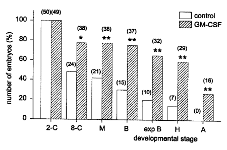
Figure 2: The effect of GM-CSF on the development of embryos to
blastocyst, hatching and attachment stages. Data is the number of
embryos developed to or beyond each stage, from experiments 1,2
and 3 combined, expressed as a percentage of the initial number of
cleaved (2-4 cell) embryos. 2-C = 2-cell embryos; 8-C = 8-cell
embryos; M = morulla; B = blastocyst; Exp B = expanded
blastocyst; H = hatching; A = attached with trophectoderm
outgrowth.
Figure 3: The effect of GM-CSF on the rate of development of embryos to
blastocyst. Data is the number of blastocysts at each time point,
from experiments 1,2 and 3 combined, expressed as a percentage of
the total number of blastocysts at 144 h post insemination.
Figure 4 RT-PCR analysis of GM-CSF receptor mRNA expression in human
blastocysts. Total cellular RNA was extracted from TF-1 cells and
each of two cohorts of blastocysts (B4)1 and B4 2), reverse
transcribed by random priming and amplified by PCR with GM-
Ra, GM-RP or actin-specific primers using conditions listed in
Table 7.
Figure 5 The effect of GM-CSF on the rate of development of embryos to the
blastocyst stage; (A) an early blastocyst (day 5, 112 h post-
insemination) from the control group; (B) an expanded blastocyst
(day 5, 112 h post-insemination) cultured in rhGM-CSF; (C) a fully
hatched blastocyst attached to the culture dish (day 6, 144 h post-
insemination); (D) an attached blastocyst cultured in rhGM-CSF
showing trophectoderm outgrowth (arrow; day 8, 200 h post-
insemination).
Figure 6 The effect of GM-CSF on the number of total cells (TCN), inner cell
mass (ICM) cells and trophectoderm (TE) cells in day 5 blastocysts
(120-124 h post-insemination). Values are mean SD of blastocysts
CA 02331890 2000-12-19
WO 99/67364 PCT/AU99/00499
9
cultured in 2 ng / ml rhGM-CSF (n=11) and blastocysts cultured in
media alone (n=10).
DETAILED DESCRIPTION OF THE INVENTION
EXAMPLE 1.
Measurement of embryonic viability and development
Materials and Methods
The embryos used in this study were donated by couples undergoing IVF
treatment at
Fertilitetscentrum AB, Goteborg, Sweden. Embryos frozen at the 2-4 cell stage
were
thawed at or beyond their one year storage limit in liquid nitrogen. Ethics
approval for the
study was obtained from the research ethics committee at University of
Goteborg (number
700-96).
Ovarian stimulation and in vitro fertilisation
Patients received 300 g buserelin gonadotrophin-releasing hormone agonist
(GnRHa;
Suprecur; Hoechst, Frankfurt, Germany) three times daily intranasally,
starting 1 week
before expected menses and lasting for two weeks. Down-regulation was
confirmed by a
serum estradiol content of <0.2nmol/l. Patients were then given recombinant
follicle
stimulating hormone (r-FSH; Gonal-F; Serono Laboratories, Aubonne,
Switzerland; 150-
225 IU / day sub-cutaneously). The starting dose was dependent on the patient
age and/or
previous response during ovarian stimulation (Wikland et al., 1994). The
ovarian response
was monitored by ultrasound and serum estradiol levels as previously described
(Bergh et
al., 1997). GnRHa and rFSH were administered until there was at least one
follicle >18
mm in mean diameter and two others >16 mm. Finally, oocyte maturation was
triggered
by one sub-cutaneous injection of 10 000 IU of hCG (Profasi; Serono
Laboratories).
Oocytes were retrieved 36-38h after hCG administration, assessed
morphologically and
fertilised in vitro. The embryos were cultured in IVF-50 (Scandinavian IVF
Science AB,
Goteborg, Sweden) and frozen on day 2 using a 3-step propanediol cryo-
preservation kit
(Freeze Kit 1, Scandinavian IVF Science) according to the manufacturers
instructions.
Recombinant GM-CSF
Recombinant human (rh)GM-CSF was obtained from R&D Systems Europe Ltd, Oxon,
UK. The biological activity of the recombinant cytokine preparations was
measured in a
bioassay employing a GM-CSF responsive cell line (human myeloid TF-1 cell
line),
essentially as described by (Kitamura et al. 1989). Duplicate serial 1:2
dilutions were
CA 02331890 2009-11-05
incubated with 2000 TF-1 cells in 200 ul of RPMI-1640 (Gibco) supplemented
with 10%
fetal calf serum (FCS; Commonwealth Serum Laboratories, Australia), 5 x 10⁻⁵ M
B-
mercaptoethanol and antibiotics. After 2 days, cultures were pulsed with 1 uCi
of 3H
thymidine (Amersham, Arlington Heights, IL) for 6 hours, harvested onto glass
fibre paper
5 using a Titretech (Trade Mark) automated cell harvester and radioactivity
measured in a
liquid scintillation beta counter.
Embryo thawing, allocation and culture
Frozen 2-4 cell embryos were thawed in four steps using a propanediol method
for embryo
10 thawing (Thaw Kit 1, Scandinavian IVF Science) following instructions given
by the
manufacturer. The viable embryos were classified and graded according to
criteria listed in
Table 1.
Table 1: Embryo classification criteria
Embryo Grade Morphology
A Regular blastomeres without fragments
B Regular or irregular blastomeres, up to 30% fragments
C Regular or irregular blastomeres, more than 30% fragments
D 50% of the blastomeres dead after thawing
To avoid bias the embryos were randomly allocated, with regard to patient and
embryo grade,
into the different culture groups (Table 2). The embryos were cultured in
groups of five
embryos per drop. To avoid the toxic effects of ammonium, released due to
metabolism and
breakdown of amino acids, the culture media was renewed every 48 h until
hatching occurred.
In two experiments the embryos were cultured in 20 l drops of IVF-50
(Scandinavian IVF
Science) containing 2 ng/ml rhGM-CSF (diluted 1:25 000 from stock material) or
carrier (2
ng/ml BSA, diluted 1:1000 from stock material). Culture drops were covered by
4 nil
Ovolil-200 (Trade Mark) (Scandinavian IVF Science) in Falcon 3004 (Trade Mark)
dishes
(Becton-Dickinson Labware, Franklin Lakes; NJ, USA). When blastocysts were
detected
these were transferred into 1 ml of S2 (Scandinavian IVF Science) in Falcon
3037 (Trade
Mark) dishes, containing 5% FCS and 2 ng/ml rhGM-CSF or carrier. Developmental
stage
was scored every 8h from thawing until 2300 h on day 8 (200 h post-
insemination).
In a third experiment the embryos were transferred from IVF-50 into S2 medium
(Scandinavian IVF Science) at the 6-8 cell stage. Additions of GM-CSF and
carrier were the
same as in the two previous experiments. When blastocysts were detected they
were
CA 02331890 2009-11-05
11
transferred to Falcon 3037 (Trade Mark) dishes, coated 24 h previously with
Biomatrix EHS
(Trade Mark) (Boehringer Ingelheim Bioproducts, Heidelberg, Germany).
Developmental
stage was scored every 8 h from thawing until 2300 h on day 8 (200 h post-
insemination).
Embryo scoring in each of the experiments was performed by the same person
(CS).
Statistical analysis was performed using Fisher's exact test and independent
samples t-test
(StatSoft, Inc.). Differences in data were considered significant when P<0.05.
Table 2: Distribution of grades amongst thawed 2-4 cell embryos
Embryo grade Control (%) GM-CSF (%)
A 20 20
B 22 27
C 32 33
D 26 20
N 50 49
Grades are defined in Table 1.
Results
The rate and extent of development of 2-4 cell embryos to the blastocyst and
hatching
blastocyst stages was significantly increased by the addition of rhGM-CSF to
culture medium
(Table 3).
Table 3: Rate and extent of embryo development in the presence or absence of
rhGM-CSF
n %BO T50 %H N % BO T50
Expt Control RhGM-CSF
1 16 38 122 50 15 60 121 89
2 16 38 116 50 16 81 98 100
3 18 17 127 33 18 83 105 53
Total 50 31 122 47 49 768 108b 78c
%B 0= % of viable thawed 2-4 cells reaching blastocyst stage. ap < 0.0001
T50 = number of hours post-insemination at which 50% blastocysts develop.
hp=0.0002 at 112h PI
%H = % of blastocysts which fully or partially hatch. cp = 0.009
A comparison between the proportion of embryos reaching blastocyst stage and
beyond in
experiment 1 and 2 (culture media containing 5% FCS from day 5) and experiment
3 (serum-
free media) are presented in Table 4. There are no significant differences
between
CA 02331890 2000-12-19
WO 99/67364 PCT/AU99/00499
12
the two groups, showing that the beneficial effect of GM-CSF is not dependent
on the
presence of FCS.
Table 4: Percent embryos developing up to or beyond each developmental stage
in
experiment 1 and 2 (FCS added) compared with experiment 3 (no FCS added).
N % B4 % Exp B(b % Hatching Attached
Control (exp 1+2) 32 37 28 19 0
GM-CSF (exp 1+2) 31 71 68 68 38
Control (exp 3) 18 17 6 6 0
GM-CSF (exp 3) 18 83 61 44 22
Bfi= blastocyst; Exp BO= expanded blastocyst
Although fewer poor quality embryos (grades C& D) reach blastocyst stage than
good
quality (grades A & B), GM-CSF exerted a comparable effect in all groups, with
similar or
slightly higher increases in the proportion of poor compared with good quality
embryos
achieving blastocyst stage (Fig. 1).
The majority of embryos grown in media alone were lost at the 4-16 cell stage.
The
beneficial effect of GM-CSF on blastocyst development appeared to result from
rescue of
this loss, with an 80% increase in the numbers of embryos reaching the morula
stage of
development (Fig. 2). Furthermore, the developmental potential of blastocysts
was
increased by culture in GM-CSF, since the rate of hatching was greater for
embryos
grown in GM-CSF. Similarly, blastocysts grown in GM-CSF (15/29), but not in
control
media (0/15), attached to the culture dish and showed trophoblast outgrowth
(Fig. 2 and
Fig. 5).
Finally, embryos cultured in the presence of rhGM-CSF had a significantly
higher rate of
development, with 50% blastocyst development achieved 14 hours earlier in GM-
CSF
compared with the control group (Fig. 3).
Conclusions
These results support the hypothesis that GM-CSF secreted into the female
reproductive
tract during early pregnancy promotes embryo growth and development. The
addition of
GM-CSF to culture media promotes the formation of blastocysts even with poor
post thaw
quality embryos. Our results also show a beneficial effect of GM-CSF on
blastocyst
expansion, hatching, attachment and trophectoderm outgrowth. Although the
functional
significance of hatching in vitro is unknown, blastocyst expansion is one of
the best
criteria for blastocyst viability and developmental potential.
CA 02331890 2009-11-05
13
The cleavage rate of embryos is suggested to be an indicator of embryo quality
(Shoukir et
at., 1997), and the rate of embryo development is known to be higher in vivo.
Importantly,
development of embryos to blastocysts was achieved significantly faster in the
presence of
rhGM-CSF.
EXAMPLE 2
Measurement of embryonic viability and development - variation of media and
source of
GM-CSF
Materials and Methods
The embryos used in this study were donated by couples, after ovarian
stimulation and in
vitro fertilisation, as described in Example 1. For culture experiments,
embryos frozen at the
2-4 cell stage were thawed at or beyond their one year storage limit in liquid
nitrogen. The
blastocysts used for the differential staining experiment were cultured from
excess embryos,
surplus to treatment and freezing requirements.
Recombinant GM-CSF
Two different commercial sources of recombinant human (rh)GM-CSF were used in
these
experiments. A laboratory grade preparation was obtained from R&D Systems
Europe Ltd,
Oxon, UK, and a pharmaceutical grade preparation, Molgramostim (Leucomax
(Trade Mark))
was obtained from Schering & Plough, Madison, NJ, USA. The biological activity
of both
recombinant cytokine preparations were measured in a bioassay employing a GM-
CSF
responsive cell line (human myeloid TF-1 cell line), as described in Example
1.
Embryo thawing. allocation and culture
Frozen 2-4 cell embryos were thawed and allocated randomly to experimental
groups as as
described in Example 1. Embryo culture was performed as described in Example
1, in two
different sequential media systems using two different commercial sources of
rhGM-CSF.
After thawing, the embryos were cultured first in G1.2 (Scandinavian IVF
Science) or IVF-
50. At 6-8 cell stage the embryos were transferred into G2.2 (Scandinavian IVF
Science) or
S2. The experiment included 6 groups: (a) G1.2/G2.2 alone, (b) G1.2/G2.2
containing 2
ng/ml rhGM-CSF (R&D Systems) (c) G1.2/G2.2 containing 2 ng/ml Molgramostim
(Schering & Plough; diluted 1:75 000 from stock material), (d) IVF-50/S2
alone, (e) IVF-
50/S2 containing 2 ng/ml rhGM-CSF (R&D Systems) (f) IVF-50/S2 containing 2
ng/ml
Molgramostim. Developmental rate was scored every eighth hour until expanded
blastocyst
stage. Blastocysts were scored on day 5 at 120 h post-insemination according
to criteria
described previously (Dokras et al., 1993). Briefly, grade A
CA 02331890 2009-11-05
14
blastocysts exhibited an expanded cavity with a distinct trophectoderm (TE)
and an
eccentrically located inner cell mass (ICM); grade B blastocysts were not yet
expanded but
otherwise morphologically identical to A; and grade C blastocysts exhibited
poor morphology
characterised by a number of degenerative foci in the ICM and TE and a poorly
developed
blastocoel cavity. Embryo scoring in each of the experiments was performed by
the same
person (CS).
Statistical analysis was performed using Fisher's exact test and independent
samples t-test
(StatSoft, Inc.). Differences in data were considered significant when P<0.05.
Differential labelling of blastocysts
Differential labelling was performed using a modification of a protocol
described previously
(Handyside and Hunter, 1984). Human blastocysts were cultured from excess
embryos,
surplus to treatment and freezing. On day 5 of culture (120 - 128 h post-
insemination) the
zona was removed in Acid Tyrodes solution containing 4 mg/ml PVP (360 000 Mw)
and
embryos were washed once in Gamete-100 (Scandinavian IVF Science) and three
times in
albumin-free S2 containing 4 mg/ml PVP (S2-PVP). The blastocysts were
incubated in
trinitro-benzene sulfonic acid (TNBS, Sigma Chemical Co., St Louis, MO, USA;
10 mM in
S2-PVP pH 8.5, 4 C / 20 min in the dark) and washed three times in Gamete-100
(Trade
Mark). TNBS-treated blastocysts were incubated in anti-dinitro-phenyl antibody
(anti-DNP;
Sigma, 0.2 mg/ml diluted in Gamete-100; 37 C / 30 min) Embryos were then
washed and
incubated in guinea pig complement serum (Sigma; diluted 1:10 in Gamete-100;
37 C / 30
min). Embryos were washed again and labelled with flourochromes (Sigma; 0.05
mM
bisbenzimide and 10 ug/ml propidium iodide in Gamete-100, 37 C / 30 min).
After extensive
washing embryos were fixed briefly in 1% paraformaldehyde and 0.5%
glutaraldehyde in
PBS, mounted under cover-slips in 20% glycerol in PBS and examined by
fluorescence
microscopy using a 400 nm exitation filter. Nuclei stained pink were scored as
lysed
trophectoderm cells (TE) and blue nuclei were scored as viable inner cell mass
cells (ICM).
Results
This experiment demonstrates the effect of culture media and source of
recombinant cytokine
on GM-CSF stimulated blastocyst development. Cytokine formulations in two
different
sequential culture media systems were found to have equivalent bioactivities
in the TF- 1 cell
proliferation assay (data not shown). There were no significant differences
between the
blastulation rates achieved in the two different culture media systems (Table
5). Both the rate
and extent of development of 2-4 cell embryos to blastocysts was significantly
increased by
the addition of 2 ng/ml rh GM-CSF. The effect was comparable
CA 02331890 2009-11-05
in extent in both G1.2/G2.2 and IVF-50/S2 sequential media combinations.
Furthermore, the
improvement in blastocyst development was achieved irrespective of the
formulation of
recombinant cytokine. The results also show that although culture in rhGM-CSF
gives rise to
more blastocysts, the distribution in morphological grade was comparable in
treatment and
control groups (Table 5).
Table 5: The effect of culture media and source of recombinant cytokine on GM-
CSF
stimulated blastocyst development.
N % BO A/B/C (%)
G1.2/G2.2 alone 23 30 57/29/14
G1.2/G2.2 + rhGM-CSF (R&D) 21 71** 67/20/13
G1.2/G2 + Moigramostim 19 63* 67/17/17
IVF-50/S2 alone 38 37 57/29/14
IVF-50/S2 + rhGM-CSF (R&D) 38 79*** 67/26/7
IVF-50/S2 + Molgramostim 20 65* 70/15/15
* P<0.05; ** P<0.01; *** P<0.001
The effect of culture in GM-CSF on blastomere number and allocation.
To investigate the effect of culture with GM-CSF on cell number and allocation
to inner cell
mass and trophectoderm cell lineage, blastocysts cultured with and without
rhGM-CSF were
analysed by immunosurgery and differential staining. Blastocysts cultured in
the presence of
rhGM-CSF had a significantly higher total cell number compared to blastocysts
cultured in
media alone (Fig. 6). An increase in the number of trophectoderm cells, and
particularly in
the number of inner cell mass cells, each contributed to the greater cell
number in GM-CSF
stimulated blastocysts.
EXAMPLE 3
IVF Media
The techniques and media used for embryo culture in IVF procedures have not
changed a
great deal since the 1980s. These procedures are set out most particularly in
Kerin et al
(1983), Trouson et al (1980), Trouson et al (1982), and Quinn et al (1985).
CA 02331890 2009-11-05
16
The media in which this invention might be used can be any media suitable for
use for the in
vitro support of embryo development and growth. These media might include but
are not
limited to HTF medium (Quinn, 1985a), Modified Whitten medium (Trounson,
1984),
whittinghams T6 medium (Trounson, 1984), Hams F 10 (Trounson et al,1982a),
Earles
solution (Edwards and Purdy, 1982), IVF50 (Scandinavian IVF Science), S2
(Scandinavian
IVF Science), G1.2 (Scandinavian IVF Science) and G2.2 (Scandinavian IVF
Science).
EXAMPLE 4
Method of IVF treatment
IVF procedures have not changed a great deal since the 1980s. The procedures
for IVF
treatment used in this invention are standard procedures that are set out most
particularly in
Kerin et al (1983), Trouson et al (1980), Trouson et al (1982), and Quinn et
al (1985) which
references are incorporated herein by references in relation to those
procedures.
EXAMPLE 5
The expression of GM-CSF receptors by human pre-implantation embryos in vitro
Material and Methods
The embryos used in this study were donated by couples, after ovarian
stimulation and in
vitro fertilisation, as described in Example 1. Excess human 2-4 cell embryos
surplus to
patients' requirements were cultured in 20 ml droplets of IVF-50 overlayed
with paraffin oil.
On day 3 (72 h post insemination) the embryos were transferred to S2. Embryos
were
collected at blastocyst stage of development. The embryos were washed in PBS,
snap frozen
in liquid nitrogen and stored at -70 C prior to RNA extraction.
Total cellular RNA was extracted from human GM-CSF responsive myeloid cells
(TF-1 cell
line), and from two cohorts each of twenty blastocysts using a method
described by (Arcellana-
Panlilio & Schultz,1993). Residual chromosomal DNA was removed by treatment
with RNAse-
free DNAse (Boehringer Mannheim) for 60 min at 37 C. First strand cDNA
synthesis was
achieved by reverse transcription (RT) of RNA primed with random hexamers
using a
Superscript (Trade Mark) RNase H-reverse transcriptase kit (Gibco) essentially
according to the
manufacturer's instructions. Detection of mRNA by RT-PCR was performed using
primer pairs
specific for the a-chain and 0-chain of the GM-CSF receptor (GM-Ra and GM-
R(3), and P-actin
(detailed in Table 7) and reagents supplied in
CA 02331890 2009-11-05
17
a Taq DNA polymerase kit (Biotech International Ltd., Perth), essentially as
described
previously. The number of cycles and annealing temperature used for each
primer pair are also
given in Table 7. To increase the sensitivity of the GM-R 0 PCR, a nested
primer design was
employed, wherein cDNA was amplified by 30 cycles with GM-Rb 'external'
primers followed
by 25 cycles with GM-RD 'internal' primers. Each PCR product was analysed by
electrophoresis
through a 2% agarose gel containing EtBr, and visualised by trans-illumination
with UV-light.
Gels were photographed and the size of the PCR products was verified by
comparison of their
relative mobility to molecular weight markers.
Table 7. Primer sequences
Target Genebank Primer sequence Position in Cycle number/ Product
accession sequence annealing temp size
number
P-actin M12481 5' tgtgatggtgggtatgggtc 48-67 35 / 62 C 372 bp
3' tagatgggcacagtgtgggt 400-419
GM-Ra M64445 5'catgcttctcctggtgacaa 162-181 40 / 60 C 279 by
3' gtgactccttcatgcagaca 421-440
GM-R(3 M59941 / external: 142-161 30 / 65 C 428 bp
M38275 5' ctacaccagccacatcacct 550-569
3' agtcctgaagccgcttgtag 239-258 25 / 65 C 230 by
internal: 449-468
5' gagccagtgtcctgtgacct
3' tggtcctggtcggtgctgat
Results
The expression of GM-CSF receptor expression by in vitro generated blastocysts
was
examined by RT-PCR. Each of two preparations of blastocysts were found to
express mRNA
for the a-chain of the GM-CSF receptor, but mRNA for the 0-chain was not
detected, even
when a highly sensitive nested PCR protocol was used (Fig. 4).
Conclusions
Expression of GM-CSF receptor a-chain mRNA was detected in each of two
blastocyst
cDNA preparations. These results indicate that human blastocysts have the
molecular
capacity to bind and respond to GM-CSF. The expression of the a-subunit in the
absence of
the 0-chain may benefit blastocyst glucose transport and thus optimise the
culture
environment. Increased glucose uptake is likely to promote blastomere
metabolic activity,
and hence cell division, and may also prolong cell survival through the
prevention of
apoptosis.
CA 02331890 2000-12-19
WO 99/67364 PCT/AU99/00499
18
REFERENCES
Arcellana-Panlilio & Schultz, 1993. Methods Enzymol. 225; 303-28.
Armstrong & Chaouat , 1989. Biol Reprod 40; 466-474
de Moraes & Hansen, 1997, Biol Reprod. 57; 1060-1065
Ding et al., 1994 Proc Natl Acad Sci USA. 91(7); 2537-41
Drake & Head, 1994, J Reprod Immunol 26; 41-56
Dunglison et al., 1996, Hum Reprod. 11; 191 196
Edwards and Purdy, 1982, (eds) 1982 Human conception in vitro Academic Press,
London
Giacomini et al., 1995. Hum-Reprod. 10; 3259-63
Hill et al., 1987. J Immunol 139; 2250-2254
Jokhi et al., 1994, 26; 147-164
Kerin et al, 1983, Clin Reprod. Fertil, 2; 129-142
Lea & Clark, 1993. Biol Reprod 48;930-953
Lenton et al., 1988, Ann NYAcad Sci, 541; 498-509
Loke et al., 1992, J Reprod Immunol, 22; 33 - 45
Menezo et al., 1990, Biol Reprod, 40; 301-306
Menezo et al., 1992, Hum Reprod, 7; 101-106
Oliveness et al., 1955, Hum Reprod, 9; 2367 - 2373
Plachot et al., 1955 In Aburumieh et al (eds) IXth World Congress on In Vitro
Fertilisation and Assisted Reproduction Monduzzi Editore, Bologna p 37
Quinn et al 1985, In Annals of N.Y. Acad. Sci. 442; 195-204.
Quinn et al 1985a, Fertil. amd Steril. 44; 493-498
Robertson et al., 1991, pp191-206 in Molecular and Cellular Immunobiology of
the
Maternal Fetal Interface, Wegmann et al eds Oxford University Press )
Robertson et al., 1992. Biol Reprod 46; 1069-79.
Robertson et al., 1996 Biol Reprod 54; 183-196.
Robertson et al., 1998 The effect of GM-CSF deficiency on early embryonic
development
in mice. Proceedings of the 29th Annual Conference of the Australian Society
for
Reproductive Biology.Shoukir et al., 1997.Hum. Reprod. 7: 1531-1536
Robertson et al., 1999 Biol Reprod 60; 251-261.
Tartakovsky & Ben-Yair, 1991. Dev Biol 146; 345-352
Trouson et al, 1980, Fertil. Steril. 34; 431-438
Trouson et al, 1982, J reprod. Fertil. 64; 285-294
Trounson et al 1982a) In: Edward and Prudy (eds) Human conception in vitro.
Academic
Press, London, p201-205
CA 02331890 2000-12-19
WO 99/67364 PCT/AU99/00499
19
Trounson 1984. in Invitro Fertilization and Embryo Transfer, Churchill
Livingstone,
(Trounson & Wood eds) pp 111 - 130
Weinberg et al., 1988, Fert Steril, 50; 993 - 5
Zhao & Chegini, 1994. J Clin Endocrinol Metab. 2; 662-5.