Note: Descriptions are shown in the official language in which they were submitted.
CA 02332775 2000-11-14
WO 99/61901 PCT/US99/11275
ELECTROPHORESIS APPARATUS AND METHOD
Field of the Invention
The present invention relates to methods and apparatus for electrophoretically
separating and characterizing sample analytes. In particular, the invention
relates to
multidimensional electrophoresis.
References
Alfonso, E.S., et al., J. Chromatogr. 689:85-96 (1995).
Anderson, N.L., et al., Electrophoresis 12:907-930 (1991).
Andrews in Electrophoresis. Theory. Techniques and Biochemical and Clinical
Anylications, Oxford U. Press, Oxford, p. 276 (1986).
Bjellqvist et al., Biochem. Biophys. Meth. 6:317-339 (1982).
Busch, M.H.A., et al., J. Chromatogr. 695:287-296 (1995).
Bushey et al., J. Chrom. 480:301 (1989).
Capelli, L., et al., J. Biochem. Biophys. Meth. 32:109 (1996).
Chen et al., J. Lig. Chrom. 15:1143 (1992).
Fung, E.N., et al., Anal. Chem. 67:1913-1919 (1995).
Ganzler, K., et al., Anal. Chem. 64:2665-2671 (1992).
Gilges, M., et al., Anal. Chem. 66:2038-2046 (1994).
Lauch., T., et al., J. Chromatoar. 680:375-381 (1994).
Li in Capillarv Electrophoresis. Principles, Practice, and Aolications,
Elsevier,
Amsterdam, p. 160 (1992).
Liao, J.L., et al., J. Cap. Elec. 2:191 (1995).
Molteni, S., et al., Electrophoresis 15:22-30 (1994).
Ng, C.L., et al., J. Chromatocr. 659:427-434 (1994).
Peterson et al., EP Publ. No. 494686 Al (1992).
Righetti, P.G., Immobilized pH Gradients: Theory and Methodology, Elsevier,
Amsterdam (1990).
I
CA 02332775 2000-11-14
WO 99/61901 PCT/US99/11275
Righetti, P.G., et al., in Gel Electronhoresis of Proteins, B.D. Hames and D.
Rickwood, Eds., IRL Press, Oxford, p.149 (1996).
Sandoval and Pesek, U.S. Pat. 5,017,540 (1991).
Sandoval and Pesek, U.S. Pat. 5,326,738 (1994).
Schans, M.J., et al., J. Chromatogr. 680:511-516 (1994).
Sun, P., et al., J. Microcol Sen. 6:403-407 (1994).
Wang, T., et al., J. Chromatogr. 594:325-334 (1992).
Werner, W.E., et al., Anal. Biochem. 212:253-258 (1993).
Wiktorowicz, J., U.S. Pat. Nos. 5,015,350 (1990) and 5,181,999 (1991).
Wilkens, M.R., et al., Eds., Proteome Research: New Frontiers in Functional
Genomics, Springer Verlag, Berlin, Germany (1997).
Wong, S.S., Chemistry of Protein Conjugation and Cross-Linking, CRC Press,
Boca
Raton, FL (1991).
Zhu et al., J. Chrom. 516:123 (1990).
Background of the Invention
For decades, electrophoretic separation methods have been central to
identifying and
characterizing chemical and biochemical samples. In the usual procedure, an
electrophoresis
tube or slab is filled with a fluid electrophoresis medium, and the fluid
medium is covalently
cross-linked or temperature-solidified to form a non-flowable, stabilized gel
separation
medium. A sample is loaded into one end of the tube, or into one or more wells
of the slab
gel, and an electric field is generated to draw the samples through the
medium. Electro-
phoretic separation may depend predominantly on molecular size, e.g., in the
cases of
nucleic acids and SDS-bathed proteins, or on a combination of size and charge,
as in the case
of non-denaturing gel electrophoresis of polypeptides or polysaccharides, for
example.
Isoelectric focusing (IEF) is an electrophoresis method based on the migration
of a
molecular species in a pH gradient to its isoelectric point (pI). The pH
gradient is
established by subjecting an ampholyte solution containing a large number of
different-pI
species to an electric field, usually in a crosslinked matrix. Analytes added
to the
equilibrated ampholyte-containing medium will migrate to their isoelectric
points along the
pH gradient.
2
CA 02332775 2000-11-14
WO 99/61901 PCT/US99/11275
For complex samples, multidimensional electrophoresis methods have been
employed
to better separate species that comigrate when only a single electrophoresis
dimension is
used. The conventional approach to two dimensional electrophoresis is to
perform the first
dimension in a rigid, usually crosslinked matrix. For analysis of proteins,
for example, the
sample is usually fractionated first by IEF in a tube or strip gel to exploit
the unique
dependence of each protein's net charge on pH. Next, the gel containing the
separated
proteins is extruded from the tube, dried (these two steps can be bypassed
using a strip gel)
and laid horizontally along one edge of a slab gel, typically a crosslinked
polyacrylamide gel
containing sodium dodecylsulfate (SDS). Electrophoresis is then performed in
the second
dimension, perpendicular to the first, and the proteins separate on the basis
of molecular
weight. Thus, proteins having similar net charges, and which are not separated
well in the
first (IEF) dimension, will separate according to their different masses in
the second
dimension. Since these two separation methods depend on independent properties
(net
charge and mass), the overall resolution is approximately the product of the
resolution in
each dimension.
A significant drawback of traditional methods for two-dimensional
electrophoresis is
that two separate devices are used to accomplish electrophoresis in the two
dimensions.
These protocols can be very time-consuming and cumbersome to practice.
Moreover,
traditional methods are susceptible to significant run-to-run variation
because of variability in
standard IEF and SDS gels, which cannot be re-used.
Accordingly, there is a need for a new multidimensional electrophoresis method
that
is faster and easier to use, which allows the identification and
characterization of hundreds or
thousands of components in complex mixtures, and which is highly reproducible.
Ideally,
the method will employ a single separation apparatus for electrophoresis in
both dimensions.
The method preferably involves a flowable (liquid-state) separation medium
that can be
easily replaced with fresh media, so that a single apparatus can be used
repetitively for
multiple samples. Ideally, the apparatus is adaptable for automation.
3
CA 02332775 2000-11-14
WO 99/61901 PCT/US99/11275
Summary of the Invention
The present invention is directed to methods and apparatus for conducting
multidimensional electrophoresis of samples within a single apparatus, such
that sample
components that have been resolved in a first electrophoretic dimension can be
directly
electrophoresed in a second dimension that is substantially perpendicular to
the first, without
needing to move or manipulate the sample between the first and second
electrophoretic steps.
In one aspect, the invention includes a two-dimensional electrophoresis
system. In
one embodiment, the system includes an electrophoresis plate assembly that
defmes (i) a
sample separation cavity, (ii) a sample loading port positioned at a corner of
the upper
portion, for introducing a sample into the electrophorusis region, and (iii)
optionally, one or
more fluid passageways positioned along the lower portion of the cavity, for
introducing or
removing liquid from the cavity.
The cavity defmed by the assembly is bounded by opposing major first and
second
surfaces, each having a defmed width and length. These major surfaces are
spaced apart by
an interfacial distance substantially shorter than the width and length of the
cavity. The
cavity further comprises (1) a first electrophoresis region located along the
upper portion of
the cavity for performing charge and/or size-based electrophoresis in a first
dimension along
said upper portion, and (2) below the first electrophoresis region, a second
electrophoresis
region for performing electrophoresis in a second dimension in a direction
substantially
perpendicular to the first dimension, such that the basis of migration of
sample components
in the second dimension depends on sample properties that are different from
the sample
properties that determine the basis of migration in the first dimension.
In one embodiment, the second electrophoresis region is an isoelectric
focusing
region containing a pKa gradient immobilized on at least one of the major
opposing surfaces,
for isoelectric focusing in a direction substantially perpendicular to the
first dimension. The
pKa gradient may span any attainable range, such as a pKa range of about 4 to
10, or 4 to 6,
for example.
In another embodiment, the second electrophoresis region does not contain a
pKa
gradient and is used to perform charge and/or size-based electrophoresis under
condition.s
different from the electrophoretic conditions used for the first dimension.
The system may further include electrode means for generating a first voltage
potential across the first electrophoresis region between the loading port and
the lateral
4
CA 02332775 2000-11-14
WO 99/61901 PCT/US99/11275
portion across from the loading port, electrode means for generating a second
voltage
potential between the upper portion and lower portion, and an aqueous medium
occupying
the cavity.
In one embodiment, the loading port is joined to the upper corner portion of
the
cavity by an elongate sample transport channel.
In another embodiment, the one or more fluid passageways are provided as a
single
slot which passes through one of major surfaces near the lower portion. In an
alternative
configuration, the one or more fluid passageways comprise a plurality of
openings positioned
along the lower portion of the cavity.
In another embodiment, the second electrophoresis region contains a plurality
of
elongate separation channels, which are substantially parallel to the lateral
portions of the
cavity and prevent flow of the medium between adjacent separation channels.
The second
electrophoresis region preferably contains at least 30 separation channels,
and preferably
more than 100 separation channels.
In another embodiment, the plates define a fan-shaped region that abuts the
upper
portion or the lower portionof the separation cavity, for introducing or
removing liquid from
the cavity.
In another embodiment, the upper edge of the first electrophoresis region
and/or the
lower edge of the second electrophoresis region is bounded by an ion-permeable
membrane,
which can be used to separate the separation cavity from an electrode
reservoir.
In another aspect, the invention includes an electrophoresis plate assembly,
such as
described above.
In another aspect, the invention includes a method for separating one or more
components of a sample mixture, using an electrophoresis plate assembly or
system such as
those described above. In the method, a sample mixture is applied to the
sample loading
port. A first voltage potential is applied across the first electrophoresis
region under
conditions effective to cause different components in the mixture to migrate
towards the
opposite portion of the electrophoresis region, such that different components
become
separated at least partially on the basis of size. After electrophoresis in
this first dimension,
a second voltage potential is applied across the upper and lower portions of
the cavity, in a
direction substantially perpendicular to the first dimension, under conditions
effective to
separate different components on the basis of physical properties (e.g.,
charge and/or size
5
CA 02332775 2000-11-14
WO 99/61901 PCT/US99/11275
features) that are different from those used in the first dimension. In one
embodiment, the
second electrophoresis region is an isoelectric focusing region, and the
electrophoretic
conditions are effective to generate a pH gradient in the isoelectric focusing
region in a direc-
tion substantially perpendicular to the direction of the first voltage
potential. Sample
components migrate into the isoelectric focusing region and separate on the
basis of their
isoelectric points.
In a second embodiment, the second electrophoresis region does not contain a
pKa
gradient, and the second electrophoretic conditions are effective to perform
electrophoresis
under conditions different from the electrophoretic conditions used for the
first dimension.
After electrophoresis in the second dimension is complete, sample components
can be
detected and imaged to obtain information about the composition of the sample.
To facilitate detection, sample components preferably contain a detectable
label, such
as a fluorescent label or radiolabel. In a preferred embodiment, the sample
comprises one or
more polypeptides to be detected.
In one embodiment, after electrophoresis in the second dimension is complete,
one or
more sample components can be collected from the separation cavity for further
characterization. Preferably, the second electrophoresis region includes a
plurality of
elongate separation channels as above, to facilitate collection of sample
components.
In another embodiment, the invention includes an electrophoresis plate,
channel, or
tube having a surface which is coated with a pKa gradient immobilized thereon.
Preferably,
the gradient spans at least one pH unit. The tube or channel preferably has a
diameter the is
200 m or less, preferably 100 m or less, or 50 m or less.
These and other features and advantages of the present invention will become
more
clear from the following detailed description together with the appended
drawings.
Brief Description of the Drawings
Fig. 1 shows an overhead view of a plate assembly in accordance with the
invention;
Fig. 2 shows a perspective view of the assembly of Fig. 1;
Fig. 3 illustrates another exemplary plate assembly in accordance with the
invention,
which additionally includes a liquid loading region, a plurality of separation
channels, and an
elongate sample transport channel;
Fig. 4 shows a perspective view of the assembly of Fig. 3;
6
CA 02332775 2000-11-14
WO 99/61901 PCT/US99/11275
Fig. 5 shows a cross-section of the channel region of the assembly from Figs.
3 and
4;
Figs. 6-9 show exemplary modifications of the triangular region of the device
from
Fig. 3;
Figs. 10 and 11 show overhead and perspective views of another exemplary plate
apparatus in accordance with the invention;
Fig. 12 shows an enlarged view of sample loading channels of the apparatus
from
Figs. 10-11;
Fig. 13 illustrates a modification of the apparatus of Figs. 10-12;
Fig. 14 illustrates an enlarged view of an electrode reservoir from Fig. 13.
Detailed Description of the Invention
As noted above, the present invention is directed to methods and apparatus for
conducting multidimensional electrophoretic separations of sample mixtures
within a single
separation cavity, such that the separation conditions in the first dimension
are different from
the separation conditions in the second dimension.
The invention is adaptable to a variety of separation conditions, including
conditions
for (1) isoelectric focusing, and (2) denaturing or non-denaturing size-based
separations in
flowable sieving media. Moreover, since electrophoresis can be accomplished in
both
d'unensions with flowable (i.e., liquid) media, the medium can be replenished
after each
sample separation without having to separate the plates.
I. Annaratus
In one aspect, the invention provides apparatus for conducting two-dimensional
electrophoresis of selected analytes, particularly polypeptides. In general,
the apparatus
includes a plate assembly that defines a cavity bounded by opposing major
first and second
surfaces, each having a defmed width and length. These major surfaces are
spaced apart by
an interfacial distance substantially shorter than the width and length of the
cavity. The
cavity further comprises (1) a first electrophoresis region located along the
upper portion of
the cavity for performing charge and/or size-based electrophoresis in a first
dimension along
said upper portion, and (2) below the first electrophoresis region, a second
electrophoresis
region for performing electrophoresis in a second dimension in a direction
substantially
7
CA 02332775 2000-11-14
WO 99/61901 PCT/US99/11275
perpendicular to the first dimension, such that the basis of migration of
sample components
in the second dimension depends on sample properties (e.g., molecular weight,
molecular
shape, hydrophobicity and/or hydrophilicity, and/or charge) that are different
from the
sample properties that determine the basis of migration in the first
dimension.
The following discussion is directed to a first embodiment of the invention
wherein
electrophoresis in the first dimension is performed on the basis of size and
charge, resulting
in different migration rates, and electrophoresis in the second dimension is
performed on the
basis of isoelectric point. Other, alternative embodiments are discussed
subsequently.
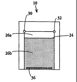
Figs. 1 and 2 show an overhead view and perspective view, respectively, of an
electrophoresis plate assembly 10 in accordance with the present invention. A
pair of plates
20,22 are disposed such that inner plate surfaces 20a and 22a are juxtaposed
face-to face to
form an enclosed separation cavity 24, for holding a separation medium through
which the
sample is electrophoresed.
Plate 20, which is also referred to arbitrarily as the bottom plate, defines a
recessed
region 26 which defmes five of the six walls of cavity 24. As seen with
reference to Fig. 2,
region 26 further includes a first sample separation surface, designated
surface 26a, for
electrophoresis along the lateral dimension of this region, and a second
sample separation
surface 26b, for electrophoresis in a direction perpendicular to the first-
mentioned
dimension.
For an IEF embodiment of the invention, surface 26b is further characterized
by the
presence of a plurality of buffering moieties, or immobilines, which defme a
pKa gradient
leading away from region 26a. The properties of region 26b are discussed
further below.
Plate 22, which is also referred to arbitrarily as the cover plate, includes
an inner
surface 22a which is substantially flat, for providing the sixth wall of
separation cavity 24.
As seen particularly with reference to Fig. 2, plate 22 defmes, in its upper
left-hand corner,
a sample loading port 30, at or through which sample is introduced into the
separation
cavity, and which also provides access to the separation cavity for a first
electrode 30a (not
shown) for establishing a voltage potential at that site. Electrodes can be
made of any
appropriate conductive material, such as platinum, nichrome, or gold, etc.,
with platinum
being preferred. An electrode port 32 is defmed in the upper right-hand corner
of plate 22,
for providing a second electrode 32a (not shown) in electrical contact with
the separation
cavity.
8
CA 02332775 2000-11-14
WO 99/61901 PCT/US99/11275
The apparatus may include an optional third electrode 34a (not shown), which
is
electrically separate from the first and second electrodes mentioned above,
and which
extends laterally across the top of cavity 24. This third electrode can be
admitted into the
separation cavity through port 30 or 32, and can be immobilized in the cavity
by being
affixed, for example, to the bottom plate along the upper edge of recessed
region 26.
Plate 22 also contains an elongate slot 36 which extends laterally along the
bottom of
the plate, for providing (i) an electrode 36a (not shown) extending along the
bottom of the
separation cavity, and (ii) a passageway for ingress and egress of separation
media and wash
solutions into and out of the separation cavity before or after
electrophoresis.
Preferably, the surfaces 26a and 26b of recessed region 26, and the inner
surface 22a
of cover plate 22, are substantially planar, to facilitate the creation of
undistorted electric
field lines and enhance sample separation during electrophoresis.
The inner surface 22a of the cover plate preferably includes a surface region
40b
whose dimensions are congruent with and opposite to surface region 26b in
plate 20, for
generating an isoelectric focusing pH gradient in the separation medium
located in that
region. Like region 26b, for isoelectric focusing, region 40b is preferably
coated with a
plurality of buffering moieties, or immobilines, which define a pKa gradient
leading away
from the region 26a.
Plates 20,22 can be formed of any material suitable for electrophoresis of the
selected
sample. Preferably, at least one of the plates is formed of a material that is
transparent with
respect to a type of signal that is used to visualize or locate sample
components in the
separation cavity. Typically, the plates are formed out of a silicon dioxide-
based glass, such
as borosilicate, although other materials such as plastics, such as
polycarbonate, or metals
rendered non-conductive by a suitable coating, are also contemplated. Visually
opaque
materials such as TEFLONT"' and MYLART', for example, can also be used, e.g.,
with
radioactive sample detection.
In a preferred embodiment, both plates are made from borosilicate glass. Glass
is
advantageous because it is transparent to a broad range of wavelengths, e.g.,
for
fluorescence detection and visual inspection, it can be cut easily, and fme
features on the
order of 1 to 100 m or greater can be formed in glass by standard
photolithographic etching
techniques. For example, a recess region 26 having a substantially planer
surface with a
9
CA 02332775 2000-11-14
WO 99/61901 PCT/US99/11275
depth of 100 m can be formed readily by conventional hydrofluoric acid
etching.
Similarly, ports 30,32 in cover plate 22 can be formed by mechanical drilling,
for example.
In the assembled apparatus (plate assembly), plates 20 and 22 are joined
together by
any suitable means that are sufficient to ensure a liquid-tight seal with
respect to separation
cavity 24. For example, glass plates can be fusion-welded together using
methods known in
the art, i.e., by holding the opposing faces of the plates together at an
elevated temperature
that is below the softening point of the plates, such that the inner surfaces
20a and 22a of the
plates become bonded together. Alternatively, the plates can be joined
together by anodic
bonding, or simply by using one or more clamps along the edges of the plates.
Figs. 3 and 4 illustrate another exemplary embodiment of an apparatus (100) of
the
invention which additionally includes a liquid loading region 160, a plurality
of separation
channels 170, and an elongate sample transport channel 180.
In this example, bottom plate 120 (Fig. 4) contains a recessed region 126
which
defines five of the six walls of separation cavity 124. Region 126 includes a
first sample
separation surface 126a which defmes a first electrophoresis region 126a, for
electrophoresis
along the lateral dimension of this region, and a second sample separation
surface 126b
which defmes a second electrohporesis region for electrophoresis in a second
direction, such
as for isoelectric focusing. Edge region 120a surrounds region 126.
Region 126 also encompasses a fan-shaped (triangular) liquid loading region
160 at
the upper end of the plate, for conveniently introducing and removing
separation media to
and from the separation cavity before and after electrophoresis. Region 160 is
also useful
for forming a pKa gradient coating on the inner surfaces of the plates for
isoelectric
focusing, as detailed below.
With continued reference to Figs. 3 and 4, surface 126b comprises a plurality
of
parallel separation channels 170 that are aligned in a direction perpendicular
to the bottom
edge of the plate. For an IEF embodiment of the invention, the channels are
each coated
with a plurality of buffering moieties, forming a pKa gradient in the vertical
direction with
respect to the top and bottom edges of the plates. Each channel 170 is
separated by a
partition 172 (Fig. 5) having a height that is flush with inner surface 120a
of plate 120. This
is preferred in order to form liquid-tight seals between the channels when
plates 120 and 122
are assembled together. The depths of the channels are preferably the same,
and are also
preferably the same as the depths of regions/surfaces 126a and 160.
CA 02332775 2000-11-14
WO 99/61901 PCT/US99/11275
The number of channels 170, and their dimensions, will vary depending on
sample
complexity and the desired resolution. Generally, sample resolution will
increase as the
number of channels is increased, subject to the limit of resolution achieved
by
electrophoresis in the first, lateral, dimension. Preferably, the channels are
dimensioned so
that the channel resolution is at least twice the sample resolution in the
lateral dimension of
the separation medium, so that each sample band partitions into from one to
three channels.
Plate 120 also contains an elongate sample transport channel 180 extending
from the
lower left hand area of the plate to the upper left hand comer of surface
126a, for size-based
electrophoretic separation of the sample during transit to separation cavity
124. Channel 180
preferably has a depth in plate 120 equal to the depth of recess 126. The
width of channel
180 is preferably no more than about 10 times the channel's depth, and is
preferably equal to
or less than about 5 times the channel depth.
Plate 122, which is also referred to arbitrarily as the cover plate, includes
an inner
surface 122a which is substantially flat, for providing the sixth wall of
separation cavity 124.
As seen in Fig. 3, and particularly with reference to Fig. 4, plate 122
defmes, in its lower
left-hand corner, a sample loading port 130, at or through which sample is
introduced into
the separation cavity, and which also provides access to the separation cavity
for a first
electrode 130a (not shown) for establishing a voltage potential at that site.
An electrode port
132 is defined in the upper right-hand comer of plate 122, for providing a
second electrode
132a (not shown) in electrical contact with the separation cavity. The first
and second
electrodes are useful for perfomzing electrophoresis of the sample along a
first dimension
stretching from port 130 to port 132, to generate a series of separated sample
components
along the upper edge of 126a after the first electrophoresis step is complete.
Plate 122 also
contains an elongate slot 140 which extends laterally along the bottom of the
plate, like slot
40 mentioned above.
Port 135 in plate 122 is included for transporting separation media and wash
fluids
into and out of the separation cavity, and also for forming the IEF coating
gradient.
To facilitate sample loading, port 130 can be accompanied by a waste port 131
defined in plate 122, such that both ports are in fluid communication with
elongate sample
transport channel 180. This allows a precise amount of sample to be injected
into channel
180, as discussed further below.
11
CA 02332775 2000-11-14
WO 99/61901 PCT/US99/11275
Returning to Fig. 4, plates 120 and 122 further define channel egress ports
136 and
138, which are located slightly above slot 140, for collecting resolved sample
components
individually from one or more channels after the IEF step is complete. In the
embodiment
illustrated in the Figure, each plate contains an egress port for alternating
channels, so that
ports 136 and 138 are staggered relative to each other. The staggering of
egress ports, while
not required, facilitates the connection of capillary tubes to the ports for
collecting fluids
from each channel.
Figs. 6-9 illustrate additional embodiments that may be utilized in the
devices of the
present invention, particularly for maintaining a relatively uniform
electrical field during
electrophoresis in the first dimension. Fig. 6 shows a modification in which
triangular liquid
loading region 160 from Fig. 3 can be modified to include a vertical barrier
150 that extends
from beneath port 135 to the top of surface 126a. A benefit of barrier 150 is
that during the
first dimension of electrophoresis, the electric field lines are constrained
to the region
bounded by surface 26a, so that band distortion may be reduced.
Fig. 7 shows a modification in which one of plates 120 and 122 contains an
additional port 152 which is closable with a suitable plug (not shown). After
regions 126a
and 126b have been filled with a desired medium or media, a low conductance
medium that
preferably has an ionic strength at least 5 times, and more preferably at
least 10 times lower
than that of the surrounding medium, is introduced into triangular region 160
through port
152 with egress through port 135, so that a vertical "wall" or region of low
conductance
medium is created in region 160 between ports 152 and 135. After loading of
the low
conductance buffer, ports 135 and 152 are closed, and electrophoresis is
performed as
described herein. Field lines in the first dimension of electrophoresis are
thus constrained to
region 126a.
In Fig. 8, the first electrophoresis region includes a trench 154 extending
across the
lateral dimension of region 126 and which provides a deeper cross-section for
this region
relative to region 126b (e.g., twice as deep as the region 126b). After
regions 126a and
126b have been filled with a desired medium or media, region 126a (trench 154)
is
preferably filled with a high conductance/high viscosity medium (e.g., due to
the presence of
a selected polymer) via port 130 or 131 and port 132, to help constrain
electrical field lines
to region 126a during electrophoresis in the first dimension. By providing a
greater cross-
12
CA 02332775 2000-11-14
WO 99/61901 PCT/US99/11275
section than the cross-sections of adjacent regions 160 and 126b, trench 154
also helps limit
mixing between regions 126a and 126b.
Fig. 9 shows another modification in which plate 122 additionally includes a
horizontal slot 156 located just above region 126a. Initially, slot 156 is
filled with a plug
(not shown) which closes the slot and has an inner surface that is flush with
the inner surface
of plate 122. After the inner surfaces of regions 126a and 126b have been
prepared as
described herein, and after the chamber has been filled with the desired
electrophoresis
medium or media, the plug is pressed further through the slot until the end of
the plug
snuggly contacts surface 126a of plate 120, thereby creating a horizontal
barrier across the
top of region 126a which constrains electric field lines during the first
dimension of
electrophoresis.
Figs. 10-12 illustrate another plate configuration in which the triangular
region from
Fig. 3 is moved to the other end of the device, adjacent the edge of the
second
electrophoresis region. Apparatus 200 includes a bottom plate 220 and top
plate 222.
Bottom plate 220 includes a first sample separation surface 226a, defming a
first
electrophoresis region 226a, for electrophoresis along the lateral dimension
of this region, and
a second sample separation surface 226b, for electrophoresis in a direction
perpendicular to the
first dimension. Plate 220 further defines a lateral channel 232a for
providing fluid
communication with port 232 in plate 222.
Plate 220 also contains an elongate sample transport channe1280 extending
along the
left edge of region 226b. Channel 280 terminates with a peripheral channe1231a
which links
port 230a to channe1280. Peripheral channels 231b, 231c, and 231d meet at
junction 231e,
and are linked in fluid communication with channel 280 via channe1231 f.
Channels 231 b,
231c, and 231d place ports 230b, 230c, and 230d in fluid conununication with
channe1280.
The operation of these channels is discussed further below. Also, it will be
appreciated that
any other suitable channel arrangement can be used in the apparatus to
facilitate loading of
sample.
Optionally, surface 226b comprises a plurality of parallel separation channels
270 that
are aligned in a direction perpendicular to the bottom edge of the plate, and
which are
separated by partitions 272 as with apparatus 100 discussed above. At the end
of those
channels, plate 220 further a fan-shaped (triangular) liquid loading region
defined by surface
260a, for conveniently introducing and removing liquids into and from the
separation cavity.
Triangular region 260a is bordered at its lateral edges by channels 233a and
233b, which
13
CA 02332775 2000-11-14
WO 99/61901 PCTIUS99/11275
provide fluid communication with ports 234a and 234b, respectively, in plate
222. Port 235 in
plate 222 provides a convenient site for for transporting separation media and
wash fluids into
and out of the separation cavity, and also for forming an IEF coating gradient
in region 226b.
Plates 220 and 222 further define pluralities of alternating channel egress
ports 236
and 238, for collecting resolved sample components individually from one or
more of
channels 270, as with apparatus 100 above.
The various ports, particularly ports 230a, 230b, 230c, 230d, 232, 234a, and
234b
may also be provided with electrodes in order to control movement of sample
(ports 230a-d),
for electrophoresis in the first dimension (ports 230a and 232), and for
electrophoresis in the
second dimension (ports 230a, 232, 234a and 234b),
The dimensions of the apparatus and assembly are a matter of design choice and
are
selected for convenience of use. For example, the separation cavity preferably
has a length
dimension of about 1 to 20 cm (e.g., 12 cm); a width dimension of about 1 to
50 cm (e.g.,
10 cm); and a depth dimension (interfacial distance between the major opposing
surfaces of
plates) of about 50 to 200 m (e.g., 100 m); the first electrophoresis region
preferably has
a path width of about 0.1 to 2 cm (e.g., 0.25 cm); channels 170 preferably
have a widths of
about 0.25 to 1 mm (e.g., 0.67 mm), depths that are preferably the same as the
above-
mentioned interfacial distance, and are spaced apart by partitions having a
width of about 0.1
to 0.5 mm (e.g., 0.33 mm); elongate channel 180 preferably has a length of
about 0.5 to 15
cm (e.g., 12 cm), a width of about 0.2 to 1 mm (e.g., 0.5 mm), and a depth
that is
preferably the same as the above mentioned interfacial distance); slot 140
preferably has a
length spanning all channels, and a width of about 0.5 to 3 mm (e.g., 2 cm);
ports 130, 131,
132, 135 and 136,138 preferably have diameters of 0.5 to 3 mm (e.g., 1 mm);
and ports 130
and 131 are spaced apart by a center-to-center distance of 0.5 to 2.5 cm
(e.g., 1.2 cm). Of
course, dimensions outside the above preferred dimensions can also be used.
With reference to the embodiments illustrated in the preceding figures, the
inner
surfaces of the separation cavity are preferably inert with respect to the
sample, to minimize
adsorption of the sample to the inner surfaces during electrophonesis. Such
adsorption is
generally undesirable because it can disrupt band resolution particularly in
the first
dimension of electrophoresis. Additionally, materials such as silicate glasses
tend to have
charged groups on their surfaces that can cause electroendosmotic flow (EOF)
of the
separation medium during electrophoresis. EOF is a phenomenon in which a bulk
flow of
14
CA 02332775 2000-11-14
WO 99/61901 PCT/US99/11275
the electrophoresis medium arises due to the effect of the electric field on
counterions
adjacent to charged surfaces of a separation cavity. In the case of a surface
that is negatively
charged, such as a silicate glass surface, there is a build-up of positive
counterions (cations)
in the solution adjacent to the surface. In an electric field, this shell of
cations can cause the
medium to migrate toward the cathodic electrode at an EOF rate dependent on
the thickness
of the cationic shell.
The rate of EOF can provide an important variable that can be optimized to
improve
the separation of two or more closely migrating species. In particular, when
electrophoresis
is carried out under conditions in which EOF and the migration of species to
be separated are
in opposite directions, the effective path length for separation can be made
extremely long by
making the rate of EOF in one direction nearly equal to the electrophoretic
migration rate of
the analyte attracted most strongly in the opposite direction by the electric
field. In the
present invention, EOF may or may not be desirable for the first dimension of
electrophoresis, depending on the nature of the sample and the degree of
desired separation.
However, for the second (IEF) dimension of electrophoresis, EOF is preferably
avoided so
that the uniformity of the IEF pH gradient is not disturbed.
If the materials from which the plates are made are not inherently
sufficiently inert
towards the sample, the inner surfaces of the plates and all other inner
surfaces of the
separation cavity can be coated with any suitable coating material, to reduce
sample
adsorption to an acceptable level. Since electrophoresis is usually performed
in an aqueous
separation medium, adsorption of sample can usually be reduced by covering the
inner
surfaces of the separation cavity with a hydrophilic coating that masks
potentially adsorptive
surface regions.
Exemplary reagents for coating adsorptive surfaces include polyacrylamide,
polyvinyl alcohol, polyethers, cellulose acetate, polyalkylene oxides,
poly(vinylpyirolidone),
and other materials as are known in the art. Preferably, such coatings are
attached to
interior surfaces covalently, although coating by adsorption may also be
suitable.
Coating reagents for reducing sample adsorption can also be used to control
the
magnitude of EOF. For example, EOF along glass silicate surfaces can be
substantially
reduced by coating them with a neutral reagent that masks a substantial
percentage of surface
silanol groups. The magnitude of EOF can be further controlled by using
coating reagents
that include positively or negatively charged groups. Positively charged
coatings can be used
CA 02332775 2000-11-14
WO 99/61901 PCT/US99/11275
to nullify surface negative charges to give a net surface charge of zero, so
that EOF = 0.
Coatings with higher positive charge densities can be used to reverse the
direction of EOF
for charged surface materials. This can be useful for slowing the net
migration rates of
positively charged sample species. Conversely, negatively charged coatings can
be used to
impart to or increase the magnitude of negative charge on surfaces, to slow
the net migration
rates of negatively charged species. Representative positively charged
coatings include
polyethyleneimine, quaternized polyethyleneimine, and chitosans, for example.
Represent-
ative negatively charged coatings include carboxylate and sulfonate containing
materials,
such as poly(methylglutamate) and 2-acrylamido-2-methylpropanesulfonate
polymers, for
example. It will be recognized that charged coatings can also effectively
reduce sample
adsorption, especially for samples having the same charge polarity as the
coating.
Sample adsorption and EOF can also be adjusted by including suitable reagents
in the
separation medium and running buffers. For example, negative surface charges
can be
masked by including a cationic additive in the medium, such as metal amine
complexes,
amines and polyamines such as propylamine, triethylamine, tripropylamine,
triethanolamine,
putrescine, spermine, 1,3-diaminopropane, morpholine, and the like.
Zwitterionic species
comprising both negatively and positively charged groups that are isoelectric
at the pH of
electrophoresis can also be used, such as trialkylammonium propyl sulfonates,
where alkyl is
methyl, ethyl, propyl, etc. (Peterson et al., 1992, Zhu et al., 1990, Bushey
et al., 1989, and
Chen et al., 1992).
The choice of additives in the separation medium will depend in part on the
sample
and the nature of the interior surfaces, as well as other factors. In some
applications, it may
be desirable to use both a covalent surface coating and soluble buffer agents
to control
sample adsorption and EOF.
According to a preferred embodiment, the second electrophoresis region
includes an
isoelectric focusing region that contains a pKa gradient immobilized on at
least one of the
major opposing surfaces, for isoelectric focusing in a direction substantially
perpendicular to
the first dimension. The pKa gradient is effective to produce an isoelectric
focusing pH
gradient when the apparatus is filled with an aqueous medium, to promote
migration of
sample components to locations in the gradient where the local pH is equal to
the pI of each
component.
16
CA 02332775 2000-11-14
WO 99/61901 PCT/US99/11275
The immobilized pKa gradient is formed on one or both major inner surfaces by
any
method suitable for forming a gradient having a desired pKa range, resolution,
and buffering
capacity. Generally, forming the pKa gradient entails exposing plates to a
solution
containing a gradient of immobilines under conditions effective to promote
covalent
attachment of the immobilines to the plates such that the pKa gradient of the
solution is
transferred to the plates.
In one approach, the immobilized pKa gradient is formed by pumping a solution
containing a gradient of immobiline molecules into the bottom of a vertically
oriented
separation cavity. The gradient can be formed using a simple binary gradient-
forming
assembly consisting of first reservoir and second reservoirs connected to a
pump. The first
reservoir contains a first solution containing low pKa immobilines, and the
second reservoir
contains a second solution containing high pKa immobilines. Initially, the
gradient solution
is drawn only from reservoir A. As filling progresses, the pump draws an
increasing
amount of solution from reservoir B instead of A. Although a linear gradient
is preferred in
most situations, it will be appreciated that curved gradients can also be
formed, depending on
the design chosen by the user. Also, while a mechanical pump may be most
convenient for
loading the gradient, other methods such as gravity-based loading can also be
used.
The solution in the second reservoir preferably has a mass density greater
than that of
the solution in the first reservoir, to help maintain the resolution and
continuity of the
gradient during the loading and attachment of the immobilines to the IEF
region. This is
accomplished, for example, by including glycerol or a mono- or disaccharide
such as glucose
in the second solution.
The sample capacity of the isoelectric focusing region will depend in part on
the
buffering capacity of the immobilized buffering groups on the major inner
surface(s) of the
plates. Buffering capacity generally increases as the density of buffering
groups on the
surface is increased. Thus, it is preferable to attach as high a density of
buffer groups to the
surface as possible, to allow the plates to accommodate higher concentrations
of sample
components to be separated on the basis of pI.
The buffering groups for creating the IEF gradient are attached to the plates
by any
suitable method known in the art. In particular, suitable compounds will
include (i)
buffering groups having desired pKa values and (ii) reactive groups for
covalently binding to
17
CA 02332775 2000-11-14
WO 99/61901 PCT/US99/11275
chemically complementary reactive groups on the inner surface of the
isoelectric focusing
region.
A large number of buffering compounds have been developed over the past
several
decades for creating IEF gradients. Exemplary descriptions of such buffering
compounds
can be found in Righetti (1990), Bjellqvist et al. (1982), Andrews (1986), and
Righetti et al.
(1996). The buffering compounds may be coupled directly to the IEF regions of
the plates
using suitably activated plates or buffering compounds, or may be attached via
a crosslinking
reagent (e.g., see crosslinking groups reviewed by Wong, 1991). Also, a
variety of
buffering compounds suitable for covalent attachment to solid phase surfaces
are
commercially available (e.g., the "IMMOBILINE" compounds sold by Amersham-
Pharmacia Biotech, Uppsala, Sweden).
For silicate glass plates, buffering compounds can be attached directly to
surface
silanol groups, as reviewed in Li (1992) or can be attached via an
intermediary coating that
provides other reactive groups. A variety of intermediary coating compounds
have been
described in the literature, such as "bind silane" (3-(trimethoxysilyl)propyl
methacrylate,
Sigma Chemical Co., St. Louis, MO; Capelli et al., 1996), a-glycidoxypropyl
trimethoxy
silane (Liao et al., 1995), chlorination followed by Grignard or organolithium
reaction
(Sandoval and Pesek, 1994), and allyl attachment via silane-hydride chemistry
(Sandoval and
Pesek, 1991, 1994). Inunobilization methods that produce Si-C bonds of
attachment are
generally preferred, to enhance longevity of covalent attachment of the
buffering groups to
the surface.
Conveniently, IEF buffering groups are attached to the IEF surface region
using the
two-step silane-hydride chemistry taught by Sandoval and Pesek (1991, 1994).
In the first
step, a solution of (EtO)3SiH is washed slowly over the plate surface to
deposit Si-H groups
(silyl hydrides) over existing silanol groups. It is usually necessary to
maintain a constant
flow rate of the hydride reagent over the plates to prevent formation of solid
deposits inside
the separation cavity. Next, the hydride groups are reacted with a dialkene
such as allyl
methacrylate in the presence of a hexachloroplatinic acid catalyst to bind
allyl groups to the
surface via a stable Si-C bond. Preferably, this 2-step coating procedure is
performed on the
entire separation cavity, to coat all free silanol groups. Also, the allyl
methacrylate reaction
can be repeated to increase overall yield. Exemplary reaction conditions are
provided in
Example 1.
18
CA 02332775 2000-11-14
WO 99/61901 PCT/US99/11275
After the surface silanol groups have been converted to surface allyl groups,
the IEF
region is contacted with a selected pKa gradient solution. The orientation of
the IEF
gradient is selected in accordance with (i) the nature of the analytes being
separated, and (ii)
the expected pH for electrophoresis in the first dimension. If the pH of the
electrophoresis
medium in the first dimension is acidic (e.g., pH 2), the IEF gradient is
usually oriented
such that the more acidic pKa buffering groups are proximal to the first
separation region,
and the more basic pKa buffering groups are distal to the first separation
region.
Conversely, if the pH of the first dimension will be relatively basic (e.g.,
pH 10), the
orientation of the IEF gradient is reversed, such that the more basic pKa
buffering groups are
proximal to the first separation region.
To attach the pKa buffering groups to the IEF region, the plate assembly can
be
positioned vertically (upright) such that the IEF region of the separation
cavity is at the
bottom for an assembly as shown in Figs. 1 and 2. If an assembly having an
upper fan-
shaped loading region 160 is used, such as shown in Figs. 3 and 4, the
assembly is inverted
so that region 160 is at the bottom. After the pKa buffer compounds are
activated (if
necessary), e.g., with a radical-initiator reagent such as TEMED plus ammonium
persulfate
(APS), the gradient is loaded into the IEF region from below until the IEF
region is filled.
Preferably, glycerol is included in the second buffer reservoir to minimize
mixing of the
buffering compounds during loading and immobilization. Also, after the
gradient solution
has been loaded, the gradient solution can be "chased" with a high-density
liquid containing a
visible dye, such as bromphenol blue, to ensure that the end of the gradient
solution reaches
the IEF region. The assembly is then allowed to incubate at a suitable
temperature until the
pKa buffering compounds become bound to the surface-attached allyl groups.
After
attachment of the buffering compounds is complete, the reaction mixture can be
replaced
with water.
In a separate step, which can be carried out before or after the pKa buffering
compounds are attached to the IEF region, the surface-attached allyl groups in
the first
separation region can be reacted with an inert coating material to reduce
sample adsorption
in this region. Preferably, the allyl groups are reacted with a hydrophilic
material, such as
linear polyacrylamide, to impart hydrophilicity to the inner surfaces in this
region (Example
2). For embodiments that do not require an IEF gradient region, the inner
surfaces of the
cavity can be coated with a suitable coating as discussed above, to control
EOF and reduce
19
CA 02332775 2000-11-14
WO 99/61901 PCT/US99/11275
sample adsorption. The separation cavity can then be stored, preferably in
water or another
suitable fluid, until use.
Similarly, the plate surfaces above and/or below IEF region 126b can be coated
with
buffering molecules defming a selected pKa, to stabilized the pKa gradient in
the IEF region.
Preferably, the pKa for the coating is selected to be outside the pKa range
defmed by the IEF
gradient. Example 2 provides a protocol for coating the triangular region and
sample
transport channel of apparatus 100 (Fig. 3) with Immobilines having an average
pKa of
about 3.5. The presence of this coating can help stabilize the anodic end of
the IEF gradient.
A similar procedure can be used to coat the plate surfaces at the cathodic end
of the IEF
gradient, if desired, using suitably basic Immobilines.
II. Method and System
In another aspect, the invention includes a method for separating one or more
components of a sample mixture, using an electrophoresis plate assembly or
apparatus such
as described above. The method is useful for identifying and characterizing a
variety of
samples, and for monitoring changes in sample composition over time.
The sample can be any substance for which electrophoretic separation by the
present
invention may be useful. Preferred sample-types include polypeptides,
glycopolypeptides,
proteoglycans, charged polysaccharides, and synthetic polymers, for example,
although other
substances, especially from biological sources, are also contemplated. Also,
the sample may
be derived from cellular or tissue extracts (e.g., Anderson et al., 1991), or
biological fluids,
such as blood, urine, semen, synovial fluid, saliva, or fractions thereof,
prepared by known
methods.
If necessary, the sample components can be modified to include one or more
detectable labels to facilitate detection and quantification in the separation
medium. In one
approach, the sample is labeled with a fluorescent label, such as a
fluorescein, rhodamine,
eosin, or "BODIPY"'" group, according to methods well known in the art. The
reactive
functionality on the label is selected to ensure labeling of most or all of
the components of
interest in the sample. Preferably, the label contains a reactive
functionality that reacts with
a limited set of complementary reactive groups. For proteins, for example,
cysteine-
selective reagents are preferred, such as iodoacetamide and maleimide
functionalities, since
most proteins (z 90-95%) contain at least one. Tyrosine and amine-reactive
labels can also
CA 02332775 2000-11-14
WO 99/61901 PCT/US99/11275
be used. Generally, hydrophilic labels are preferred, to help avoid sample
precipitation.
Fluorescent compounds suitable for labeling proteins and the like are well
known, and are
commercially available from Sigma Chemical Co. (St. Louis, MO) and Molecular
Probes,
Inc. (Eugene, OR). Preferred derivatized labels include functionalized eosin
or "BODIPY",
and monobromobimane.
It will be appreciated that although fluorescent derivatization of sample
components
may alter the pI values of some components, such alterations are acceptable
since they do not
interfere with detecting and monitoring the components.
A chemical labeling reaction is carried out for a time sufficient to label
uniformly
most or all labelable components in the sample. Unbound label can be removed
by
quenching with an excess amount of a scavenger substrate, such as free
cysteine, followed by
passing the reaction mixture through a size-exclusion gel, such as SephadexTM
G-25 or G-50
(Amersham-PharmaciaBiotech).
Alternatively, the sample may include a detectable radioisotope, such as 125I,
32P, 355,
14C, or 3H. Chemical and biochemical methods for introducing such isotopes
into samples
are well known in the art.
In operation, a plate assembly having the desired dimensions is selected which
contains a pKa gradient in the isoelectric focusing region spanning a desired
pI range. For
example, for analysis of proteins in a pI range of 4 to 9, the isoelectric
focusing region
contains a continuous buffer gradient spanning a pKa range of less than or
equal to 4 to
greater than or equal to 9. The assembly is encased in a device that includes
valved
inlet/outlet ports and electrodes which form liquid-tight connections with
corresponding ports
and slot(s) in the cover plate.
Prior to sample loading and separation, the separation cavity of the system is
filled
with one or more flowable separation media. In a preferred embodiment, the
separation
medium consists of two solutions, one for each separation region.
For embodiments that utilize IEF in the second dimension, the medium in the
second
electrophoresis region preferably has a low ionic strength, in order not to
interfere with the
IEF step, although soluble ampholines can also be included if desired to
strength the
buffering capacity of the pl gradient. Also, for embodiments in which the
first dimension of
electrophoresis is performed at an acidic pH, the pH of the medium is
typically more acidic
than the lowest pKa of the IEF region. This ensures that sample components
that have pI
21
CA 02332775 2000-11-14
WO 99/61901 PCT/US99/11275
values within the pH range of the IEF gradient will migrate into that region
during the IEF
step. Exemplary acidic buffers for the first dimension of electrophoresis
include citrate,
formate, and acetate, typically at a concentration of about 1 to 50 mM, and
preferably about
to 20 mM.
5 To reduce precipitation of sample components during electrophoresis, and
particularly during isoelectric focusing, the separation medium may
additionally include one
or more neutrally charged denaturing agents or detergents to reduce non-
covalent interactions
between sample molecules and wall interactions. Exemplary denaturing agents
include urea,
thiourea, and dimethylformamide (DMF). Exemplary neutral detergents include
polyoxy-
ethylene ethers ("tritons"), such as nonaethylene glycol octylcyclohexyl ether
("TRITON" X-
100), polyglycol ethers, particularly polyalkylene alkyl phenyl ethers, such
as nonaethylene
glycol octylphenyl ether ("NONIDET" P-40 or NP-40), polyoxyethylene sorbitan
esters,
such as polyoxyethylene sorbitan monolaurate ("TWEEN"-20), polyoxyethylene
ethers, such
as polyoxyethylene lauryl ether (C,2E23) ("BRIJ"-35), polyoxyethylene esters,
such as 21
stearyl ether (C,$E23) ("BRIJ" 721), N,N-bis[3-gluconamidopropyl]cholamide
("BIG-
CHAP"), decanoyl-N-methylglucamide, glucosides such as octylglucoside, and the
like.
neutral, zwitterionic detergents can also be used. The optimal concentration
of a denaturing
agent or detergent will depend on the particular detergent used. Urea is
typically used at a
concentration up to about 10M, for example, with a concentration of 4M to 8M
being pre-
ferred. Generally, the detergent concentration will range from 0.01 % to
5%(v:v), and more
typically between 0.025 and 2%, although these ranges are not limiting.
The separation medium may also include soluble agents for coating the walls of
the
separation cavity, to help reduce endosmotic flow during electrophoresis. Such
soluble
coating agents include quaternary ammonium-containing polymers (Wiktorowicz
(1990,
1991), methyl cellulose derivatives (Molteni et al., 1994), cellulose acetate
(Busch et al.,
1995), polyethylene oxide (Fung et al., 1995), chitosan (Sun et al., 1994),
polyvinyl alcohol
(Gilges et al., 1994), polyethylene glycol (Wang et al., 1992),
polyethylenimine (Ibid.), and
polyethylene oxide-polypropylene oxide-polyethylene oxide triblock copolymers
(Ng et al.,
1994), for example. Typically, soluble coating agents can be included at
concentrations of
about 0.05 % to about 4%, and more preferably of about 1% to about 2%.
In a particularly preferred embodiment, the separation medium contains a
polymer
material (also referred to as "entangled polymer") that differentially impedes
sample
22
CA 02332775 2000-11-14
WO 99/61901 PCT/US99/11275
components on the basis of their sizes. A variety of polymer materials that
promote size-
based separation of analytes are known in the art, such as linear
polyacrylamide (Werner et
al., 1993), polyethylene oxide (Schans et al., 1994), dextran (Lauch et al.,
1993),
polyethylene glycol (Ganzler et al., 1992), and polyvinyl alcohol (Alfonso et
al., 1995). The
appropriate concentration and size of the polymer material included in the
medium will
generally depend at least in part on the physical properties and complexity of
the sample
being analyzed, the properties of the selected polymer(s), and the desired
range of
component molecular weights to be resolved. For example, if only components
with a high
molecular weight are of interest, a higher concentration of polymer is used,
which allows
low molecular weight components to pass through quickly while larger
components migrate
more slowly. Preferably, the separation medium remains flowable, that is,
substantially in
liquid form, so that the medium can be easily removed from or replaced in the
apparatus by
moderate pressure differentials (e.g., less than 50 psi). Further guidance
regarding the
choice of polymer material, size and concentration can be found in the
references cited
above. In a preferred embodiment, the polymer material is linear
polyacrylamide, e.g., 3%
w/v with an average molecular weight (MW) of 100-300 kDa.
The inclusion of such polymer materials is useful for enhancing the level of
sample
separation in the first dimension of electrophoresis, wherein sample
components can be
separated on the basis of a combination of their sizes and net charges. Such
polymer
materials may also be useful for reducing convection currents and EOF in the
separation
medium during and after electrophoresis. A further advantage is that these
polymers do not
interfere with the isoelectric focusing step.
The invention will now be further illustrated with respect to an embodiment
wherein
electrophoresis in the first dimension is based on charge and size of the
sample components,
and electrophoresis in the second dimension is based on pl (by IEF). With
reference to the
assembly illustrated in Figs. 3 and 4, a low-ionic strength solution,
preferably containing one
or more denaturing reagents such as urea and thiourea, is loaded into the
separation cavity
via ports 130 and 135, with egress through elongate slot 138 until residual
air bubbles have
been removed from the cavity. Slot 138 is then closed, and valves at ports
135, 130 and 132
are opened to admit a second electrophoresis solution via port 135 into
regions 160 and 126a
and elongate channel 180. This second solution is preferably a low pH,
flowable entangled
polymer solution as above, which preferably includes a denaturant, for
effecting size-based
23
CA 02332775 2000-11-14
WO 99/61901 PCT/US99/11275
separation in the first dimension. Some diffusion between the two solutions in
the cavity
may occur, but this should not significantly affect performance.
For electrophoresis, the plate assembly is preferably oriented horizontally,
to
minimize convection currents in separation medium. To control temperature, the
plates can
be placed on a constant-temperature heating/cooling device, such as a Peltier
device, to
maintain the separation medium at a selected temperature (e.g, 0 to 40 C) and
prevent
overheating. The device preferably includes an elongate sample transport
channel, such as
channel 180 in Figs. 3 and 4 or channel 280 in Figs. 10-12, to lengthen the
migration
distance in the first dimension, and to increase spacing between bands.
With the plate equilibrated with the appropriate solutions, sample injection
can be
accomplished by hydrodynamic or electrophoretic means. Referring to the
embodiment in
Figs. 3 and 4 for illustrative purposes, hydrodynamic injection is performed
by closing all
ports and slot(s) except ports 130 and 131, and pumping a selected volume of
sample
through the injection chamber (i.e., the portion of channel 180 located
between ports 130
and 131). The sample can be moved into the portion of channel 180 beyond port
131 by
closing port 131, opening port 132, and pumping the appropriate volume of
buffer solution
through port 130. The injection chamber can then be purged of residual sample
by closing
port 132 and opening port 131 again, to wash a selected amount of solution
through the
injection chamber. Electrophoretic sample injection can be accomplished by
filling the
injection chamber with a selected amount of sample via ports 130 and 131 as
above, closing
those ports, and then applying an electric field between ports 130 and 132 for
a selected time
(e.g., 5 kV for 1 to 5 seconds) so that a small aliquot of positively charged
sample
(components with pI values greater than the pH of the medium) migrates into
the separation
channel upstream of port 131, and then optionally removing any residual sample
via ports
130 and 131. Note that if desired, sample migration can be monitored, for
example, by
fluorescence detection.
The advantages of hydrodynamic over electrophoretic injection schemes are well-
known and are mainly concerned with the oversampling of faster migrating
components in
electrophoretic injection. Hydrodynamic injection does not suffer from this
shortcoming
since all components are injected within the solution. Higher sensitivity may
be experienced
with electrophoretic injection, however, since only sample molecules (not
buffer or water)
enter the separation path, and sample components are more highly concentrated
at the start.
24
CA 02332775 2000-11-14
WO 99/61901 PCT/US99/11275
However, sensitivity can be improved for hydrodynamic injection by using a
sample buffer
that has lower ionic strength than the buffer solution in channel 180, or by
interposing a
volume of low ionic strength buffer (preferably having an ionic strength at
least 5 times, and
more preferably at least 10 times lower than that of the surrounding medium)
between the
sample and the separation buffer, to promote sample stacking. The amount of
sample
injected for analysis will vary according to the complexity of the sample, the
type of
detection, etc. By way of illustration, a sample volume may consist of 40 nL
of a 100
g/mL sample mixture of 1,000-10,000 fluorescently labeled polypeptides.
With reference to the embodiment in Figs. 10-12, the configuration of ports
230a-
230d and channels 231a-231d and 231f is useful for loading samples by
different methods.
In one approach (T-injector mode), sample solution is pumped into port 230b,
with egress
out of port 230d, so that sample is placed at junction 231e. The sample at
junction 231e can
then be moved into channel 180 by imposing an electric field between ports
230c and 232.
After the sample has reached channel 180, the electric field between ports
230c and 232 can
be replaced with a field between ports 230a and 232, to reduce leakage into
channel 180
from channels 231b-231d. Preferably, channel 231f is preloaded with a low
conductance
buffer (conductance lower than that of the surrounding buffer) to promote
sample stacking
immediately downfield of the low conductance buffer when the sample reaches
channel 180.
In a second approach, sample is pumped into port 230b, with egress out of port
230c, in
order to fill channel 231c with a selected amount of sample. Channel 231b is
optionally
purged of residual sample by pumping buffer into port 230d, with egress out of
port 230b.
An electric field is then applied between port 230c and port 232 to transport
the sample into
channel 180. Again, channel 231f is preferably preloaded with a low
conductance buffer to,
promote sample stacking immediately downfield of the low conductance buffer
when the
sample reaches channel 180.
After sample loading is complete, electrophoresis is performed across region
126a
(first dimension) by applying an electric field between ports 130 and 132
(e.g., 5 to 30 kV),
so that injected sample components migrate through channel 180 and into region
126a
towards a cathodic electrode at port 132. This process may be monitored in
real-time, e.g.,
by fluorescence or chemiluminescence detection. A constant field or pulsed
field can be
used, depending on the sample and the desired resolution.
CA 02332775 2000-11-14
WO 99/61901 PCT/US99/11275
When the fastest migrating component reaches port 132, or the desired amount
of
separation has occurred, the field is turned off, and a new field is applied
across region 126
in a direction substantially perpendicular to the first dimension. This field
can be generated
by balancing the electric potentials at ports 130 and 132 to establish a
substantially uniform
field vertically across regions 126a and 126b towards slot 140. In another
approach, this
field is generated using an elongate wire electrode which (i) is electrically
isolated from point
electrodes located at ports 130 and 132, (ii) enters region 126a via port 132,
and (iii) spans
the upper edge of region 126a. In a third approach, the field is generated
using electrodes
located at port 135 and slot 140.
The first dimension of electrophoresis is usually completed within a few
minutes,
depending on the magnitude of the field. For example, cationic polypeptides
can migrate
approximately 20 cm within 10 minutes in a field of 250 V/cm. Longer
electrophoresis
times in the first dimension, or lower concentrations of entangled polymers,
can be used to
select for slower-migrating components. Focusing in the second dimension is
typically
complete in less than 10 minutes in a field of 500 V/cm (5 kV field over 10
cm). Two-
dimensional separations can thus be performed well within one hour.
The maximum field permissible is dependent on the ability of the device to
dissipate
Joule heat, which is typically facilitated by contact-cooling (e.g., using a
Peltier device) or
by convective cooling (e.g., high Reynolds number air-flow).
Figs. 13 and 14 illustrate another embodiment wherein the first
electrophoresis region
is bordered by a membrane that segregates the first electrophoresis region
from an external
electrode for use in the'second electrophoresis step. For example, apparatus
200 from Figs.
10-12 can be modified so that the upper edges of region 226a are bordered by a
membrane
290 which defines the upper surface of region 226a. Membrane 290 separates
region 226a
from an electrode reservoir 292 that contains an electrode 294 which is
linkable to a voltage
source 296. The membrane is preferably permeable to small ions but not to the
sample
components of interest. Thus, the membrane preferably has a molecular weight
cutoff (pore
size) that is smaller than the smallest sample component of interest.
Preferably the molecular
weight cutoff is less than or equal to 3000 MW, and more preferably less than
or equal to
1000 MW. Any appropriate membrane material can be used, such as a cellulose or
cellulose
acetate, for example, and many such membranes are available commercially.
26
CA 02332775 2000-11-14
WO 99/61901 PCT/US99/11275
For electrophoresis in the first dimension, regions 226a and 226b are filled
with
appropriate separation media, and electrode reservoir 292 is filled with a
sufficient amount of
buffer to keep membrane 290 wetted while keeping electrode 294 dry or
otherwise
electrically insulated from the separation chamber. After electrophoresis in
the first
dimension is complete (e.g., by imposing an electric field between ports 230a
and 232),
more buffer is added to reservoir 292 so that electrode 294 is submerged in
the buffer.
Electrophoresis in the second dimension can then be performed by applying an
electic field
between electrode 294 and an electrode located at port 235 or between ports
234a and 234b.
Conveniently, plates can be manufactured for this embodiment by cutting off
the upper ends
of plates 220 and 222 using a rotory saw, to produce relatively smooth, flush
ends for
contacting membrane 290. If desired, gaskets, such as gasket 291 in Fig. 14,
can be
included between the membrane surfaces and the plate ends and reservoir 292,
to help ensure
a liquid-tight seal therebetween. A similar membrane/electrode/reservoir
structure can be
included at the lower end of the second electrophoresis separation region, to
provide an
external electrode at that end.
Other two-dimensional embodiments encompassed by the invention include the
following first-dimension/second-dimensioncombinations:
(1) Non-SDS Denaturing/Non-SDS Denaturing. In this embodiment, the separation
cavity contains different media in the first and second electrophoresis
regions such that the
basis of sample migration in the second dimension is different from that of
the first
dimension (i.e., sample migration in the second dimension depends on sample
features
different from those in the first dimension). For example, the first
separation region can
contain a medium with an acidic pH (e.g., 2.5) and a low concentration of
sieving
components (e.g., 2% linear acrylamide), and the second region can contain a
medium with
a more basic pH (e.g., 8) and a higher concentration of sieving components
(e.g., 4% linear
acrylamide).
(2) IEF/SDS-Electrophoresis In this embodiment, the first electrophoresis
region
contains an immobilized pKa gradient within region 26a, with an appropriate
low-ionic
strength buffer, the second electrophoresis region contains an SDS-containing
buffer (but not
a pKa gradient), and plate 122 additionally contains a lateral slot 190 (not
shown) in loading
region 160, just above region 26a, which is similar in dimensions to lateral
slot 140. After
electrophoresis in the first dimension, an SDS-containing buffer is loaded
into region 160 via
27
CA 02332775 2000-11-14
WO 99/61901 PCT/US99/11275
port 135, with egress through slot 190. An electric field is then applied
between slot 190 and
slot 140 so that SDS molecules diffuse into region 26a. The SDS molecules
associate with,
and impart negative charge to, the neutrally charged sample components in
region 26a, so
that the components are drawn into region 26b for size-based separation.
It will be appreciated that the invention can be adapted to other combinations
of
separation modes, according to the needs of the user.
After electrophoresis has been performed for a selected time, separated bands
can be
detected, localized, and/or quantified in a variety of ways, including
photography, confocal
fluorescence scanning, phosphor imaging plates (for radioactivity detection),
and by CCD
(charge-coupled detection), depending on the nature of the signal being
detected. According
to one advantage of the invention, signal acquisition can be performed while
the electric field
is maintained across the IEF region, to allow integration of signal over time
while bands are
maintained in position by the field. The components are preferably indexed and
recorded
using an automated recording device, such as a computer-controlled digital
imaging device
(e.g., CCD) linked to a low-power microscope. Optical absorbance densitometry
techniques
can also be used.
If both plates are optically transparent, transmission-type signal detection
can be
used, as in the case of fluorescence detection involving excitation or
illumination from
beneath, and detection above, the plates. Alternatively, signal generation (if
necessary) and
detection can take place above a single plate surface, e.g., for confocal
fluorescence
detection or radiolabel detection.
For embodiments involving separation channels in the second electrophoresis
region,
selected sample lanes can be collected through one or more egress ports. For
each selected
channel, a small-diameter capillary tube is inserted into the egress port, and
fluid is
withdrawn through the capillary by positive pressure, vacuum, piezoelectric
pumping, or
electroosmotic pumping. A diode detector can be used at the end of the
capillary tube to
monitor sample bands entering the tube. The withdrawn sample can be
transferred to
collection vials or membrane if desired. For configurations having channels
that terminate
with open ends at the lower ends of the plates, samples may be collected on a
membrane by
dragging the membrane across the ends of the plate assembly, or by attaching
tubes to the
channel termini to withdraw sample.
28
CA 02332775 2000-11-14
WO 99/61901 PCT/US99/11275
III. Utili
The present invention provides methods and apparatus for characterizing,
detecting
and/or identifying hundreds or thousands of components in a sample on the
basis of different
charge and mass properties. The invention therefore has utility in a number of
applications,
including "fmgerprinting" samples, e.g., for differential display to
facilitate identification
and differentiation of samples; detecting and/or monitoring compositions of
normal and
diseased cells and tissues; diagnosing or monitoring disease; characterizing
or monitoring
molecular expression levels of gene products; characterizing the effects of
the addition,
mutation, deletion or truncation of genes; detecting, identifying,
distinguishing, or otherwise
characterizing viruses, bacteria, fungi, and other microbes, or components or
products
thereof; monitoring analyte levels over time as a function of environmental
change, life
cycle, or exposure to exogenous chemicals or stimuli; toxicity testing; and
testing drug
candidates for therapeutic efficacy. The method is particularly of interest in
studying and
characterizing protein components of biological samples, and therefore is
useful in proteome
research (Wilkens et al., 1997).
From the foregoing, it can be seen how the objectives and features of the
invention
are met. The invention provides a method that permits two-dimensional
electrophoresis in a
single apparatus. The apparatus is simple to use and can generate analytical
results more
rapidly than previous two-dimensional methods. The method permits
characterization of
samples containing hundreds or thousands of components under a variety of
different
separation conditions. The method does not require a crosslinked matrix, and
therefore is
easily refilled with the same or different media for separating additional
samples. By using a
chemically stable pKa gradient that is immobilized on one or more interior
surfaces of the
separation cavity, the apparatus can be reused with high reproducibility,
especially with
respect to the IEF dimension. Thus, there is no need to form a new pKa
gradient for each
sample. Furthermore, the use of separation channels in the second dimension
facilitates
collecting selected lanes or individual analytes for further characterization,
after the pattern
of analytes has been imaged and indexed.
The invention can be further understood in light of the following examples,
which are
not intended to limit the scope of the invention.
29
CA 02332775 2000-11-14
WO 99/61901 PCT/US99/11275
Materials and Methods
All solvents were obtained in he highest purity available and then passed
through a
0.22 m filter. Triethoxysilane (TES, Huls America, Bristol, PA), hydrogen
hexachloroplatinate IV (Aldrich Chemical), and allyl methacrylate (Aldrich
Chemical) were
used as received from the manufacturer. Immobilines (pKa = 3.6, 4.6, 6.2, 7.0,
8.5 and
9.3) and TEMED were used as received from the manufacturer (Amersham-Pharmacia
Biotech). Water was purified with a Milli-Q System and filtered through a 0.45
m filter.
Example 1
Isoelectric Focusing Region
A plate assembly made from etched borosilicate glass having a configuration
substantially in accordance with Figs. 1 and 2 was used, with a separation
cavity having
dimensions of 10 cm x 10 cm x 100 m. Prior to derivatization with
immobilines, the
channel region was preconditioned at room temperature by treatment with an
approx. 6 mM
ammonia solution, pH 10. After rinsing with deionized water and flushing with
0.1 M HCl
to remove adsorbed ammonia, the channels were rinsed with deionized water, and
the
separation cavity was dried with nitrogen at 100 C for 20 hours or more.
Attachment of immobilines was accomplished as follows. First, allyl
methacrylate
moieties were attached to the entire separation cavity in a two-step process
described below.
Both steps were conducted in a gas chromatography heating oven (Hewlett
Packard) under
nitrogen atmosphere. Solvents and chemical reagents were delivered to the
bottom ends of
the channels using nitrogen pressure (50 psi). Solvents and reagents were
removed by
vacuum.
The reactions in the two derivatization steps is described by the following
reaction
schemes:
(1) -Si-OH + (EtO)3Si-H -~ Si-O-Si-H + 3 EtOH
(2) -Si-H + CHz = CH-R -~ Si-CHZ CHZ R
where R = -OC(=O)CHZC(CH3)=CH2
CA 02332775 2000-11-14
WO 99/61901 PCT/US99/11275
In the first derivatization step, a silicon-hydride monolayer was formed by
washing
the channels with dioxane, and then treating them with a steady flow of 1.0 M
solution of
triethoxysilane (TES) in dioxane at 90 C for 60 minutes. Constant flow was
necessary to
prevent reagent deposition and clogging of the channels. The hydride-modified
surface was
then washed sequentially at room temperature with several volumes of dioxane,
tetra-
hydrofuran (THF), and then toluene. Toluene was maintained over the channels
for the next
step.
The hydride-modified surface was washed with dry toluene. A mixture -of 25 mL
pure allyl methacrylate plus 25 mL toluene was mixed with 70 L of 10 mM
Spiers catalyst
(hexachloroplatinic acid in 2-propanol) and heated to 60-70 C for one hour
before being
flushed through the channels at 100 C for 5 to 24 hours. This allyl
methacrylate treatment
procedure was repeated one more time, after which the surface was washed
successively
with toluene and THF.
Immobilines were attached to the surface allyl methacrylate groups in the IEF
region
of the separation cavity as follows. A gradient of immobilines was delivered
to the coated
region from the bottom of the electrophoresis apparatus via a quaternary HPLC
pump
connected to the inlet of a fan-shaped delivery element. The gradient was
generated using
two solutions, A and B.
Solution A Amount ( L)
Immobiline pK 3.6 ---
Immobiline pK 4.6 105
Immobiline pK 6.2 46
Immobiline pK 7.0 449
Immobiline pK 8.5 144
Immobiline pK 9.3 330
acrylamide (10% w/v) 433
water 3496
glycerol 750
31
CA 02332775 2000-11-14
WO 99/61901 PCT/US99/11275
Solution B Amount ( L)
Immobiline pK 3.6 413
Immobiline pK 4.6 ---
Immobiline pK 6.2 171
Immobiline pK 7.0 34
Immobiline pK 8.5 126
Immobiline pK 9.3 ---
acrylamide (10% w/v) 216
water 1771
glycerol 144
Solutions A and B were degassed and the pH was adjusted to 7.0 by adding 2
drops
of 0.1 M HC1 to solution A and 2 drops of 0.1 M NaOH to solution B. The
electrophoresis
apparatus was first filled from the bottom with water at a flow rate of 0.1
mL/min, to
minimize capillary action. Next, 5.5 L each of TEMED and APS were added to
solution
A, and 3 L each of TEMED and APS were added to solution B. The gradient was
then
started, going from 0% to 100% A over 2.3 min. at a flow rate of 0.3 mL/min.
At the end
of the gradient, flow was stopped briefly, after which a solution of 40%
glycerol/0.05%
bromphenol blue (gradient-chasing solution) was pumped in at 0.1 mL/min. When
the front
of the dye solution reached the bottom of the plates, the flow was stopped,
and the
immobiline coating reaction was allowed to proceed for 5 hours with the plates
in an upright
position. The immobiline mixture was then removed, and the apparatus was
filled with
water. The resulting immobiline gradient was sufficient to create a
substantially linear pH
gradient spanning pH 3 to 9, from the top to the bottom of the channel region.
Example 2
Coating Method with Acrylamide Capping Step
The following procedure can be used to create a separation chamber coated with
a
continuous pKa gradient such that residual activated regions are capped with
acrylamide.
32
CA 02332775 2000-11-14
WO 99/61901 PCT/US99/11275
The plates (see Fig. 3) are activated with 0.1 M NaOH (overnight), then washed
with water, 0.1 M HCI, and again water followed by washing with acetone and
drying 1
hour under N, at 90 C. The TES solution is loaded into the plates using a
peristaltic
pump equipped with PEEK tubing, Teflon fittings, VITON 0-rings (DuPont), and a
bubble trap between the pump and the plates. Preferably, prior to loading the
plates,
PEEK tubing containing the TES solution is heated in an oven at 90 C, to
minimize
temperature differences. The TES reaction is carried out for 90 minutes at 90
C. The
plate is then washed with dioxane and emptied.
After the VITON 0-rings are changed, the reaction with allyl methacrylate (10
mL
allyl methacrylate + 10 mL toluene + 280 L of 10 mM catalyst) is performed at
90 C
for 48 hours. After completion of the reaction, the plate is washed with
toluene (using a
washing bottle and vacuum) followed by dioxane, acetone, and drying under N,
for 1 day
or more at 50 C.
For creating a pKa gradient, solutions A and B are made as in Example 1,
except
that solution A is modified to contain 8% glycerol (use 3784 L water and 461
L
glycerol) and solution B is modified to contain 13 % glycerol (use 1542 L
water and 373
L glycerol). A third solution, solution D, is prepared containing 40% glycerol
in a
solution of Coomassie Brilliant Blue, Sigma Catalog #B7920. Solutions A and B
are
degassed and the pH is adjusted to 7.0 by adding 2-3 drops of 1 M HCl acid to
solution A
and 2 crystals of Tris to solution B. Then 1.0 mL of each solution (A and B)
is placed in
separate tubes and 1.0 L of APS (ammonium persulfate) and TEMED are added to
each
tube. The gradient is carried out from 100% solution A to 100% solution B,
using an
HPLC pump with a total gradient volume of 650 L and the following time line:
Time (min.) % % % Flow
of A of B of D (mL/min)
0.00 100.00 0.00 0.00 0.05
13.00 0.00 100.00 0.00 0.05
13.05 0.00 0.00 100.00 0.05
until dye 0.00 0.00 100.00 0.05
reaches
channels
33
CA 02332775 2000-11-14
WO 99/61901 PCT/US99/11275
The gradient is stopped when the blue 40% glycerol solution reached the
channels
(about 10 mm). Total run time is 26 minutes. The plate is left in a vertical
position
overnight. In the morning the plate is emptied by vacuum and washed with
deionized
water.
The next step is to modify the fan-shaped region and sample transport channel
with
low pH (-- 3.30) Inunobiline solution. The solution is prepared as follows: in
2 mL
volumetric flask add.
Immoboline Amount mM
pK L
3.1 57.0 5.7
6.2 25.2 2.52
Acrylamide (40% 160 --
stock solution)
Water/dye 1768 --
The pH of the solution is adjusted to pH = 7.0 with solid Tris. 2.0 L of APS
(ammonium persulfate) and TEMED are added to 2 mL of the above solution. With
the
plates oriented vertically with the fan-shaped region at the bottom, the fan-
shaped region
and the rdgion for electrophoresis in the first dimension are filled by
syringe with water
and 0.1 % Triton X-100 (to wet the interior surfaces of the plates). Next, the
sample
transport channel is filled from distal port of the channel by syringe with
Immobiline
solution. Using vacuum and argon pressure the fan-shaped region is filled with
the
Inunobiline solution through the port at the apex of the fan-shaped region.
The plate is
left overnight at room temperature (RT). In the morning it is emptied and
washed with
water. Then the plate is endcapped by flowing continuously a solution of 0.1 %
NN-
dimethylacrylamide (DMA) (including 1 L each of TEMED and APS per mL of DMA
solution) through the plates, with the plated heated to 50 C in a gas
chromatography oven
for 2 hours with a flow rate. of 0.1 to 0.5 mL/min (e.g., 0.2 mL/min). During
this
procedure, the container from which the endcapping solution is delivered to
the plates is
kept in an ice bath to reduce polymerization. Optionally, the capping step is
repeated
34
CA 02332775 2000-11-14
WO 99/61901 PCT/US99/11275
using a solution containing a higher amount of acrylamide, e.g., 1 to 2%
acrylamide
monomer, to promote capping further.
In a further embodiment, the gradient solution can be followed directly by a
low
pKa Immobiline solution (which may contain a high glycerol concentration).
Likewise a
high pKa Immobiline solution can precede the gradient-forming solution, to
provide a
high pH barrier at the end of the pI (IEF) gradient zone. Finally, the plate
is emptied by
vacuum, washed with deionized water, and stored.
Although the invention has been described with respect to particular
embodiments
and examples for purposes of illustration, it will be appreciated that various
modifications
can be made without departing from the scope and spirit of the invention.