Note: Descriptions are shown in the official language in which they were submitted.
CA 02333104 2000-11-20
WO 99/59668 PCT/US99/10787
DEVICE FOR INSERTING A FLEXIBLE INTRAOCULAR LENS
Background and Summary of the Invention
The present invention pertains to a device for inserting a flexible membrane
(e.g.,
a flexible intraocular lens) into an eye of a patient, and in particular, to a
device which
enhances the surety of properly loading and advancing the membrane.
Intraocular lenses are implanted into eyes to improve a patient's vision. The
intraocular lens may be a replacement for a natural crystalline lens or
designed to function
in conjunction with the natural lens. To minimize the size of the incision in
the eye,
intraocular lenses are ordinarily focmed to be flexible. In this way, the lens
can be folded
or otherwise compressed to pass through a small incision. The intraocular lens
is then
permitted to expand to its natural size for proper placement within the eye.
Many devices have been developed for the insertion of a flexible intraocular
lens
into an eye. These devices typically include a tubular member into which the
lens is
placed and a plunger for advancing the lens through the passage and into the
eye. In a
number of these inserters, the lens is first folded into a cartridge which is
then loaded into
a holder with a plunger for advancing the folded lens into an eye. See, for
example, U.S.
Patent No. 5,494,484 to Feingold. These devices, however, require several
steps to
achieve (oading and positioning of the lens for advancement into an eye. In
other devices,
the tubular member containing the plunger directly receives a generally
unstressed lens
into a staging area of the central passage via a lateral opening in the
device. See, for
1
SUBSTITUTE SHEET (RULE 26)
CA 02333104 2000-11-20
WO 99/59668 PCT/US99/10787
example, Intemational Patent Application No. PCT/US95/09973 to Figueroa et al.
In this
device, the lens is folded as it is advanced toward the eye by an intemal
contour of the
passage. Acxordingly, this device reduces the number of steps needed to load a
lens into
an insertion device.
When a generally unfolded lens is placed directly into a tubular member, it is
usually important for the plunger to engage the lens in a particular manner to
effect proper
compressing and advancement of the lens into an eye. The plunger may be
specially
configured to grasp or engage the lens in a particular way. As an exaniple,
the plunger
in the noted Figueroa application is provided with a slot which is dimensioned
to grasp and
hold the lens in order to prevent undesired twisting of the lens and to better
control the
expansion of a lens inserted into an eye. An improper engagement between the
lens and
the plunger may result in damage to the lens, a loss of control in folding the
lens, or an
inability to properly advance the lens.
The components of these non-cartridge insertion devices have in the past been
composed of a natural, uncolored dear or translucent plastic material.
Consequently, the
appearance of the plunger tends to blend into the staging area which may
result in the
surgeon failing to notice the improper position of the plunger. Accordingly,
the plunger has
at times been inadvertently advanced too far into the insertion device such
that the dista(
tip of the plunger is improperly positioned in the staging area when the lens
is loaded. In
this position, the lens is set onto the distal tip of the plunger such that
the end of the
plunger cannot properly engage the lens.
In the present invention, the distal end of the plunger has a visual indicator
which
2
SUBSTITUTE SHEET (RULE 26)
CA 02333104 2000-11-20
WO 99/59668 PCT/US99/10787
provides a contrasting image as compared to the staging area for supporting
the lens. The
visual indicator thus enables the surgeon to easily see whether the distal tip
of the plunger
has encroached into the staging area before loading the lens. The visual
indicator may
consist of providing the distal end of the plunger with a contrasting color.
While cartridge
type inserters have been produced with different colored plungers, these
devices in no way
offer a visual indicator for the surgeon during loading of the lens. Rather,
the lens in these
inserters is loaded and folded in a cartridge separate and apart from the
holder containing
the plunger.
Brief Description of the Drawings
Figure 1 is a perspective view of a device for inserting an intraocular lens
in
accordance with the present invention.
Figure 2 is a side elevational view of the plunger of the device.
Figure 3 is a top plan view of the plunger.
Figure 4 is a cross-sectional view taken along line 4-4 in Figure 3.
Figure 5 is a side elevational view of the distal end of the plunger.
Figure 6 is a front view of the distal end of the plunger.
Figure 7 is a top plan view of the distal end of the plunger.
Figure 8 is a partial top plan view of the tubular unit of the insertion
device with an
intraocular lens in the staging area and with the cover and cannula omitted.
Figure 9 is a partial cross-sectional view of the tubular unit with the cover
open and
the cannula omitted.
Figure 10 is a cross-sectional view taken along line 10-10 in Fig. 9, without
the
3
SUBSTITUTE SHEET (RULE 26)
CA 02333104 2000-11-20
WO 99/59668 PCT/US99/10787
cover.
Figure 11 is a plan view of the inside of the cover.
Figure 12 is a plan view of the inside of the deck of the tubular member.
Figure 13 is an enlarged top plan view of the distal tip of the plunger
holding an
intraocular lens.
Figure 14 is a front end view of the device with the plunger extended to the
distal
end of the cannula.
Figure 15 is a cross-sectional view of an eye illustrating the insertion and
placement
of an intraocular lens into an eye.
Figure 16 is a perspective view of an altemative inserter in accordance with
the
present invention.
Figure 17 is a side view of a plunger of the alternative inserter.
Figure 18 is an enlarged top view of the distal end of the plunger of the
altemative
inserter.
Figure 19 is-an enlarged side view of the distal end of the plunger of the
altemative
inserter.
Figure 20 is a cross-sectional view taken along line 20-20 in Figure 17.
Detailed Description of the Preferred Embodiments
The present invention is directed to an improved insertion device which
includes a
tubular member having a passage for directing a flexible intraocular lens into
an eye and
a plunger received within the passage for advancing the lens through the
passage. In a
preferred embodiment, the present invention is incorporated into an insertion
device
4
SUBSTITUTE SHEET (RULE 26)
CA 02333104 2007-03-19
having the construction and operation as disclosed in U.S. Patent No.
6,336,932
issued January 8, 2002. Nevertheless, the invention is not limited to this
particular
construction of an inserter. Rather, the invention has general applicability
for inserters
wherein the intraocular lens is loaded into the passage of a tubular member
which
contains a plunger.
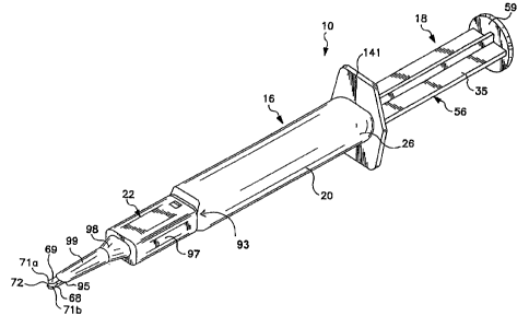
In a preferred embodiment, the present invention includes a device 10 for
inserting a flexible membrane, such as a flexible intraocular lens 12, into an
eye 14 of
a patient (Figs. I and 15). The device comprises a tubular member 16 having a
passage 17 and a plunger 18 movably received within the passage. The tubular
member preferably includes a base member 20, a cover 21, and a cannula 22
which
are coupled together (Figs. I and 9). The components of the device are
preferably
composed of a plastic material.
Base member 20 is an elongate tubular element defining a passageway 24 which
is provided with a relatively large opening at proximal end 26 and an opening
27 of
reduced size near, but spaced from, distal end 28 (Figs. 1, 8, 9 and 12).
Passageway
24 of base member 20 is adapted to movably receive and guide plunger 18. A
longitudinal groove 34 is preferably positioned along one of the side walls 32
of
passageway 24 to receive a flange 35 of the plunger and prevent twisting of
the
plunger during use.
A forwardly extending deck 29 projects beyond opening 27 to form a staging
area
45 for initially receiving the lens. A cover 21 is pivotally attached to base
member 20
and is movable between an open position to facilitate loading of a lens onto
the deck,
and a closed position where the cover overlies the deck and encloses the lens.
Cover
21 preferably includes a pair of rearwardly extending arms 36 provided with
knobs 37
CA 02333104 2007-03-19
on their free ends. The free ends of the arms 36 are fit into sockets 42 in
base
member 20 to form a hinge for the cover. Of course other connections could be
used
to pivotally attach the cover for movement about either a longitudinal or
transverse
axis. The internal surfaces of deck 29 and cover 21 are configured to control
the
folding of the intraocular lens as the lens is advanced toward the eye. The
shapes and
functions of these surfaces are described in the above-noted U.S. Patent No.
6,336,932.
Plunger 18 is an elongate member which is adapted to move through the passage
17 of tubular member 16 (Figs. 1-7). The plunger comprises a main body 56
preferably shaped with a cross-shaped cross section, although other
constructions
could be used. As discussed above, one flange 35 of the body is received into
groove
34 (Figs. 2-4). A flat thumb pad 59 is provided on the proximal end of body 56
for
manual operation of the device (Figs. 1-3). Other constructions, however, may
be
provided to effect advancement of plunger 18 through tubular member 16. The
forward end of body 56 includes a pair of spaced apart 0-rings 60 (Figs. 2-3).
The 0-
rings provide a level of resistance to enable a more controlled manual
operation of the
plunger. The 0-rings further help to prevent the plunger from inadvertent
movement
when the surgeon manipulates device 10 during the surgical procedure. Other
constructions, such as friction fit flanges, could be used in place of the 0-
ring. A
slender rod 62 projects forwardly beyond the main body 56 of plunger 18 (Figs.
1-3
and 5-7). The rod engages the lens at the staging area 45 and advances the
lens into
an eye.
The distal tip 68 of rod 62 is preferably bifurcated to define a pair of
prongs 71 a,
6
CA 02333104 2000-11-20
WO 99/59668 PCT/US99/10787
71b separated by a slot 72 (Figs. 1-3 and 5-7). The slot is shaped to receive
and hold a
proximal plate haptic 49 and optic 48 of lens 12. The ends of prongs 71 a, 71b
are
chamfered to form a pair of walls 77a, 77b which collectively form a generally
V-shaped
configuration. Depending on the sturdiness o1%the proximal haptic, walls 77a,
77b may or
may not engage the proximal end of optic 48. Of course, the distal tip 68 of
plunger 18
may altematively be formed with other structural configurations to engage the
disclosed
lens as well as other types of lenses (including lenses with loop haptics)
when the lens is
pushed toward the eye.
In accordance with the present invention, the distal end 68 of plunger 18 is
provided
with a visual indicator 69 which can be easily seen by the surgeon when
extended beyond
opening 27 (Figs. 2-3, 5-7, and 12). The visual indicator is preferably formed
by providing
the distal end with a color which contrasts with the color of the staging area
so as to be
easily seen by a surgeon if the end of the plunger is improperly advanced into
the staging
area. With such an indicator, the surgeon is much less likely to load a lens
overtop of the
distal end of the plunger. In addition, opening 27 adjacent staging area 45
preferably
conforms closely to the size of rod 62 so that the visual indicator is
substantially hidden
from view when the plunger is properly positioned. While the relationship of
opening 27
with rod 62 is not essential, it enables the surgeon to more easily identify
when the plunger
is improperly positioned. The visual indicator will also help the surgeon see
whether the
plunger properly engages the lens. For instance, in the present embodiment,
the surgeon
will be better able to see that the lens is engaged in slot 72 when the
plunger is advanced
after loading of the lens.
7
SUBSTITUTE SHEET (RULE 26)
CA 02333104 2000-11-20
WO 99/59668 PCT/US99/10787
As an example, the visual indicator 69 can be provided by forming the tubular
base
member 20 with deck 29 with a natural, uncolored clear or translucent
appearance (which
for purposes of this application is considered a color), and the distal end of
the plunger to
be dark blue. As a manufacturing expedient, the entire plunger may be formed
as a
uniform color, but only the distal end acts a visual indicator for the
surgeon. Altematively,
the indicator may consist of colored markings or regions on the distal end of
the plunger,
as opposed to the entire distal end, so long as the markings or regions are
plainly and
strikingly visible to the surgeon when the cover is open.
Once the lens has been properly loaded, the cannula 22 is fit over the cover
21 and
deck 29 (Fig. 1). Cannula 22 is an elongate tubular member with an open
proximal end
93 and an opposite open distal end 95. The proximal section 97 of the cannula
has a
generally rectangular configuration which defines a cavity to matingly receive
the
assembled deck 29 and cover 21. The medial section 98 of cannula 22 is smaller
than
proximal section 97 so that a shoulder is placed in abutment with the aligned
distal ends
28, 111 of base member 20 and cover 21. The inner wall of medial section 98
converges
to define a funnel shaped passageway. This funnel section causes the lens to
become
substantially curled and compressed for entry into the eye. The distal section
99 of
cannula 22 is a long, narrow tube which defines a lumen. Distal section 99 is
adapted to
be inserted through the narrow incision made in the eye.
To load the lens into insertion device 10, the cover 21 is opened to expose
the
staging area 45 on the upper side of deck 29. With the distal segment of the
plunger
having a visual indicatorwhich contrasts with the staging area, the surgeon
can easily see
8
SUBSTITUTE SHEET (RULE 26)
CA 02333104 2007-03-19
if the plunger has been advanced beyond opening 27 and into the staging area
(Fig.
12). If the plunger is in the staging area, the surgeon retracts the plunger
into
passageway 24 prior to loading the lens. After the lens has been loaded onto
deck 29,
the plunger is advanced so that the distal end 68 engages the lens 12. The
visual
indicator enables the surgeon to better see this engagement and thereby avoid
operational problems due to faulty loading. Thereafter, the cover is closed
and the
cannula fit over the deck and the cover. A viscoelastic material, typically
used for such
surgical procedures as a lubricant for the insertion process, is placed in the
cannula
22 prior to attachment of the cannula 22 to the assembly.
In use, the surgeon inserts the distal end of cannula 22 into the incision 142
in the
eye 14 (Fig. 15). The surgeon grasps lateral flanges 141 and pushes on pad 59
to
move plunger 18 in a forward motion. The plunger 18 acts to push lens 12
through
open end 95 and beyond cannula 22. In the preferred construction, plunger 18
is
pushed manually forward in a controlled manner, although other means, such as
an
electric motor or pneumatic drive, may be used.
As another example, a visual indicator as described above can be provided on
the distal end of the plunger of U S Patent No. 5,944,725 issued August 31,
1999.
As seen in Figs. 16 and 17, device 150 comprises a tubular member 152
having an axial passage 154, and a plunger 156 movably received in the
passage. The tubular member 152 is preferably composed of a base member
158 and a movable compressor 160 for compressing the lens. A staging area
162 is provided in passage 154 for initially receiving the lens through
9
CA 02333104 2000-11-20
WO 99/59668 PCT/US99/10787
an opening 164 defined in the device. A cover 166 is attached to the
compressor 160 for
movement between an open position to permit placement of the lens in the
staging area
and a closed position to enclose the passage for use in implanting a iens in
an eye. As
with device 10, a visual indicator 168 is provided on the distal end of
plunger 156 so that
the surgeon can more easily see whether the distal tip of the plunger has been
properly
positioned in the staging area before loading the lens 169, and to more easily
see that the
plunger has properly engaged the lens after loading of the lens into the
staging area.
In the preferred construction, plunger 156 has an elongate, slender distal end
167
adapted to engage and advance an intraocular lens through the narrow passage
154 and
into an eye. The distal end wall 170 of plunger 156 is formed with a concave
configuration
to engage and hold the proximal edge of the lens optic (Fig. 19). End wall 170
preferably
has a forwardly opening, generally V-shaped slot 174 with tapered opposing
faces 176,
177 and an inner face 178. Tapered faces 176, 177 are symmetrically positioned
about
axis 180 of the plunger at about a 30 angle relative to each other to guide
the lens into
the slot. The tapered faces extend inward about 1 mm (although- other depths
could be
used) to hold the proximal edge of the optic in the slot while the lens is
compressed and
advanced. The slot construction and dimensions could be much different.
The distal end of the plungerfurther includes a transverse recess 171 for
receiving
the trailing loop haptic 173 of.the lens (Figs. 18 and 19). The recess 171
protects the
trailing haptic from being damaged as the plunger pushes against the optic.
One side 182
of the plunger tapers inwardly as it extends toward end wall 170 to provide
clearance for
haptic 173 extending from the optic. The front wall 184 of recess 171 is
inclined toward
SUBSTITUTE SHEET (RULE 26)
CA 02333104 2000-11-20
WO 99/59668 PCT/US99/10787
the end wall 170 as it extends toward side 182 at an angle (e.g., about 600
to axis 180)
to minimize the recess and provide sufficient clearance for the haptic when
the lens is first
loaded into devioe 150. Front wall 184 is also inclined along the axis at an
angle 0 (e.g.,
of about 35 0 to axis 180) to form a ramp and ease the release of the haptic
from the recess
171. The ramp-shaped front wall 184 alleviates catching the trailing haptic on
the front
wall when the lens is released into the eye. The noted angular dimensions are
merely
examplary and could vary widely.
Plunger 156 also preferably includes a projection or bump 186 along the distal
end
of the plunger wtiich presses against an intemal face of passage 154. The bump
eliminates lateral play in the plunger and aids in aligning the slot with the
loaded lens.
Although many shapes could be used, the bump preferably has an outer arcuate
surface
to minimize the additional friction caused by the bump.
In use, a surgeon first checdcs device 150 to ensure that the plunger 156 is
properly
positioned for loading of the lens into the staging area 162. As discussed
above, the
visual indicator 168 enables the operator to. easily see if the plunger is
properly positioned.
In this case, the plunger is intended to be positioned slightly into the
staging area so that
the trailing haptic can be set in recess 171 when loading the lens. However,
advancement
of the plunger too far into the staging area could result in the optic of the
lens being loaded
overtop of end wall 170 such that engagement with slot 174 is not effected. In
proper
operation, the lens is loaded so that the optic is placed forward of end
surface 170 and the
trailing haptic 173 is set in recess 171. The plunger is then advanced so that
the optic is
received into the slot 174 in end surface 170. The compressor 160 is pressed
inwardly to
11
SUBSTITUTE SHEET (RULE 26)
CA 02333104 2000-11-20
WO 99/59668 PCT/US99/10787
compress the lens and move cover 166 to its closed position. In this position,
the device
is ready to move the enclosed lens into an eye.
The above discussion concems the preferred embodiments of the present
invention.
Various other embodiments as well as many changes and alterations may be made
without
departing from the spirit and broader aspects of the invention as described in
the claims.
12
SUBSTITUTE SHEET (RULE 26)