Note: Descriptions are shown in the official language in which they were submitted.
CA 02333529 2000-11-27
WO 99/60949 PCT/US99/11822
BONE ANt:HOR AND DEPLOYMENT DEVICE THEREFOR
REFERENCE TO EELATED APPLICATIONS
This application claims the benefit of priority of 09/086,508, filed on May
28, 1998 which is a continuation-in-part of U.S. Application Serial No.
08/595,772, filed ori February 2, 1996 (Attorney Docket: 0383311-0040) and of
Application Serial No. 08/814,149, filed on March 10, 1997 (Attorney Docket:
0383311-0070) and, thereby, of U.S. Application Serial No. 08/163,130 (now
U.S. Patent No. 5,725,529) filed on December 6, 1993 (Attorney Docket:
0383311-0019) and, thereby, of U.S. Application Serial No. 08/765,445 (now
U.S. Patent No. 5,268,001) filed on September 25, 1991 (Attorney Docket:
0383311-0018) and, thereby, of U.S. Application Serial No. 08/588,025 (now
abandoned) filed on September 25, 1990 (Attorney Docket: 0383311-0043).
BACKGROUND OF THE INVENTION
The present invention is directed to a bone anchor for attaching tissue to
bone, and to a device for deploying such an anchor in bone. More specifically,
the invention is directed to a bone anchor which employs a "floating" washer
that
conforms to the angile of the bone surface to hold tissue in place, and to a
deployment device having a plunger-like configuration that facilitates
deployment
of such an anchor.
Soft tissue, siuch as tendon, may become detached from a patient's bone as
a result of injury or a medical procedure. In either case, the tissue must be
re-
attached in order to permit healing. Medical devices used to perform this
function are known as bone anchors.
Traditionally, bone anchors were merely tacks or nails that were
hammered through a. patient's soft tissue and directly into the patient's
bone.
Anchors of this type, however, had many deficiencies. For example, they were
CA 02333529 2000-11-27
WO 99/60949 PCT/US99/11822
prone to coming out of the bone, particularly in cases where patients were
relatively active. In addition, because the anchors were hammered directly
into
the bone, deployment: was difficult and could sometimes result in hairline
fractures.
In recent times, more sophisticated bone anchors have been developed
which alleviate some of the problems mentioned above. For example, many bone
anchors now include prongs or something similar, which reduce the chances that
the anchor will dislodge from the bone. Likewise, bone anchors have now been
developed which can be inserted into pre-formed holes in bone, rather than
being
hammered.
When affixing tissue directly to bone, it is often desirable to deploy the
bone anchor at an angle that is normal to the bone surface. If this is not
done,
then sufficient contact may not be achieved with the surrounding tissue to
hold the
tissue in place. Understandably, achieving such placement can be difficult
when
the anchors are placed endoscopically.
In addition to the foregoing, the force required to deploy a conventional
bone anchor often makes deployment problematic. Driving a tack or nail-like
anchor into bone, for example, is difficult if the surgeon does not have
adequate
leverage on the bone to counterbalance the force of impact.
Thus, there exists a need for a bone anchor which does not need to be
deployed at a precise angle in order to attach tissue to bone reliably, and a
device
for deploying such an. anchor which does not require a surgeon to impart
substantial counterforce against the device during bone anchor deployment.
In view of the foregoing, an object of the invention is to provide improved
bone anchors and methods for deployment thereof. A related object is to
provide
improved devices for deploying such anchors.
2
- ---- ---- - - ---------
CA 02333529 2000-11-27
WO 99/60949 PCT/US99/11822
A further object is to provide anchors that attach tissue firmly and reliably,
regardless of the angle of deployment.
A still further object is to provide anchors, methods and devices for
deployment thereof suitable for use in endoscopic procedures.
Yet a still further object is to provide such anchors, methods and devices
that can be deployed easily and without application of unnecessary leverage or
counterforces.
SUMMARY OF THE INVENTION
The present irivention addresses the foregoing objects by providing a rivet-
like bone anchor haviing a floating washer at its head that can adapt to an
angled
bone surface and, thereby, better secure a tissue thereto. The invention also
provides a deployment tool that permits the anchor to be deployed without
application of unnecessary counterforce.
Thus, according to one aspect, the present invention is a bone anchor
which includes a rivet, an expandable sleeve, and a washer. The rivet includes
a
head and an elongate body having proximal and distal ends, the head being
mounted on the proximal end of the elongate body. The expandable sleeve has an
inner bore adapted to receive the rivet body. The washer "floats" at a
proximal
end of the sleeve. As the rivet is inserted into sleeve, the sleeve expands
into an
interference fit with the bone. The head of the rivet, moreover, forces the
floating washer into contact with the tissue at an angle that conforms to that
of the
underlying bone surface.
In other aspects of the invention, an external surface of the expandable
sleeve and/or the rivet has one or more annular ribs, threads or protrusions.
These increase the strength of the interference fits between the rivet and the
sleeve, as well as between the sleeve and the bone, thereby reducing the
chances
3
CA 02333529 2000-11-27
WO 99/60949 PCT/US99/11822
that the anchor will dislodge. The washer can likewise include ridges, teeth
or
other protrusions that enhance fixation of the tissue upon deployment.
In still other aspects of the invention, the bone anchor includes a housing
which is frangibly coupled to the expandable sleeve via breakable flanges. The
housing, which can be used to affix the anchor to the delivery device, can
also
protect and store the rivet prior to deployment. During deployment, the head
of
the rivet breaks the flanges of the housing, thereby freeing the housing from
the
anchor.
According to another aspect, the present invention provides an apparatus
for deploying a bone anchor of the type described above. The apparatus
includes
an outer tube, the distal end of which can hold the anchor housing, e.g., via
a
screw fit. A rod, which is slidably disposed within the bore of the tube, can
be
used to push the rivet: into the expandable sleeve so that the sleeve expands
into
the bone, so that the floating washer is forced into position against the bone
surface, and so that anchor is broken away from the housing. This can be
effected, for example, by squeezing the proximal ends of the outer tube and
the
rod together, e. g. , in the manner that the end of a syringe is squeezed.
By virtue of this design, the counterforce which a surgeon must impart to
the apparatus during Ibone anchor deployment is eliminated. More specifically,
because the outer tube is coupled to the expandable sleeve and because the rod
pushes on the rivet, there are no net forces that must be counterbalanced
during at
least the initial stage of deployment.
,
According to another aspect, the present invention is a method of
deploying a bone anchor of the type described above using a deployment device
of the type described above. The method includes pulling the tissue into
position
over the bone with a guide wire, e.g., a pin or K-wire, and drilling a hole in
the
bone, e.g., with a drill bit fitted over the end of the guide wire. With the
guide
wire holding the tissue in place over the bone hole, the bone anchor is then
4
CA 02333529 2000-11-27
WO 99/60949 PCT/US99/11822
threaded down the wire and into position for deployment (e.g., with the sleeve
in
the bone hole and the: floating washer resting against the tissue and bone
surface).
The deployment device, which is preferably threaded to the anchor, is
simultaneously slid inito position for deployment.
According to yet another aspect of the invention, the tissue is pierced and
pulled into position over a predrilled bone hole via a guide or probe integral
to,
and extending from, ithe distal end of the outer tube. With the guide or probe
holding the tissue in place over the bone hole, the bone anchor is passed
through
the outer tube, threaded through the tissue and into the bone hole.
Still further aspects of the invention provide for securing the sleeve into
the bone hole by threading it through clockwise (or counter-clockwise)
rotation of
the outer tube.
The rivet is deployed by squeezing the proximal end of the outer tube and
inner rod together in a syringe-like motion,' or put another way, by applying
downward force to the inner rod and upward force to the outer tube thereby
forcing the rivet out of the housing and into the expandable sleeve. Continued
application of these forces, at least partially sets the floating washer in
conformity
to the angle of the underlying bone surface and causes the frangible flanges
to
break, thereby freeing the housing from the sleeve. Once the anchor is at
least
partially deployed, the deployment device and attached housing (sans anchor)
are
removed from the site. The inner tube of the deployment device is then
rethreaded down the K-wire so that its distal end re-abuts the head of the
rivet.
By applying force (e. g. , tapping) to the proximal end of the inner rod, the
rivet is
fully set, fully fixing the washer at the angle of the underlying bone
surface.
A more complete understanding of the invention can be obtained by
reference to the following detailed description of the preferred embodiments
thereof in connection with the attached drawings.
5
CA 02333529 2000-11-27
WO 99/60949 PCT/US99/11822
BRIEF DESCR:IPT][ON OF THE DRAWINGS
A more complete understanding of the invention may be attained by
reference to the drawings, in which:
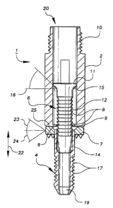
Figure 1 is a close-up view of a bone anchor in accordance with the
present invention, in which the bone anchor's rivet is not deployed;
Figure 2 is a close-up view of a bone anchor in accordance with the
present invention, in which the bone anchor's rivet is deployed;
Figure 3 shov!is a bone anchor deployment device in accordance with the
present invention;
Figure 4 shows an outer tube of the bone anchor deployment device shown
in Figure 3;
Figure 5 shows an inner rod of the bone anchor deployment device shown
in Figure 3; and
Figures 6 through 12 show a process for deploying the bone anchor shown
in Figure 1 into a bone using the bone anchor deployment device shown in
Figure
3.
DETAILED DESCRIPTION OF THE PREFERRED EMBODIMENTS
Figures 1 and 2 are close-up, cut-away views of a bone anchor according
to the present invention. As shown in Figure 1, bone anchor 1 includes housing
2, expandable sleeve 4, rivet 6, floating washer 7, breakable flanges 9, and
threading 10. In preferred embodiments of the invention, some or all of these
components are made: of a bioabsorbable material which dissolves in a
patient's
body over a period of time leaving little or no trace. Alternatively, bone
anchor
6
CA 02333529 2000-11-27
WO 99/60949 PCT/US99/11822
1 may be made of other biocompatible materials, such as conventional plastics
or
the like.
Rivet 6 is coniprised of head 11, elongate body 12, tapered tip 14, and a
centerbore (not show:n) running therethrough. Head 11 is located at the
proximal
end of rivet 6 and has a diameter which is greater than that of either
elongate
body 12 or tapered tip 14. Head 11 also includes undersurface 15. Undersurface
can be formed with a spherical radius or, alternatively, angled relative to a
longitudinal axis of the elongate body. Thus, as shown in the illustration,
the
10 undersurface is angled relative to plane 16 which, as shown in Figure 1,
bisects
elongate body 12 at a. right angle. Preferably, undersurface 15 is angled at
45
degrees; although other angles may be used. Elongate body 12 includes annular
ribs 8 on an outer surface thereof, which are adapted to aid in maintaining
rivet 6
in sleeve 4. It shoulti be noted, however, that elongate body 12 need not
include
15 annular ribs in order to perform its function. Tapered tip 14 is located at
the
distal end of rivet 6, and has a diameter which decreases gradually from the
diameter of elongate body 12 to less than that of inner bore 19 of expandable
sleeve 4.
Prior to deployment of bone anchor 1, rivet 6 is housed in housing 2, as
shown in Figure 1. Housing 2 is preferably cylindrical in shape and includes
threading 10 at first open end 20. Threading 10 is adapted to connect bone
anchor 1 to a bone anchor delivery device, such as that described in detail
below.
Housing 2 is integrally coupled with expandable sleeve 4 via flanges 9, which
are
sized to break in response to either downward or upward pressure against head
11
so as to disconnect (i., e. , free) housing 2 from expandable sleeve 4.
Expandable sleeve 4 includes inner bore 19, as noted above, and is
fabricated of a material which is capable of expanding into an interference
fit with
a bone hole. Inner bore 19 has a diameter that is less than the diameter of
elongate body 12, but: which is greater than or equal to a diameter of tapered
tip
14. As a result of these dimensions, expandable sleeve 4 is able to receive
rivet
7
CA 02333529 2000-11-27
WO 99/60949 PCT/US99/11822
6, distal-end-first. As described in more detail below, expandable sleeve 4
expands as elongate body 12 moves into expandable sleeve 4, eventually
resulting
in the arrangement shown in Figure 2, in which expandable sleeve 4 is fully
expanded. When bone anchor 1 is deployed in a hole in a bone, this expansion
results in an interference fit between expandable sleeve 4 and the bone. To
aid in
expansion, expandabile sleeve 4 also may include radial or longitudinal slots
(not
shown) which runs a:ll or part-way along expandable sleeve 4. In addition, a
membrane may also Ibe included on the slot, which permits expansion while, at
the same time, guarding against breakage of expandable sleeve 4.
The outer suriFace of expandable sleeve 4 includes annular ribs or threads
17 along at least a portion thereof. Annular ribs or threads 17 contact sides
of a
bone hole into which bone anchor 1 is implanted and, when expandable sleeve 4
is expanded, assist in maintaining bone anchor 1 in the bone. This feature of
the
invention is described in more detail below.
Disposed around the outer surface of expandable sleeve 4 is washer 7.
Washer 7 may be of any shape, i.e., it may be elliptical, circular, etc., and
may
include ridges or other protrusions on its undersurface for improved contact
with
tissue or bone. Whein bone anchor 1 is not deployed in a bone, washer 7
"floats", meaning that it is capable of at least longitudinal motion and
limited
angular motion relative to expandable sleeve 4. Washer 7 may be capable of
lateral motion relative to expandable sleeve 4 as well. Thus, referring to
Figure
1, washer 7 is capable of longitudinal motion along line 22 and of angular
motion
at, e.g., angles 23 and 24. This longitudinal motion and angular rotation is
possible when expandable sleeve 4 is fully unexpanded and, preferably, also
when
it is fully expanded. 'Washer 7 also has a top surface 25 which abuts with
undersurface 15 of rivet 6, and which may or may not be angled or radiused
relative thereto. In the embodiment shown in Figures 1 and 2, top surface 25
of
washer 7 is not angled or radiused.
Bone anchor 1. is deployed into a hole in a bone using a plunger-like
8
CA 02333529 2000-11-27
WO 99/60949 PCT/US99/11822
deployment device, such as that shown in Figure 3. As shown in Figure 3, bone
anchor deployment device 26 is comprised of tube 27 and rod 29. These
components may be fabricated of any material; although stainless steel is used
in
the preferred embodiment. Tube 27, which is shown in Figure 4, has a bore (not
shown) therethrough for receiving rod 29. At distal end 30, the inner surface
of
tube 27 includes threading (not shown). This threading is adapted to mate with
threading of a bone anchor, such as bone anchor 1, in order to hold the
housing
of the bone anchor substantially inunobile during deployment. Tube 27 also
includes "T"-shaped handle 31 at its proximal end. Handle 31 interacts with
knob 32 of rod 29 so as to limit the motion of rod 29 relative to tube 27.
In this regard, rod 29, which is shown in Figure 5, is removably disposed
within the bore of tube 27 and is slidable therein. That is, rod 29 can be
removed from tube 27 merely by sliding rod 29 out of tube 27. In addition, rod
29 is capable of sliding within tube 27 such that distal end 34 of rod 29
extends
out from tube 27 (see Figure 3). This feature of rod 29 makes it possible for
rod
29 to move within at least a portion of the housing of a bone anchor connected
to
tube 27, as described! in more detail below. As noted above, rod 29 also
includes
knob 32, which contacts handle 31 of tube 27 in order to limit the motion of
distal end 34 relative to tube 27. Finally, rod 29 includes a centerbore (not
shown) which is capable of receiving a guide wire, such as a K-wire and/or the
like.
Figures 6 through 12 explain operation of both bone anchor 1 and
deployment device 26 in the context of deploying a bone anchor into a bone
during an endoscopic: or other surgical procedure. To initiate such a
procedure, a
K-wire is inserted through the tissue (e.g., tendon) and into the bone at
which a
bone anchor is to be deployed. A hole is then formed in the bone at that
location,
e.g., via a drill bit or other cutting device disposed at the distal end of
the K-
wire. In this regard, hole 37 can be formed by any conventional means. In
preferred embodimerits of the invention, however, a drill bit or other cutting
device (not shown) is slid over K-wire 36 so as to secure the drill bit to K-
wire
9
CA 02333529 2000-11-27
WO 99/60949 PCT/US99/11822
36 in order to drill hole 37. Thereafter, the drill is removed from K-wire 36.
In
these embodiments oi' the invention, a drill guide which is used with such a
drill
may take the place of' the K-wire entirely. The following, however, assumes
that
a K-wire is used. The result of these steps is shown in Figure 6, namely K-
wire
36 in hole 37.
After, before or during formation of hole 37 in bone 39, a bone anchor,
such as that shown in Figure 1, is installed in a bone anchor deployment
device,
such as that shown in Figure 3. Specifically, with reference to Figure 6,
housing
2 of bone anchor 1 is screwed into inner threads of tube 27 on deployment
device
26. Once this has been done, bone anchor deployment device 26 and bone anchor
1 are slid over K-wire 36 via respective throughbores of sleeve 4, rivet 6 and
rod
29. This is shown in Figure 6.
Next, as shown in Figure 7, expandable sleeve 4 on bone anchor 1 is
inserted through soft tissue 40 and into hole 37 of bone 39. Since expandable
sleeve 4 is not expancied at this stage of the deployment process, little
actual
pressure is required on the part of the surgeon in order to insert expandable
sleeve 4 into hole 37. As shown in Figure 7, rod 29 may also be moved into
contact with rivet 6 at this point by pressing down on knob 32. This pressure
may be applied by any means, such as by a surgeon "squeezing" handle 31 and
knob 32 together using one hand in a syringe-like manner. Alternatively,
hammer
blows may be applied to knob 32 while holding tube 27 in place.
In any event, iPollowing insertion of expandable sleeve 4 into hole 37,
additional pressure is applied to knob 32 to begin initial deployment of rivet
6
into expandable sleeve 4. Specifically, downward pressure is applied to rivet
6
by sliding rod 29 downwards relative to tube 27 so as to force rod 29 into
housing 2 and to force rivet 6 out of housing 2 and into expandable sleeve 4.
Since housing 2 is helld substantially immobile by tube 27, and since housing
2 is
connected to sleeve 4., the process of deploying rivet 6 has little or no
effect on
the bone. That is, the downward force applied to rivet 6 is not significantly
CA 02333529 2000-11-27
WO 99/60949 PCT/US99/11822
imparted to bone 39 because rivet 6 moves within housing 2 and sleeve 4, both
of
which are held in place by tube 27. As a result, bone 39 moves little during
initial deployment of rivet 6.
As shown in Figures 8 and 9, as rivet 6 is deployed into expandable sleeve
4, expandable sleeve 4 expands within hole 37. This expansion results in an
interference fit between bone anchor 1(meaning rivet 6 and expandable sleeve
4)
and bone 39. Annular ribs 17 on expandable sleeve 4 strengthen this
interference
fit by coming into reilatively tight contact with the sides of hole 37. In
addition,
annular ribs 18 on rivet 6 also enhance the reliability of the interference
fit by
reducing the chances that rivet 6 will come out of expandable sleeve 4.
The initial deployment process depicted in Figures 6 through 9 causes rivet
6 to be moved substantially, but not completely, into hole 37 of bone 39. That
is, as shown in Figure 9, following initial deployment, there is still a space
42
between rivet 6 and a bottom of hole 37. Moreover, as also shown in Figure 9,
following initial deployment, washer 7 is not firmly sandwiched between head
11
of rivet 6 and tissue 40 on bone 39. Since washer 7 is preferably firmly
sandwiched between head 11 and tissue 40 in order for bone anchor 1 to
function
properly, additional steps are performed in order to complete deployment.
Specifically, in order to complete deployment of bone anchor 1,
deployment device 26 must be removed from K-wire 36 and housing 2 must be
disconnected from expandable sleeve 4. One advantage of the present invention
is
that these actions may be performed concurrently. More specifically, as noted
above, bone anchor 1 includes flanges 9 which break when sufficient force is
applied thereto. In the present invention, this force may comprise either
contact
with head 11 during downward movement of rivet 6 into expandable sleeve 4, or
contact with head 11 during upward movement of tube 27. In this regard, to
break flanges 9 during upward movement of tube 27, downward pressure is
retained on rod 29 and upward pressure is applied to tube 27.
11
CA 02333529 2000-11-27
WO 99/60949 PCT/US99/11822
In any event, as shown in Figure 10, once flanges 9 break, deployment
device 26, with hous;ing 2 still attached thereto, can be slid off of K-wire
36,
leaving rivet 6, washer 7, and expandable sleeve 4 within bone 39. Final
deployment then can begin by disconnecting rod 29 from tube 27 (i.e., by
sliding
rod 29 out of tube 27) and sliding rod 29 back over K-wire 36, as shown in
Figure 11. Washer ' 7 is then set in place by applying a force to knob 32 of
rod
29 by way of hammer blows or the like. Because this additional force can be
applied via hammer blows or the like, the need for a surgeon to exert
counterforce during final deployment is reduced.
The additional force applied above causes rivet 6 to move substantially all
the way into hole 37, thereby resulting in firm contact between head 11 and
washer 7. As a result of this contact, head 11 forces washer 7 against tissue
40
so that washer 7 is fiirmly sandwiched between head 11 and tissue 40. In this
regard, since washer 7 floats relative to bone surface, head 11 forces washer
7
against tissue 40 so that washer 7 is substantially parallel to a surface of
bone 39,
i. e. , so that washer 7 is disposed at an angle that substantially conforms
to a
surface of the underlying bone (see Figure 12) . The angle or radii of the
undersurface of head 11 described above facilitates this placement. As a
result,
washer 7 is able to hold tissue 40 in place. Thus, by using a floating washer
in
this manner, the invemtion reduces the need to implant the bone anchor at a
precise angle relative to the bone.
The present invention has been described with respect to particular
illustrative embodiments. It is to be understood that the invention is not
limited
to the above-describeci embodiments and modifications thereto, and that
various
changes and modifications may be made by those of ordinary skill in the art
without departing froin the spirit and scope of the appended claims.
12
- ------- --- --- -