Note: Descriptions are shown in the official language in which they were submitted.
CA 02334750 2000-12-08
WO 99/63889 PCT/AU99/00450
1
TITLE
"METHOD AND APPARATUS FOR DIAGNOSIS OF A MOOD DISORDER
OR PREDISPOSITION THEREFOR"
FIELD OF THE INVENTION
THIS INVENTION relates generally to mood disorders. In
particular, the present invention is concerned with a method and apparatus for
diagnosis of a mood disorder, particularly unipolar and/or bipolar mood
disorder, or
predisposition therefor. The invention also relates to a process of using the
diagnostic method to prevent mood disorders, to identify therapeutic compounds
for
alleviation of mood disorders, and to identify genetic markers associated with
such
disorders.
BACKGROUND ART
A variety of mood disorders exist which compromise to varying
degrees the social integration and quality of life of affected individuals.
The major
forms of mood disorder include bipolar disorder (manic depression) and
unipolar
disorders (major depression and unipolar mania). Other mood disorders include
dysthymic disorder, cyclothymic disorder, seasonal affective disorder and
substance-
induced mood disorder.
Bipolar disorder is a common condition with a lifetime prevalence of
1.2% to 1.6% (Weissman et al. 1988, Psych. Med. 18:141-153; Kessler et al.
1994,
Arch. Gen. Psych. 51:8-19). It is characterised by recurrent episodes of mania
and
depression with symptomatic recovery between episodes.
The pathophysiology of bipolar disorder remains poorly understood
despite considerable research (Goodwin et al. 1998, Arch. Gen. Psych. 55:23-
25).
Although it is strongly heritable, the genetics are complex, with less than
full
concordance in monozygotic twins (Mitchell et al. 1993, Aust. & New Zeal. J.
Psych.
27:560-580). At least four different susceptibility loci have been identified
(Adams
et al. 1998, Am. J. Hum. Genet. 62:10841091). A trait-dependent biological
marker
would assist genetic linkage studies (which are dependent upon the
identification of
the clinical phenotype) and would potentially lead to an understanding of the
CA 02334750 2000-12-08
WO 99/63889 PCT/AU99/00450
2
underlying molecular defect in bipolar disorder.
In unipolar depression, there are recurrent episodes of depression
with symptomatic recovery, but there are no episodes of mania. In unipolar
mania
there are recurrent episodes of mania but no episodes of depression. Like
bipolar
disorder, the pathophysiology and specific genetic defects underlying unipolar
disorders remain poorly understood.
Current techniques for diagnosing mood disorders rely entirely on
subjective interpretation of a patient's condition based on clinical
interview.
However, apart from being relatively time-consuming, the subjective nature of
this
technique in interpreting a psychiatric profile does not provide consistently
accurate
determinations of clinical phenotype. Consequently, misdiagnosis of mood
disorders
may occur which can thereby affect the prescribed pharmacological and non-
pharmacological therapy.
In the 1930s, Hunt and Guilford (1933, J. Abnormal and Social
Psychology 28:443-452) found that hospitalised manic-depressive patients
displayed
slow alternation rates when viewing an ambiguous figure (ie. Wheatstone cube)
compared to normal controls. The mean passive viewing number of alternations
per
minute was 4.25 for manic-depressives and 18.06 for normal controls. A strong
implication from this study is that such slower alternation rates may be the
result of
clinical progression. Moreover, the data from this study support the use of
this test
to confirm the presence of manic-depressive illness in hospitalised
individuals with
a life history of illness at least as long as that for the individuals in the
study.
SUMMARY OF THE INVENTION
The present invention arises from the unexpected discovery of
differential rates of binocular rivalry between subjects with mood disorders
(particularly unipolar and bipolar mood disorders), and non-clinical controls.
In this
respect, it was found that euthymic subjects affected by these mood disorders
have
a statistically significant slower rate of rivalry compared to non-clinical
control.
Surprisingly, the inventors also discovered that slow rates of binocular
rivalry are
present in some relatives of subjects with mood disorders. These findings
suggest
that slow binocular rivalry alternation rate is an alternative phenotypic
expression of
CA 02334750 2000-12-08
WO 99/63889 PCT/AU99/00450
3
the bipolar and/or unipolar genotype and is not the result of one or more
clinical
episodes.
The inventors have also found from unilateral caloric, and
transcranial magnetic, stimulation during binocular rivalry (as hereinafter
described)
that binocular rivalry is likely to be an interhemispheric switching
phenomenon, ie.
the perceptual alternations relate to altemating activation of the left and
right
hemispheres of the brain. Thus, the inventors consider that slow binocular
rivalry is
likely to correspond to slow rate of interhemispheric switching. The inventors
have
also shown that unilateral caloric stimulation also alters the perceptual
alternations
of the Necker cube, thus supporting interhemispheric switching as the neural
mechanism of ambiguous figures. The similarly abnormal (slow) alternation
rates
in binocular rivalry and the Necker cube in subjects with bipolar disorder
suggest that
these perceptual phenomena share a common neural mechanism. The similar
effects
of caloric stimulation on binocular rivalry and Necker cube alternations
suggest that
this common neural mechanism is interhemispheric switching.
Accordingly, the inventors have devised a method of diagnosing
mood disorders or predisposition therefor based on the above candidate trait-
dependent biological marker. The current method therefore may also have
utility in
genetic linkage studies for the identification of the molecular defect(s)
underlying
these disorders, and for the identification of compounds which may alleviate
such
disorders. Other aspects of the invention will become apparent from the
following
description.
Thus, in one aspect, the invention broadly resides in a method for
diagnosis of a mood disorder or predisposition therefor in a test subject,
said method
including the steps of:
(a) determining an interhemispheric switch rate of the test subject, wherein
the test
subject has not been diagnosed previously with the mood disorder; and
(b) comparing the switch rate with a corresponding reference switch rate to
diagnose presence or absence of the mood disorder or predisposition therefor.
Suitably, the test subject has had less than two episodes of the
disorder or is asymptomatic.
CA 02334750 2000-12-08
WO 99/63889 PCT/AU99/00450
4
Preferably, the interhemispheric switch rate is determined by
measuring a rate of perceptual rivalry in the test subject.
The rate of perceptual rivalry may be determined by measuring a rate
of reversal of perspective for ambiguous optical stimuli.
Preferably, the rate of perceptual rivalry is determined by measuring
a rate of binocular rivalry.
Alternatively, the interhemispheric switch rate may be determined by
measuring a rate of the nasal cycle.
Suitably, the rate of perceptual rivalry is measured by: -
(a) displaying at least one image to the test subject, wherein the at least
one image
invokes perceptual alternation;
(b) signalling respective incidences of perceptual alternation in the test
subject
during a predetermined period to provide a number of signals; and
(c) dividing the number of signals by the predetermined period to provide the
rate
of perceptual rivalry.
Preferably, the method is characterised in that said signalling is
effected by the test subject or by a suitable detection means.
Preferably, the method is further characterised by the step of
processing each of the signals relating to interhemispheric alternation to
convert
these signals into digitised signals, and storing the digitised signals for
subsequent
use.
Suitably, presence of the mood disorder is diagnosed, or a
predisposition therefor is suggested, when the interhemispheric switch rate of
the
subject is equal to a corresponding reference switch rate associated with the
mood
disorder or predisposition therefor. In contrast, absence of the mood disorder
may
be diagnosed, or predisposition therefor discounted, if the above criteria are
not
satisfied and/or when the interhemispheric switch rate of the subject is equal
to a
corresponding reference switch rate associated with normal or control
phenotype.
In the case of an interhemispheric switch rate determined by
binocular rivalry, presence of bipolar disorder is diagnosed, or a
predisposition
therefor is suggested, preferably when the rate of perceptual alternation in
the subject
CA 02334750 2000-12-08
WO 99/63889 PCT/AU99/00450
is less than 0.40 Hz, more preferably less than 0.35, and most preferably less
than
0.30. Preferably, the stimulus for binocular rivalry is moving gratings.
Conversely, absence of bipolar disorder may be diagnosed, or a
predisposition therefor discounted, when the rate of perceptual alternation is
greater
5 than 0.35 Hz, more preferably greater than 0.40 Hz, and most preferably
greater than
0.45 Hz. Preferably, the stimulus for binocular rivalry is moving gratings.
Suitably, presence of unipolar disorder is diagnosed, or a
predisposition therefor suggested, when the rate of perceptual alternation in
the
subject is in the range of between 0.25 Hz and 0.45 Hz. Preferably, the
stimulus for
binocular rivalry is moving gratings.
In another aspect, the invention provides a method for diagnosis of a
mood disorder or predisposition therefor in a test subject, said method
including the
steps of:
(a) determining an interhemispheric switch rate of the test subject; and
(b) comparing the switch rate with a corresponding reference switch rate to
diagnose presence or absence of the mood disorder or predisposition therefor;
wherein the interhemispheric switch rate is not determined by reversal of
perspective
of ambiguous optical stimuli.
In yet another aspect of the invention, there is provided a method for
diagnosis of a mood disorder in a test subject, said method including the
steps of:
(a) determining binocular rivalry rate in the subject; and
(b) comparing said rivalry rate with a corresponding reference rivalry rate to
diagnose presence or absence of the mood disorder or predisposition therefor.
In still yet another aspect, the invention provides a method for
assessing the clinical state of a test subject with a mood disorder, said
method
including the step of comparing measurements of current relative hemispheric
activation to corresponding measurements obtained when said subject was
euthymic
to thereby ascertain the clinical state.
Preferably, the relative hemisphere activation is measured by:
(a) recording binocular rivalry in the test subject;
(b) calculating a ratio of total time spent perceiving left eye's presented
image
CA 02334750 2000-12-08
WO 99/63889 PCT/AU99/00450
6
versus right eye's presented image;
(c) determining which eye's presented image is represented in which
hemisphere;
and
(d) interpreting which hemisphere has greater relative activation from the
results
of the aforementioned steps.
Suitably, the step of determining which eye's presented image is
represented in which hemisphere is carried out by the steps of:
(a) stimulating one of said hemispheres;
(b) calculating a post-stimulation ratio of total time spent perceiving left
versus
right eye's presented image; and
(c) comparing pre- and post-stimulation ratios to determine whether left eye's
image or right eye's image is represented in said stimulated or opposite
hemisphere.
Suitably, said stimulation is effected by unilateral caloric vestibular
and/or unilateral transcranial magnetic stimulation as, for example,
hereinafter
described.
In a further aspect of the invention, there is provided an apparatus for
diagnosing mood disorder, said apparatus comprising:
(a) a monitoring means for monitoring interhemispheric switching in a test
subject;
and
(b) processing means for determining an interhemispheric switch rate and for
comparing said switch rate with a predetermined data set for providing
diagnosis of presence or absence of the mood disorder or predisposition
therefor.
The monitoring means suitably comprises means for presenting
different viewing images separately to each eye and recordal means for
recording
when the subject perceives a change in the viewed image.
Suitably the different viewing images comprise a moving horizontal
grating presented to one eye and a moving vertical grating presented to the
other eye.
Alternatively, the different viewing images may be a stationary horizontal
grating
presented to one eye and a stationary vertical grating presented to the other
eye.
CA 02334750 2000-12-08
WO 99/63889 PCT/AU99/00450
7
The monitoring means preferably incorporates a liquid crystal shutter
before each eye.
The recordal means for recording perceived change may suitably be
a subjective device in the form of an indicator means activated by the test
subject
when a change is perceived.
Preferably, the recordal means is an objective device that records eye
movements as an indicator of which image is being perceived. Alternatively,
steady
state visual evoked potentials may be measured to provide an objective
indication of
the perceptual alternation.
The processing means suitably includes timing means and means for
receiving signals from the recordal means indicative of perceptual change.
The apparatus may also include change means for inducing a change
in ratio of total time spent perceiving left eye's presented image versus
right eye's
presented image.
In yet a further aspect of the invention, there is provided a process for
identifying one or more genetic markers associated with a mood disorder, said
process including the steps of:
(a) testing respective members of one or more pedigrees affected by the mood
disorder using the method of the invention;
(b) identifying members having the mood disorder or predisposition therefor;
and
(c) conducting genetic linkage analysis on the identified members to identify
the
or each genetic marker associated with the mood disorder.
Preferably, the mood disorder is bipolar disorder or unipolar disorder.
In a still yet a further aspect, the invention provides a method of
treating a patient with unipolar disorder, said method comprising the steps
of: -
(a) determining an interhemispheric switch rate of the patient ;
(b) comparing said interhemispheric switch rate with a range of reference
interhemispheric switch rates associated with bipolar disorder; and
(c) administering to said patient a pharmaccologically-effective dosage of a
mood-
stabilising drug when said interhemispheric switch rate is in said range.
Suitably, the mood stabilising drug is lithium.
CA 02334750 2000-12-08
WO 99/63889 PCT/AU99/00450
8
Preferably, the interhemispheric switch rate is determined by
perceptual alternation more preferably binocular rivalry. In the latter case,
the drug
is administered to the patient when the alternation rate is below 0.25 Hz,
more
preferably below 0.20 Hz, and most preferably below 0.15 Hz.
BRIEF DESCRIPTION OF THE DRAWINGS
In order that the invention may be readily understood and put into
practical effect, preferred embodiments of the invention will be described
with
reference to the attached drawings, in which: -
FIG. 1 is one embodiment of a diagnostic apparatus according to the
invention. Psychophysical set-up used to examine binocular rivalry. To avoid
problems with binocular fixation and alignment, the rivalrous stimuli are
presented
at the same location. By alternating rapidly between the rivalrous stimuli in
phase
with liquid crystal shutters, each eye's view can be restricted to the
stimulus intended
for it. The subject reports the perceived stimulus by depressing one of three
keys for
horizontal, vertical, or mixed/indeterminate percepts.
FIG. 2 shows a flow chart for a block or module of testing to
determine a subject's binocular rivalry rate.
FIG. 3 shows the distribution of rivalry switch rate in normal and
bipolar subjects. Distribution of rivalry switch rate in normal and bipolar
subjects.
The height of each column gives the mean rivalry switch rate for one
individual over
two blocks of trials totalling 20 min. Only a few individuals have short
intervals,
with a distinct plateau of common rates around 0.6 Hz and a long tail reaching
out
to slow rates. Bipolar subjects (n=26, median=0.26 Hz) have slow rates and are
highly significantly different from normals (n=63, median=0.57 Hz, p<0.0001).
FIG. 4 shows test-retest correlation of rivalry alternation rate in
bipolar and control subjects. Test-Retest Correlation of Rivalry Alternation
Rate in
bipolar and control subjects. There is a high correlation (rZ=0.83) between
the rates
obtained from the same individual on different occasions, indicating that this
is a
stable trait that would lend itself to genetic analysis.
FIG. 5 shows the rivalry rates of bipolar subjects (open bars) and first
degree relatives of bipolar subjects (black bars) tested with stationary
vertical and
CA 02334750 2000-12-08
WO 99/63889 PCT/AU99/00450
9
horizontal gratings. The distribution is bimodal with approximately half the
first
degree relatives having rates faster than the bipolar subjects and the
remainder, with
rates as slow as bipolar subjects. Those slow switching relatives are likely
to have
inherited the genotype that predisposes to developing bipolar disorder while
the
faster switching relatives have not inherited this susceptibility.
FIG. 6 shows the correlation of binocular rivalry rates in 16 pairs of
monozygotic twins. The high correlation of switch rates (r-0.72) suggests that
there
is indeed a genetic contribution to binocular rivalry switch rate. Twins were
tested
with moving horizontal and vertical gratings using the same apparatus for
testing and
recording as in FIG. 1.
FIG. 7 shows a graph of nasal cycle length versus binocular rivalry
cycle length. Individuals with slow nasal cycle also have slow binocular
rivalry
cycle and vice versa. The correlation of nasal cycle length to rivalry cycle
length is
grater than 0.6. This suggests coupling of interhemispheric oscillators
similar to the
coupling seen in circadian and ultradian oscillators in Drosophila.
Detennination of
the nasal cycle length was by subjective report of nasal patency but can be
objectively observed using nasal thermistors (Werntz et al., 1983), which is
incorporated by reference herein.
FIG. 8 shows a set-up for caloric stimulation and binocular rivalry
experiments (a) and the perceptual interference effects predicted by the
interhemispheric switch hypothesis (b, c). The rivalry set-up (a) shows a
right-
drifting vertical grating being presented to the left eye and an upward-
drifting
horizontal grating being presented to the right eye using liquid crystal
shutters to
restrict the presentation of each image to its intended eye. The orthogonal,
gratings
induce binocular rivalry and the subject's report their perceptual
alternations using
response keys on a keyboard. The caloric stimulation procedure involves
irrigating
the external ear canal with iced water until subjects report vertigo and
examiners
observe nystagmus. The stimulation acts via the semicircular canals and
brainstem
and results in activation-of contralateral structures (indicated in red) known
to be
involved in attentional processing and binocular rivalry (see text). The
expected
interference effects on rivalry alternations from such unilateral hemisphere
activation
CA 02334750 2000-12-08
WO 99/63889 PCT/AU99/00450
(according to the interhemispheric switch hypothesis) are depicted in the
theoretical
frequency histograms shown in (b) and (c). These represent the frequency (y-
axis)
of horizontal and vertical perceptual intervals in seconds (x-axis) during the
rivalry
viewing period. In (b), there is no baseline predominance of either horizontal
or
5 vertical percepts so unilateral hemisphere activation might be expected to
induce
either a horizontal (bottom left) or vertical (bottom right) predominance. In
(c), there
is a baseline predominance of the horizontal percept that might be expected to
disappear (bottom left) or even reverse to a vertical predominance (bottom
right)
following unilateral hemisphere activation by caloric stimulation. Actual
rather than
10 theoretical frequency histograms are shown in FIG. 8.
FIG. 9 shows a set-up for transcranial magnetic stimulation (TMS)
and binocular rivalry experiments and the perceptual interference effects
predicted
by the interhemispheric switch hypothesis. (a) The circular coil delivers a
single
pulse to the temporo-parietal region of the left hemisphere. The subject views
orthogonal stationary gratings (see methods) and reports their perceptual
alternations
using two response keys, one of which triggers the magnetic stimulation. (b)
The
time course of perceptual alternations shows the predicted pattern of
interference
effects when the TMS is triggered by a switch to the horizontal percept. The
interhemispheric switch hypothesis suggests that if the left hemisphere adopts
the
horizontal image, TMS applied to this hemisphere when the horizontal image is
perceptually dominant will disrupt this representation and allow the vertical
percept
to assume dominance. The theoretical frequency histogram depicts very short
horizontal interval durations. (c) When the stimulation is delivered to the
same
hemisphere but at the opposite phase of the perceptual switch (ie. triggered
when the
subject reports a switch to the vertical percept), disruption of the left
hemisphere
should have little effect since the vertical representation resides in the
right
~
hemisphere. Thus the theoretical frequency histogram for this contingency
shows
normal interval durations. Actual rather than theoretical frequency histograms
for
TMS's effect on rivalry alternations are shown in FIG. 13.
FIG. 10 shows an analysis procedure for caloric stimulation and
binocular rivalry experiments. There are six blocks of rivalry each
representing
CA 02334750 2000-12-08
WO 99/63889 PCT/AU99/00450
11
approximately seven minutes of viewing. Each block contains four 100-second
trials
separated by 30-second rest periods and each block is separated by a 2-minute
rest
period. The first block is considered training and discarded before analysis.
Blocks
2 and 3 are pre-stimulation blocks, while 4,5 and 6 are post-stimulation
blocks. The
predominance ratio is calculated by dividing the total time spent perceiving
the
vertical gratings by the total time spent perceiving the horizontal gratings,
excluding
mixed percepts. The resulting V/H ratio is log-transformed before analysis.
Since
there is random variation in this V/H ratio between two pre-stimulation
blocks, to
show an effect of caloric stimulation, there must be greater absolute
magnitude of
change in the V/H ratio between blocks 3 and 4 (random variation plus
experimental
effect) compared with the random change seen between blocks 2 and 3.
Comparison
of the V/H 2-3 change with the V/H 3-5 change (and the V/H 3-6 change) allows
an
indication of the stimulation's decay. The left hemisphere activation group
(n=18)
demonstrated a greater V/H change following stimulation compared with that
seen
prior to stimulation (p>0.05) and the effect was largely diminished by the
fifth block
(though not in all subjects). We were less able to demonstrate greater than
baseline
shifts in predominance for the right hemisphere activation group (n=14,
p=0.72).
Results were not significant for a control group of twelve subjects who
underwent
the entire protocol minus the caloric stimulation (p=0.21).
FIG. 11 shows the effects of caloric vestibular stimulation on two
individuals' perceptual alternations in binocular rivalry. In both cases the
predominance of one perceptual alternative is shifted by left hemisphere
activation
(right caloric stimulation). These changes are demonstrated in the frequency
histograms of interval durations for each rivalling image. The shifts are also
reflected
by the predominance ratios shown in the top right hand comer of each of the
histograms. In the first subject's case, (a) a baseline horizontal bias of
V/H=0.93 was
increased to (b) V/H=0.54 following caloric stimulation. This was the usual
direction of change for left hemisphere activation. The second subject also
illustrates
a post-stimulation change, beginning with (c) a horizontal grating bias of
V/H=0.94
which was reversed to (d) a vertical bias of V/H=1.26 following stimulation.
The
direction of shift for this subject is exceptional but suggests that
designation of image
CA 02334750 2000-12-08
WO 99/63889 PCT/AU99/00450
12
to hemisphere may not always be fixed. This subject's results along with a
number
of other reasons (see text) also makes interpretations based on residual eye-
movements, difficult to accept. The effect of caloric vestibular stimulation
on the
predominance of rivalling images supports the interhemispheric switch
hypothesis
and the data across all subjects (see text) suggests that the horizontal
percept is
usually adopted by the left hemisphere.
FIG. 12 shows the effect of caloric vestibular stimulation on
perceptual alternations of a reversible figure, the Necker cube. Right caloric
stimulation (left hemisphere activation) shifts a baseline perspective bias
(A=lower
square face closer to observer; B=upper square face closer to observer; ratio
calculated as for binocular rivalry and excludes indeterminate percepts) of
A/B=1.3
(a) to A/B=0.85 (b). Overall, subjects demonstrated shifts in both directions
following stimulation, indicating that, unlike for binocular rivalry,
designation of
perceptual configuration to hemisphere is arbitrary. Also shown (c) are the
raw data
for a single subject demonstrating normal baseline perceptual alternations,
with
roughly equal time spent experiencing each perspective, and the effect of
caloric
stimulation which virtually eliminated the ability to perceive one perceptual
alternative. The subject alternated between perspective A and the `undecided'
response option (where no depth was perceived) following left hemisphere
activation. This dramatic effect may be related to the fact that this subject
received
prolonged iced water irrigation compared with other subjects. The effect of
unilateral hemisphere activation on Necker cube alternations is further
evidence that
binocular rivalry and reversible figures share a common neural mechanism and
suggests to us that this mechanism is interhemispheric switching.
FIG. 13 shows the effect of left hemisphere transcranial magnetic
stimulation on a single subject's binocular rivalry alternations. There is a
marked
interference with percepts when left hemisphere TMS is contingent on one
direction
of perceptual switch but not when the contingency is at the opposite phase.
(a) If
TMS was delivered when the subject signalled a switch from the vertical to the
horizontal percept, there was an immediate reversion to the vertical percept,
indicated
by a dramatic shortening of the horizontal interval durations. The histogram
only
CA 02334750 2000-12-08
WO 99/63889 PCT/AU99/00450
13
plots the vertical intervals just before stimulation and the horizontal
intervals just
after stimulation. (b) When TMS was administered at the opposite phase (when
the
subject signalled a switch from the horizontal to the vertical percept), it
had minimal
influence upon interval durations. This histogram only plots the vertical
intervals
just after stimulation and horizontal intervals just before stimulation. That
one
stimulation contingency but not the other affects rivalry alternations
supports the
interhemispheric switch hypothesis. Phase-specific interference effects such
as those
shown here are difficult to explain using a within-hemisphere competition
model of
binocular rivalry.
FIG. 14 shows the rivalry rates of non-clinical controls (open bars)
and unipolar subjects (black bars) tested with moving vertical and horizontal
gratings. There appears to be two groups: ie. one group has normal alternation
rates
and the other group has slower than normal alternation rates but generally not
as slow
as bipolar subjects.
DETAILED DESCRIPTION
1. Defnitions
Unless defined otherwise, all technical and scientific terms used
herein have the same meaning as commonly understood by those of ordinary skill
in
the art to which the invention belongs. Although any methods and materials
similar
or equivalent to those described herein can be used in the practice or testing
of the
present invention, preferred methods and materials are described. For the
purposes
of the present invention, the following terms are defined below.
By "ambiguous optical stimuli" is meant those stimuli able to elicit
different perceptions which alternate during continued observations of the
same
stimulus. Suitable ambiguous stimuli of this type include ambiguous figures
such
as the Necker cube and Schroder staircase.
The term "binocular rivalry" refers to the alternating perceptual states
that arise when viewing different images, presented separately to each eye, in
the
same retinal location. In this regard, it is well known that when
corresponding
regions of the two eyes are stimulated by sufficiently different patterns, the
stimuli
CA 02334750 2000-12-08
WO 99/63889 PCT/AU99/00450
14
rival in terms of conscious perception, rather than fuse into a composite
pattern.
Accordingly, a perceptual alternation or switch between these nonfusible
dichoptic
stimuli results.
The term "genetic marker" includes within its scope a region of a
chromosome, locus, allele or fragment thereof that is associated with a
particular
phenotype.
By "interhemispheric switch rate" is meant the rate of
interhemispheric alternation in one or more regions of the brain inclusive of
temporo-
parietal cortex, hypothalamus, prefrontal, and limbic regions of the brain.
Preferably,
the interhemispheric switch rate relates to the rate of interhemispheric
alternation of
the temporo-parietal cortex.
Throughout this specification, unless the context requires otherwise,
the words "comprise", comprises" and "comprising" will be understood to imply
the
inclusion of a stated integer or group of integers but not the exclusion of
any other
integer or group of integers.
2. Diagnosis of mood disorders using interhemispheric switch rates
The present invention arises from the unexpected discovery that the
rate of perceptual alternation in binocular rivalry is slow in euthymic
subjects with
mood disorders, particularly those with bipolar or unipolar disorders.
Surprisingly,
the inventors also discovered that slow rates of binocular rivalry are present
in
relatives of subjects with mood disorders. These finding suggests that slow
binocular
rivalry alternation rate is an altemative phenotypic expression of the bipolar
and/or
unipolar genotype and is not the result of one or more clinical episodes.
As will be more fully described hereinafter, it has also been found
unexpectedly, from two unilateral hemispheric activation techniques in human
subjects, that perceptual alternations result from competition between rather
than
within the cerebral hemispheres. Accordingly, the inventors have proposed an
interhemispheric switching mechanism for these perceptual phenomena. It is
believed that the interhemispheric switch rate provides a trait-dependent
biological
marker for mood disorders and in particular, bipolar disorder and unipolar
disorder.
From the foregoing, the inventors have devised a method of
CA 02334750 2008-01-10
diagnosing mood disorders or predisposition therefor based on the above
candidate trait-dependent biological marker. The method includes the steps of
determining an interhemispheric switch rate of the test subject, and comparing
the
switch rate with a corresponding reference switch rate to diagnose presence or
5 absence of the mood disorder or predisposition therefor. In this regard, the
invention broadly encompasses diagnosing a mood disorder or predisposition
therefor when the interhemispheric switch rate is aberrant relative to a
normal
range of interhemispheric switch rates.
The interhemispheric switch rate of the subject may be determined by any
10 suitable technique and, in this regard, techniques that indirectly measure
a
particular interhemispheric switch rate are also contemplated by the
invention. For
example, an interhemispheric switch rate relating to the temporo-parietal
cortex
may be determined by measuring the rate of perceptual rivalry (or perceptual
alternation) in the subject. Alternatively, an interhemispheric switch rate
relating
15 to hypothalamic activity may be determined by measuring the rate of
alternating
sympathetic and parasympathetic activity in the nasal turbinates, also known
as
the nasal cycle (Shannahoff-Khalsa, 1993, Intern. J Neuroscience 70:285-298).
Preferably, the step of determining interhemispheric switch rate is
characterised by subjecting the test subject to a stimulus which invokes
interhemispheric alternation therein. Any suitable stimulus having such
characteristics is contemplated by the invention and in this regard, the
stimulus
may comprise images that are sufficiently different that rivalry is induced
rather
than fusion; ie. perceptual rivalry is induced.
The rate of perceptual rivalry may be determined by measuring the rate of
reversal of perspective for ambiguous optical stimuli. Exemplary methods which
may be used to measure the rate of reversal of perspective include, but are
not
limited to, those disclosed in George (1935, J. Gen. Psychol. 39-59), Washburn
and Manning (1933, In Studies from the psychological laboratory of Vassar
College 632-633), Washburn et al (1933, ibid 633-636), Washburn et al (1933,
ibid 636-637), and Borsellino et al (1972, 10.Bd., Heft 3:139-144).
CA 02334750 2008-01-10
16
Preferably, the rate of perceptual rivalry is determined by measuring the
rate of binocular rivalry. Examples of binocular rivalry techniques include,
but are
not limited to, those disclosed in Howard and Rogers (1995, "Binocular fusion
and rivalry", In Binocular Fusion and Stereopsis, eds Mackintosh et al, Oxford
University Press), Logothetis et al (1996, Nature 380:621-624), Kovacs et al
(1996, Proc. Natl. Acad. Sci. USA 93:15508-15511), Sheinberg and Logothetis
(1997, Proc. Natl. Acad. Sci. USA 94:3408-3413), and Andrews and Purves
(1997, Proc. Natl. Acad. Sci. USA 94:9905-9908).
Suitably, the rate of perceptual rivalry is measured by displaying an image
to the test subject which image invokes perceptual alternation, signalling
respective incidences of perceptual alternation in the test subject during a
predetermined period to provide a number of signals and dividing the number of
signals by the predetermined period to provide the rate of perceptual rivalry.
Preferably, the method is characterised in that said signalling is effected by
the test subject or by a suitable detection means. In the case of a subject
effecting
the signalling, the subject preferably signals a perceptual alternation or
switch. In
this context, the subject may signal visually, audibly, or by touch wherein
the
signal is registrable by a suitable sensor. For example, the subject may
depress a
button that is suitably operably connected to a signal registration means that
registers the signal.
Alternatively, a perceptual alternation may be signalled by a suitable
detection means. For example, the detection means may be adapted to measure
visually evoked potentials (VEP). In this regard, reference may be made for
example, to Brown and Norcia (1997, Visions Res. 37:2401-2408) which teach a
real-time, steady-state VEP based on labelling each eye's image with a
slightly
different temporal frequency so that the record generated by each can be
recovered by an electroencephalogram (EEG) by spectrum analysis. In this way,
it
is possible to track the "waxing" and "waning" of the VEP amplitudes for each
eye's image simultaneously during spontaneous rivalry, permitting an analysis
of
the relative dominance of each eye's image in real-time and to determine
alternation rate.
CA 02334750 2008-01-10
17
Alternatively, the detection means may be adapted to monitor eye
movement. For example, Blackwood et al (1996, Br. J. Psych. 168:85-92) teach a
smooth-pursuit eye tracking procedure in which a subject visually tracks an
image
and an electro-oculograph is recorded in the horizontal plane via electrodes
attached adjacent to the outer canthus of each eye. Reference also may be made
to
Sweeney et al (1998, Biol. Psychiatry 43:584-594) who disclose the use of
infrared recordings to monitor eye movements. Such procedures that monitor eye
movements have particular utility in binocular rivalry methods that rely on
moving dichoptic stimuli, such as moving vertical and horizontal gratings.
Alternatively, the interhemispheric switch rate may relate to the rate of
interhemispheric alternation of hypothalamic activity as mentioned above. Such
rate may be determined by measuring the rate of alternating sympathetic and
parasympathetic activity in the nasal turbinates, otherwise known as the nasal
cycle, as for example disclosed in Shannahoff-Khalsa (1993, supra) and Werntz
et
al (1983, Human Neurobiol. 2:39-43).
Alternating cerebral hemisphere activation may be determined by EEG
recordings as for example disclosed in Shannahoff-Khalsa (1993, supra) and
Werntz et al (1983, supra).
Also contemplated, as a measure of interhemispheric switch rate is
alternation of performance in hemisphere specific functions such as verbal and
spatial abilities (Shannahoff-Khalsa, 1993, supra; Klein and Armitage, 1979,
Science 204:1326-1328).
Suitably, the method is further characterised by the step of processing each
of the signals relating to interhemispheric alternation to convert these
signals into
digitised signals, and storing the digitised signals for subsequent use.
In preference, the step of determining the rate of interhemispheric
switching is further characterised by dividing the number of signals
corresponding
to interhemispheric alternation by the total time the subject is under test.
For
example, in the case of perceptual rivalry referred to above, the
interhemispheric
switch rate may be calculated by dividing the number of perceptual switches by
the
CA 02334750 2000-12-08
WO 99/63889 PCT/AU99/00450
18
total time of rivalry. Preferably, in the case of binocular rivalry such
calculation
excludes mixed or indeterminate percepts.
The step of determining interhemispheric switch rate may further
include a practice period wherein the subject becomes familiarised with the
test.
Suitably, this period is not taken into account when determining the rate of
interhemispheric switching.
Suitably, presence of the mood disorder is diagnosed, or a
predisposition therefor is suggested, when the interhemispheric switch rate of
the
subject is equal to a corresponding reference switch rate associated with the
mood
disorder. In such a case, the corresponding reference switch rate may
correspond to
a predetermined average range of interhemispheric switch rates in subjects
having
the mood disorder. In contrast, absence of the mood disorder may be diagnosed,
or
predisposition therefor discounted, if the above criteria are not satisfied
and/or when
the interhenzispheric switch rate of the subject is equal to a corresponding
reference
switch rate associated with normal or control phenotype. In such a case, the
corresponding reference switch rate may correspond to a predetermined average
range of interhemispheric switch rates in non-clinical control subjects.
In the case of an interhemispheric switch rate determined by
binocular rivalry, presence of bipolar disorder is diagnosed, or a
predisposition
therefor is suggested, preferably when the rate of perceptual alternation in
the subject
is less than 0.40 Hz, more preferably less than 0.35, most preferably less
than 0.30.
In this regard, a predisposition for bipolar disorder is suggested suitably
when the
rate of perceptual alternation in a relative of a bipolar subject is less than
0.40 Hz,
more preferably less than 0.35, most preferably less than 0.30.
Conversely, absence of bipolar disorder may be diagnosed, or a
predisposition therefor discounted, when the rate of perceptual alternation is
greater
than 0.35 Hz, more preferably greater than 0.40 Hz, and most preferably
greater than
0.45 Hz.
Suitably, presence of unipolar disorder is diagnosed, or a
predisposition therefor suggested, when the rate of perceptual alternation in
the
subject is in the range of between 0.25 Hz and 0.45 Hz.
CA 02334750 2000-12-08
WO 99/63889 PCT/AU99/00450
19
For the above diagnoses or predispositions, the stimulus which
invokes binocular rivalry in the test subject preferably comprises moving
gratings.
The method may further include the step of repeating the method on
at least one separate occasion to validate the diagnosis. In this regard, the
subject
tested is suitably euthymic.
3. Assessing clinical state of a subject with a mood disorder
The invention also contemplates a method for assessing the clinical
state of a subject with a mood disorder including the step of comparing
measurements of current relative hemispheric activation to corresponding
measurements obtained when said subject was euthymic to thereby ascertain the
clinical state. In this regard, it will be appreciated that mood shifts seen
in bipolar
disorder are associated with relative hemisphere activation: left hemisphere
activation being associated with mania, while right hemisphere activation is
associated with depression.
Preferably, the relative hemisphere activation is measured by
recording binocular rivalry in the subject calculating a ratio of total time
spent
perceiving left eye's presented image versus right eye's presented image,
determining which eye's presented image is represented in which hemisphere;
and
interpreting which hemisphere has greater relative activation from the results
of the
aforementioned steps.
Suitably, the step of determining which eye's presented image is
represented in which hemisphere is carried out by the steps of stimulating one
of said
hemispheres, calculating a post-stimulation ratio of total time spent
perceiving left
eye's presented image versus right eye's presented image; and comparing pre-
and
post-stimulation ratios to determine whether left or right eye's image is
represented
in said stimulated or opposite hemisphere. Preferably, said stimulation is
effected
by unilateral caloric vestibular and/or unilateral transcranial magnetic
stimulation as,
for example, hereinafter described.
Alternative methods of measuring relative hemispheric activation
may be effected by indirect measures of other interhemispheric switching
phenomena
such as the nasal cycle and higher functions such as verbal and spatial
abilities.
CA 02334750 2000-12-08
WO 99/63889 PCT/AU99/00450
4. Apparatus for diagnosis of mood disorders
The invention also provides an apparatus for diagnosing mood
disorder, comprising a monitoring means for monitoring interhemispheric
switching
in a test subject; and processing means for determining an interhemispheric
switch
5 rate and for comparing said switch rate with a predetermined data set for
providing
diagnosis of presence or absence of the mood disorder or predisposition
therefor.
The monitoring means suitably comprises means for presenting
different viewing images separately to each eye and recordal means for
recording
when the subject perceives a change in the viewed image.
10 Suitably the different viewing images are a moving horizontal grating
presented to one eye and a moving vertical grating presented to the other eye.
Alternatively, the different viewing images comprise a stationary horizontal
grating
presented to one eye and a stationary vertical grating presented to the other
eye.
Other visually distinct images, such as mentioned in the prior art relating to
binocular
15 rivalry, can also be employed.
The monitoring means preferably incorporates a liquid crystal shutter
before each eye. The liquid crystal shutters allow the field of view of each
eye to be
superimposed so that the different viewing images are presented at the same
retinal
location.
20 The recordal means for recording perceived change is suitably a
subjective device in the form of an indicator means activated by the test
subject when
a change is perceived. Preferably, the recordal means is an objective device
that
records eye movements as an indicator of which image is being perceived.
Alternatively, steady state visual evoked potentials may be measured to
provide an
objective indication of the perceptual alternation.
The processing means suitably includes timing means and means for
receiving signals from the recordal means indicative of perceptual change.
Interhemispheric switch rate is calculated by dividing the number of
perceptual
switches by the total time of perceptual rivalry.
The apparatus may also include change means for inducing a change
in ratio of total time spent perceiving left eye's presented image versus
right eye's
CA 02334750 2008-01-10
21
presented image. The change means may be a caloric vestibular stimulation
means, trans-cranial magnetic stimulation means, contrast altering means, or
other
means known to produce a change in the interhemispheric switch rate.
5. Use of Diagnostic Method to Identify Genetic Markers Linked to Mood
Disorders
Also contemplated is a process for identifying one or more genetic
markers associated with a mood disorder, including the steps of testing
respective
members of one or more pedigrees affected by the mood disorder using the
method of the invention, identifying members having the mood disorder or
predisposition therefor; and conducting genetic linkage analysis on the
identified
members to identify the or each genetic marker associated with the mood
disorder.
Linkage analysis is well known to those of skill in the art. Exemplary
protocols which may be used for this purpose include, but are not limited to,
those
disclosed in Dracopoli et al (1994, "Current Protocols in Human Genetics",
John
Wiley and Sons Inc., USA), Ott, J. (1991, "Analysis of Human Genetic Linkage"
Johns Hopkins University Press), and Adams et al.(1998, Am. J. Hum. Genet.
62:1084-1091).
The invention also contemplates linkage studies carried out on non-
affected individuals ie. non-pedigree members. In this regard, one subset of
the
non-affected individuals will have fast interhemispheric switch rates and
another
subset will have slow interhemispheric switch rates. The application of
linkage
analysis to these subsets will be advantageous in identifying molecular
markers
linked to switch rate (a quantitatively varying trait). These markers may then
be
employed for the identification of molecular markers linked to mood disorders
such as bipolar disorder and unipolar disorder.
The invention also extends to the genetic marker(s) obtained by the
aforementioned process.
6. Use of Diaanostic Method to Identify Candidate Therapeutic Azents
The invention also provides a process for identifying a candidate
therapeutic agent for alleviating a mood disorder, including the steps of
measuring
first interhemispheric switch rate in a test specimen, administering or
applying a
CA 02334750 2008-01-10
22
test compound to said test specimen, measuring second interhemispheric switch
rate in the test specimen, and identifying a candidate therapeutic agent if
said
second interhemispheric switch rate is faster than said first interhemispheric
switch rate. Suitably, the test specimen includes, but is not limited to, a
human, an
animal, brain tissue thereof or brain cell(s) thereof.
Any suitable method may be used to determine interhemispheric switch
rate. For example, methods hereinbefore described may be used in the case when
the test specimen is a human or animal. Alternatively, when the test specimen
is
an animal, electrical activity of brainstem or hypothalamic neurones
associated
with interhemispheric switching may be measured. This particular technique may
also be used when the test specimen is brain tissue or brain cells. An example
of a
method which uses such measurement of electrical activity in vitro and/or in
vivo
is described by Schaap et al (1997, Brain Res. 753:322-327).
Alternatively, interhemispheric switch rate may be determined by
inducing, in the test specimen, spontaneous nystagmus that alternates in
direction
(periodic alternating nystagmus (PAN)). In this regard, reference may be made
to
methods of inducing PAN respectively in humans (Baloh et al., 1976, Brain
99:11-26), monkeys (Waespe et al., 1984, Science 228:199-202) and goldfish
(Dow and Anastasio, 1997, NeuroReport 8:2755-2759). In PAN, the cycle length
of the alternating eye movements is known to vary (Baloh et al., supra).
Accordingly, the period of alternation can be monitored in experimental
animals
using electronystagmography (Waespe et al., supra). Decreases in cycle (ie.
increased rate) following administration of pharmacological agents would
provide
means for detecting lead compounds that may be therapeutic for mood disorders.
In this respect, the test specimen is preferably a monkey, and PAN is
preferably
induced by cerebellar uvula and nodulus destruction as for example described
by
Weaspe et al. (supra).
The invention also extends to a process for identifying a candidate
therapeutic agent for alleviating a mood disorder, including the steps of
measuring
first perceptual rivalry rate in a test specimen, administering or applying a
test
compound to the test specimen, measuring second perceptual rivalry rate in the
test specimen, and identifying a candidate therapeutic agent if said second
perceptual rivalry rate is faster than said first perceptual rivalry rate.
Preferably,
CA 02334750 2008-01-10
23
said perceptual rivalry is binocular rivalry. Suitably, the test specimen is a
human
or animal.
In the case of the test specimen comprising an animal, the animal is
suitably a cat, and in this regard, the rate of binocular rivalry is
preferably
determined by measurement of optokinetic nystagmus as for example described
by Fries et al (1997, Proc. Natl. Acad. Sci. USA 94:12699-12704).
Alternatively,
the animal may be a monkey and in such a case, the binocular rivalry rate is
measured by optokinetic nystagmus or by the method of Sheinberg and Logothetis
(1997, supra).
7. Use of DiaQnostic Method to Treat Unipolar Patients
The invention further provides a method of treating a patient with unipolar
disorder, said method comprising the steps of determining an interhemispheric
switch rate of the patient, comparing said interhemispheric switch rate with a
range of reference interhemispheric switch rates associated with bipolar
disorder;
and administering to said patient a pharmaccologically-effective dosage of a
mood-stabilising drug when said interhemispheric switch rate is in said range.
Preferably, the mood stabilising drug is lithium. Preferably, the
interhemispheric
switch rate is determined by perceptual alternation more preferably binocular
rivalry. In the latter case, the drug is administered to the patient when the
alternation rate is below 0.25 Hz, more preferably below 0.20 Hz, and most
preferably below 0.15 Hz.
The inventors have found that there appears to be two unipolar disorder
groups: ie. one group has normal alternation rates and the other group has
slower
than normal alternation rates but generally not as slow as bipolar subjects.
These
two groups may reflect different underlying biological abnormalities and
therefore
switch rates may be used to subtype unipolar disorder, preferably unipolar
depression.
CA 02334750 2000-12-08
WO 99/63889 PCT/AU99/00450
24
EXAMPLES
EXAMPLE 1
Apparatus,for measuring rate of binocular rivalry
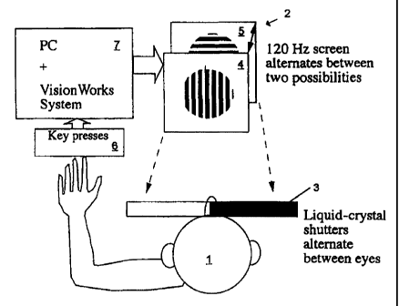
In one embodiment shown in FIG. 1, binocular rivalry is used as an
indicator of mood disorder or predisposition therefor. Binocular rivalry is
tested with
a subject I seated three metres from a computer monitor 2 and wearing liquid
crystal
shutter goggles 3 to enable the presentation of vertical moving lines 4 to the
left eye
and horizontal moving lines 5 to the right eye. The stimuli subtend 1.5
degrees of
visual angle with a spatial frequency of 8 cycles/degree moving at 4
cycles/second.
The subject 1 presses one of three response buttons 6 to indicate their
perceptual state
(horizontal, vertical or mixed/indeterminate percepts). Rivalry testing is
conducted
over half an hour. There are three blocks of testing, each containing four 100
second
trials interspersed with 30 second rests. Each block is interspersed with a
two-minute
rest. A non-limiting example of a block of testing is given in FIG. 2.
Alternation rate per second is calculated in a processing means such
as a personal computer 7 by dividing the number of perceptual switches by the
total
time of rivalry, excluding mixed/indeterminate percepts. Other calculated
indicators
include a measure of image bias (total time spent viewing vertical lines
divided by
the total time spent viewing horizontal lines), autocorrelation (a measure of
the
independence of successive interval durations) and fit to the gamma
distribution (a
known feature of binocular rivalry). The latter two indicators may be used to
verify
reliability of subjects' subjective reports, as they are well known features
of rivalry
that are difficult to fabricate.
The inventors have found that binocular rivalry using orthogonal
moving lines is particularly simple to implement and consistent in result.
However,
the use of other images has been reported in literature relating to binocular
rivalry
and the inventors are aware that other image combinations would be suitable
and
could be employed in the invention as for example described herein. Indeed,
other
devices for measuring interhemispheric switch rate can be used, such as
monitors of
the nasal cycle as described herein.
CA 02334750 2000-12-08
WO 99/63889 PCT/AU99/00450
EXA.MPLE 2
Linkage studies
The inventors' discovery of slow rivalry alternation rate in
individuals with bipolar disorder, and in particular, relatives of individuals
with such
5 disorder, is potentially a major breakthrough in understanding the genetics
of the
disorder. There is no doubt that bipolar disorder is strongly heritable
(Mitchell et al,
1993, Aust. & New Zeal. J. Psych. 27:560).
The identification of a trait-dependent marker for bipolar disorder
will assist genetic linkage studies, and may lead to the identification of the
10 underlying molecular defect. Gershon and Goldin (1986, Acta Psych Scand
74:113)
proposed four basic criteria for markers to be considered risk factors for
disease:
i. The biological variable should be associated with the disease
at the population level and should clearly separate patients from controls;
ii. Family and twin studies should confirm that the variable is
15 heritable;
iii. It should be a trait rather than a state marker, present in both
the acute phase and in remission from the illness; and
iv. It should be abnormal in relatives, including some
asymptomatic relatives who would otherwise be considered to be carriers.
20 The data presented herein suggest that slow rivalry alternation rate in
bipolar disorder may satisfy the first of these criteria. The heritability of
the
phenomenon in bipolar families will be examined by comparison of rivalry
alternation rates in non-affected relatives and controls. Any demonstration of
abnormal rates or bimodality would suggest the possibility that this would
represent
25 an alternative phenotypic marker for this illness.
Bipolar Families
Large bipolar pedigrees will be sought. Respective family members
will be interviewed with the Diagnostic Instrument for Genetic Studies (DIGS;
Nurnberger et al 1994, Arch. Gen. Psych. 51:849) and will have blood taken for
genotyping.
CA 02334750 2000-12-08
WO 99/63889 PCT/AU99/00450
26
Inheritance
The inheritance of rivalry alternation rates in the bipolar pedigrees
will be examined. The major aim of this experiment is to determine whether
rivalry
alternation rates in the non-affected blood relatives of the bipolar families
(i.e. those
without bipolar or unipolar illness) differ from the rates found in control
subjects.
First, rivalry alternation rates in affected individuals, non-affected
relatives and
controls will be compared. Second, evidence of bimodality of rates in
unaffected
relatives will be examined.
While slower switch rates in affected individuals would be expected
from studies described herein, slow switch rates in non-affected relatives
would be
confirmatory evidence of an alternative expression of the genetic trait for
bipolar
disorder. Twin pairs will also be examined to assess inheritance of rivalry
alternation
rate. Any demonstration of concordance of rivalry alternation rates in such
pairs
would be confirmatory evidence that this represented an alternative phenotypic
expression of the bipolar genotype. A preliminary analysis of 16 pairs of
monozygotic twins (FIG. 6) shows that there is a high correlation of switch
rates
(r=0.72) and this suggests that there is indeed a genetic contribution to
binocular
rivalry switch rate.
EXAMPLE 3
Identifting a candidate tlzerayeutic agent for alleviating or preventing mood
disorders (I)
Binocular rivalry in monkeys detected by their behavioural response.
Macaca mulatta monkeys are trained, while wearing liquid crystal
shutter goggles, to fixate on a light spot on a computer monitor. They are
then taught
to pull and hold one of two levers if vertical moving lines are presented to
both eyes,
and to pull and hold a different lever if horizontal moving lines are
presented to both
eyes. In addition, they are trained not to respond to the presentation of
different
blends of horizontal and vertical moving lines. The stimuli subtend 1.5
degrees of
visual angle with a spatial frequency of 8 cycles/degree moving at 4
cycles/second.
Rewards of juice are provided for successful identification of visual stimuli
during
CA 02334750 2000-12-08
WO 99/63889 PCT/AU99/00450
27
periods of random presentation of horizontal moving lines, vertical moving
lines, or
blended horizontal and vertical moving lines. When animals can accurately
identify
the different visual stimuli, binocular rivalry is induced using the liquid
crystal
shutters to present vertical moving lines to the left eye, and horizontal
moving lines
to the right eye. Eye position is monitored, and excursion significantly
outside 3
degrees of visual angle aborts the observation period. Observation periods of
up to
60s are used. Altemation rate per second is calculated by dividing the number
of
perceptual switches by the total time of rivalry. Other calculated indicators
include
a measure of image bias (total time spent viewing vertical lines divided by
the total
time spent viewing horizontal lines), autocorrelation (a measure of the
independence
of successive interval durations) and fit to the gamma distribution (a known
feature
of binocular rivahy). Repeated observations are made over several days to
determine
the mean and standard deviation of the alternation rate. Putative compounds
for the
treatment or prevention of manic or depressive episodes may be given to
monkeys
acutely (eg intravenously or orally) or chronically, over weeks. Effects of
these
compounds on accuracy in identifying vertical, horizontal and blended moving
lines
will be determined as a control. Rivalry alternation rate during exposure to
test
compounds will be detennined, and compared to the altemation rate before, and
after
exposure to the test compounds. Dose response curves will be constructed.
During
chronic exposure to test compounds, the time course of the onset and recovery
of
effects on alternation rate will be determined. To obtain reliable test
results, a test
should be conducted twice and the results averaged if comparable. A third test
should be carried out if results are different, and the results from each test
subsequently averaged.
EXAMPLE 4
IdentifyinP a candidate therapeutic aQent for alleviating or preventing mood
disorders (II)
Binocular rivalry in monkeys detected by optokinetic nystagmus.
Macaca mulatta monkeys are trained, while wearing liquid crystal
shutter goggles, to attend to various visual stimuli (presented on a computer
monitor)
CA 02334750 2000-12-08
WO 99/63889 PCT/AU99/00450
28
which subtend 1.5 degrees of visual angle. While anaesthetised with ketamine
and
xylazine, silver/silver chloride electrodes are implanted subcutaneously
lateral to
each eye, and above and below one eye to record horizontal and vertical
movements
respectively. These electrodes are then used to detect Optokinetic nystagmus
(OKN)
in the awake-trained monkeys. Optokinetic nystagmus correlates well with the
perception of direction of motion of moving vertical lines. The accuracy of
this
assumption is tested in each animal by randomly presenting visual stimuli with
vertical lines moving either left to right, or right to left. The stimuli
subtend 1.5
degrees of visual angle with a spatial frequency of 8 cycles/degree moving at
4
cycles/second. Binocular rivalry is then established using liquid crystal
shutters, and
altemation rate calculated from the OKN. Pharmacological studies are conducted
as
described in Example 3.
EXAMPLE 5
Identlfying a candidate therapeutic agent for alleviatinF or preventing mood
disorders (III)
Periodic Alternating Nystagmus
A Macaca mulatta monkey is anaesthetised, and its cerebellum
exposed by opening the dura and pia mater. A complete nodulo-uvulectomy is
performed by suction ablation. This ablation includes removal of the lateral
2mm of
the nodulus located rostrally. Silver/silver chloride electrodes are implanted
subcutaneously lateral to each eye, and above and below one eye to record
horizontal
and vertical movements respectively. These electrodes are subsequently used to
detect periodic alternating nystagmus. Postoperatively animals receive
analgesics
and antibiotics, and receive prophylactic promethazine HCl to prevent
vomiting.
Postural instability resolves over weeks. Periodic alternating nystagmus
develops
in the majority of awake animals so prepared minutes after they are placed in
the
dark. The rate of alternation is determined by recordings from the implanted
electrodes. Pharmacological studies are conducted on the rate of alternation
as
described in Example 3.
CA 02334750 2000-12-08
WO 99/63889 PCT/AU99/00450
29
EXAMPLE 6
Identifying a candidate theraneutic agent for alleviating or preventing mood
disorders (IV)
Brainstem slice preparation.
Brains are removed from deeply anaesthetised rats or mice, and 200-
400 micron transverse or horizontal slices are cut from the brain stem using a
vibratome. Single slices are placed in a chamber and continuously perfused
with
physiological saline (ACSF, 32 C). Bilateral recordings are made from single
neurones using either low impedence extracellular electrodes, or intracellular
glass
electrodes. Low impedence electrodes may also be used to make bilateral
recordings
of field potentials reflecting the discharge of populations of neurones.
Bilateral,
simultaneous recordings are sought from neurones that exhibit spontaneous
bistability. Such recordings are sought in paired nuclei including the locus
coeruleus,
pedunculopontine nucleus, periaqueductal gray nuclei, and the serotonergic
raphe
nuclei and any of multiple other brainstem or subcortical nuclei.
Pharmacological
studies are performed on bistable paired recordings by superfusing test
compounds
at appropriate concentrations. The reversible effects of compounds on the rate
of
discharge of bistable neurones is determined. Dose response curves are
calculated.
Intracellular recordings are used to determine the effects of test compounds
on
membrane properties of single neurones. To obtain reliable test results, a
test should
be conducted twice and the results averaged if comparable. A third test should
be
carried out if results are different, and the results from each test
subsequently
averaged.
EXAMPLE 7
Identifying a candidate therapeutic aPent for alleviating or preventing mood
disorders (V)
In vivo single unit studies
In vivo experiments are performed on rats or mice anaesthetised with
pentobarbitone sodium (intraperitoneal), or ketamine and xylazine
(intramuscular).
Anaesthesia is maintained respectively by bolus intravenous injections of
CA 02334750 2000-12-08
WO 99/63889 PCT/AU99/00450
pentobarbitone, or intramuscular ketamine. A heating pad is used to maintain
body
temperature at 36-37 degrees, as monitored by a rectal probe. Animals are
placed in
a stereotaxic apparatus, and a craniotomy performed to permit access to brain
stem
structures. Sites of recording is determined by sterotaxic coordinates, and
where
5 appropriate, by recording field potentials elicited by stimulating
antidromically. At
the end of each experimental session, recording sites are marked by use of
electrolytic lesions, and identified histologically. Bilateral recordings are
obtained
from paired brainstem nuclei, including the locus coeruleus, pedunculopontine
nucleus, periaqueductal gray nuclei, and the serotonergic raphe nuclei and any
of
10 multiple other brainstem or subcortical nuclei. Extracellular
microelectrodes are
used to record either single units, or field potentials. Bilateral,
simultaneous
recordings exhibiting spontaneous bistability are sought from single neurones
or
from populations of neurones from paired nuclei.. Pharmacological studies are
performed by administering test compounds intravenously. The reversible
effects of
15 compounds on the rate and characteristics of paired bistable recordings are
determined.
EXPERIMENTAL
A Sticky Interhemispheric Switch In Bipolar Disorder
The present invention was stimulated by work that emphasises the
20 contrasting cognitive styles of the cerebral hemispheres (Ramachandran,
1994, Int.
Rev. Neurobio. 37:291-333). Stroke patients with anosognosia (denial of
disease)
usually have right-sided parietal lesions (McGlynn & Schacter, 1989, J. Clin.
Exp.
Neuropsych., 11:143-205). Patients with similar left-sided lesions rarely
exhibit
anosognosia and are usually fully aware of their deficits. Ramachandran (1994,
25 supra) therefore suggested that the left hemisphere's cognitive style is
goal-directed
with a coherent plan of action that denies or smooths over discrepancies,
while the
right hemisphere's style is that of a "devil's advocate" that monitors and
seeks to
raise discrepancies. If the lesioned hemisphere permits the opposite
hemisphere to
engage its preferred cognitive style unopposed, this would explain the
observed
30 hemispheric asymmetries associated with anosognosia.
CA 02334750 2000-12-08
WO 99/63889 PCT/AU99/00450
31
Antithetical viewpoints of each hemisphere would pose problems for
a neural executive that tried to act upon them simultaneously. From our
observations
of a fish with an interhemispheric switch that is apparent to visual
inspection of its
eye movements (Wallman et al. 1995, "Hemispheric Switching of Eye Movements
in Sandlances". Abstract in Nervous Systems and Behaviour: Proc. of the IV
Int.
Congress of Neuroethology, Thieme Medical Publishers, New York), we
hypothesised that in humans the complementary viewpoints of the hemispheres
are
adopted successively. In this way we could explain the mood shifts seen in
bipolar
disorder in terms of the cognitive style associated with the activated
hemisphere: left
hemisphere activation being associated with confidence, elation, or mania,
according
to the intensity and/or duration of activation, while an increasing degree of
right
hemisphere activation would be associated with caution, apprehension, or
depression.
Binocular rivalry
To study the putative interhemispheric switch in bipolar subjects we
have used binocular rivalry: - ie. the alternating perceptual states that
arise when
viewing different images, presented separately to each eye, in the same
retinal
location. We have suggested that competition for awareness during rivalry
occurs
between rather than within hemispheres (Miller et al. 1997, Proc. Aust.
Physio.
Pharm. Soc., September, 68P; Pettigrew et al. 1998, "A Hemispheric Switch in
Binocular Rivalry? " Proc. Aust. Neurosci. Soc. Abstract). Rivalry has been
thought
to be mediated by reciprocal inhibition of neurones in the separate channels
for each
eye, in early visual cortex (Blake, 1989, Psychological Review, 96:145).
Recent
single-unit (Sheinberg & Logothetis, 1997, supra) and psychophysical
(Logothetis
et al. 1996 supra; Kovacs et al. 1996, supra; Andrews & Purves, 1997, supra)
studies however, support the notion that rivalry is a high level attentional
process that
cannot be explained by neural activity early in the visual pathway.
Since it has been suggested that in both normal and split brain
subjects, the cerebral hemispheres can function independently of each other
during
perceptual and attentional tasks (Luck et al. 1989, Nature 342:543-545;
Zaidel, 1995,
"Interhemispheric Transfer in the Split Brain: Long term Status Following
Complete
Cerebral Commissurotomy" In Davidson and Hugdahl (eds) Brain Asymmetry, MIT
CA 02334750 2000-12-08
WO 99/63889 PCT/AU99/00450
32
Press, London, 491-532), we hypothesised that the resolution of the
conflicting visual
information in binocular rivalry might be resolved by independent hemispheric
function. Thus alternating visual awareness during rivalry would correlate
with
alternating hemispheric activation. To test this hypothesis we assessed the
effect of
two unilateral hemisphere stimulating techniques, caloric vestibular
stimulation and
transcranial magnetic stimulation (Miller et al. 1997, supra; Pettigrew et al.
1998,
supra). The changes in rivalry characteristics that occurred following such
hemispheric stimulation can be understood if rivalry is a between-hemisphere
competition phenomenon. Within-hemisphere competition at any level would not
predict an effect from unilateral hemisphere stimulation.
Methods
Normal subjects aged 19-55 (22 females and 27 males) were drawn
from university students and employees. Subjects were screened by a medical
practitioner for symptoms of mood disorder. Bipolar patients, aged 27-74 (9
females
and 9 males), were recruited with the help of local psychiatrists and
hospitals. They
underwent an OPCRIT diagnostic classification (McGuffin et al 1991, Arch. Gen.
Psych. 48:764-770) or had already been extensively evaluated for prior
research
purposes using a structured clinical interview for DSM-IIIR. Inclusion
criteria were
at least one admission for mania or an OPCRIT diagnosis of either bipolar
disorder
or bipolar disorder with psychotic features. All bipolar subjects were
euthymic at the
time of testing. The remaining bipolar patients were on one or a combination
of the
following medications: lithium, clonazepam, valproate, carbamazepine, a
variety of
antidepressants, haloperidol and risperidone. There was no significant
difference
between the rates of unmedicated subjects (n=3), the rates of subjects on
lithium only
(n=6), and the rates of subjects on combination therapy (n=9). Accordingly, we
have
pooled the data for all bipolar subjects. Subjects were paid for their
participation and
gave written informed consent as part of a protocol approved by the University
of
Queensland Medical Research Ethics Committee.
In the case of corresponding studies concerning unipolar subjects,
criteria for inclusion of these subjects were history for antidepressant
medication and
no previous history of manic episodes.
CA 02334750 2000-12-08
WO 99/63889 PCT/AU99/00450
33
The aforementioned method is given only by way of example and in
this regard, alternative means for diagnosing unipolar disorder, bipolar
disorder or
predispositions respectively therefor, are also contemplated. For example,
such
diagnosis may be effected by measuring the number or fraction of particular
perceptual intervals of unusually long duration relative to the mean
perceptual
interval duration.
Results
Bipolar subjects were clustered on the tail of the distribution
representing slower alternation rate. This is shown in FIG. 3 which gives the
distribution of alternation rates in bipolar (median=0.27 Hz) and non-clinical
(median=0.60 Hz) subjects. These results are highly significant (Mann-Whitney
U-
test, Z=-4.569, p<0.0005) and indicate that a rivalry alternation rate of less
than 0.40
Hz is indicative of bipolar disorder or predisposition therefor. However, as
with
most clinical tests, there is a possibility of false positives or false
negatives.
The time intervals collected from a single subject undergoing rivalry
form a gamma distribution (Logothetis et al 1996, supra). While different
subjects
all have gamma-like distributions, the scale of the abscissa varies
considerably
between subjects. The distribution of rivalry intervals across our population
of
subjects is also gamma-like with only a small number of individuals having
intervals
that are shorter than the mode of the distribution, compared with the extended
tail of
the distribution where individuals have long intervals. A given individual has
a fairly
repeatable distribution of intervals when retested. Although the bias for one
of the
two alternative rivalry states may vary from trial to trial, the overall rate
is relatively
constant. When subjects were retested several weeks or months later the test-
retest
correlation coefficient was around 0.80 (see FIG. 4.).
Subjects with unipolar disorder (N=9) exhibit a slower than usual
rivalry rate though to a lesser extent than bipolar subjects. In this regard,
unipolar
disorder may be diagnosed, or a predisposition therefor suggested, when the
rate of
perceptual alternation in the subject is in the range of between 0.35 Hz and
0.45 Hz
(Mann-Whitney U-test, Z=-2.642, P=0.008).
CA 02334750 2000-12-08
WO 99/63889 PCT/AU99/00450
34
Discussion
Genetics
We have suggested elsewhere (Miller et al 1997, supra) that an
interhemispheric switching mechanism in binocular rivalry may be mediated by
bistable oscillators (Marder et al 1996, Physiological Reviews 76:687-717)
located
in the brainstem. While the switch is likely to have top-down influences, the
fundamental rhythm may be determined intrinsically, in the same way as for
other
bistable oscillators, by the number of cationic currents that drive the rate
of
depolarisation following the hyperpolarisation phase, with the rate
proportional to
the number of cationic currents present (Rowat & Selverston, 1997, J. Comp.
Neurosi. 4:103-107; Marder, 1998, Annu. Rev. Neurosci. 21:25-45). If the
slowed
rivalry rate that we have observed in bipolar patients proves to be a reliable
trait
marker for the disorder, we would predict that the relevant genes would be
associated
with the many cationic currents that have been described so far. There are
multiple
different cationic channels, each of which might contribute to the rhythm of
the
switch. This functional multiplicity could explain the well recognised failure
of
linkage studies to settle on a single chromosomal locus (e.g. Adams et al.
1998, Am.
J Hum. Genet. 62:1084-1091; McGue & Bouchard, 1998, Annun. Rev. Neurosci.
21:1-24). A quantitative trait such as slow rivalry rate may be more revealing
in
genetic studies than the more limited, qualitative information available from
the
presence or absence of clinical episodes.
A Model of Bipolar Disorder
Slow switches are "sticky" switches, because the intrinsic cationic
currents driving the slower oscillator are reduced, making the switch more
likely to
be held down in one state by external synaptic inputs (Rowat & Selverston,
1997,
supra). At first sight, the suggestion that the primary defect is a reduction
in cationic
channels, which would have a general effect of decreased neuronal sensitivity,
conflicts with evidence of increased neuronal sensitivity in the form of
elevated
levels of G proteins (Mitchell et a11997, Am. J. Psych. 154:218-223) and
increased
responsiveness of cAMP processes (Andreopoulos et al 1997, Neuroscience
CA 02334750 2000-12-08
WO 99/63889 PCT/AU99/00450
Abstracts 23:1676) in bipolar subjects. These apparent contradictions can be
resolved if the primary effect of the decreased cationic channels on the
timing of the
oscillator is distinguished from the "downstream" effects on other parts of
the brain,
such as the cerebral hemispheres, where compensatory mechanisms may be
5 employed to restore normal levels of excitability in the face of reduced
cationic
channel function. In other words, the hemispheres may be concerned more with
neuronal excitability than with clock rate. Since many effective medications
for
bipolar disorder (eg. lithium) are known to decrease excitability via G-
protein and
cAMP mediated processes, we suggest that the mechanism of action may be upon
10 these downstream effects rather than on the defect in the oscillator per
se.
Since the cerebral hemispheres provide an important contralateral
"top-down" synaptic input to the brainstem switch, a compensatory increase in
sensitivity would lead to increased hemispheric output (in response to a
stressor) and
might therefore increase the likelihood that the switch will be held down
("stuck")
15 on the side favouring that hemisphere. We therefore envisage a manic or
depressive
episode being the result of a stressor that causes the switch to be "stuck" in
one of
two positions:- unrelieved left hemisphere activation being associated with
mania,
in line with that hemisphere's cognitive style; unrelieved right hemisphere
activation
being associated with depression, in line with its style.
20 In view of the above, compounds which directly increase switch rate
rather than acting on downstream effects of reduced cationic channels would
represent a new mechanism of action for mood stabilisation. In this regard,
reference
may be made to a wide variety of possible modulatory agents of bistable
oscillators
described in Harris-Warrick (1991, Annu. Rev. Neurosci. 14:39-57) which is
hereby
25 incorporated by reference.
Hemispheric Asymmetries of Mood and Mood Disorder
Hemispheric asymmetries of mood and mood disorder have been
widely discussed (Kinsbourne (ed), 1988, "Cerebral Hemisphere Function in
Depression " American Psychiatric Press, Inc. Washington; Davidson & Hugdahl
30 (eds), 1995, "Brain Assymetry" MIT Press, London, Ch. 13, 361-387). Imaging
studies suggest that there is greater relative right prefrontal activation in
depression
CA 02334750 2000-12-08
WO 99/63889 PCT/AU99/00450
36
- ie. left prefrontal'hypometabolism' - which was not present when subjects
were re-
scanned following clinical remission (Bench and Dolan 1995,Psych. Med. 25:247-
251; Martinot et a11990, Am. J. Psych. 147:1313-1317). EEG studies also
support
greater right activation in depression (Henriques & Davidson, 1991, J. Abn.
Psych.
100:535-545). Activation asymmetries favouring the left hemisphere have been
reported in mania (Migliorelli et al 1993, J Neuropsychia. Clin. Neurosci.
5:379-
383). In keeping with these activation asymmetries, it has been shown that TMS
of
prefrontal cortex is therapeutic for depression when administered on the left
(George
et al 1994, Am. J. Psych. 154:1752-1756; Pascual-Leone et al 1996, The Lancet
348:233-237).
Unilateral hemisphere inactivation using sodium amobarbitol has also
been associated with asymmetric mood sequelae. Inactivation of the left
hemisphere
has been shown to more commonly induce negative moods on subjective measures
(Christianson et al 1993, Brain and Cognition 23:127-144) while objective
measures
of affect showed crying to be related to left hemisphere injections and
laughter/elation to right-sided injections (Lee et al 1990, Brain and
Cognition
12:267-280). Lesion studies (Robinson & Downhill, 1995, "Lateralization of
Psychopathology in Response to Focal Brain Injury" In Davidson and Hugdahl,
supra, Ch. 23, 693-711) have been particularly illuminating with respect to
asymmetries. Robinson and Downhill (1995, supra) report that left-sided
lesions in
prefrontal and basal ganglia regions are more commonly associated with
depression
than similar lesions on the right; and secondary mania more commonly follows
right-
sided lesions (basotemporal cortex, orbitofrontal cortex, basal ganglia,
thalamus)
than similar left-sided lesions.
Robinson and Downhill (1995, supra) suggest that lesion
asymmetries may be the result of asymmetric pathophysiologic responses to
injury.
While such mechanisms may be relevant, studies of emotion and mood in normal
subjects (Davidson, 1995, supra; Heller & Nitschke, 1997, Cognition and
Emotion
11:637-661) support the notion of underlying physiological asymmetries which
would also explain the lesion data. This interpretation does not exclude
asymmetric
response to injury since asymmetries of physiologic function may be reflected
in
CA 02334750 2000-12-08
WO 99/63889 PCT/AU99/00450
37
neurochemical asymmetries and subsequent pathologic response asymmetries.
Slowed Oscillator for Frontal and Limbic Regions
The notion of alternating hemispheric activation has been suggested
before and is supported by electrophysiological and psychological studies of
ultradian rhythms (<20 hrs duration) of cerebral dominance (for a review see
Shannahoff-Khalsa, 1993, supra). The typical period for such rhythms is in the
minutes to hours range. The oscillator for binocular rivalry targets regions
at high
stages of visual processing in temporo-parietal cortex, based on
neurophysiological
evidence from monkeys undergoing rivalry (Sheinberg & Logothetis, 1997,
supra).
An interhemispheric switch for cognitive style and mood, would be likely to
engage
frontal and limbic regions and to have a period similar to that of reported
ultradian
rhythms of cerebral dominance (ie. minutes to hours). A slowing of the
oscillator for
rivalry, from 1-2 seconds to 10-20 seconds, would not account for any of the
clinical
phenomenology of bipolar disorder. The slowing of an oscillator for temporo-
parietal cortex might also be accompanied by a proportionate slowing of the
putative
oscillators that govern interhemispheric switching in other regions. There is
a
precedent for such coupling in Drosophila where a single mutation may
simultaneously reduce the rate of both fast (ultradian) and slow (circadian)
oscillators
(Hall & Rosbash, 1988, Annu. Rev. Neurosci. 11:373-393). The question of
coupled
oscillators is clearly relevant to variants of bipolar disorder such as
seasonal affective
disorder (Teicher et al 1997, Arch. Gen. Psych. 54:124-130; Madden et al 1996,
Arch. Gen. Psych. 53:47-55; Corbera, 1995, Bio. Rhythm Res. 26:253-260).
Conclusion
In the present specification, there is presented a readily testable
neurophysiological model of bipolar disorder. It is based on our studies of
interhemispheric switching and binocular rivalry, as well as a substantial
body of
evidence on hemispheric asymmetries of mood and mood disorders. Our model also
incorporates the possible molecular defects of different cationic channels
whose
multiplicity may help explain the difficulties encountered in genetic linkage
studies.
Identification of the molecular defects may in future lead to novel
therapeutic
CA 02334750 2000-12-08
WO 99/63889 PCT/AU99/00450
38
approaches.
Interhemispheric Switching Mediates Perceptual Rivalrv
Binocular rivalry refers to the alternating perceptual states that occur
when different images, such as orthogonal contours, are presented
simultaneously,
one to each eye. For each image, periods of perceptual dominance alternate
with
periods of perceptual suppression, usually every few seconds. Until recently,
this
phenomenon was thought to result from reciprocal inhibition between monocular
neurones (ie. neurones responsive to input from one eye only) in separate
channels
in primary visual cortex (V1) (Blake R, 1989, Psychological Review, 96:145-
167).
This model of binocular rivalry is inconsistent with the findings of Leopold
and
Logothetis (Leopold and Logothetis, 1996, Nature, 379:549-553) who demonstrate
that only a small percentage of neurones in V 1 exhibit activity that
correlates with
a monkey's perceptual reports during rivalry. Moreover, of those neurones in V
I
whose activity was correlated with the monkey's reports, all but one were
binocular
(ie. responsive to input from either eye). Sheinberg and Logothetis (1997,
Proc Natl
Acad Sci USA, 94:3408-3413) then showed that high in the visual pathway in
inferotemporal cortex and the superior temporal sulcus, around 90% of neurones
demonstrate activity that is dependent on the perception of an effective
visual
stimulus.
Psychophysical studies are also inconsistent with the low-level or
eye-competition models of binocular rivalry which postulate that rivalry
occurs at a
stage prior to binocular convergence. Our group has replicated and quantified
the
convincing demonstration by Diaz-Caneja in 1928 (Diaz-Caneja E, 1928, Annales
D'Oculistique, 721-731) that eye-competition cannot alone explain the
psychophysics
of binocular rivalry. These and related experiments (Kovacs et al, 1996, Proc
Natl
Acad Sci USA, 93:15508-15511) show that the brain is able to organise aspects
of
each eye's presented image into two rivalling coherent images. Such perceptual
periods in which there is re-organisation of components of each eye's image
into
coherent rivalling patterns, cannot be accounted for by eye-competition models
of
rivalry.
In accordance with the compelling Diaz-Caneja (1928, supra)
CA 02334750 2000-12-08
WO 99/63889 PCT/AU99/00450
39
evidence from psychophysical and single-unit studies, that binocular rivalry
is a high
level phenomenon between stimulus representations (or organised coherent
representations), two recent fMRI studies of humans undergoing binocular
rivalry
demonstrate brain activation in regions of the visual processing hierarchy
beyond V 1
(Lumer et al., 1998, Science, 280:1930-1934; Tong et al., 1998, Neuron, 21:753-
759). Despite this evidence, there are no models of the neural mechanisms that
mediate this high-level competitive process. Here we present a readily
testable
model, the interhemispheric switch hypothesis, which suggests that each
cerebral
hemisphere adopts one of the rivalling representations and that competition
for
awareness occurs between, rather than within, each hemisphere.
Neuropsychological studies with normal and split-brain subjects
support the notions of hemispheric independence and dynamic modularity (.
Zaidel
E, Clarke JM, Suyenobu B: Hemispheric independence: a paradigm case for
cognitive neuroscience. In Neurobiology of Higher Cognitive Function 1990,
eds.
Scheibel AB, Wechsler A: The Guilford Press, New York, 297-355; Luck et al.,
1989, Nature, 342:543-545). The contrasting cognitive styles of the
hemispheres
(Ramachandran VS, 1994, Int Rev Neurobiol, 37:291-333) further suggest that
behavioural and perceptual conflict might be resolved by alternating
hemispheric
activation. Evidence for such hemispheric alternations in humans can be found
in
the literature on ultradian rhythms of cerebral dominance (Shannahoff-Khalsa
D,
1993, Int J Neurosci, 70:285-298) but a periodicity in minutes-hours is
suggested
rather than the seconds-long periodicities seen in binocular rivalry. Birds
demonstrate interhemispheric switching in song production (Suthers RA, 1997, J
Neurobiol, 33:632-652) and brainstem mediated oculomotor alternations are
evident
in fish (Pettigrew et a1.,1999, Curr Biol, 9:421-424) and humans (Baloh et
al., 1976,
Brain, 99:11-26).
To test the interhemispheric switch hypothesis of binocular rivalry,
we first examined the effect of caloric vestibular stimulation on the
perception of
rivalling vertical and horizontal drifting gratings. PET (Bottini et al.,
1994, Exp
Brain Res, 99:164-169) and fMRI (Vitte et al., 1996, Exp Brain Res, 112:523-
526)
studies have shown that caloric stimulation causes activation in contralateral
CA 02334750 2000-12-08
WO 99/63889 PCT/AU99/00450
hemispheric structures that are involved in attentional processing (Posner and
Petersen, 1990, Ann Rev Neurosci, 13:25-42) and binocular rivalry (Lumer et
al.,
supra) (e.g. temporo-parietal, insular and anterior cingulate cortex). In a
clinical
context, this technique can temporarily ameliorate left-sided neglect and
anosognosia
5 (denial of disease) associated with right hemisphere damage (Ramachandran
VS,
supra; Vallar et al., 1993, Brain, 116:71-86). The ability of caloric
stimulation to
activate contralateral structures implicated in attentional processing and
binocular
rivalry suggests that, if rivalry is a between-hemisphere competition
phenomenon,
caloric stimulation should alter the temporal properties of the competitive
process.
10 Specifically, the baseline perceptual predominance of one image relative to
the other,
should be altered by unilateral hemisphere activation of regions involved in
binocular
rivalry (see FIG. 11). Within-hemisphere competition at any level does not
predict
an effect from such unilateral hemisphere activation.
We next tested predictions that binocular rivalry occurs at the same
15 level as other reversible figures, by assessing the effect of caloric
stimulation during
viewing of the Necker Cube. Similar effects of caloric stimulation on both
binocular
rivalry and Necker Cube alternations would be further support for the notion
that
these phenomena share a common neural mechanism (Logothetis NK, 1998, Phil
Trans R Soc Lond B, 353:1801-1818; Walker P, 1975, Perception and
20 Psychophysics, 18:467-473). A stimulation-induced change in the baseline
predominance of either perceptual configuration of the Necker Cube would
indicate
that interhemispheric switching also mediates the alternations of these
bistable
perceptual phenomena. In these experiments, we were also interested to see
whether
sham caloric stimulation with body temperature water would have any effect on
25 perceptual alternations.
Finally, since the longer time course of caloric stimulation in relation
to rivalry does not allow a direct assessment of the switching process itself,
we used
unilateral single pulse transcranial magnetic stimulation (TMS), with its high
temporal precision, to assess whether this would perturb the rivalry process
in a
30 manner that supports the interhemispheric switch hypothesis. The
predictions for
this experiment are: (i) unilateral hemisphere disruption due to TMS would
disrupt
CA 02334750 2000-12-08
WO 99/63889 PCT/AU99/00450
41
that hemisphere's image representation and would thus cause perceptual
disruption
if the TMS is applied during perceptual dominance of that image; (ii)
disruption of
a hemisphere's image representation should have little effect on perceptual
alternations if the TMS is applied when that image is perceptually suppressed.
Thus
a phase-specific pattern of interference effects is expected from unilateral
hemisphere
disruption with TMS if binocular rivalry is indeed, an interhemispheric
switching
phenomenon (see FIG. 9).
Results
Results of caloric stimulation's effect on two subjects' rivalry
alternations are presented in FIG. 11, where it can be seen from the frequency
histograms that caloric stimulation produces a change in predominance
(reflected by
the V/H ratio, the ratio of total time spent perceiving the vertical and
horizontal
gratings, excluding mixed percepts). In individuals, the effect ranged from
strong to
absent. Our group analysis (see FIG. 10) compared the absolute magnitude of
change in the V/H ratio between pre-stimulation block 2 and pre-stimulation
block
3 (a measure of the normal fluctuation in V/H ratio) with the absolute
magnitude of
change between pre-stimulation block 3 and post-stimulation block 4 (a measure
of
the experimental effect plus normal variation). This comparison yielded
statistical
significance for the right caloric (left hemisphere) stimulation group
(Wilcoxon
signed-ranks test, n=18, p<0.05) and was not significant for a control group
of twelve
subjects who underwent the entire protocol minus the caloric stimulation
(p=0.21).
The delta V/H comparisons, in the left hemisphere stimulation group, between
blocks
2-3 and 3-5 (p=0.31) and between blocks 2-3 and 3-6 (p<0.72) indicate that the
effect
of caloric stimulation was largely diminished by the fifth and sixth blocks of
rivalry.
Further analysis demonstrated that left hemisphere activation
consistently increases the predominance of the horizontal grating (p<0.05).
This
effect was due primarily to a reduction in mean vertical interval durations
(p<0.05)
while the mean horizontal interval duration did not change significantly
(p=0.59).
These temporal changes are reminiscent of Levelt's finding (Levelt WJM, 1966,
Br
JPsychol, 57:225-238) that increasing the stimulus strength of one presented
image
has little effect on its mean interval duration but decreases the mean
interval duration
CA 02334750 2000-12-08
WO 99/63889 PCT/AU99/00450
42
of the other image. The same effect was observed in preliminary experiments in
which the eye-of presentation was reversed suggesting that the left hemisphere
bias
for horizontal may be linked to the stimulus itself rather than the
(contralateral) eye-
of-presentation. It is possible that this bias is due to the link between a
cultural bias
for horizontal scripts and the known left hemisphere bias for language
processing.
Left caloric stimulation (right hemisphere activation), was less effective at
producing
shifts in predominance above baseline fluctuation (n=14, p=0.72). Alternation
rate
(Hz) increased following both right (p<0.05) and left (p<0.05) caloric
stimulation.
Since the effect of caloric stimulation on predominance was most
striking for left hemisphere activation, we concentrated on this hemisphere
for the
Necker Cube experiments. The effect of right ear caloric stimulation on Necker
cube
alternations was dramatic in one subject (FIG. 12c) who had normal baseline
perceptual alternations, but demonstrated an almost complete inability to see
one
perspective following caloric stimulation. His post-stimulation responses
alternated
between one clear perspective and the 'undecided'/indeterminate option where
no
depth was perceived. Other subjects showed a reversal of predominance
following
right ear caloric stimulation (FIG. 12a, 12b) similar to the effect seen with
binocular
rivalry. While the magnitude of the changes varied with the subject, perhaps
according to the duration of the stimulation, left hemisphere activation
caused a
change in perspective predominance greater than baseline fluctuations
(Wilcoxon
signed-ranks test, n=10, p<0.05). This result did not occur for control (n=10,
p=0.41)
or sham stimulation (n=10, p=0.59) conditions. The caloric stimulation shifts
in
Necker cube perspective bias occurred in both directions, suggesting that
designation
of perspective to hemisphere is more arbitrary than for binocular rivalry
where the
data support a preferential association between the horizontal percept and the
left
hemisphere. Alternation rate of Necker cube reversals did not change
significantly
following caloric stimulation. The predominance results demonstrate that, as
for
binocular rivalry, unilateral (left) hemisphere activation by caloric
stimulation affects
the relative time spent perceiving the two Necker cube perspectives and thus
supports
interhemispheric switching as the neural mechanism of reversible figures.
FIG. 13 shows that application of a TMS pulse to one hemisphere
CA 02334750 2000-12-08
WO 99/63889 PCT/AU99/00450
43
(temporo-parietal region) had a disruptive effect on binocular rivalry which
was, as
predicted, phase-specific. In particular, left hemisphere stimulation, applied
just as
the percept was switching from vertical to horizontal, caused a reversion to
vertical
(brief or enduring, depending on the subject and stimulation intensity). There
was
minimal effect of the same stimulation when the TMS pulse was timed to occur
at
the opposite perceptual switch. Phase-specific interference effects occurred
in five
of the seven subjects tested. We attribute the variability between subjects to
the
difficulty of simultaneously establishing both a threshold stimulation
intensity and
an optimal location. These difficulties aside, the hypothesis of
interhemispheric
switching is supported by the striking positive findings that one stimulation
contingency can alter the rivalry process while the opposite phase of
stimulation has
little effect even though delivered to the same hemisphere.
Discussion
Evidence for the interhemispheric switch hypothesis of perceptual rivalry
Our results demonstrate that unilateral caloric vestibular stimulation,
particularly lefft hemisphere stimulation, influences the alternation patterns
of
binocular rivalry and reversible figures. We have also demonstrated a phase-
specific
effect of unilateral transcranial magnetic stimulation on rivalry
alternations. The
findings that unilateral hemisphere activation alters the predominance of
perceptual
states in binocular rivalry and reversible figures, and that unilateral
hemisphere
disruption has phase-specific interference effects in binocular rivalry,
suggest to us
that between-hemisphere competition is the neural correlate of these
perceptual
phenomena. We suggest that each hemisphere preferentially represents one image
or perspective, and perceptual alternation reflects competition between the
hemispheres for visual awareness. This view is in keeping with the suggestion
that
it is the stimulus representations rather than the eyes that rival during
binocular
rivalry.
The asymmetry we observed in caloric-induced interference effects
should be interpreted with caution at this stage. The lack of a change in
predominance above baseline fluctuations for the right hemisphere activation
group
CA 02334750 2000-12-08
WO 99/63889 PCT/AU99/00450
44
may be explained by the following interpretation. A recent fMRI study of
humans
undergoing binocular rivalry found right-sided activation asymmetry during
rivalry
(Lumer et al., supra). This study did not separately analyse regional
activation for
each direction of perceptual switch and hence could not assess the
interhemispheric
switch hypothesis. Since activation patterns were assessed by combining both
directions of perceptual switch in this study, the finding of asymmetric
activation in
right fronto-parietal regions suggests that these regions may be important in
gating
perceptual alternations and may be functionally quite distinct from the
regions
responsible for the image representations themselves. Reports that right-sided
frontal
lesions cause the perception of only one of the two possible perspectives of
the
Necker cube (Meenan and Miller, 1994,: Neuropsychologia, 32:1145-1149) support
this notion of a right-lateralised anterior gating region. Thus activation of
the right
hemisphere by left caloric stimulation might interfere with this gating
process and
may thus mitigate any expected shifts in predominance.
Hemifields and hemispheres
In thinking about our model of interhemispheric switching, it is
important not to be limited by spatially-symmetric notions of hemifield
representations in Vi. Although the patchwork contour rivalry used by Diaz-
Caneja
shows that rivalry does not occur between the cerebral hemispheres
(Logothetis,
1998, supra) at a level in which the hemifields are represented separately,
the single-
unit data (Leopold and Logothetis, 1996, supra; Sheinberg and Logothetis,
1997,
supra) and fMRI studies (Lumer et al., 1998, supra; Tong et al., 1998, supra)
demonstrating high-level processing regions as the most likely site of
rivalry, suggest
that the hemispheres cannot be considered irrelevant a priori to the
resolution of
rivalling images. The binocular neurones in inferotemporal cortex whose
activity
correlates with monkeys' reported percepts, can process information presented
to
either hemifield as indicated by their properties of bilateral receptive
fields and
ipsilateral field loss following section of the posterior corpus callosum and
anterior
commissure (Gross et al., Inferior Temporal Cortex as a Pattern Recognition
Device.
In Computational Learning and Cognition: Proceedings of the 3rd NEC Research
Symposium 1993, ed. Baum E, 44-73). Indeed, involvement of the hemispheres may
CA 02334750 2000-12-08
WO 99/63889 PCT/AU99/00450
help to explain the phenomenon of coherence. Diaz-Caneja's experiments
[supra],
our replication of these experiments and the chromatic patchwork experiments
of
Kovacs et al. (1996, Proc Natl Acad Sci USA, 93:15508-15511), all suggest that
the
brain is able to group or bind image segments that fit together irrespective
of their
5 eye-of-origin. The interhemispheric switch model suggests that the brain
groups or
binds the segments of each coherent image in separate hemispheres. This
suggestion
is compatible with the principles of image selection in rivalry elucidated by
Logothetis (1998, supra) however our model has the selection process occurring
independently, and in alternation, for each hemisphere.
10 Eye movements
It is at least possible that the observed stimulation-induced
predominance shifts actually result from residual nystagmic eye movements
rather
than the effect of contralateral cortical activation. We feel this is highly
unlikely for
a number of reasons. Firstly, the subject illustrated in FIG. 11b demonstrates
that
15 although most left hemisphere activation subjects have stimulation-induced
shifts
that decrease the mean vertical interval durations, not all subjects exhibit
this pattem.
Residual (horizontal) eye movements could not explain this shift towards a
reduction
in mean horizontal interval duration. Moreover, it has been shown that
microsaccades do not alter the activity of neurones in inferotemporal cortex
(Leopold
20 and Logothetis, 1998, Exp Brain Res, 123: 341-345).
Results for the Necker Cube experiments also can't be explained by
eye movements because the shifts in configuration predominance occurred
equally
in both directions yet residual eye movements could only be occurring in one
direction. The TMS results, which strongly support the interhemispheric switch
25 hypothesis, may or may not involve eye movements. However since the TMS was
delivered to the same hemisphere for both stimulation-contingencies, any
effect due
to eye movements should be seen in both stimulation-contingencies. This was
clearly not the case as illustrated in FIG. 13.
It has been shown that patients with bipolar disorder (manic
30 depression) who are treated with lithium have increased saccades during
smooth
pursuit tasks (Holzman et al., 1991, Biological Psychiatry, 29:1001-1015). If
it is
CA 02334750 2000-12-08
WO 99/63889 PCT/AU99/00450
46
suggested that residual saccades account for our observed stimulation-induced
reduction in the mean vertical interval duration, one might assume that the
increased
saccadic activity during smooth pursuit in bipolar subjects would cause them
to have
faster than usual alternation rates due to reduction in mean interval
durations of one
or both images. We have shown exactly the opposite. Bipolar disorder is
associated
with slowed rivalry rates (a trait marker, independent of medication) and many
of our
bipolar subjects were on lithium treatment at the time of testing (Pettigrew
and
Miller, 1998, Proc R Soc Lond B, 265:2141-2148).
Brainstem oscillator or corpus callosum?
The highly-developed corpus callosum connecting the human
hemispheres may immediately suggest itself as a site for the proposed
interhemispheric switch. We think that this is an unlikely site, and predict
that split-
brain subjects will nevertheless show perceptual alternations. We suggest that
the
primary mechanism of interhemispheric switching involves different subcortical
bistable oscillator circuits related either to the short period perceptual
alternations
studied here or long-period alternating hemispheric activity (Pettigrew and
Miller,
1998, supra; Shannahoff-Khalsa D, 1993, supra). The suggestion that a
subcortical
bistable oscillator mediates interhemispheric switching is based on both
comparative
considerations and clinical evidence in humans. Bistable oscillators are well-
studied
in invertebrates (Marder and Calabrese, 1996, Physiological Reviews, 76:687-
717)
and interhemispheric switching has been observed in the brains of birds
(Suthers,
1997, supra) and fish (Pettigrew et al., 1999, supra) that lack a corpus
callosum.
Moreover, in human patients with midline cerebellar or brainstem damage, a
roughly
90-second oscillator has been described that shows side-to-side alternation of
eye
movements (Baloh et al., 1976, supra). This oculomotor alternation, known as
periodic alternating nystagmus, is believed to be a brain stem phenomenon and
is
accompanied by perceptual alternations during binocular rivalry consistent
with our
proposals concerning interhemispheric switching (Miller and Pettigrew, in
preparation). The role of the brainstem in mediating synchronous neural
activity
(Munk et al., 1996, Science, 272:271-274) will be particularly interesting if
temporal
correlation (Engel et al., 1997, Cerebral Cortex, 7:571-582) of units with
similar
CA 02334750 2000-12-08
WO 99/63889 PCT/AU99/00450
47
preferred stimuli is shown to be important at high levels of the visual
pathway during
binocular rivalry.
Conclusions
We have presented a readily testable neurophysiological model of
perceptual rivalry based on the perceptual interference effects of unilateral
hemisphere activation and disruption. Our results suggest that during
perceptual
rivalry, each hemisphere represents one of the competing images or perceptual
configurations and perceptual alternations correspond to brainstem-mediated
hemispheric alternations. The interhemispheric switching model also has
clinical
relevance because of the findings that patients with bipolar disorder have a
slower
switch rate for both binocular rivalry (Pettigrew and Miller, 1998, supra) and
reversible figures (Hunt and Guilford, 1933, supra). Finally, in relation to
neural
mechanisms of conscious experience, the hypothesis of interhemispheric
switching
suggests that awareness of visual information during perceptual rivalry is not
uniformly distributed in both hemispheres at the same time.
Materials and Methods
Binocular rivalry
Thirty-two right-handed male and female subjects ranging from 18 to
54 years of age underwent cold caloric stimulation of either the right (n=18)
or left
(n=14) ear. Twelve control subjects underwent the full protocol minus the
stimulation. Written, informed consent was obtained according to a protocol
approved by the University of Queensland's Medical Research Ethics Committee.
A
Vision WorksTM display with liquid crystal shutters was used to present an
upward-
drifting horizontal (square-wave) grating to the right eye while
simultaneously
presenting a right-drifting vertical grating to the left eye (see FIG. 8).
This
arrangement was reversed in some subjects to test whether biases were based on
eye-
of-origin or on the stimulus. The liquid crystal shutters allow the fields of
view for
each eye to be superimposed, with both horizontal and vertical targets
occupying the
same spatial location, so no training in fixation was required. The stimuli
were
presented in a circular patch and subtended 1.5 degrees of visual angle with a
spatial
CA 02334750 2000-12-08
WO 99/63889 PCT/AU99/00450
48
frequency of 8c/deg moving at 4c/sec. Contrast of the gratings was 0.9.
Subjects sat
three metres from the monochrome (green) computer monitor and recorded their
perceptual alternations by pressing one of three keyboard response buttons for
vertical, horizontal or mixed percepts. The latter were removed before
analysis.
Baseline perceptual alternations were recorded for half an hour. This was
followed
by caloric stimulation and a further half-hour of rivalry data collection.
Each half-
hour session was divided into three blocks, consisting of four 100-second
trials.
Each trial was separated by a 30-second rest period, and each block by a two-
minute
rest period. The first block was considered a training block and was discarded
before
analysis. The V/H ratio values (see text) were log transformed before
statistical
analysis.
Necker cube
Ten right-handed males (and a further subject shown in FIG. 8c),
aged 18-25, were tested on three separate occasions. The Necker cube was
presented
on a matt white surface 120cm from the subject and at eye level. The cube
subtended
7.20 x 8.0 (height x width) of visual angle and had a central fixation cross
(0.5 x
0.5). Subjects were asked to maintain gaze on the fixation point and to record
their
perceptual alternations using a keyboard with a response key for each of the
percepts
and a third option for `undecided' or indeterminate percepts or if their gaze
strayed
from the fixation point. The latter were removed before analysis. Alternations
were
recorded for half an hour, divided into three blocks each with three 100-
second trials.
Each trial was separated by a 60-second break, and each block by a 4.5-minute
break.
Subjects then had (i) five minutes rest, (ii) sham stimulation using water at
body
temperature, or (iii) cold caloric stimulation of the right ear. A further
half-hour of
data was then collected. The A/B ratio values (see FIG. 12 legend) were log
transformed before statistical analysis.
Caloric stimulation
Cold water irrigation was administered by a medical practitioner
using a 50 mL syringe and soft silastic tubing from a butterfly cannula. Head
position was 30 degrees from horizontal bringing the lateral semicircular
canal into
CA 02334750 2000-12-08
WO 99/63889 PCT/AU99/00450
49
the vertical plane. The tubing was inserted into the external auditory canal
until it
was adjacent to the tympanum. Iced water was then instilled until the subject
reported vertigo and the examiner observed nystagmus (usually following 10-30
mL
of iced water irrigation). Subjects demonstrated nystagmus with the brisk
phase in
the direction contralateral to the ear stimulated. Post-stimulation data
collection
began when all signs of nystagmus and subjective vertigo had ceased.
Transcranial magnetic stimulation
Single pulse TMS was applied to the left temporo-parietal cortex
using a 90nun circular coil (Magstim 200TM, The Magstim Company). Magnetic
stimuli were triggered when the subject signalled a perceptual switch either
to the
vertical percept in one trial or to the horizontal percept in the other. The
intensity of
stimulation was varied between 0.66 and 1.1 T according to the subject. The
rivahy
apparatus used in these experiments consisted of two lcm (diameter) by 2cm
translucent plastic tubes each with a 50d lens at the proximal end, viewing a
lnun
(diameter) square wave grating (8 cycles) on translucent paper at the distal
end (see
FIG. 9). The tubes were positioned by the subject on the face-plate of a
safety mask
so that the gratings viewed by each eye were orthogonal in orientation and
viewed
Throughout the specification the aim has been to describe the
preferred embodiments of the invention without limiting the invention to any
one
embodiment or specific collection of features. Those of skill in the art will
therefore
appreciate that, in light of the instant disclosure, various modifications and
changes
can be made in the particular embodiments exemplified without departing from
the
scope of the present invention. All such modifications and changes are
intended to
be included within the scope of the appendant claims.