Note: Descriptions are shown in the official language in which they were submitted.
CA 02335315 2002-O1-16
WO 00/03685 PC'T/U599/16366
NITRILASE HOMOLOGS
FIELD OF THE INVENTION
The present invention generally relates to the field of oncology and tumor
suppressor genes, and more particularly to the structure and function of the
NITI
gene, the structure of its encoded proteins, and the use of NITI genes and the
NITI
related genes and their encoded proteins and vectors containing the NITI
coding
sequence as diagnostic and therapeutic reagents for the detection and
treatment of
cancer.
BACKGROUND OF THE INVENTION
Introduction
The present invention relates to nucleotide sequences of the NITl gene and
amino acid sequences of its encoded proteins, as well as derivatives and
analogs
thereof. Additionally, the present invention relates to the use of nucleotide
sequences of NITI genes and amino acid sequences of their encoded proteins and
vectors containing the NITI coding sequence, as well as derivatives and
analogs
thereof and antibodies thereto, as diagnostic and therapeutic reagents for the
detection and treatment of cancer. The present invention also relates to
therapeutic
compositions comprising Nitl proteins, derivatives or analogs thereof,
antibodies
thereto, nucleic acids encoding the Nitl proteins, derivatives, or analogs,
and NITI
antisense nucleic acids, and vectors containing the NITI coding sequence.
CA 02335315 2002-O1-16
wo ooro~s 2 Pcrnrss~ns~ss
Approaches to Elucidation and racterization o NITI
The tumor suppressor gene FHIT encompasses the common human
chromosomal fragile site at 3p14.2 and numerous cancer cell bi-allelic
deletions.
To study Fhit function, Fhit genes in D. melanogaster and C. elegans were
cloned
and characterized. The Fhit genes in both of these organisms code for fusion
proteins in which the Fhit domain is fused with a novel domain showing
homology
to bacterial and plant nitrilases; the D, melanogaster fusion protein
exhibited
-diadenosine triphosphate (ApppA) hydrolase activity expected of an authentic
Fhit
homolog.
In human and mouse, the nitrilase homologs and Fhit are encoded by two
different genes, FNI?'and NITI, localized on chromosomes 3 and 1 in human, and
14 and 1 in mouse, respectively. Human and marine NITI genes were cloned and
characterized, their exon-intron structure, their patterns of expression, and
then
alternative mRNA processing were determined.
The tissue specificity of expression of marine FHIT and NITI genes was
nearly identical. Typically, fusion proteins with dual or triple enzymatic
activities
have been found to carry out specific steps in a give biochemical or
biosynthetic
pathway; Fhit and Nitl, as fusion proteins with dual or triple enzymatic
activities,
likewise collaborate in a biochemical or cellular pathway in mammalian cells.
Imwortance o_fFHIT
The human FHIT gene at chromosome 3p 14.2, spanning the constitutive
chromosomal fragile site FRA3B, is often altered in the most common forms of
human cancer and is a tumor suppressor gene. The human FHIT gene is greater
than one megabase in size encoding an mRNA of 1.1 kilobases and a protein of
147 amino acids.
The rearrangements most commonly seen are deletions within the gene.
These deletions, often occurring independently in both alleles and resulting
in
inactivation, have been reported in tumor-derived cell lines and primary
tumors of
CA 02335315 2002-O1-16
WO 00/03685 3 PCTNS99/16366
lung, head and neck, stomach, colon, and other organs. In cell lines derived
from
several tumor types, DNA rearrangements in the FHIT locus correlated with RNA
and/or Fhit protein alterations.
Because the inactivatian of the FHi'T gene by point mutations has not been
demonstrated conclusively and because several reports have shown the
amplification of aberrant-sized FHIT reverse transcription-PCR (RT-PCR)
products from normal cell RNA, a number of investigators have suggested that
the
FHIT gene may not be a tumor suppressor gene. On the other hand it has been
reported. that re-expression of Fhit in lung, stomach and kidney tumor cell
lines
lacking endogenous protein suppressed tumorigenicity in vivo in 4 out of 4
cancer
cell lines. This suggests that FAIT is indeed a tumor suppressor gene. It is
noted
that a report has suggested that Fhit enzymatic activity is not required far
its tumor
suppressor function.
Fhit protein is a member of the histidine triad (HIT) superfamily of
nucleotide binding proteins and is similar to the Schizosaccharomyces pombe
diadenosine tetraphosphate (Ap4A) hydrolase. Additionally it has been reported
that, in vitro, Fhit has diadenosine triphosphate (ApppA) hydrolase enzymatic
activity.
Neither the in vivo function of Fhit nor the mechanism of its tumor
suppressor- activity is known. Nonetheless, genetic, biochemical and
crystallographic analysis suggest that the enzyme-substrate complex is the
active
form that signals for tumor suppression. One approach to investigate function
is to
investigate Fhit in model organisms such as Drosophila melanogaster and
Caenorhabditis elegans.
The present invention involves the isolation and characterization of the
NITI gene in these organisms. Fhit occurs in a fusion protein, Nit-Fhit, in D.
melanogaster and C. elegans, but FHIT and NITI are separate genes in
mammalian cells. The human and mouse NITI genes are members of an
uncharacterized mammalian gene family with homology to bacterial and plant
nitrilases, enzymes which cleave nitriles and organic amides to the
corresponding
carboxylic acids plus ammonia.
CA 02335315 2002-O1-16
wo ooio~ses ~ PCT/US99116366
SUMMARY OF THE INVENTION
Accordingly, it is an object of the present invention to purify a NITI gene.
It is a further object of the present invention to purify a NITI gene, wherein
the purified gene is a human gene.
It is an object of the present invention to purify a NIT1 gene, wherein the
purified gene is a mammalian gene.
It is an object of the present invention to purify a Nitl protein.
. It is .another object of the present invention to purify a Nitl protein,
wherein the purified protein is a human protein.
It is another object of the present invention to purify a Nitl protein,
wherein the purified protein is a mammalian protein.
Yet another aspect of the present invention is a purified protein encoded by
a nucleic acid having a nucleotide sequence consisting of the coding region of
SEQ ID NO:1 (Figure 6).
Another aspect of the present invention is an antibody capable of binding a
Nitl protein.
It is another object of the present invention to isolate a nucleic acid of
less
than 100 kb, comprising a nucleotide sequence encoding a Nitl protein.
Another object of the present invention is a pharmaceutical composition
comprising a therapeutically effective amount of a Nitl protein; and a
therapeutically acceptable carrier.
Another object of the present invention is a method of treating or
preventing a disease or disorder in a subject comprising administering to said
subject a therapeutically effective amount of a molecule that inhibits Nitl
function.
Another aspect of the present invention is a method of treating or
preventing a disease or disorder in a subject comprising administering to said
subject a therapeutically effective amount of a molecule that enhances Nitl
function.
It is yet another aspect of the present invention to diagnose or screen for
the
presence of or a disposition for developing a disease in a subject, comprising
CA 02335315 2002-O1-16
WO 00/03685 5 PCT/US99/16366
detecting one or more mutations in NITl DNA, RNA or Nitl protein derived from
the subject in which the presence of said one or more mutations indicates the
presence of the disease or disorder or a predisposition for developing the
disease or
disorder.
It is yet another aspect of the present invention to treat a disease or
disorder
with a vector containing the coding segment of the NITI gene.
- 10 BRIEF DESCRIPTION OF THE DRAWINGS
Fig. 1. A sequence comparison of human, marine, D. melanogaster, and C.
elegans Nitl and Fhit proteins. Identities are shown in black boxes,
similarities
are shown in shaded boxes. For human and mouse FHIT GenBank accession
I S numbers are U46922 and AF047699, respectively.
Fig. 2. Northern blot analysis of expression of NITI and FHIT mRNAs in
marine and human tissues, as well as in D. melanogaster, and C. elegans. (A)
Mouse multiple tissues Northern blot. Lanes 1-8: heart, brain, spleen, lung,
Liver,
skeletal muscle, kidney, and testis. (Top) Fhit probe; (Middle) Nitl probe;
20 (Bottom) actin probe. (B) Human blot, NITI probe. Lanes 1-8: heart, brain,
placenta, lung, liver, skeletal muscle, kidney, and pancreas. (C) Lanes 1'and
2: D.
melanogaster adult, D. melanogaster embryo; D. melanogaster Nit-Fhit probe.
Lane 3: C. elegans adult; C. elegans Nit-Fhit probe.
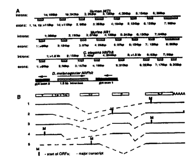
Fig. 3. Genomic organization of human and marine NITI genes and D.
25 melanogaster and C. elegans Nit-Fhit genes. (A) Exon-intron structure of
the
genes. (B) Alternative processing of human NITI gene.
Fig. 4. Cleavage of ApppA by D. melanogaster Nit-Fhit. At indicated
times of incubation, samples were spotted on TLC plates with appropriate
nucleotide standards.
30 Fig. 5. Analysis of alternative transcripts of human NITI by RT-I'CR RT-
PCR of HeLa RNA was performed with primers in different exons. Lanes 1-6:
PYlInC 1 anr~ 't ltrancnrint 71~ Pxnnc 1C'. anal 'i ttranccrint 51: exons 1A
and 3
CA 02335315 2002-O1-16
WO 00/03685 6 PCT/US99I16366
(transcripts 3, upper band and 4, Iowa band): exons 2 and 3 (scripts 2-4); .
exons 1 and 1 C (transcript 5); and exans 1 and 2 (transcript 2).
Fig. 6. Highly conserved sequence of human, marine, D. melanogaster,
and C. elegans NITI gene. (SEQ ID NO:1 ).
DETAILED DESCRI,1'T10N
Genomic and cDNA clones
One million plaques of a mouse genomic library (bacteriophage library
from strain SVJ129, Stratagene, La Jolla, CA) and one hundred thousand plaques
of a D. melanogaster genomic library were screened with corresponding cDNA
probes. Clones were purified and DNA was isolated. Sequencing was carried out
using Perkin Elmer thermal cyclers and ABI 377 automated DNA sequencers.
DNA pools from a human BAC library (Research Genetics, Huntsville, AL) were
screened by PCR with NITI primers (TCTGAAACTGCAGTCTGACCTCA (SEQ
ID N0:2) and CAGGCACAGCTCCCCTCACTT (SEQ ID N0:3)) according to
the supplier's protocol. The DNA from the positive clone, 31 Kl 1, has been
isolated using standard procedures and sequenced. Chromosomal localization of
the human NIT! gene was determined using a radiation hybrid mapping panel
(Research Genetics) according to the supplier's protocol and with the same
primers as above. To map marine Nitl gene, Southern blot analysis of genomic
DNA from progeny of a (AEJlGn-a bpHla bpH x M. spretus)F1 x AEJlGn-a bp~l a
bp" backcross was performed using a full length marine Nitl cDNA pmbe. This
probe detected a unique 2.0 kb DraI fragment in AEJ DNA and a unique 0.75 kb
fragment in M. spretus DNA. Segregation of these fragments were followed in
180 N2 offspring of the backcross. Additional Mit markers (DIMit34, DIMit35,
and DIMit209) were typed from DNA of 92 mice by using PCR consisting of an
initial denaturation of 4 minutes at 94°C followed by 40 cycles of
94°C for 30
seconds. 55°C for 30 seconds and 72°C for 30 seconds. Linkal~e
analysis was
CA 02335315 2002-O1-16
WO 00/03685 ~ PCTNS99/16366
performed using the computer program SPRETUS MADNESS: PART DEUX. '
Human and mouseNITl expressed sequence tag (EST) clones were purchased
form Research Genetics. The sequences of human and marine NITI genes and
cDNAs and D. melanogaster and C. elegans Nit-Fhit cDNAs have been deposited
in GenBank.
In situ hvbridization
D. melanogaster polytene chromosome spreads were prepared from
salivary glands of third-instar larvae as described. NitFhit DNA fragments
were
labeled with digoxigenin-11-dUTP using a random-primed DNA labeling kit
(Boeringer Mannheim, Indianapolis, IN), and were used as probes for the
chromosomal in situ hybridization. Hybridization was for 20 hours at
37°C in
hybridization buffer: 50% formamide, 2x standard saline citrate (SSC), 10%
dextran sulfate, 400 mglml salmon sperm DNA. Antidigoxigenin-fluorescein
antibodies (Boehringer Mannheim) were used for detection of hybridizing
regions.
DNA was counterstained with Hoechst 33258 (Sigma, St. Louis, MO). The slides
were analyzed by fluorescence microscopy. For in situ hybridization, embryos
were fixed and processed as described previously, except that single-stranded
RNA probes were used. Full length NitFhit cDNA was cloned into BluescriptII
KS+ vector and used to synthesize antisense RNA probes with the Genius 4 kit
(Boehringer Mannheim).
RT PCR. Northern and RACE analysis
Human and mouse multiple tissue northern blots (Clontech, Palo Alto, CA)
were hybridized with corresponding NITI cDNA probes and washed using the
supplier's protocol. For the HeLa cell line, total RNA was isolated from 1-S x
10a
cells using Trizol reagent (Gibco BRL, Gaithersburg, MD). D. melanogaster
PolyA+ RNA was purchased from Clontech. Three pg of polyA+ RNA or 15 pg
of total RNA were electrophoresed in 0.8% agarose in a borate buffer
containing
CA 02335315 2002-O1-16
WO 00103685 g PCTIUS99i1b366
formaldehyde, transferred to HybondN+ membrane (Amersham, Arlington
Heights, IL) using standard procedures and hybridized as described above. For
RT-PCR, 200 ng of polyA+ RNA or 3 pg of total RNA were treated with DNaseI
5 (amplification grade, Gibco BRL) following the manufacturer's protocol.
DNase-
treated RNA was used in reverse transcription (RT) reactions as follows: 10 nM
each dNTP, 100 pmoles random hexamers (oligo (dT) priming was used in some
cases), DNaseI treated RNA, and 200 units of marine leukemia virus (MuLV)
reverse transcriptase (Gibco BRL), in total volume of 20 Pl were incubated at
10 42°C for 1 hour followed by the addition of 10 pg RNase A and
incubation at
37°C for 30 min. One p1 of the reaction was used for each PCR reaction.
PCR
reactions were carried out under standard conditions using 10 pmoles of each
gene-specific primer and 25-35 cycles of 95° 30", 55-60° 30",
72° 1'. Products
were separated on 1.5% agarose gels and sometimes isolated and sequenced or
15 cloned and sequenced. Oligo (dT}-primed double-stranded cDNA was
synthesized
by using procedures and reagents from the Marathon RACE cDNA amplification
kit (Clontech); the cDNA was ligated to Marathon adapters (Clontech). 3' and
5'
RACE products were generated by long PCR using gene-specific primers and the
APl primer (Clontech). To increase the specificity of the procedure, the
second
20 PCR reaction was carried out by using nested gene-specific primers and the
AP2
primer (Clontech). PCR reactions were performed according to the Marathon
protocol using the Expand long template PCR system (Boehringer Mannheim) and
30 cycles o~ 94° 30", 60° 30", b8° 4'. RACE products were
electrophoresed,
identified by hybridization and sequenced. Degenerate FHIT primers were:
25 GTNGTNCCNGGNCAYGTNGT (SEQ ID N0:4) and
ACRTGNACRTGYTTNACNGTYTGNGC (SEQ ID NO:S). D. Melanogaster
Fhit RACE and RT-PCR primers were: GCGCCTTTGTGGCCTCGACTG (SEQ
ID N0:6) and CGGTGGCGGAAGTTGTCTGGT (SEQ ID N0:7). C. elegans
Fhit RACE and RT-PCR primers were: GTGGCGGCTGCTCAAACTGG (SEQ
30 ID N0:8) and TCGCGACGATGAACAAGTCGG (SEQ ID N0:9). Human NIT!
RT-PCR primers were: GCCCTCCGGATCGGACCCT (SEQ ID NO:10) (exon
1 ); GACCTACTCCCTATCCCGTC (SEQ ID NO:11 ) (exon 1 a);
CA 02335315 2002-O1-16
wo ooro~sss 9 pcrms~n ~~s
GCTGCGAAGTGCACAGCTAAG (SEQ ID N0:12) and
AAACTGAAGCCTCTTTCCTCTGAC (SEQ ID N0:13) (exon lc);
TGGGCTTCATCACCAGGCCT (SEQ ID N0:14) and
CTGGGCTGAGCACAAAGTACTG (SEQ ID NO:15) (exon 2);
GCTTGTCTGGCGTCGATGTTA (SEQ ID N0:16) (axon 3).
Protein expression and enzvmatic characterization
The NIT FHIT cDNA was amplified with primers
TGACGTCGACATATGTCAACTCTAGTTAATACCACG (SEQ ID N0:17) and
TGGGTACCTCGACTAGCTTATGTCC (SEQ ID N0:18), digested with Nde1
and KpnI, and cloned into plasmid pSGA02 as a Ndel-Kpnl fragment.
Escherichia coli strain SG100 transformants were grown in Luria-Hertani with
100 p.g/ml of ampieillin and 15 pg/ml of chloramphenicol at I S°C. When
the
culture reached an optical density (600 nm) of 0.25, isopropyl Q-D-
thiogalactoside
was added to a final concentration of 200 pM. NitFhit protein was purified
from
inclusion bodies as described. Briefly, the cell pellet from a 1-liter culture
was
resuspended in 50 ml of 20 mM Tris~HCl (pH 7.5), 20% sucrose, 1mM EDTA and
repclleted. Outer cell walls were lysed by resuspension in ice-water.
Spheroblasts
'~ were pelleted, rcsuspended in 140 mM NaCI, f:7 mM ICI, 12 mM Na~P04 {pH
7.3), SmM EDTA, SOOmM phenylmethylsulfonyl fluoride, 1 pg/ml leupeptin and
20 pglml of aprotinin, and sonicated. The resulting inclusion body preparation
was washed and solubilized in 5 M guanidinium hydrochloride, SOmM Tris~HCl
(pH 8.0), SmM EDTA. Soluble NitFhit protein was added dropwise to 250m1 of
SOmM Tris~HCI (pH 8.0), 1mM DTT, 20% glycerol at 40°C. After a 14
hour
incubation, the 13-kg supernatant was concentrated 100-fold with a Centricon
filter. A 1-liter culture yielded approximately 200 p.g of partially purified,
soluble
NitFhit. ApppA hydrolase activity was assayed at 30°C in 20 p1 of
SOmM
Na~HEPES pH 7.5, 10% glycerol, 0.5 rnM MnCl2, 4mM ApppA, 1 ~,M NitFhit.
TLC plates were developed as described.
CA 02335315 2002-O1-16
WO 00/03685 1 ~ PCT/U599/16366
Cloning and characterization of D. melanoQaster and C. elegans
Fhit homologs
To obtain D. melanogaster Fhit sequences, degenerate primers were
designed in the conserved regions of axons 5 and ? of human FH~T. RT-PCR
experiments with these primers and D, melanogaster RNA resulted in an 200 by
product, which when translated showed ~50% identity to human Fhit protein.
This
sequence was used to design specific D. melanogaster Fhit primers. 5' and 3'
RACE with these primers resulted in ~1.5 kb full length cDNA (including
polyadenylation signal and Poly(A) tail) encoding a 460 amino acid protein
with a
145 amino acid C-terminal part homologous to human Fhit (40% identity and 47%
similarity) and a 315 amino acid N-terminal extension (Fig. l). Northern
analysis
(Fig. 2C) showed a singer band of ~1.5 kb in both embryo and adult D.
I S melanogaster confirming that the full length cDNA has boon clonod.
The 460 amino acid predicted protein sequence was used in a BLASTP
search. Of the top SO scoring alignments, 22 aligned with the 145 residue C-
terminal segment (Fhit-related sequences) and 28 aligns with the 315 residue N-
terminal segment. The 28 sequences aligning with the N-terminus were led by an
uncharacterized gene from chromosome X of Saccharomyces cerevisiae (P-value
of 1.4 x 10'~s), followed by uncharacterized ORFs of many bacterial genomes
and
a series of enzymes from plants and bacteria that have been characterized as
nitrilases and amidases. Thus, the 460 amino acid predicted protein contains
an N-
terminal nitrilase domain and a C-terminal Fhit domain and was designated
NitFhit.
The D. melanogaster Nit-Fhit cDNA probe was used to screen a D.
melanogaster lambda genonue library. Sequencing of positive clones revealed
that the gene is inironless and, interestingly, the 1.5-kb Nit-Fhit gene is
localized
within the 1.6-kb intron 1 of the D. melanogaster homolog of the marine
glycerol
kinase (Gyk) gene. The direction of transcription of the Nit-Fhit gene is
opposite
to that of the Gyk gene (Fig. 3A). It is not known if such localization
affects
transcrintional regulation of these two ttenes.
CA 02335315 2002-O1-16
W0 00/03655 11 PCTIUS99f16366
The cytological position of the Nit-Fhit gene was determined by in situ
hybridization to salivary gland polytene chromosomes. These experiments
showed that there is only one copy of the sequence which was localized to
region
S 61A, at the tip of the left arm of chromosome 3. Digoxigenin-labeled RNA
probes
were hybridized to whole-mount embryos to determine the pattern of expression
during development. Nit-Fhit RNA was uniformly expressed throughout the
embryo suggesting that NitFhit protein could be important for most of the
embryonic cells.
. Because human Fhit protein and the D. melanogaster Fhit domain were
only 40% identical, to show that the authentic D. melanogaster Fhit homolog
was
cloned, its enzymatic activity was tested. Fig. 4 shows that recombinant D.
melanogaster Nithhit is capable of cleaving ApppA to AMP and ADP and
therefore possesses ApppA hydrolase activity.
. el ans
Fhit genomic sequences were obtained from the Sanger database (contig
Y56A3) by using BLAST searches. 5' and 3' RACE with C. elegans Fhit specific
primers yielded a 1.4-kb cDNA {including polyadenylation signal and Poly(A)
tail) coding for a 440 amino acid protein (Fig. 1). Northern analysis (Fig.
2C)
showed a single band of a similar size in adult worms. Similarly to D.
melanogaster, the C. elegans protein contained an N-terminal nitrilase domain
and
a C-terminal Fhit domain (Fig, l) with 50% identity and 57% similarity to
human
Fhit. Comparison between C. elegans Nit-Fhit cDNA and genomic sequences
from the Sanger database revealed that the C. elegans Nit-Fhit gene comprises
8
exons and is more than 6.5 kb in size (Fig. 3A); the nitrilase domain is
encoded by
exons 1-6, and the Fhit domain is encoded by exons 6-8. D. melanogaster and C.
elegans NitFhit proteins are 50°10 identical and 59% similar and
exhibit several
conserved domains (Fig. l).
CA 02335315 2002-O1-16
wo oonu36ss 12 ~crius~n~s
Cloning and ra t~~rized of human and murlne NIT cDNAs anil genes
Because Fhit and nitrilase domains are part of the same polypeptides in D.
melanogaster and C. elegans, it is reasonable to suggest that they may be
involved
in the same biochemical or cellular pathways) in these organisms. Because
nitrilase homologs are conserved in animals, the mammalian nitrilase homologs
were cloned as candidate Fhit-interacting proteins.
To obtain human and marine NITI sequences, the D. melanogaster nitrilase
10 domain sequence was used in BLAST searches of the GenBank EST database.
Numerous partially sequenced human and marine NITI ESTs were found. All
mouse Nitl ESTs were identical, as were all human NITI ESTs, suggesting the
presence of a single NITI gene in mouse and human. To obtain the full-length
human and mouse cDNAs, several human and mouse ESTs and human 5' and 3'
15 RACE products were completely sequenced. This resulted in the isolation of
a
~1.4-kh full-length human sequence encoding 327 amino acids and a ~1.4-kb
mouse full-length sequence coding for 323 amino acids (FIg. 1), although
several
alternatively spliced products were detected in both cases (see below and Fig.
3B).
Both cDNAs are polyadenylated, but lack polyadenylation signals, although AT-
20 rich regions arc present at the very 3' aid of each cDNA. Mouse and human
Nitl
amino acid' sequences were 90°l° identical; the human Nitl amino
acid sequence
was 58% similar and 50% identical to the C. elegans nitrilase domain and 63%
similar and 53% identical to the D. melanogaster nitrilase domain (FIg. l).
Marine lambda and human BAC genomic libraries were screened with the
25 corresponding NITI cDNA probes, yielding one mouse lambda clone and one
human BAC clone containing the NITI genes. The human and marine NITI
genomic regions were sequenced and compared to the corresponding cDNA
sequences. The genomic structure of human and mouse NITI genes is shown in
Fig. 3A. Both genes are small: the human gene is ~3.2 kb in size and contains
7
30 exons; the marine gene is --3.6 kb in size and contains 8 exons. Southern
analysis
confirmed that both human and mouse genomes harbor a single NITl gene.
CA 02335315 2002-O1-16
WO 00/03685 ~ 3 QCfNS99116366
A radiation hybrid mapping panel (GeneBridge 4) was used to detenmine
the chromosomal localization of the human NITI gene. By analysis of PCR data
at
the Whitehead/MIT database (hrip;!lwww-genome.wi.mit.edu), the NITI gene was
localized 6.94 cR from the marker CHLC.GATA43A04, which is located at 1q21-
1 q22.
A full length marine Nitl cDNA probe was used to determine the
chromosomal location of the marine gee by linkage analysis. Interspecific
backcross analysis of 180 NZ mice demonstrated that the Nitl locus
cosegregated
with several previously mapped loci on distal mouse chromosome 1. The region
to which Nitl maps was further defined by PCR of genomic DNA from 92 NZ mice
using the markers DIMit34, DlMit35 and DlMit209 (Research Genetics). The
following order of the genes typed in the cross and the ratio of recombinants
to Nz
mice was obtained: centromere - DIMit34 - 7178 - DIMit35 - 8190 - Nitl - 11/91-
DIMit209 - telomere. The genetic distances given in centiMorgans (tS.E.) are
as
follows: centromere - DlMit109 - 9.0 ~ 3.2 - DlMit35 - 8.9 t 3.0 - Nit7 - 12.1
X3.4 - DIMit209 - telomere. This region of mouse chromosome 1 (1q21 - 1q23)
is syntenic to human chromosome 1q and is consistent with the localization of
the
human ortholog of Nitl.
expression and altern4tive splicing ofhuman and marine Nitl genes
For the human gene, Northern analysis revealed two major transcripts of
~1.4 kb and ~2.4 kb in all adult tissues and tumor cell lines tested. A third
band of
~1.2 kb was observed in adult muscle and heart (Fig. 2B). The longest cDNA
(~1.4 kb) corresponds to the ~1.4-kb transcript observed on Northern blots.
The
1.2-kb band corresponds to transcript 1 on Flg. 3B (see below). It is not
known if
the --2.4-kb RNA represents an additional transcript or an incompletely
processed
mRNA. No significant variation in human NITI mRNA levels was observed in
different tissues (Fig. 2B). On the contrary, different mouse tissues showed
different levels of expression of Nttl mRNA (Flg. 2A). The highest levels of
Nitl
mRNA were observed in mouse liver and kidney (Fig. 2A, Middle, lanes 5 and 7).
CA 02335315 2002-O1-16
WO 00/03685 14 PC'1'/US99/16366
Interestingly, the pattern of Nitl expression was almost identical to the
pattern of.
the expression of Fhit (Fig. 2A, Top and Middle), supporting the hypothesis
that
the proteins may act in concert or participate in the same pathway.
Analysis of mouse Nitl ESTs revealed that some transcripts lack exon 2
and encode a 323 amino acid protein. An alternative transcript containing exon
2
encodes a shorter, 290 amino acid protein starting with the methionine 34
(Fig. l).
Analysis of human ESTs and 5' RACE products from HeLa and testis also
suggested alternative processing. To investigate this; a series of RT-PCR
experiments was carried out. Fig. 5 shows the results obtained from HeLa RNA
(similar results were obtained using RNAs from the MDA-MB-436 breast cancer
cell line and adult liver). The alternatively spliced transcripts are shown on
Fig.
3B. Transcript 1, lacking exon 2, was represented by several ESTs in the
Genbank
EST database. This transcript probably corresponds to the ~1.2-kb transcript
observed on Northern blots in adult muscle and heart. Transcript 2 encoding
the
327 amino acid Nitl protein (Fig. l) is a major transcript of human NITI at
least in
the cell lines tested. This transcript lacks exons la and 1b. Transcript 3 has
exon
la and 1b; transcript 4 has exon la but lacks exon 1b (Fig. 3B). It is not
known if
transcript 5 (lacking exon 2) starts from exon 1 or lc.
The alternative initiating methionines of different transcripts are shown on
Fig. 3B. Data suggest that at least in COS-7 cells transfected with a
construct
containing transcript 2, the methionine in exon 3 (shown in transcripts l and
3,
Fig. 3B) initiates more efficiently than the methionine in exon 2 (Fig. 3B,
transcript 2).
Discussion
Although the frequent loss of Fhit expression in several common human
cancers is well documented, and results supporting its tumor suppressor
activity
have been reported, the role of Fhit in normal and tumor cell biology and its
mechanism of its action in vivo are unknown. The Ap3A hydrolytic activity of
Fhit seems not to be required for its tumor suppressor function, and it has
been
CA 02335315 2002-O1-16
WO 003685 ~ 5 PCTIUS99116366
suggested that the enzyme-subtract complex is the active form of Fhit. To.
facilitate an investigation of Fhit function, a model organisms approach was
initiated by cloning and characterization of D. melanogaster and C. elegans
Fhit
genes.
Surprisingly, in flies and worms, Fhit is expressed as a fusion protein with
the Fhit domain fused into a "Nit" domain showing homology to plant and
bacterial nitrilases. Human and marine NfTI genes were further isolated. Nit
and
Fhit are expressed as separate proteins in mammals but, at the mRNA level, are
coordinately expressed in mouse tissues.
In several eukaryvtic biosynthetic pathways multiple steps are catalyzed by
multifunctional proteins containing two or more enzymatic domains. The same
steps in prokaryotes frequently are carried out by monoenzymatic proteins that
are
hornologs of each domain of the comsponding eukaryotic protein. For example,
Gars, Gart and Airs arc domains of the same protein in D. melanogaster and
mammals. These domains catalyze different steps in de novo synthesis of
purines.
In yeast, Gart homolog (Ade8) is a separate protein and Gars and Airs homologs
(AdeS and Ade7} are domains of a bienzymatic protein; in bacteria, all three
homologs (PurM, PurN and PurD) are separate proteins. De novo pyrimidine
biosynthesis illustrates a similar case. Recently, a fusion protein of a
lipoxygenase
and catalase, both participating in the metabolism of fatty acids; has been
identified in corals. In all of these examples, if domains of a multienzymatic
protein in some organisms are expressed as individual proteins in other
organisms,
the individual proteins participate in the same pathways. This observation and
the
fact that lrhit and Nitl exhibit almost identical expression patterns in
marine
tissues suggest that Fhit and Nitl participate in the same cellular pathway in
mammalian cells.