Note: Descriptions are shown in the official language in which they were submitted.
CA 02335831 2000-12-21
WO 00/00091 PCTIUS99/09861
ECCENTRIC SURGICAL FORCEPS
FIELD OF THE INVENTION
The present invention relates to surgical instruments, such as forceps having
a distal
assembly for performing internal surgical procedures involving cutting,
grasping, and/or removing
internal tissue. More particularly, the present invention relates to biopsy
forceps, having
articulated end effectors, for cutting and/or removing tissue samples.
BACKGROUND OF THE INVENTION
Surgical procedures involving the cutting, grasping, and/or removal of
internal tissue are
frequently performed using various forms of surgical forceps which may be
passed into the body
through an elongated, usually flexible tube, such as an endoscope, catheter,
cannula, or trocar.
Examples of endoscopes used in these procedures are laparoscopes, thorascopes,
and
arthroscopes. Typical of such forceps is a bioptome used to remove biopsy
specimens internally.
The bioptome includes a long flexible coil having a pair of opposed jaws at
the distal end and
manual actuator at the proximal end. Manipulation of the actuator opens and
closes the jaws.
During biopsy tissue sample procedures, the bioptome, or other surgical
instrument, is inserted
into the narrow lumen of an endoscope while viewing the biopsy site through an
optical scope.
The surgeon guides the endoscope to the biopsy site until the opposed jaws
arrive at the biopsy
site. The surgeon then positions the jaws of the bioptome around the tissue to
be sampled and
manipulates the actuator so that the jaws close around the tissue. A sample of
the tissue is then
cut or torn away from the biopsy site while it is trapped between the jaws of
the bioptome.
Keeping the jaws closed, the surgeon then withdraws the bioptome from the
endoscope and opens
the jaws to disgorge the biopsy tissue sample.
- -- ---------- - --------
CA 02335831 2000-12-21
WO 00/00091 PCT/US99/09861
-2-
Because the lumen of the endoscope is usually relatively small, the bioptome
inserted
through the lumen must also be small and of compact configuration. Further, in
order to achieve
the necessary manipulation required to remove the sample and extract it
through the lumen of the
endoscope, the bioptome frequently has a fairly complex structure. Because of
the size constraint
and other limitations associated with the bioptome, the jaws typically open to
a relatively narrow
extent, thereby severely limiting the size and type of specimen that can be
extracted. Some
conventional bioptomes, for example, frequently employ a fixed jaw and a
single movable jaw
which closes against the fixed jaw. Other conventional bioptomes employ two
movable jaws
which are separately activated by complex, multi-link systems and which can
only be opened to a
relatively narrow extent.
The same considerations and constraints generally apply for other surgical
forceps used
for cutting, grasping, and/or removing internal tissue.
SUMMARY OF THE INVENTION
It is therefore an object of the invention to provide a surgical instrument
for internally
cutting, grasping, and/or removing tissue, the instrument having end effectors
with jaws that can
open relatively widely once inserted into an internal body area.
It is a further object of the invention to provide a surgical instrument for
internally cutting,
grasping, and/or removing tissue having end effectors with jaws that can be
closed into a compact
configuration to facilitate passage through the lumen of a surgical scope.
Additional objects and advantages of the invention will be set forth in the
description
which follows and, in part, will be apparent from the description or may be
learned by practice of
the invention. The objects and advantages of the invention will be realized
and attained by means
of the elements and combinations particularly pointed out in the appended
claims.
CA 02335831 2006-04-18
-3-
To achieve the objects and in accordance with the purpose of the invention, as
embodied and broadly described herein, the invention comprises a surgical
instrument, such
as a surgical forceps, having a distal assembly for cutting, grasping, and/or
taking internal
body tissue that includes a proximal actuator assembly and a distal end
effector assembly
having first and second end effectors in which the proximal end of the second
end effector
pivotally engages the first end effector intermediate its proximal and distal
ends. The two end
effectors thereby form a pair of movable jaws in which the second end effector
pivotally
engages with the distal end of a clevis at a point intermediate its length.
The clevis has an
axial passage therethrough to accommodate a slidable, elongated actuator whose
distal end is
pivotally attached to the proximal end of the first end effector which is also
slidably disposed
in the distal end of the clevis. The proximal end of the clevis is adapted for
attachment to the
actuator assembly, which includes a tubular sheath containing a slidable,
elongated cable that
engages the proximal end of the actuator to permit axial displacement of the
actuator and the
end of the end effector to which it is attached, to cause the two jaws of the
device to open or
close.
In one aspect there is provided a distal assembly for grasping or cutting
internal body
tissue, the distal assembly comprising first and second end effectors
pivotally engaging one
another; a clevis having a distal end in pivotal engagement with said second
end effector, said
clevis having a longitudinal passage therethrough; and a slidable actuator
disposed in said
passage of said clevis wherein a distal end of the actuator is pivotally
attached to a proximal
end of said first end effector in the passage of said clevis actuator.
In another aspect there is provided surgical forceps for grasping or cutting
internal
CA 02335831 2006-04-18
-3a-
tissue, comprising a sheath having a distal end and a proximal end; an
elongated member
having a distal end and a proximal end, slidably disposed within said sheath;
an end effector
assembly disposed at the distal end of said sheath and comprising first and
second end
effectors pivotally engaging one another; a clevis having a proximal end
engaging the distal
end of the sheath and a distal end in pivotal engagement with said second end
effector, said
clevis having a longitudinal passage therethrough; a slidable actuator
disposed in the passage
and having a proximal end attached to the distal end of said elongated member,
wheresin a
distal end of the actuator is pivotally attached to the proximal end of said
first end effector in
said the passage of said clevis; and a manipulator attached at the proximal
end of said sheath
for causing axial displacement of said elongated member relative to said
sheath to cause
articulation of said first and second end effectors.
In another aspect there is provided an end effector assembly for use in a
surgical
forceps, comprising first and second articulated end effectors, each having a
proximal end
and a distal end adapted for opposing engagement with the distal end of the
other, the
proximal end of the first effector being adapted for engagement with a distal
end of an axially
displaceable actuator and the proximal end of the second effector pivotally
engaging said first
effector at a point intermediate the proximal and distal ends of the first
effector; the proximal
end of said second end effector being adapted for pivotal engagement with the
distal end of a
clevis having an axial passage for accommodating said actuator.
It is to be understood that both the foregoing general description and the
following
detailed description are exemplary and explanatory and are intended to provide
furthe;r
explanation of the invention as claimed.
CA 02335831 2006-04-18
-3b-
The accompanying drawings are included to provide a further understanding of
the
invention and are incorporated in and constitute a part of this specification,
illustrate
embodiments of the invention and together with the description serve to
explain the
principles of the invention.
BRIEF DESCRIPTION OF THE DRAWINGS
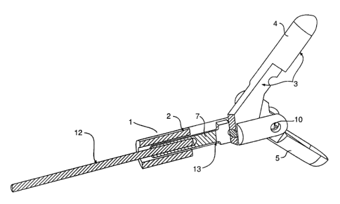
Figure 1 is a partial cut-away perspective view showing one side of the distal
assembly of the present invention with the respective jaws in an open
configuration.
CA 02335831 2000-12-21
wo 00/00091 PCT/US99/09861
-4-
Figure 2 is a side view illustrating an opposite side of the distal assembly
shown in
Figure 1.
Figure 3 is a side internal view of the component parts of the distal assembly
of Figure 1
with the jaws in the open configuration.
Figure 4 is a side internal view of the internal component elements of the
distal assembly
of Figure 1 with the assembly in the closed configuration.
Figure 5 is a bottom view of the assembly of Figure 4.
Figure 6 is a side internal view illustrating the opposite side of the
assembly of Figure 1
with the jaws in the open configuration.
Figure 7 is a top view of the distal assembly of Figure 1.
Figure 8 is an exploded top view of the assembly of Figure 1.
Figure 9 is a side view of the entire surgical forceps of the invention with
the distal end
effector assembly in the open configuration.
Figure 10 is a side view of the entire surgical forceps of the invention with
the distal end
effector assembly in the closed configuration.
Figure 11 is a front view of the assembly of Figure 1 with the jaws in open
configuration.
Figure 12 is a side view of an alternative embodiment of the distal assembly
of the
invention.
DESCRIPTION OF THE PREFERRED EMBODIMENTS
Reference will now be made in detail to the present preferred embodiments of
the
invention, examples of which are illustrated in the accompanying drawings.
Whenever possible,
the same reference numbers will be used throughout the drawings to refer to
the same or like
parts.
CA 02335831 2000-12-21
WO 00/00091 PCTIUS99/09861
-5-
The present invention is directed to an improved distal assembly for a
surgical instrument
and more particularly to surgical forceps having a distal assembly, for
example, for taking biopsy
specimens, and including a pair of end effectors that are interconnected to
form a pair ofjaws that
can be widely opened and retracted to form a compact, linear unit that is
compatible with the
lumen of a surgical scope while also permitting wide opening of the jaws to
facilitate their use
once in place internally in a body.
The device of the invention can be employed in connection with various
surgical scopes,
trocars, cannulas, or catheters for removing, grasping, or cutting tissue
specimens internally.
Further, the distal assembly described herein is not limited in its
application to surgical forceps,
but can also be incorporated in other surgical devices having opposed jaws or
cutting surfaces.
Typical of devices useful in the invention are various graspers, surgical
scissors, dissectors,
clamps, and forceps
Figure 1 of the drawings illustrates the distal assembly incorporated in
surgical forceps in
accordance with the present invention. The distal assembly 1 includes a clevis
2 having an
actuator rod 7 slidably disposed within it and two end effectors 3 and 5 with
opposing end
portions 4 and 6 which, in the illustrated embodiment, are cup-shaped jaws
having sufficiently
sharp edges to facilitate the cutting, grasping, and removing of tissue
samples when the respective
end portions 4 and 6 are brought together by axial displacement of the
actuator rod 7 whose distal
end pivotally engages the proximal end of end effector 3 disposed within the
clevis.
Figure 2 of the drawings illustrates the side of the surgical forceps of the
invention
opposite that shown in Figure 1 and, in particular, illustrates the off-set,
pivotal engagement of
the proximal end of end effector 5 with end effector 3 at pivot point 8
intermediate the proximal
and distal ends of end effector 3 resulting in pivot point 8 being out of
axial alignment with
actuator 7 in clevis 1 when the end effectors 3 and 5 are open as shown. When
the end effectors
CA 02335831 2006-04-18
-6-
3 and 5 are closed together as shown in Figures 4 and 5, pivot point 8 is in
axial alignment
with the actuator 7 within the clevis 2.
The internal configuration of the distal assembly of the invention is shown in
Figures
3 and 6, which are internal side views corresponding to Figure 1. Distal
assembly 1 includes
an elongated clevis 2 having a distal end 14 and a pair of engaging end
effectors 3 and 5. The
shorter of the two end effectors 5 is pivotally attached at pivot point 10 to
the distal end 14 of
the clevis and also pivotally attached at pivot point 8 to the other end
effector 3. As shown in
at least Figure 6, a cut-out portion 16 in effector 3 accommodates pivot point
10 when the
end effectors are closed. End effector 3, which is the longer of the two end
effectors, has a
pivotal engagement at pivot point 9 with the distal end of actuator rod 7
which is slidably
disposed within the clevis 2. Both of the end effectors 3 and 5 are provided
with opposing
end cups 4 and 6. The distal end of flexible, tubular sheath 11 is adapted to
engage by any
appropriate, known means, such as threading or fixed attachment, with the
proximal end of
clevis 2 and contains an elongated actuator cable 12 slidably disposed
therein. Flexible cable
12 engages within channel 17 of actuator rod 7 to enable axial sliding
movement of actuator
rod 7 within the clevis 2 in response to axial movement of the actuator cable
12.
Movement of actuator rod 7 in the proximal direction (the direction of the
arrow)
causes movement in the proximal direction of the proximal end 15 of end
effector 3. This
axial movement by end effect or 3, in the direction of the arrow, also causes
pivotal rotation
of end effector 3 around pivot point 8 and pivotal rotation of end effector 5
around pivot
point 10 thereby causing the end effectors cups 4 and 6 to close towards one
another until end
effectors 3 and 5 are in opposing alignment in a compact linear configuration
as illustrated in
CA 02335831 2006-04-18
-6a-
Figure 4. Similarly, movement of the actuator rod 7 in the opposite or distal
direction with
respect to clevis 2 causes a sliding movement of proximal end 15 of end
effector 3 which
opens the jaws or end cups 4 and 6 of the device so that the opposing end
effectors 3 and 5
are eventually widely separated. The
CA 02335831 2000-12-21
WO 00/00091 PCT/US99/09861
-7-
various pivot points 8, 9, 10, and 22 are otherwise of conventional
construction and can include,
for example, any form of pin, bolt, or rotatable bearing.
As shown in Figures 9 and 10, the proximal ends of sheath 11 and cable 12
terminate
conveniently in a manipulator 18 having a hand-grip 23 including fixed grip 19
attached to barrel
24 and moveable grip 20 which pivotally engages the barrel 24 at pivot point
22. The distal end
(not shown) of grip 20 engages the proximal end of cable 12 (not shown) within
barrel 24 such
that movement of the proximal end of grip 20 toward or away from grip 19
results in axial
movement of cable 12 within barre124 and tubular sheath 11 whose proximal end
is attached by
retainer 21 to barrel 24. Other forms of manipulators known in the art can
also be employed to
effect axial movement of cable 12 thereby opening or closing the jaws of the
end effectors. For
example, handgrip 23 may be a conventional bioptome actuation device whose
construction and
operation are fully described in U.S. Patent No. 5,542,432 to Slater et al.,
the complete disclosure
of which is hereby incorporated by reference. Similarly, sheath 11 may be any
conventional long,
flexible coil-like section well known in the art for connecting a proximal
handle to a distal end
effector assembly. For example, the bioptome disclosed in the Slater et al.
patent uses a long,
flexible, and hollow coil with a control wire extending therethrough to couple
the handle to an
end effector assembly. The hollow coil may be manufactured from 304 steel wire
by forming the
Wire over a mandrel. The use of the distal assembly described herein, however,
is not limited to
the actuation assembly disclosed in Slater et al. or any specific type of
coil, and may be used in
connection with various other actuation assemblies and coils known in the art.
As noted, Figure 4 of the drawing is an internal view of the surgical forceps
of the
invention in collapsed configuration. The two opposing cups or jaws 6 and 4
are pressed against
one another and the two end effectors 3 and 5 are in close alignment
substantially within the
confines of the clevis 2.
CA 02335831 2000-12-21
WO 00/00091 PCTIUS99/09861
-8-
Figure 5 of the drawings further illustrates from the bottom the closed
configuration of the
surgical forceps of the invention. (Figures 4 and 5 do not show the tubular
sheath 11 shown in
Figures 8 and 9.)
Figure 11 of the drawings illustrates the surgical biopsy forceps of the
invention in the
open configuration as viewed from the front of the device.
Figure 12 of the drawings illustrates an alternate embodiment of the invention
whereby the
distal end 15 of end effector 3 is further stabilized and guided during its
axial displacement within
clevis 2 by providing a slot 16 in the side of the clevis to accommodate a pin
17 on the proximal
end of the effector 3.
The distal assembly of the invention, because of its unique engagement of the
end
effectors with each other and with the actuator rod within the clevis, whereby
the first effector is
directly attached to the actuator rod while the second end effector has a
pivotal attachment
between the ends of the first end effector, achieves a compact, linear
collapsed configuration,
compatible with an interior lumen of surgical scopes or similar devices. The
extremely wide
opening of the end effectors in the open configuration of up to about one
hundred twenty degrees
as shown, for example, in Figure 3, greatly facilitates the taking of tissue
specimens. Further, the
avoidance of a direct connection between the actuator rod and the second
effector while achieving
opening and closing movement of both effectors permits fewer and less complex
linkages and
reduced size while preserving the advantages of two widely opening, actuated
jaws.
The improved distal assembly of the invention has been described in connection
with a
bioptome having jaws for taking biopsy samples. It is to be understood that
other types of end
effectors, including end effectors for clamping or cutting tissue or
performing any other
appropriate surgical procedure, may be modified for use in accordance with the
present invention.
CA 02335831 2000-12-21
WO 00/00091 PCT/US99/09861
-9-
The end effector described herein is illustrative only of one preferred
embodiment of the
invention.
It will be apparent to those skilled in the art that various inodifications
and variations can
be made in the assembly of the present invention without departing from the
scope or spirit of the
invention. Other embodiments of the invention will be apparent to those
skilled in the art from
consideration of the specification and examples should be considered as
exemplary only, with a
true scope and spirit of the invention being indicated by the following
claims.