Note: Descriptions are shown in the official language in which they were submitted.
CA 02335937 2000-12-21
WO 00/00810 PCT/US99/14620
1
AZAFTIG, A PROTEOGLYCAN
FOR MONITORING CACHEXIA AND FOR CONTROL OF OBESITY
Chandan Prasad, Julio E. Figueroa II, and Parakat Vijayagopal
TECIINICAL FIELD
This invention pertains to the detection of a propensity for cachexia and to
the
control of obesity.
BACKGROUND ART
Cachexia is defined as significant weight loss. It occurs commonly in cancer
patients
and HIV-infected individuals, but can also be caused by hypercatabolism due to
cardiac
failure (especially, right-sided or biventricular failure), hepatic failure,
renal failure, bums,
inflammation (including sepsis), infection or tuberculosis. See R.B. Verdery,
"Reversible
and irreversible weight loss (cachexia) in the elderly," in Textbook of
Internal Medicine, 2d
Edition (V.T. DeVita et al. eds.), Ch. 523, pp. 2424-2425 (1992); K.I. Marton,
"Approach
to patient with unintentional weight loss," in Textbook of Internal Medicine,
2d Edition
(V.T. DeVita et al. eds.), Ch. 444, pp. 2113-2115 (1992); R. M. Jordan et al.,
"Weight
loss," in Internal Medicine, 4th Edition (J.H. Stein ed.), Ch. 152, pp. 1260-
1262 (1994);
C.P. Artz et al., "Burns: Including cold, chemical, and electrical injuries,"
in Textbook of
Surgery, 11th Edition (D.C. Sabiston, Jr. ed.), Ch. 15, pp. 295-322 (1977); E.
Braunwald,
"Heart Failure," in Harrison's Principles of Internal Medicine, 13th Edition
(K.J. Isselbacher
ed.), Ch. 195, pp. 998-1009 (1994); and D.W. Foster, "Gain and loss in
weight," in
Harrison's Principles of Internal Medicine, 13th Edition (K.J. Isselbacher
ed.), Ch..40, pp.
221-223 (1994). Over 50% of cancer and HIV-infected patients experience an
unintended
weight loss of greater than 10% of their baseline weight. Moreover, this
weight loss is
associated with an increase in morbidity and mortality. Many cachectic
patients manifest
multiple physiological problems involving the immune system, muscular system,
and hepatic
function that can be directly related to loss of body weight or wasting.
Therefore,
understanding the mechanisms of cachexia in patients can lead to better
treatment and
CA 02335937 2000-12-21
WO 00/00810 PCT/US99/14620
2
consequently can have a substantial impact on the quality of life and survival
of many cancer
and HIV/AIDS patients. See G.O. Coodley et al.,"The HIV Wasting Syndrome: a
Review," Journal of Acquired Immune Deficiency Syndromes, vol. 7, pp. 681-694
(1994);
L.M. Hecker et al., "Malnutrition in patients with AIDS," Nutrition Reviews,
vol. 48, pp.
393-401 (1990); N.M. Graham et al., "Clinical factors associated with weight
loss related to
infection with Human Immunodeficiency Virus Type 1 in the rnulticenter AIDS
cohort study,
" American Journal of Epidemiology, vol. 137, pp. 439-46 (1993); and K.A.
Nelson et al.,
"The cancer anorexia-cachexia syndrome," Journal of Clinical Oncology, vol.
12, pp. 213-25
(1994).
Despite the prevalence of weight loss in cancer patients, the mechanisms
underlying
the weight loss are unknown. Current explanations for cancer or AIDS-
associated weight
loss are divided into two general categories--(1) mechanisrns that decrease
food intake
(anorexia); and (2) mechanisms that increase energy expenditure through
altered or increased
metabolism. Hecker et al., 1990. Any mismatch between energy intake and
expenditure
will result in a change in weight.
Many cancer or AIDS patients have decreased oral intake and, therefore,
decreased
energy consumption. Accordingly, despite normai or even decreased energy
expenditures in
these patients, they may lose weight. Other patients experience anorexia due
to the
cancerous tumor itself (either by a mechanical obstruction or a change in
tissue function) or
due to the therapy used to treat the tumor, e.g., chemotherapy. Graham et al.,
1993; Nelson
et al., 1994. Similarly, many HIV/AIDS patients experience significant weight
loss that
correlates with decreased caloric intake. See C. Grunfeld et al.,"Metabolic
disturbance and
wasting in the acquired immunodefrciency syndrome," The New England Journal of
Medicine, vol. 327, pp. 329-337 (1992). Thus, anorexia plays a major role in
weight loss
for the majority of both cancer and HIV/AIDS patients.
Factors that have been identified as causing anorexia in patients include
opportunistic
gastrointestinal infections or tumors, side effects of treatment, enteropathy,
central nervous
system disease, and psychiatric disorders. In addition, numerous physiological
mediators of
anorexia have been reported in the literature, including tumor necrosis
factor, interleukin-1,
interleukin-6, y-interferon, and a-interferon. Coodley et al., 1994; Nelson et
al., 1994; and
Grunfeld et al., 1990. Yet the mechanisms by which these or other mediators
induce
anorexia remain unknown.
Another proposed mechanism contributing to the weight loss seen in cancer or
AIDS
patients is an increased or ineffective metabolism. It has beeri reported, and
disputed, that
CA 02335937 2000-12-21
WO 00/00810 PCT/US99/14620
3
resting energy expenditures in some patients rise throughout the course of the
disease and
increase even more at the end stage. See Coodley et al., 1994; Nelson et al.,
1994; and
Grunfeld et al., 1990. However, alterations in resting or total energy
expenditures do not
correlate with weight loss. Therefore, it is unlikely that increased energy
demands alone
account for wasting. Even with decreased energy use, patients may lose weight
due to ineffective
metabolism. It is hypothesized that during episodes of weight loss, patients
fail to switch
from carbohydrate and protein oxidation to the fatty acid oxidation that would
normally
occur under conditions of starvation. This failure explains the observation
that patients lose
predominantly muscle mass rather than fat tissue. It has also been suggested
that futile
cycling of lipid metabolism can waste energy, thus accelerating the necessity
of carbohydrate
and protein breakdown, despite a decrease in total energy expenditure. See
Coodley et al.,
1994; Nelson et al., 1994; and Grunfeld et al., 1990.
Recently, alterations in hormone metabolism have been proposed as possible
etiologies of HIV/AIDS or cancer-related weight loss, particularly due to
muscle wasting.
During severe or chronic infections, patients, particularly HIV/AIDS patients,
demonstrate
resistance to the actions of growth hormone. Because growth hormone acts to
maintain
muscle mass, it has been hypothesized that this resistance leads to muscle
wasting and weight
loss in HIV/AIDS patients. Recently, researchers demonstrated that HIV/AIDS
patients with
the wasting syndrome have a decreased response to exogenous growth hormone
compared
with a control group. In particular, the effects of growth hormone on insulin-
like growth
factor-I (IGF-I, a major mediator of growth hormone action) secretion was
studied. When
IGF-I was exogenously administered to patients with the wasting syndrome, the
patients
experienced a transient increase in nitrogen retention, but returned to
baseline after 8-10
days. See S.A. Lieberman et al., "Anabolic effects of recombinant insulin-like
growth
factor-I in cachectic patients with the acquired immunodeficiency syndrome,"
Journal of
Clinical Endocrinology and Metabolism, vol. 78, pp. 404-410 (1994). Thus,
alterations in
the growth hormone/IGF-I system may play an important role in HIV/AIDS
cachexia.
In cancer patients, growth hormone resistance has been seen, but also other
important hormones, including insulin and its antagonist glucagon, appear to
be abnormally
produced. Since these hormones are essential to normal metabolism, it has been
postulated
that abnormalities in these pathways explain the wasting syndrome in these
patients. See
Nelson et al., 1994. Unfortunately, the mechanisms by which cancer or HIV
infection
causes these alterations in hormone metabolism are poorly understood at best.
CA 02335937 2000-12-21
WO 00/00810 PCT/US99/14620
4
The control of caloric intake and body weight maintenance is very complex. The
search for endogenous mediators over several decades has led to the
identification of a
variety of substances ranging from simple amino acids to large proteins and
glycoproteins.
However, it has been difficult to establish an unequivocal association between
the amount of
any one of these factors and human disease states such as anorexia/cachexia
and anorexia
nervosa.
Three glycoproteins or proteoglycans that modulate appetite or body weight
have
been identified: satietin, satiomem, and MAC16 mouse protein. A glycoprotein
is a protein
that contains attached carbohydrates that are not polymers of repeating units.
In contrast, a
proteoglycan is a protein that contains repeating units of glycosaminoglycans
covalently
attached to a core protein.
Satietin is a glycoprotein with a molecular weight of 50,000 Dalton that has
been
isolated from human and animal sera. Satietin is known to suppress food intake
in mammals.
See J. Knoll, "Satietin, a blood-borne, highly selective and potent anorectic
glycoprotein,"
Biomed. Biochim. Acta, vol. 44, pp. 317-328 (1985); and J. Knoll, "Satietin: a
50,000
Dalton glycoprotein in human serum with potent, long-lasting and selective
anorectic
activity," J. Neural Transmission, vol. 59, pp. 163-194 (1984).
Satiomem is a proteoglycan with a molecular weight of 50,000 Dalton that has
been
isolated from plant and animal membranes, including human erythrocyte
membrane.
Satiomem has been shown to suppress food intake and cause weight loss. See
R.K. Upreti et
al., "A step towards developing the expertise to control hunger and satiety:
Regulatory role
of satiomem--A membrane proteoglycan," Neurochemical Research, vol. 20, pp.
375-384
(1995); A.M. Kidwai et al., "A Novel Plant membrane proteoglycan which causes
anorexia
in animals," Molecular and Cellular Biochemistry, vol. 120, pp. 111-117
(1993); and A.M.
Kidwai et al.,"Isolation of an anorexigenic protein from membranes," Molecular
and
Cellular Biochemistry, vol. 91, pp. 117-122 (1989).
The MAC 16 protein is a sulfated, phosphated glycoprotein of 24 kDa initially
identified from the urine of mice with the MAC 16 tumor. Using a monoclonal
antibody to
the mice MAC16 protein, a similar protein was also found in the urine of human
cachectic
cancer patients. The mouse MAC 16 protein causes weight loss in rodents,
primarily due to a
decrease in the lean body mass. The primary bioactivity of this protein is to
increase muscle
proteolysis and decrease protein synthesis. The MAC16 protein binds tightly to
muscle cell
membranes. The MAC16 protein also causes some lipolytic activity and does not
affect food
intake. The protein core of the mouse MAC 16 protein has been identified to
have at least 18
CA 02335937 2000-12-21
WO 00/00810 PCT/US99/14620
amino acids and digestion with chondroitinase AC results in a single fragment
of 14 kDa.
The human protein identified with the monoclonal antibody ("human MAC16") to
MAC16
also increases proteolysis in muscle cells. The first 14 amino acids of "human
MAC16" are
identical to those of mouse MAC16 protein. The human MAC16 has been found only
in the
5 urine of cachectic cancer patients, not in patients suffering extreme weight
loss from other
diseases such as sepsis, burns or major surgery. See P.T. Todorov et al.,
"Structural
Analysis of a Tumor-produced Sulfated Glycoprotein Capable of Initiating
Muscle Protein
Degradation," The Journal of Biological Chemistry, vol. 272, pp. 12279-88
(1997); P.
Cariuk et al., "Induction of Cachexia in Mice by a Product isolated from the
urine of
cachectic cancer patients," British Journal of Cancer, vol. 76, pp. 606-613
(1997); M.J.
Lorite et al.,"Induction of muscle protein degradation by a tumour factor,"
British Journal
of Cancer, vol. 76, pp. 1035-1040 (1997); P. Todorov et al., "Characterization
of a cancer
cachectic factor," Nature, vol. 379, pp. 739-742 (1996); P.T. Todorov et al.,
"Induction of
muscle protein degradation and weight loss by a tumor product," Cancer
Research, vol. 56,
pp. 1256-1261 (1996); T.M. McDevitt et al., "Purification and Characterization
of a Lipid-
mobilizing Factor Associated with Cachexia-inducing Tumors in Mice and
Humans," Cancer
Research, vol. 55, pp. 1458-63 (1995); J.E. Belizario et al., "Bioactivity of
skeletal muscle
proteolysis-inducing factors in the plasma proteins from cancer patients with
weight loss,"
British Journal of Cancer, vol. 63, pp. 705-710 (1991); S.A. Beck et al.,
"Lipid mobilising
factors specifically associated with cancer cachexia," British Journal of
Cancer, vol. 63, pp.
846-850 (1991); P. Groundwater et al., "Alteration of serum and urinary
lipolytic activity
with weight loss in cachectic cancer patients," British Journal of Cancer,
vol. 62, pp. 816-
821 (1990); and S.A. Beck et al., "Alterations in serum lipolytic activity of
cancer patients
with response to therapy," British Journal of Cancer, vol. 62, pp. 822-825
(1990).
At present there is no rational therapy for cachexia, i.e., one based on the
etiology of
the disease. Since conunon symptoms of anorexia/cachexia syndrome include loss
of
appetite, fat deposit, and muscle mass, all existing therapies for cachexia
include agents
known to increase appetite (e.g., cyproheptadine (PERIACTIN ), facilitate
energy storage
(e.g., megestrol acetate (MEGACE )), or increase muscle mass (androgenic
agents). While
these therapies work for some patients, for many nothing works. Since time is
very
important for these patients, until a rational therapy can be found, a need
exists to predict
which patients might respond to which of the various available therapies.
CA 02335937 2008-02-13
6
Obesity plays a major role in the etiology of many chronic diseases,
including cardiovascular diseases, cancer, and diabetes. Therefore, a national
goal has
been to reduce the prevalence of obesity in the U.S. population to no more
than 20%.
Unfortunately, there has been a substantial rise in obesity in U.S. during the
last decade.
Obesity is generally classified into two groups based on the site of fat
deposition-visceral and nonvisceral, also known as upper-body/android (apple-
shaped)
and lower-body/gynoid (pear-shaped) obesity, respectively. It is well-
established that
visceral adipose tissue is associated with greater morbidity and mortality,
particularly
hypertension, hyperlipidemia, and insulin resistance. Data also show that
weight loss by
diet, exercise, or pharmacotherapy generates a decrease in visceral adipose
tissue and
improvements in hypertension, hyperlipidemia, and insulin resistance. See F.X.
Pi-
Sunyer, "Medical Hazards of Obesity," Annals of Internal Medicine, vol. 119,
pp. 655-
660 (1993); and G.A. Bray, "Pathophysiology of Obesity," American Journal of
Clinical
Nutrition, vol. 55, pp. 488S-494S (1992).
A pharmacologic treatment to reduce body fat, particularly visceral fat,
would be of great health significance. Currently there is no available
pharmacotherapy
that will facilitate a decrease in fat deposit. Agents like REDUXTM and
Fen/phen have
been successful in obesity treatment; however, these agents have been removed
from the
market due to serious side effects.
DISCLOSURE OF INVENTION
We have discovered a proteoglycan ("azaftig") with a molecular weight of
approximately 24,000 Dalton that has been isolated and characterized from the
urine of
cachectic cancer and non-cancer patients. Azaftig has been shown to bind to
receptors
on fat cell membranes and to cause lipolysis. Azaftig does not bind to muscle
cell
membranes or cause proteolysis. Azaftig detection in urine will allow early
identification of patients in whom weight loss may become a problem. Azaftig
may also
aid fat loss in humans in whom obesity is a threat to health.
In accordance with an embodiment of the present invention there is
provided substantially pure azaftig; wherein the azaftig is a proteoglycan
having a
molecular weight of about 24 kDa as determined by sodium dodecyl sulfate-
polyacrylamide gel electrophoresis; and wherein the azaftig is obtained from
or is
identical to a proteoglycan obtained from urine of cachectic cancer patients;
is a
CA 02335937 2008-02-13
7
proteoglycan as determined by partial digestion with either chondroitinase ABC
or
chondroitinase AC; is not readily digested by neuraminidase; binds to fat cell
membranes; does not bind to muscle cell membranes; and is a negatively charged
molecule as determined by DEAE-Sephacel chromography at pH 7Ø
In accordance with another embodiment of the present invention there is
provided a method for detecting a propensity to cachexia in a human,
comprising
assaying body fluids from the human for detectable quantities of azaftig;
wherein the
azaftig is a proteoglycan having a molecular weight of about 24 kDa as
determined by
sodium dodecyl sulfate-polyacrylamide gel electrophoresis; and wherein the
azaftig is
obtained from or is identical to a proteoglycan obtained from urine of
cachectic cancer
patients; is a proteoglycan as determined by partial digestion with either
chondroitinase
ABC or chondroitinase AC; is not readily digested by neuraminidase; binds to
fat cell
membranes; does not bind to muscle cell membranes; and is a negatively charged
molecule as determined by DEAE-Sephacel chromatography at pH 7Ø
Yet another embodiment of the present invention provides for use of an
effective amount of azaftig for inducing weight loss in a mammal; wherein the
azaftig is
a proteoglycan having a molecular weight of about 24 kDa as determined by
sodium
dodecyl sulfate-polyacrylamide gel electrophoresis; and wherein the azaftig is
obtained
from or is identical to a proteoglycan obtained from urine of cachectic cancer
patients; is
a proteoglycan as determined by partial digestion with either chondroitinase
ABC or
chondroitinase AC; is not readily digested by neuraminidase; binds to fat cell
membranes; does not bind to muscle cell membranes; and is a negatively charged
molecule as determined by DEAE-Sephacel chromatography at pH 7Ø
A still further embodiment of the present invention provides polyclonal
antibodies to azaftig; wherein the azaftig is a proteoglycan having a
molecular weight of
about 24 kDa as determined by sodium is obtained from or is identical to a
proteoglycan
obtained from urine of cachectic cancer patients; is a proteoglycan as
determined by
partial digestion with either chondroitinase ABC or chondroitinase AC; is not
readily
digested by neuraminidase; binds to fat cell membranes; does not bind to
muscle cell
membranes; and is a negatively charged molecule as determined by DEAE-Sephacel
chromotography at pH 7Ø
CA 02335937 2008-02-13
8
BRIEF DESCRIPTION OF DRAWINGS
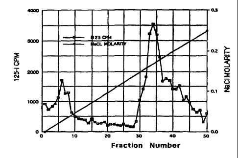
Figure 1 illustrates the DEAE-Sephacel elution profile of 125 I-azaftig.
Figure 2 illustrates the decrease in the body weight of a rat due to azaftig
injections.
Figure 3 illustrates the decrease in the body weight of mice due to azaftig
injections.
Figure 4 illustrates the time course of the weight loss of mice seen in Figure
3.
Figure 5 illustrates the time course of weight gain in mice after ceasing
azaftig
injections.
Figure 6 illustrates the decrease in percent intraperitoneal fat in azaftig-
treated
mice as measure one week after the last azaftig injection.
Figure 7 illustrates the food intake at 3 hr and 24 hr for control and azaftig-
treated mice.
Figure 8 illustrates the SephadexTM G-25 elution profile of 125 I-azaftig.
Figure 9A illustrates the specific binding of'2sI-azaftig to fat cell membrane
preparations.
Figure 9B illustrates the effect of pH on the specific binding of IZ5I-azaftig
to fat
cell membrane preparations.
Figure 9C illustrates the time dependence of specific binding of'2sI-azaftig
to fat
cell membrane preparations.
Figure l0A illustrates the effect of concentration of 125 I-azaftig on
specific
binding to fat cell membrane preparations.
Figure l OB illustrates the Scatchard analysis of the binding data from Figure
I OA.
Figure 11 illustrates the effect of azaftig on in vitro muscle cell
proteolysis.
Figure 12 illustrates the rate of blood clearance in mice of'2sI-azaftig.
Figure 13 illustrates the DEAE-SephacelTM elution profile of azaftig.
Figure 14 illustrates the Q-SepharoseTM elution profile of azaftig.
Figure 15 illustrates the binding pattern of the synthetic peptide core of
MAC16
and the MAC 16 from urine of AIDS patients.
CA 02335937 2008-02-13
8a
MODES FOR CARRYING OUT THE INVENTION
We have isolated a proteoglycan with a molecular weight of
approximately 24,000 Dalton from the urine of cachectic cancer and non-cancer
patients. We have named this proteoglycan "azaftig". Azaftig has been shown to
cause
weight loss in mammals. It has also been shown to increase lipolysis and to
bind to fat
cell membrane preparations. However, unlike the MAC 16 glycoprotein, azaftig
does not
augment proteolysis in muscle tissue or bind to muscle cell membrane
preparations.
Example 1
Isolation of Azaftig
Urine was collected for 24 hr from a patient with a diagnosis of metastatic
adenocarcinoma of unknown primary source, who had experienced a 50 lb weight
loss
over several months prior to diagnosis. The urine was treated with ammonium
sulfate
(80% saturation), and incubated overnight at 7 C. The solution was centrifuged
at 6,000
x g for I hr, and the supernatant was removed. The ammonium sulfate
precipitate was
dissolved in 50 ml of water and centrifuged again. The supematant was saved,
and the
pellet was resuspended in 5% sodium dodecyl sulfate ("SDS"). Both the
supematant and
the SDS-dissolved precipitate were subsequently separated by SDS-
polyacrylamide gel
electrophoresis. The supernatant revealed several protein bands, with two
predominant
bands at 24 kilodaltons and 70 kilodaltons. The proteins with the molecular
weight of 24
kilodaltons, or that were later determined to be its multiple (70
kilodaltons), were named
azaftigs.
Example 2
Characterization of Azaftig
DEAE-Sephacel chromatography
Azaftig of 24 kilodaltons was isolated as described above. The purified
protein was
radiolabeled with 125I using the chloramine-T method as described by F.C.
Greenwood
et al., "The preparation of 13iI-labeled growth hormone of high specific
activity,"
Biochemical Journal, vol. 89, pp. 114-123 (1963). The protein was subsequently
analyzed for charge using DEAE-Sephacel anion exchange chromatography. 125I-
azaftig
was dialyzed overnight at 4 C against a solution of 8 M urea, 0.1 M Tris, 0.3%
TritonTM
X-100, and 0.15 M NaCI (pH 7.0) containing protease inhibitors. The dialyzed
sample
was applied to a column of DEAE-Sephacel (bed volume 4 ml) that had been
CA 02335937 2008-02-13
8b
equilibrated in the same buffer as the dialyzing solution. The column was
washed with
20 ml of the same buffer at a flow rate of 10 ml/h. The column was then eluted
with a
continuous NaCI gradient (from 0.15 to 1.0 M) in the urea buffer. Fractions of
1.0 ml
were collected, and aliquots were counted in a gamma counter to determine 125
1
radioactivity. The pattern of eluting at 0.18 M NaCI demonstrated that azaftig
is a
negatively charged molecule and is likely a proteoglycan, molecules known to
have
negatively charged sulfate groups (Figure 1). Consistent with this conclusion,
chondroitinase ABC digestion as described by H. Saito et al., "Enzymatic
methods of
the determination of small quantities of isomeric chondroitin sulfate," J.
Biol.
CA 02335937 2000-12-21
WO 00/00810 PCT/US99/14620
9
Chem., vol. 243, pp. 1536-1542 (1968), of azaftig resulted in a decrease in
the azaftig band
on SDS-PAGE. Because Chondroitinase ABC is an enzynie that specifically
cleaves the
chondroitin sulfate or dermatan sulfate groups in proteoglycans, this loss in
azaftig indicated
that azaftig is a chondroitin sulfate-containing proteoglycan.
Radiolabeled azaftig was separated by SDS-PAGE. Autoradiography demonstrated
three to four distinct bands generated by purified azaftig which indicated
that azaftig had a
tendency to aggregate. To decrease aggregation of the sample, purified azaftig
was treated
with 1% Triton X-100 and subsequently chromatographed over a Sephadex G-50
column.
In addition, experiments were performed in the presence of 4 M guanidine-HCI
to minimize
aggregation. Both treatments resulted in decreased aggregation as seen by a
single band by
SDS-Page, demonstrating that azaftig forms aggregates in vitro. Subsequent
studies with
anti-azaftig antibody have also demonstrated a similar aggregation pattern, as
described in
Example 3 below.
Enzymatic digestion
'uI-azaftig was digested in separate experiments by using 50 each units of
neuraminidase, chondroitinase ABC, or chondroitinase AC. Each digestion
product was
analyzed by SDS-PAGE electrophoresis. Neuraminidase did not degrade the
proteoglycan,
while chondroitinases ABC and AC caused partial digestion. Chondroitinase AC
produced
fragments with molecular weights below 10 kDa. These data confirm that azaftig
is a
proteoglycan, because both chondroitinase ABC and AC specifically cleave the
chondroitin
sulfate or dennatan sulfate found in proteoglycans.
Example 3
Development of Western blot assay
Production of antibody to azaftig
Five Ecg of purified azaftig electroeluted from SDS-PAGE gels was injected
into New
Zealand White rabbits using complete Freund's adjuvant (Difco Laboratories,
Detroit, MI).
Subsequent immunizations were performed using the same amount of azaftig in
incomplete
Freund's adjuvant every two weeks for a total of four injections. After four
immunizations,
the rabbits were bled, and the antisera, with its polyclonal antibodies, were
tested against the
purified azaftig and the original urine samples from the patient. As
demonstrated by Western
Blot, the antiserum bound azaftig at a 1:1,000 dilution. This antiserum was
then used for the
detection of azaftig in HIV/AIDS patients with weight loss.
----- -- - - ---- -
CA 02335937 2000-12-21
WO 00/00810 PCT/US99/14620
Additional polyclonal and monoclonal antibodies to the azaftig molecule can
also be
made by a person with ordinary skill in the art using techniques well known in
the field.
Western blot methods
5 Proteins from a patient's unconcentrated urine were separated by 14% SDS-
PAGE,
and transferred to nitrocellulose by the method of H. Towbin et al.,
"Electrophoretic transfer
of proteins from polyacrylamide gels to nitrocellulose sheets: Procedure and
some
applications," Proc. Nati. Acad. Sci. U.S.A., vol 76, pp. 4350-54 (1979). The
transferred
proteins were then probed with the anti-azaftig antibody. After development
with an alkaline
10 phosphatase-conjugated goat anti-rabbit immunoglobulin, a semiquantitative
assessment was
made of the intensity of the bands present on the blot.
Example 4
Screening for Azaftig in Non-cancer, HIV Patients
Forty-two HIV-positive patients were chosen at random to provide urine samples
and
to complete a questionnaire concerning weight loss, opportunistic infections,
and other
parameters of HIV activity. All 42 were screened by the Western Blot method
discussed
above. Of the 42 patients, 17 were found not to have azaftig. Ten had large
amounts of
azaftig in the urine, while the remaining 15 had modest amounts of azaftig.
Twenty-four
patients (13 with azaftig and 11 without azaftig) completed questionnaires
that solicited
weight information. Table 1 presents the data concerning the presence of
azaftig and weight
loss in these patients.
Table 1
Azaftig in Urine
Present Absent Total
Weight Loss 9 4 13
No Weight Loss 4 7 11
Total 13 11 24
Thus in the 24 patients, 13 patients had experienced weight loss, and 9 of
these 13
(69.2%) had azaftig in their urine. Of the 11 patients that had not
experienced any weight
loss, only 4 (36.4%) had azaftig in their urine. Therefore, in this sample
population,
patients with azaftig were almost twice as likely to experience weight loss as
those without
CA 02335937 2000-12-21
WO 00/00810 PGT/US99/14620
11
azaftig. Likewise, patients with weight loss were almost twice as likely to
express azaftig as
those without weight loss. However, the sample size was not sufficiently large
to show
statistical significance (p = 0.10).
One explanation for the small number of patients who exhibited measurable
quantities of azaftig but did not experience weight loss may be differences in
the structures of
azaftig produced by different individuals.
Example 5
Concentrations of Azaftig in Cancer Patients
Twenty-three hospitalized cancer patients, seven non-cancer patients, and ten
healthy
adults were randomly selected. The non-cancer patients had been diagnosed with
diabetes,
emphysema, anemia, hypertension and coronary heart failure. The participants
were asked
to complete questionnaires detailing their eating habits and any pattern of
weight loss or
gain. Of the 23 cancer patients, seven reported weight loss, three no weight
loss, and
thirteen did not respond to the questionnaire. Of the seven non-cancer
patients, only two
patients with coronary heart failure reported weight loss. The extent of
weight loss was not
determined by the questionnaire. The total urine volume produced by each
patient over 24
hr was collected, and a portion was analyzed by SDS-PAGE. The intensity of the
azaftig
band in each sample was quantified using NIH Image software (v. 1.59). Known
concentrations of purified bovine serum albumin (BSA) were analyzed in the
same manner to
generate a standard curve. The concentration of azaftig in patient samples was
determined by
comparing the integrated densities for patient samples with band densities of
known
concentrations of BSA. The mean concentration of azaftig in the patients with
cancer was
8.37 12.51 mg/L, with a range of 0.00 to 39.25 mg/L. These data demonstrated
a great
deal of variability in the levels of azaftig in cancer patients. The non-
cancer patients and
healthy adults all had azaftig levels of 0.00 mg/L. It was interesting that
the only non-
cancer patients reporting weight loss were patients diagnosed with cardiac
failure, a disease
associated with cachexia. Without wishing to be bound by this theory, it is
possible that
these two patients were showing incipient signs of cachexia, but that azaftig
had not yet
reached measurable levels.
CA 02335937 2000-12-21
WO 00/00810 PCT/US99/14620
12
Example 6
Azaftig Causes Weight Loss in Mammals
Weight Loss in Rats
Two Sprague-Dawley rats were cannulated in the carotid artery and their
weights
allowed to stabilize after the operation. At the end of five. days, one rat
was given three
doses (at 5 hr intervals) of purified azaftig at 1 g/gram body weight,
administered in
phosphate buffered saline. The azaftig was isolated from a human cancer
patient. The other
rat received only buffered saline. As seen in Figure 2, the control rat gained
weight over 24
hr, while the azaftig-treated rat lost 10% of its body weight over the same
period. This
preliminary experiment demonstrated that the azaftig caused weight loss in
another
mammalian species.
Weight Loss in Mice
Similar studies were performed on four inbred NMRI mice (Charles River,
Willinington, MA) and four outbred NIH Swiss mice (Hilltop Laboratories,
Scottsdale, PA).
Initially, the mice were injected intraperitoneally with an elution buffer of
0.1 % SDS in 50
mM ammonium acetate for five days until their weights became stable. After
weight
stabilization, the mice were injected daily with 0.1 mg/kg of gel-purified
azaftig for five
days. Daily body weight and food intake were recorded. As shown in Figure 3,
the eight
mice lost weight at an average of 12.0 %( t 7%) of maximal measured body
weight. This
reduction is significant in a paired t-test (p=0.001) using the pre-azaftig
weight of each
animal as a control for post-treatment weight.
In Figure 4, the data are plotted to display the time course of weight loss
with the
administration of azaftig. Because the mice had different initial weights, the
data are
expressed as percentages of the maximum weight seen with each animal (mean
SD) during
the experiment. These data demonstrate that azaftig administration causes
substantial and
sustained weight loss.
To determine whether the azaftig effect was reversible, four mice of the
original
eight were allowed to recover after discontinuing the injection of azaftig. '
As shown in
Figure 5, these animals slowly regained lost weight, but did not return to
baseline. An
autopsy was performed on these four mice. The mice were found to have little
to no
intra-abdominal fat. This suggested that although the weight was regained, the
weight
increases were not due to an increase in fat mass.
CA 02335937 2000-12-21
WO 00/00810 PCT/US99/14620
13
Example 7
Effect of Azaftig on Intraperitoneal Fat
To further confirm the above observation that weight gain was not due to fat,
five
mice were given 0.1 mg/kg azaftig intraperitoneally daily for five days. One
week after the
last azaftig administration, the mice were weighed and sacrificed.
Intraperitoneal fat was
surgically removed and weighed. The percent intraperitoneal fat of total body
weight was
calculated and the results shown in Figure 6. Animals that had not received an
injection
served as controls. Azaftig-treated animals showed a significant reduction
(60%) in the
percentage of intraperitoneal fat as compared with controls. The mean
percentage
intraperitoneal fat ( standard deviation) for azaftig-treated animals was
5.75 % 1.83; for
control animals, 14.7% 2.44 (p = 0.002).
Example 8
Effect of Azaftig on Food Intake
To ascertain the effect of azaftig on appetite, experiments were performed on
fasted
mice. Eighteen female NIH Swiss mice were divided into two groups, a control
and
treatment group of nine mice each. The mice were kept from food, but not
water, for 21 hr
and then fed for 3 hr on five consecutive days. On day six, 30 min before the
scheduled
feeding time, the mice were treated intraperitoneally either with vehicle (0.1
ml/mouse) or
with azaftig (0.1 mg/kg in 0.1 ml vehicle). Thirty minutes later, food was
presented. At 3
hr and 24 hr food intake for each mice was measured. The data presented in
Figure 7 show
that azaftig did not significantly affect food intake at either 3 hr or 24 hr.
Example 9
Demonstration of Azaftig Receptors on Fat Cells
Preparation and purification of "Sl-azaftig
Azaftig was labeled using a lactoperoxidase 125I-labeling kit purchased from
ICN
Radiochemicals. Briefly, 1.5 mCi neutralized carrier-free 125 I (10 /cl) was
added to a tube
containing 30 g azaftig (100 l) and mixed thoroughly. Ten pl of
lactoperoxidase solution
(1 yg/ l) in water was added to the above mixture. The reaction was initiated
by adding 5
/cl of 3% freshly prepared H202. The addition of H202 was repeated three times
at 10 min
intervals until a total of 40 jul H2OZ was added. Ten min after the last
addition, the reaction
was terminated by dilution with 500 l of 50mM potassium phosphate buffer, pH
7.5. The
total mixture was loaded on a Sephadex G-25 column with a bed volume of 9 ml.
The
CA 02335937 2000-12-21
WO 00/00810 PCT/US99/14620
14
column was eluted with the above phosphate buffer, and 14 one-ml fractions
were collected.
The peak of radioactivity appeared between fraction 4 and 7, and was pooled.
This pooled
sample was mixed with 1/10th volume of 5% bovine serum albumin, and stored at -
20 C in
1.2 ml aliquots.
The 125 I-azaftig was further purified before receptor binding by loading a
1.2 ml
sample with 164,400 CPM on a Sephadex G-25 column (58 X 0.75 cm, bed volume of
25.6
ml). The column was eluted with 50 mM Tris-HCI (pH 7.5) containing 0.15 M
NaCI, and
148 fractions (0.25 ml/fraction) were collected and counted for radioactivity.
The data
presented in Figure 8 show three peaks of radioactivity with Peak 3 being the
free 125I.
Following SDS PAGE, Peak 1 migrated with known azaftig whereas Peak 2 appeared
to be a
degradation product. Only the Peak 1 product, at fractions 18 to 36, was used
in subsequent
receptor binding assays.
Preparation offat cell membranes
Adult female NIH Swiss mice (30-35 grams) were killed by cervical dislocation,
and
visceral fat was collected from the abdominal cavity. The fat (200-300 mg) was
suspended in
10 ml of ice-cold 50mM Tris-HCI, pH 7.4, and minced with scissors until a good
suspension
of cells was achieved. The suspension was kept cold while it was homogenized
with a Virtis
Polytron for 20 sec at a setting of 2.5. The homogenate was centrifuged at
3,000 X g for 10
min at 4 C. The supernatant was recentrifuged at 49,000 X g for 15 min at 4 C,
and the
pellet collected. The pellet was then resuspended in the homogenizing buffer
at 20 mg
original tissue/nil, and mixed with the Polytron for 5 seconds. This sample
was then used
for receptor binding.
Binding Assay
For the binding assay, 300 ul of membrane preparation, 10 icl of buffer (50mM
Tris-HCI, pH 7.4) (with or without non-radioactive azaftig), and 10 41 of 125
I-azaftig
(300-500 pmol) was incubated over ice for 15 min. The reaction was stopped by
addition of
5 ml of ice-cold buffer. The membrane-bound "5I-azaftig was inunediately
collected by
suction through a glass microfibre filter with a one-micron pore size (Whatman
Co.),
followed by two 5-ml washings with buffer. The whole process of filtration and
washing
took about 15 sec. The filters were transferred to a scintillation vial, and
radioactivity was
counted in a gamma-counter. Specific 125I-azaftig binding was calculated by
subtracting the
non-specifically bound radioactivity from the total bound radioactivity not
displaced by 1.0
M azaftig.
CA 02335937 2000-12-21
WO 00/00810 PCT/US99/14620
Optimal Conditions for12Sl-azaftig binding
Specific binding of 'uI-azaftig to fat cell membrane preparations was
dependent upon
membrane protein concentration, pH, and the duration of incubation (Figure 9A,
9B, and
9C). At a temperature of 0 to 4 C and pH 7.4, the specific binding of "I-
azaftig was
5 proportional to the membrane protein concentration, which varied from 0.5 to
5.0 mg tissue
per tube (Figure 9A). The specific binding was the highest at neutral pH
(Figure 9B). At
4 C and pH 7.4, the specific binding of 'uI-azaftig increased linearly with
time, reaching a
maximum at 15 min (Figure 9C).
Saturation of12S1-azaftig binding
10 Addition of increasing amounts of 'uI-azaftig to a fixed amount of receptor
preparation resulted in saturation of specific binding (Figure l0A). The
Scatchard analysis
of these binding data (Figure lOB) indicated the presence of a single
population of binding
sites with an apparent dissociation constant (KD) value of 85.2 nM and maximal
binding
capacity (B.) of 67.25 fmols/mg fat tissue.
Example 10
Azaftig Does Not Promote Protein Degradation in Muscles, nor Bind to Muscle
Cells
Muscle tissue incubated in vitro undergoes proteolysis, resulting in loss of
muscle
tissue and release of amino acids. This proteolysis can be augmented by the
addition of the
glycoprotein MAC16, which also increases lipolysis. Using methods as described
by P.
Todorov et al., "Structural analysis of a tumor-produced sulfated glycoprotein
capable of
initiating muscle protein degradation," J. Biol. Chem., vol. 272, pp. 12279-
12288 (1997),
azaftig, by contrast, did not augment muscle degradation.
The diaphragm muscle was dissected from two mice, cleaned of extraneous
tissue,
and weighed, 67.2 and 66.3 mg. Each diaphragm muscle was transferred into a
small vial
containing 1 ml Krebs-Ringer bicarbonate buffer, pH 7.4, with 0.1 % glucose.
The vial was
gassed with air containing 5% carbon dioxide and allowed to incubate for 30
min at 37 C.
The muscle tissue was then removed, blotted, and transferred to a clean vial
containing either
1 ml Krebs-Ringer buffer (control) or 1 ml of Krebs-Ringer buffer containing
150 Ecg of
azaftig (experimental). The vials were then gassed as above and allowed to
incubate for 2 hr
at 37 C. At the end of incubation, the muscle was removed, washed three times
with
phosphate-buffered saline, and transferred to a clean vial containing 3 ml of
the Krebs-Ringer
buffer. A 0.5 ml aliquot of the solution was removed immediately for the zero
time
determination of amino acids released by proteolysis. The inuscle was then
incubated at
CA 02335937 2000-12-21
WO 00/00810 PCT/US99/14620
16
37 C and similar aliquots drawn at 1 hr, 3 hr, and 4 hr. The amino acids were
assayed by
the ninhydrin method as described by S. Moore, "Amino acid analysis: Aqueous
dimethyl
sulfoxide as solvent for the ninhydrin reaction," J. Biol. Chem., vol. 243,
pp. 6281-6283
(1968). As shown in Figure 11, the azaftig-treated muscle released amino acids
at the same
rate as the control. Azaftig did not augment the normal proteolytic rate.
Radiolabeled "I-azaftig was incubated with membrane preparations from a
variety of
tissues, including heart, muscle, adrenal, kidney, liver, and fat cells. Only
the fat cells
showed binding indicating a high affinity receptor. Muscle cells did not bind
the "I-azaftig
and were used as controls in later receptor assays.
Example 11
Half-life of Azaftig
NIH Swiss mice were treated twice daily with azaftig (0.5 mg/kg,
intraperitoneal
injection) on five consecutive days. Food intake and changes in body weight
were measured
daily. The onset of weight loss after azaftig administration was delayed by 1-
2 days. Weight
loss, however, continued for several days after termination of azaftig
treatment. The loss of
the visceral fat deposit in mice was clearly visible several days after
termination of treatment.
To understand the mechanism underlying the azaftig-mediated weight loss and
decrease of
the fat deposit, the blood half-life of "5I-azaftig in Swiss Webster mice was
measured.
Five adult female NIH Swiss mice from Hilltop Farm were injected
intraperitoneally
with 0.1 ml "'I-azaftig (5 X 10⁶ CPM, 5Ecg azaftig). At various times 10 l
blood was
collected from the tail and radioactivity of the blood sample was determined
in a
gamma-counter. The data presented in Figure 12 show a radioactivity profile in
a typical
mouse. Radioactivity in the blood reached a maximum of about 1300 CPM/10 l in
about
20 min and remained elevated for about 30 min. Then the level of radioactivity
in the blood
declined slowly with a half-life of approximately 4 to 5 hr, indicating a slow
clearance rate
for azaftig. This slow clearance is indicative of azaftig resistance to
metabolic degradation
and makes azaftig a potent cachectic agent.
Example 12
Sequencing of azaftig
The purified azaftig was used in an initial attempt to sequence the protein
core of the
proteoglycan. Unfortunately, the amino terminus was found to be blocked. By
contrast, the
amino terminus of the MAC-16 protein is not similarly blocked. See Cariuk et
al., 1997.
CA 02335937 2000-12-21
WO 00/00810 PCT/US99/14620
17
Once 10 /cg of azaftig is purified as described below in Example 13, the
azaftig
protein core will be sequenced by first cleaving the molecule and then
sequencing the
unblocked segments by methods known in the field.
Example 13
A three step purification for azaftig
A three-step method was developed to further purify the azaftig.
Step 1: DEAE-Sephacel chromatography
Two hundred milliliters of urine from a cachectic cancer patient was passed
through
a DEAE-Sephacel column (4.0 ml bed volume) at a flow rate of 10 ml/hr. The
column was
washed with 20 ml of a 0.05 M sodium acetate buffer, pH 6.0, containing 0.5%
Triton X-
100 and 8 M urea, and then eluted with a continuous NaCI gradient from 0 to
0.3 M in the
same buffer. Fractions of 1 ml were collected, and aliquots were tested for
protein by
measuring absorbance at 280 nm. Fractions with protein were subjected to SDS-
Page,
transferred to nitrocellulose membranes, and probed with the antibody to
azaftig. As shown
in Figure 13, fractions 27-70 showed positive immunoreactivity. The highest
immunoreactivity was found between fractions 33-41. Fractions 33, 37, and 41
were pooled
for further purification.
Step 2: Q-Sepharose chromatography
The pooled fractions from Step 1 were dialyzed against 0.01 M Tris-HCI, pH 8.0
for
24 h at 4 C, and then applied to a Q-Sepharose column (8.0 ml bed volume).
The column
was washed with 20 ml of 0.01 M Tris-HCl buffer, pH 8.0, and then eluted with
a 0 to 0.3
M NaCI gradient in the same buffer at a flow rate of 10 ml/h. Fractions of I
ml were
collected and tested for protein by measuring absorbance at 280 nm. Fractions
with protein
were subjected to Western blot analysis using the azaftig antibody as
described above. As
shown in Figure 14, fractions 38-48 showed positive immunoreactivity, with the
highest
activity in fractions 41 and 42. Fractions 41 and 42 were pooled for further
purification.
Step 3: High Pressure Liquid Chromatography (HPLC)
The pooled sample of fractions 41 and 42 from Step 2 was injected into a HPLC
column (Novo-pack C'g 60A 4)um, 3.9 x 300 mm, 40 C). The column was eluted at
a flow
rate of 0.5 mI/min using a linear gradient from 0.1 % to 35% of acetonitrile
in 0.1 %
trifluoroacetic acid and 0.05% triethylamine. Each fraction was recorded for
absorbance at
214 nm and tested for immunoreactivity against azaftig antibody. The azaftig
eluted from the
column in about 6 min.
CA 02335937 2000-12-21
WO 00/00810 PCT/US99/14620
18
Example 14
Detection of azaftig as a Diagnostic Tool
A detection system for azaftig will be developed to identify patients at risk
of
experiencing cachexia from cancer, HIV infection, or other conditions, e.g.,
burns, sepsis,
or tuberculosis. In many cases the emergence of cachexia (a secondary
condition) makes it
difficult to continue appropriate therapy for the primary disease (cancer,
HIV/AIDS etc.). A
knowledge of impending cachexia would allow physicians to institute early
measures to
combat the condition and maintain body weight, thereby allowing continuation
of therapy for
the primary disease. Several detection assays can easily be developed, e.g.,
ELISA, RIA,
and antibody-impregnated "dipsticks." Biological samples appropriate for such
detection
include serum, saliva, and urine. The antibodies used in the assays may be
polyclonal or
monoclonal.
Example 15
Azaftig Use in Fat Reduction
Following an approved protocol, azaftig will be administered by peripheral
routes to
normalize body weight and reduce fat deposit in obese patients at risk for
hypertension,
cardiovascular diseases, diabetes and other ailments associated with obesity.
This method of
reducing fat deposit ('chemical liposuction') is much preferable over surgical
removal of fat,
which is not only expensive but it also poses serious risk of infection and
surgical anesthesia.
Example 16 - 18
Development of ELISA for Macl6 Glycoprotein
To further analyze differences between azaftig and MAC 16, a polyclonal
antibody
against the octadecapeptide sequence of the protein core of MAC16 glycoprotein
was
generated. This antibody was then used in an enzyme-linked immunosorbent assay
(ELISA)
to test for the presence of MAC16 in the urine of cachectic patients, as
described in D.
Shiuan et al., "Competitive enzyme-linked inununosorbent assay for protein,"
Methods in
Enzymology, vol. 279, pp. 321-26 (1997).
Generation of MAC 16 Peptide and ELISA Assay
A peptide was synthesized by Alpha Diagnostics, San Antonio, Texas to match
the
reported sequence of the peptide core of MAC 16: NHZ Tyr-Asp-Pro-Glu-Ala-Ala-
Ser-Ala-
Pro-Gly-Ser-Gly-Asp-Pro-Ser-His-Glu-Ala-Cys-COOH, as described by P. Todorov
et al.,
"Characterization of a cancer cachectic factor," Nature, vol. 379, pp. 739-742
(1996). The
CA 02335937 2000-12-21
WO 00/00810 PCT/US99/14620
19
purity of the synthesized peptides was determined by mass spectroscopy, high-
pressure liquid
chromatography, amino acid analysis, and amino acid sequence analysis. Goat
anti-rabbit
immunoglobulin G antibody (the second antibody), substrate, and all other
reagents for
ELISA were purchased from Alpha Diagnostic International, Inc., San Antonio,
TX, USA.
Production of polyclonal antibody:
The synthesized peptide was coupled to keyhole limpet hemocyanin (KHL) using
m-maleimidobenzoyl-N-hydroxysuccinimide ester (MBH) as the bifunctional agent.
Two
adult New Zealand rabbits received primary injection of peptide-KLH conjugate
(0.3-0.4
mg/rabbit) emulsified in Freund's complete adjuvant. All injections were made
at multiple
sites by subcutaneous and intramuscular routes. Multiple booster injection was
given with
peptide-KLH conjugate (0.3-0.4 mg/rabbit) emulsified in Freund's incomplete
adjuvant every
two weeks. The first blood was drawn 1 week after the 5th injection, and the
antibody titer
was measured as described below. Thereafter, animals were injected with
booster every two
weeks and bled one week after each injection.
Procedure for competitive ELISA:
The synthesized peptide (0.5 mg/ml) was diluted to 1.0 ug/ml in coating buffer
consisting of 50 mM sodium phosphate, 145 mM NaCl, pH 7.4, and an antigen
stabilizer.
The wells of high-binding microtiter plates were coated with 0.1 ml of peptide
(1.0 ug/ml)
by overnight incubation at 4 C. All further operations were performed at room
temperature
(22-23 C). To wash the wells of the microtiter plate or to remove its
contents, the plate was
rapidly inverted and the contents forcefully dashed into a tray. Each well was
washed 3
times with 0.3 ml wash buffer (50 mM sodium phosphate, 145 mM NaCI, 0.05%
Tween,
0.1 % NaN3, pH 7.4 containing an antigen stabilizer), blocked for 3 hr with
0.2 ml of
blocking buffer (10% bovine serum albumin, 50 mM sodium phospate, 145 mM NaCI,
pH
7.4, and an antigen stabilizer), and the buffer was then removed. To each well
was added an
unknown sample or control sample of increasing amounts of peptide in a total
volume of 50
/.cl, 50y1 of the peptide-antibody diluted (1: 400 to 1: 6,400) in ELISA
buffer (1.5% bovine
serum albumin, goat/fetal bovine serum, 0.1 % NaN3, and an antigen
stabilizer), and 150 /.cl
of ELISA buffer. This solution was incubated for 3 hours. At the end of
incubation, plates
were washed 3 times with wash buffer, and 0.1 ml of goat anti-rabbit IgG
conjugated with
horseradish peroxidase (diluted 1:2000 in ELISA buffer) was added and
incubation was
continued for an additional 30 minutes. Plates were washed 5 times with wash
buffer. The
enzymatic reaction was initiated by addition of 0.1 ml of TMB substrate
solution (50 mM
tetramethylbenzidine, 1% dimethylsulfoxide, 0.01% hydrogen peroxide, and an
antigen
CA 02335937 2000-12-21
WO 00/00810 PCT/US99/14620
stabilizer). The reaction was terminated 15 min later by the addition of 0.1
ml of stop
solution (0.2 M sulfuric acid in water). Absorbance was measured at 450 nm
using an
ELISA plate reader.
5 Immunoidentity between MAC16 glycoprotein and synthetic peptide
The reliability of the measurement of the endogenous level of MAC 16
glycoprotein
by ELISA in urine or other body fluids depends on the specificity of the
antibody used.
Using anti-peptide antibody, we have shown a close immunoidentity between
urinary MAC
16 glycoprotein and the synthetic peptide. The addition of synthetic peptide
to the assay well
10 led to a dose-dependent decrease in the binding of peptide-antibody to the
peptide attached to
the well, and therefore to a decrease in A4sonm= (Figure 15, closed circles)
Under the
conditions described above, the limit of detection was about 50 ng/ml or 1.0
ng per well.
The useful range of the standard curve, however, extended up to 1000 ng/tnl.
Urine samples were diluted twofold at a time (1:1 to 1:8), and 50 l was used
for
15 ELISA. The ability of the synthetic peptide and MAC 16 in urine to inhibit
antigen-antibody
reaction in ELISA was compared. The addition of urine from a cachectic AIDS
patient to
the assay well reduced A4,
,0,,,,, in proportion to its MAC 16 glycoprotein content in a manner
parallel to the synthetic peptide (Figure 15, open circles). These data
suggest an
itnmunoidentity between urinary MAC 16 glycoprotein-like immunoreactivity and
synthetic
20 peptide inununoreactivity.
Distribution of MAC16 glycoprotein and azaftig in urine from AIDS patients
Urine samples from 17 of the HIV-positive patients previously analyzed for the
presence of azaftig by a Western Blot assay (Example 4 above), were now
analyzed for the
presence of MAC16 by ELISA. Urine from 12 of the patients showed detectable
levels of
the MAC-16 protein (>20 ng/ml) in the urine. However, there was no correlation
(r=0.24,
p=0.35) between the amount of urinary MAC-16 glycoprotein and weight loss.
Azaftig did
show a correlation with weight loss. Urine from six patients had detectable
MAC-16 levels
(>20 ng/ml) without detectable azaftig, and 3 patients without MAC-16
glycoprotein had
azaftig.
Azaftig, combined with a pharmaceutically acceptable carrier, may be
administered
to manunals, including humans, intravenously, subcutaneously, percutaneously,
intramuscularly, or intranasally to control weight loss.
CA 02335937 2008-02-13
21
The dosage will vary depending on the specific purpose for which azaftig is
administered; appropriate dosages may readily be determined by those of skill
in the art,
an "effective amount" being that which increases (azaftig) weight loss by a
statistically
significant amount.
Reference is also made to the following two papers, which are not prior art
to the present invention: J. Figueroa et al., "Azaftig, a urinary proteoglycan
from
cachectic cancer patients, causes profound weight loss in mice," submitted for
publication in Life Sciences (1998); and J. Figueroa et al., "Abundance of a
24 KD
proteoglycan in the urine of both cachectic AIDS and cachectic cancer
patients,"
submitted for publication to AIDS Research and Human Retroviruses (1998).
20
30