Note: Descriptions are shown in the official language in which they were submitted.
CA 02336128 2000-12-21
WO 00/03272 PCT/US99/15100
OPTICAL PROBE HAVING AND METHODS FOR UNIFORM LIGHT
IRRADIATION AND/OR LIGHT-COLLECTION OVER A VOLUME
BACKGROUND OF THE INVENTION
Field of the Invention
The present invention relates to optical probes and optical methods, some
embodiments thereof being particularly related to optical probes and methods
having
utility in the examination of material, especially material in the interior of
cavities
having restricted access through orifices or passageways, and some embodiments
thereof
being particularly related to optical probes and methods having utility in the
examination
to of the epithelia and other tissues of anatomical structures within the body
cavities and
tubular organs and viscera of mammals.
Description of Related Art
Various apparatus are known for optically probing the interior of cavities of
living and non-living bodies. An early inspection apparatus that uses a
disposable sheath
is and which has particular application to the human cervix is described in
United States
Patent No. 3,945,371 entitled "Apparatus for Inspection and Sampling in
Restricted
Aperture Cavities Employing Fibre Optics," issued March 23, 1976 to Adelman.
The
disposable sheath has an upper duct terminating in a protective window for
containing
either one fiber optic bundle or two fiber optic bundles used in illuminating
tissue and
2o collecting a reflected image from the tissue. The light source is a lamp
mounted in a
reflector that concentrates the light on the end of the fiber optic bundle
being used for
illumination. By selecting the numerical aperture, or NA, of the fiber
materials used in
the image collecting fiber optics bundle, different capabilities are achieved.
Fiber
materials having an NA of 0.56 permit close inspection of the tissues at a
viewing
~s distance of 3 mm with low illumination, while fiber materials having an NA
of 0.099
permit a general vantage at a viewing distance of 2 cm with high illumination.
The
possibility of using lenses is mentioned but not elaborated on.
-1-
CA 02336128 2000-12-21
WO 00/03272 PCTNS99/15100
More recently, an optical probe for use in the diagnosis of the tissues of the
human cervix using fluorescence and Raman spectroscopies has been described in
United States Patent No. 5,697,373 entitled "Optical Method and Apparatus for
the
Diagnosis of Cervical Precancers using Raman and Fluorescence Spectroscopies,"
issued December 16, 1997 to Richards-Kortum et al. The probe, which includes 2
excitation fibers and S collection fibers, is a type know as "mufti-point
contact" because
it uses discrete collection fibers disposed a substantially fixed distance
from the tissue
surface to detect fluorescence and/or Raman emissions from tissue regions
proximate the
distal fiber ends. The fixed distance is maintained by a quartz shield or
window which
/o contacts the tissue under investigation. The probe is part of a diagnostic
or screening
system that includes electromagnetic sources for generating the excitation
energy, filters
or spectrum analyzers for isolating wavelengths of interest, and computers for
processing the wavelengths of interest to determine the tissue properties of
interest.
Another optical probe using a large number of paired excitation/collection
fibers and a
/s shaped contact window is described in United States Patent No. 5,699,795
entitled
"Optical Probe for the Detection of Cervical Neoplasia Using Fluorescence
Spectroscopy and Apparatus Incorporating Same," issued December 23, 1997 to
Richards-Kortum et al. One embodiment uses 31 fiber optic pairs in a bundle
while
another embodiment uses 357 fiber optic pairs in a bundle.
Zo One disadvantage of the mufti-point contact probe is its shallow depth of
field,
which generally necessitates that the ends of the collection fibers in the
distal end of the
probe be positioned a short fixed distance from the target. If any portion of
the distal end
of the contact probe were not properly positioned, the light energy returning
from the
target would not be accurately detected due to the critical depth-of field
properties of
zs such a probe. improper positioning of a contact probe can result from
operator error or
from a target that is angled with respect to the contact probe's distal end to
such an
extent that full contact cannot be achieved. Another disadvantage of the mufti-
point
contact probe is its limited resolution, which is a practical result of the
difficulty and
expense of assembling a large number of very fine fibers into a small probe.
Yet another
3o disadvantage of the mufti-point contact probe is the lack of uniform
excitation and
collection of emissions due to the necessary spacing-apart of the excitation
fibers and the
collection fibers at the distal end of the probe.
-2-
CA 02336128 2000-12-21
WO 00/03272 PCT/US99/15100
Optical devices using lenses avoid some of the disadvantages of point contact
optical probes in that they typically have better depth-of field and better
resolution.
However, achieving uniform light illumination has remained problematic. Many
endoscopes have offset illuminating and observing optical systems and suffer
uneven
s illumination produced by the parallax inherent in the offset arrangement.
Some
endoscopes have coaxially arranged illuminating and observing optical systems
to
eliminate the non-uniformity introduced by parallax. For example, European
Patent
Specification number 0 343 558 B1, published October 12, 1994 and entitled
"Image
Picking-Up and Processing Apparatus" describes an endoscope having an optical
fiber
/o bundle arranged such that its end surface surrounds an objective lens used
to detect
reflected light. However, the illumination achieved by this ring of discrete
optical fibers
is not uniform. Another type of endoscope described in United States Patent
No.
4,671,630 entitled "Illuminating Optical System for Endoscopes," which issued
June 9,
1987 to Takahashi, also has coaxially arranged illuminating and observing
optical
~s systems to eliminate the non-uniform illumination introduced by parallax.
To overcome
the non-uniformity of earlier coaxially-arranged illuminating and observing
optical
systems, Takahashi uses a rectangular parallelopipedal transparent body or
prism in front
of the objective lens of the observing optical system and introduces light
from the side of
the prism. Except where the illumination enters, the sides of the prism are
reflecting
Zo surfaces. Illumination light introduced into the prism is totally reflected
on the objective
surface due to the difference in the refractive indices of the prism and air
and is also
totally reflected by the reflecting side surfaces of the prism, but projects
out of the object
surface due to the higher refractive index of water relative to air in the
tissue against
which the prism is pressed during normal use. The object surface is thereby
directionally
~s illuminated, nearly obliquely so, which exaggerates shadows from
irregularities in the
tissue and permits a strong stereoscopic image to be achieved. While this type
of
illumination may be useful for observation by reflected light, its usefulness
for
observations based on light interactions with tissue other than reflectance is
not
described. Another type of endoscope described in United States Patent No.
5,700,236
3o entitled "Endoscope Attachment for Changing Angle of View," which issued
December
23, 1997 to Sauer et al., uses a sheath having a distal portion that contains
structure for
changing the angle of view and/or illumination angle of an endoscope.
Structure for
changing the view angle include a prism; and structure for changing the
illumination
-3-
CA 02336128 2000-12-21
WO 00/03272 PCT/US99/15100
angle include a prism, a curved light guide, and an angled optical fiber.
However, the
illumination achieved by the discrete optical fibers is not uniform for
typical light
interaction analysis. No measures are described for achieving uniform light
using the
alternative techniques.
SUMMARY OF THE INVENTION
A need, therefore, exists for apparatus and methods of providing uniform
irradiation for observation involving light interactions with tissue other
than reflectance
or in addition to reflectance. For example, while diagonal illumination as
described in
the aforementioned Takahashi patent may be suitable for use with optical
systems that
~o observe reflected light, it is not effective for use with optical systems
that are designed to
observe light coming from within a target. For example, the aforementioned
Richards-
Kortum '373 patent describes systems based on cell fluorescence and/or Raman
scattered light, both of which are attributable to light that emanates from
within tissue
cells and not light reflected from the tissue surface. Optical systems having
parallax or
is producing non-uniform or highly angled light relative to the target surface
are not
optimal for fluorescence and Raman -based systems, which require uniform
diffuse light
irradiation capable of penetrating into the target for quantitative or
qualitative analysis.
Accordingly, an object of the present invention in various of its embodiments
is
to front-irradiate target materials with light that is uniform and diffuse
with many near-
Zo normal rays relative to the general orientation of the target surface,
throughout a field of
view of the light detection system.
Another object of the present invention, in various of its embodiments, is to
provide an irradiation system that uses a separate optical probe section,
whether
reusable, disposable, or single use, to contact target materials. Some
components of the
zs irradiation system are incorporated into the separate section of the
optical probe while
other components of the light delivery system are incorporated into a reusable
section of
the optical probe.
-4-
CA 02336128 2000-12-21
WO 00/03272 PCT/US99/15100
Another object of the present invention, in various of its embodiments, is to
incorporate only low cost components of an irradiation system into a
disposable or
single-use section of the optical probe, while other components of the
irradiation system,
including high cost components, are incorporated into the reusable section of
the optical
probe.
These and other objects are achieved in various embodiments of the present
invention. One embodiment of the present invention is an optical probe having
a distally
disposed optical window, comprising a light collector, a light source, and a
spatial mixer.
The light collector has an axis of light collection passing through the
optical window and
~o a focal plane generally proximate the optical window. The light source has
a light
projection pattern. The spatial mixer has a proximal end in optical
communication with
the light source, a distal end in optical communication with the optical
window, and an
axis of light projection passing through the optical window. The spatial mixer
also has a
light mixing surface that is partially intersected by the light projection
pattern of the light
is source to establish a distribution of irradiation ray angles proximate the
optical window
that has a maximum away from normal and near-normal to the axis of light
projection.
In a variation thereof, the light mixing surface is partially intersected by
the light
projection pattern of the light source to establish a distribution of
irradiation ray angles
proximate the optical window that has a maximum near-parallel to the axis of
light
Zo projection.
Another embodiment of the present invention is an optical probe for examining,
through
an optical window therein, living tissue in the interior of cavities having
restricted access
through orifices or passageways, comprising a body, a lens system, a light
source, and an
elongated inside surface. The body has an elongated distal section containing
the optical
Zs window, and a proximal section. The lens system is mounted in the body and
has an
optical axis passing through the optical window of the probe and a focal plane
lying
generally proximate to the optical window. The light source is mounted in the
body
about the lens system and is coaxial with the lens system with a direction of
light
projection generally toward the optical window. The elongated inside surface
has one
3o end disposed generally about the light source and another end disposed
generally about
the optical window, the inside surface comprising a light scattering surface
and the
-S-
CA 02336128 2000-12-21
WO 00/03272 PCT/US99/15100
pattern of light projection at least partially intersecting the light
scattering surface to
establish a distribution of ray angles proximate the optical window that has a
maximum
near-parallel to the optical axis of the lens system.
Yet another embodiment of the present invention is a disposable for an optical
probe, the
disposable having a distal end to contact a target having a fluid associated
therewith and
a proximal end to mount to a reusable optical probe section. The disposable
comprises a
body having a mounting surface toward the proximal end and a light mixing
inside
surface toward the distal end, and an optical window element disposed within
the body.
The optical window element and the body proximal of the optical window element
are
/o barriers to the fluid.
BRIEF DESCRIPTION OF THE DRAWINGS
Figure 1 shows schematically the basic elements of an illustrative system for
the optical
examination of materials.
Figure 2 shows schematically the principal elements of an optical probe that
is suitable
/s for use with the system of Figure 1 to probe material in the interior of
cavities having
restricted access through orifices or passageways for other means of
examination.
Figure 3 is a plan cutaway side view of an optical probe illustrating basic
elements of an
irradiation system, the probe being suitable for viewing, analyzing and/or
treating
material in the interior of cavities having restricted access through orifices
or
2o passageways.
Figure 4 is a cross-section of the optical probe of Figure 3 taken normal to
the optical
axis thereof near a ring light source within the irradiation system, which
illustrates in
cross-section the output of the ring radiation source.
Figure 5 is a cross-section of the optical probe of Figure 3 taken along the
optical axis
2s thereof and through the irradiation optical path, the collection optical
path, and a spatial
-6-
CA 02336128 2000-12-21
WO 00/03272 PCT/US99/15100
mixer contained therein, and which shows the behavior of various exemplary
rays in the
irradiation path.
Figure 6 is a plan side cutaway view of the optical probe of Figure 3
illustrating basic
elements of a radiation collection system along with some elements of the
irradiation
system.
Figure 7 is a plan side cutaway view of an optical probe like the optical
probe of Figure
6 but illustrating alternative elements of a radiation collection system along
with some
elements of the irradiation system.
Figure 8 is a ray trace diagram showing how reflected radiation rays from an
/o intermediate window are blocked in a collection system with an aperture.
Figure 9 is a side view of an optical probe showing the relationship between a
reusable
section and a disposable section thereof.
Figure 10 is a plan cutaway side view of the reusable optical probe section of
Figure 9
that shows portions of an irradiation system and a radiation collection
system, the probe
/s being suitable for use in viewing, analyzing and/or treating material in
the interior of
cavities having restricted access through orifices or passageways.
Figure 11 is a plan cutaway side view of an alternative reusable optical probe
section
that shows portions of an irradiation system and a radiation collection
system, the probe
being suitable for use in viewing, analyzing and/or treating material in the
interior of
Zo cavities having restricted access through orifices or passageways.
Figures 12 -19 are views of various alternative light or radiation guide
components for
the optical probes of Figures 10 and 11, including cross-section views along
the optical
axes thereof and corresponding end views.
CA 02336128 2000-12-21
WO 00/03272 PCTlUS99/15100
Figures 20 - 23 are cross-sections through various disposable probe sections
suitable for
use with the reusable probe section shown in Figures 10 and 11.
Figure 24 is a side view of an optical probe showing the relationship between
a reusable
section and a disposable section thereof;
DETAILED DESCRIPTION OF THE PREFERRED EMBODIMENT
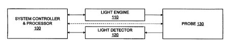
Figure 1 shows schematically the basic elements of an illustrative optical
system for the
examination of materials. As used herein, optics refers to the branch of
physics that deals
with the generation, propagation, and detection of electromagnetic radiation
having
wavelengths greater than x-rays and shorter than microwaves, and light refers
to
/o electromagnetic radiation at one or more wavelengths (narrowband,
broadband, or any
combination thereof) anywhere in the electromagnetic spectrum greater than x-
rays and
shorter than microwaves. An optical probe 130 is used to irradiate the
material being
examined (i. e. the target) and for collecting radiation from the target due
to the
irradiation. A system controller and processor 100 controls the various
operations
/s performed by the system and processes various characteristics of the
radiation image
collected from the target to obtain multispectral indications about various
properties of
the target material. Where the material is mammalian tissue which may suffer
one or
more abnormalities, the system controller and processor 100 may use
appropriate
algorithms to determine whether the tissue is normal or abnormal, including
the type of
Zo abnormality, and display the result; or use appropriate algorithms to
calculate a
probability of the tissue being normal or abnormal and, if abnormal, a
probability of the
type of abnormality, and display the result; or use appropriate algorithms to
screen the
tissue for abnormality and display the result; or control the power, duration,
and other
characteristics of light projected onto the tissue for treating tissue
abnormality; or a
Zs combination of the foregoing. A light engine 110 includes one or more
electromagnetic
energy sources for generating specific irradiation wavelengths. A light
detector 120
includes such components as filters and detectors or a spectrum analyzer for
measuring
the amplitude of wavelengths of interest in the probe image over the field of
view of the
probe 130. The system controller and processor 100 is coupled to the light
engine 110
3o and light detector 120 to control the various operations thereof. The light
engine 110 and
_g_
CA 02336128 2000-12-21
WO 00/03272 PCT/US99/15100
light detector 120 are coupled to the optical probe 130 using any suitable
means such as
fiber optic cable, although other coupling techniques such as liquid light
guides may be
used instead. If desired, various of the components of the light engine 110,
the light
detector 120, or both may be integrated into the probe 130, in which case
various
hardwired or wireless techniques may be used to couple the system controller
and
processor 100 to the probe 130. If the probe 130 contains any controllable or
powered
components, the probe 130 may be connected to the system controller and
processor 100
to receive control signals and/or power and/or furnish status signals.
Examples of
systems for the optical examination of mammalian epithelial tissues include
United
io States Patent No. 5,697,373 entitled "Optical Method and Apparatus for the
Diagnosis of
Cervical Precancers using Raman and Fluorescence Spectroscopies," issued
December
16, 1997 to Richards-Kortum et al., and United States Patent Application
Serial No.
08/666,021 entitled "Diagnostic Method and Apparatus for Cervical Squamous
Intraepithelial Lesions in Yitro and in Vivo Using Fluorescence Spectroscopy,"
filed
~s June 19, 1996 in the name of Richards-Kortum et al., which hereby are
incorporated
herein in their entirety by reference thereto.
Figure 2 shows schematically the principal elements of an optical probe 200
that is
suitable for use in the system of Figure 1 to probe material in the interior
of cavities
having restricted access through orifices or passageways, such as, in the case
of
Zo mammals, the epithelia and anatomical structures within their body cavities
and tubular
organs and viscera. For access to tissue within generally tubular cavities,
the probe 200
preferably is elongated and generally cylindrical (including round, oval, and
elliptic),
and includes a light collector 210 and an irradiator 220, which in turn
includes a light
conductor 222 and a spatial mixer 224. Other geometric shapes may be used for
the
2s probe 200 and/or for the light collector 210, the light conductor 222, and
the spatial
mixer 224, as required for the application, including triangular, rectangular,
hexagonal,
octagonal, other multiple facet geometries, and so forth. Moreover, the
principles of the
probe 200 may be used for applications such as surface applications not
requiring access
to the interior of cavities, in which event the overall shape of such probes
may be made
3o suitable for the application and need not be elongated.
-9-
CA 02336128 2000-12-21
WO 00/03272 PC'T/US99/15100
Probe output efficiency is maximized by having the irradiation and collection
paths
essentially separate except for a shared path at the optical window 240 and
through a
portion of the irradiator 220. For example, the light conductor 222 emits
Iight toward a
target 260 from around the periphery of the light collector 210, as shown in
greatly
s simplified form for illustratively a ring source by rays 230. The light
collector 210
collects light from the target 260 as represented by rays 270. Light from the
light
conductor 222 partially intersects the spatial mixer 224 as it passes through
(not shown
here; see, e.g., Figure S), which mixes the light to remove any reflected
images and
irradiation artifacts therein. The field of view of the light collector 210
preferably is such
io that any residual reflections and fluorescence from the spatial mixer 224
are excluded
from collection. While the window 240 may be just an opening, an optical
element such
as a solid flat optical window, a sheet of pliable material, a shaped lens, a
conformal
window such as a window having a nipple shaped to conform to the Os of the
cervix, or
a fluid filled sac, or a combination of one or more of such optical elements
may be used
~s at the position of the window 240 and/or inside of the spatial mixer ahead
of the light
collector 210 and light conductor 222 to achieved certain desired mechanical
and/or
optical effects. A conformal window is described in, for example, United
States Patent
No. 5,699,795, issued December 23, 1997 to Richards-Kortum et al. and entitled
"Optical Probe for the Detection of Cervical Neoplasia Using Fluorescence
Zo Spectroscopy and Apparatus Incorporating Same," which hereby is
incorporated herein
in its entirety by reference thereto. While any such solid window or lens
would be shared
by the light collector 210 and irradiator 220, which preferably are designed
to take into
account any optical effect thereof, the effect of any such solid window or
lens on optical
efficiency is minor compared to efficiency loses suffered by optical systems
that use a
Zs beam splitter or a dichoric mirror in the optical path. Moreover, beam
splitters and
dichoric mirrors tend to generate large amounts of stray light as compared
with the
partially common irradiation and collection paths of the probe 200.
During normal use, the probe 200 is brought into contact with the target
generally at the
optical window 240. The irradiator 220 projects light along an optical axis of
projection
3o coincident with an axis 250 which uniformly irradiates a specific surface
region of the
target material and penetrates into a volume of the target material through
the irradiated
surface. The light collector 210 uniformly collects light from this volume
along an
-10-
CA 02336128 2000-12-21
WO 00/03272 PCT/US99/15100
optical axis of collection coincident with the axis 250. While the optical
axis of
projection and the optical axis of collection preferably are coincident (e.g.
axis 250) to
achieve symmetry, this is not a necessary condition provided that the
irradiation is
sufficiently uniform over the collection volume.
s The light from the irradiator 220 preferably is stable, uniform, and due to
interactions
with the spatial mixer 224, diffuse (rays of the light intersecting the target
at a
multiplicity of angles and from a multiplicity of directions). The diffuse
nature of the
light improves its ability to penetrate into the target, including into areas
of the target
which are blocked from receiving normal radiation, with the distribution of
ray angles
/o relative to the axis of light projection from the irradiator 220 being
selected based on the
overall nature of the target material. For example, where the target is the
human cervix
and high irradiation eff ciency is desired for excitation of weak emissions
such as
fluorescence and Raman, preferably the distribution of ray angles has a
maximum near-
parallel to the axis of projection, with a small percentage of the rays being
parallel and
/s essentially none of the rays being highly deviant from parallel. However, a
distribution
of ray angles having a maximum at a much greater degree of deviance from
parallel is
desirable for some other applications, especially applications in which the
surface of the
target is moderately to severely irregular. A distribution of ray angles
having a
maximum near-normal to the axis of projection is undesirable, since such light
does not
zo penetrate sufficiently into the target. The specific distribution of ray
angles in the light
projected from the irradiator 220 depends on the material or materials used
for and
geometry of the spatial mixer 224 as well as the angles of the rays 230
emitted by the
light conductor 222.
The light collector 210 has a field of view of about the size of the optical
window
zs 240, a generally uniform collection efficiency over its field of view, and
a focal plane in
the vicinity of the optical window 240 having a good depth of focus.
Preferably, the light
collector 210 is a telecentric lens system or near-telecentric lens system,
which is
particularly suitable because of its uniform collection efficiency and Ionger
effective
depth of focus without appreciable distortion for applications involving low
level
3o responses such as fluorescence spectroscopy of mammalian epithelia, as
described in the
aforementioned Richards-Kortum patent documents. However, other types of
optical
-11-
CA 02336128 2000-12-21
WO 00/03272 PCT/US99/15100
collectors that have an adequate field of view may be used, if desired,
provided that the
collected light is compensated for non-uniformity across the field of view and
that any
excessive spatial distortion is also compensated for. Preferably, the light
collector 210 is
color corrected for mufti-spectral analysis, and any collection non-uniformity
is
compensated for by the use of well known normalization algorithms or by well
known
optical corrections such as the use of a bull's eye filter. The light
collector field of view
and depth of focus can vary a great deal for applications related to cervical
and other
tissues as well as non-medical applications.
Although the distal surface of the light conductor 222 is shown in Figure 2 to
be
~o in the same plane as the distal surface of the light collector 210, it may
be further
extended distally from this plane or recessed from this plane with adequate
means of
light transmission to the target.
While the probe 200 may be configured and dimensioned as desired so as to be
useful for probing different types of material, organic and inorganic, the
optical probe
~s 200 may be configured and dimensioned for use in diagnosing and/or
screening
cancerous and pre-cancerous tissues of mammalian epithelia using fluorescence
spectroscopy in the manner described in the previously cited Richards-Kortum
patent
documents. Figure 3 shows an optical probe 300 that is based on the
generalized probe
200 and is configured and dimensioned for probing tissues of the human cervix
in the
2o diagnosis of cancers and precancers using tissue fluorescence. In this
medical
application, the optical probe 300 emits a uniform light with a generally
normal but
somewhat diffuse orientation in the ultraviolet range, the visible range, or
both through
an optical window 302 which forms the distal end of the probe 300 to excite
tissue into
fluorescence within a cylindrical volume, and collects the low level tissue
fluorescence
2s through the optical window or probe distal end 302 from a cylindrical
volume that
extends into the tissue substantially concentric with the excited cylindrical
volume. In
the case of cervical examination, the field of view preferably is about 25 mm
and the
depth of focus is preferably about 8 mm.
The probe 300 has a housing (shown in cross section) that includes a generally
3o cylindrical projecting distal end section 310 and a proximal end section
316 from which
-12-
CA 02336128 2000-12-21
WO 00/03272 PCT/US99/15100
fiber optic bundles 330 and 340 extend. The distal end section 310 is
generally
cylindrical and illustratively about 10.8 inches (about 27.4 cm) in length and
about an
inch (25 mm) in internal diameter at the probe distal end 302. The distal end
section 310
is slightly flared in a direction away from the probe distal end 302 to
accommodate
bulging of the fibers of the bundle 340 about the lens system 320;
illustratively, the flare
is about 2.5 degrees beginning at a point about 10.7 cm (4.2 inches) from the
window.
Preferably, the dimensions of the distal end section 310 allow probe clearance
through a
speculum or other such devices. The distal end section 310 and the proximal
end section
316 may be constructed as one piece or separate pieces connected in any
desired manner,
/o as by being threaded and screwed together, welded, joined with adhesive,
clamped
together, and so forth. The proximal end section 316 is of any convenient
shape for
housing the fiber optics bundles 330 and 340. While optical probes generally
may be
supported in any convenient manner such as by a suitable mechanical support,
the probe
300 is designed to be hand-held and includes a suitable handle 350.
Illustratively, the
!s handle 350, which has a yoke portion and the proximal end section 316 is of
any suitable
shape for receiving the yoke portion, which is rotatably connected to the
distal end
section 316 with screws 430 and 432 (Figure 4) or any other suitable connector
and
extends illustratively about 7.5 inches from the proximal end section 316.
Alternatively,
the handle 350 may be fixed to the proximal end section 316 or may be part of
the
2o proximal end section 316. Any materials suitable for the application may be
used for the
probe 300. For example, for cervix examinations, the distal and proximal end
sections
310 and 316 may be made of commonly available stainless steel such as type 304
or
equivalent or type 6061T6 aluminum that is hard black anodized. The handle 350
may
also be made of type 6061 T6 aluminum or other suitable material with or
without plating
Zs or coating. All aluminum components may also be gold anodized or coated
with any
suitable plating or coating. Many other materials are suitable for various
parts of the
probe 300. For example, in medical applications the distal end section 310
which
contacts the patient may be made of any of various medically approved
materials,
including rigid plastics, pliable plastics, and paper, while other parts such
as the handle
so may be made of rigid plastic, dense core foam, and so forth. Moreover, the
handle 350
and/or the proximal end section 316 of the probe 300 may be coated with non-
slip
materials for easier handling, while the distal end section 310 may be coated
with
slippery materials to reduce friction during insertion.
-13-
CA 02336128 2000-12-21
WO 00/03272 PCT/US99/15100
Irradiator
Figures 3, 4 and 5 show various components of one type of irradiator for the
optical probe 300. Figure 3 is a side cutaway view of the optical probe 300.
Figure 4 is a
cross-sectional view taken normal to the optical axis of the optical probe 300
just in front
of the distal ends of numerous optical fibers of the bundle 340, two of which
are referred
to by the reference numbers 312 and 314 (Figure 3). Figure 5 is a cross-
sectional view
taken along the optical axis of the optical probe 300 and through part of the
distal end
section 310. The probe 300 terminates in a lens 306 at its distal end 302,
although the
lens 306 may be positioned anywhere in-between the distal end 302 of the probe
300 and
/o the distal end of the fibers of the bundle 340 or omitted entirely. The
fiber optics bundle
330 from a lens system 320 and the fiber optics bundle 340 pass through the
back of the
proximal end section 316 for connection to a light detector 120 (Figure 1 )
and a light
engine 110 (Figure 1) respectively. The fibers of the bundles 330 and 340
extend
continuously to the light detector 120 and the light engine 110 respectively
to achieve
is high e~ciency, although the bundles 330 and/or 340 may be segmented with
intervening
connectors located, for example, at or near the back of the proximal end
section 316.
The bundle 340 contains fiber optics for illuminating the target,
illustratively
twelve hundred fibers, each being approximately 0.2 mm in diameter and having
a
numerical aperture of, illustratively, 0.28. Suitable fibers are available
from a variety of
2o sources, including Ceramoptec Inc. of East Longmeadow, Massachusetts, under
the
product designation Optran. Illustratively, the fibers of the bundle 340 are
separated into
twenty-four groups 401-424 (Figure 4) of approximately fifty fibers each, the
groups
40I-424 being routed along the outside surface of the lens system 320 from the
fiber
bundle 340 to evenly-spaced annular positions on a toothed annular form 308
about the
2s distal end of the lens system 320 to form a ring light source. During
manufacture, the
fibers of the bundle 340 are held in place about the casing for the lens
system 320 using
various tooling and then potted in a manner well known in the art using
preferably non-
fluorescent potting material. The gathering of the fibers near the proximal
end of the lens
system 320 causes a bulge on one side of the probe 300 that is accommodated by
the
3o flaring of the distal end section 310. Note that the irradiation fibers 310
may be bundled
for connection to the light engine in other ways. For example, the fibers may
be gathered
- 14-
CA 02336128 2000-12-21
WO 00/03272 PCTNS99/15100
into two or more separate bundles rather than into the single larger diameter
bundle 340,
which would reduce the amount of bending of the individual fibers and result
in less
bulging. As a further example, the fibers may also be arranged coaxially about
the fiber
bundle 330. Note also that the use of twenty-four groups 401-424 is
illustratively, and
more or fewer groups containing more or fewer fibers may be used as desired.
The fibers
need not be grouped, but may be continuously arranged about the inside of the
body 304,
if desired. The fibers may be randomized to provide some mixing of any spatial
definition from the light engine. Note that the f hers of the bundle 340 may
be held in
place on the casing of the lens system 320 by other techniques, such as by
other suitable
~o adhesives and even mechanical retainers before being ground and polished on
the ends.
Alternatively, the fibers of the bundle 340 may be mounted on the inside
surface of the
distal end section 310 (not shown), or may be mounted on a form (not shown)
that is
disposed between the distal end section 310 and the lens system 320. The
outside
generally cylindrical surface formed by the potted fibers from the bundle 340
on the lens
~s system 320 is wrapped with Teflon tape to facilitate probe assembly,
although a variety
of other coatings and covering materials may be suitable as well.
The form 308 on the distal end of the lens system 320 serves to angle the
distal
ends of the fibers of the bundle 340 at about ten degrees toward the optical
axis of the
lens system 320. During manufacture, the angled fibers are sliced normal to
the optical
zo axis of the lens system 320 and ground and polished in a well-known manner
to achieve
surfaces that are themselves angled about ten degrees relative to the
respective axes of
the fibers of the bundle 340. Any suitable anti-reflective coating may be
applied to the
ends of the fibers to increase transmission efficiency. As a result of this
geometry, the
center of the light cone emitted from the end of each fiber of the bundle 340
is angled
Zs about fifteen degrees toward the optical axis of the lens system 320.
The optical probe 300 also includes a spatial mixer, which is implemented by
providing a particular finish to or applying a particular material to the
inside wall 304 of
the distal end section 310. Generally, the surface 304 forming the spatial
mixer is a
substantially non-fluorescing material having or having been finished to have
high
3o diffuse reflectivity in preferably the ultraviolet and visible wavelengths
and to strongly
forward-scatter the wavelengths of light exiting the distal ends of the fibers
of the bundle
-15-
CA 02336128 2000-12-21
WO 00/03272 PCTNS99/15100
340. For example, where the distal end section 310 is a stainless steel tube,
the spatial
mixer surface 304 is achieved by grinding and honing the inside of the tube to
achieve a
suitable surface finish, illustrative an 8 to 16 micron finish, and then
electropolishing or
chemically polishing the finish to improve uniformity and efficiency and to
reduce
s backscatter. Alternatively, the spatial mixer 304 may be aluminum, metal,
mylar, or
other type of foil that has suitable surface properties and is made to line
the inside of the
distal end section 310. The specific property for the spatial mixer surface
304 is
determined by balancing reflection efficiency on the one hand and uniformity
and
diffusivity on the other hand. Hence, even near-specular finishes on the order
of 4
io micron may be suitable in some arrangements, although care should be taken
when using
near-specular finishes not to re-image the output of the fibers 310 at the
target from the
spatial mixer surface 304. In other arrangements, a surface finish greater
than 16 microns
may be suitable where greater uniformity is required and efficiency is less of
a concern.
Most of the light from the distal ends of the fibers of the bundle 340 is
directed
~s toward the probe distal end 302, but the light spreads with a half angle of
about sixteen
degrees so that some light initially encounters the spatial mixer surface 304
and is
forward-scattered to augment light intensity generally in the periphery of the
field of
view of the lens system 320 and to add an additional profusion of ray angles
to the light
at the probe distal end 302, thereby causing a uniform diffuse light to occur
in the
2o vicinity of the probe distal end 302. Hence, some number of reflections of
light rays
within the spatial mixer 304 is desirable. However, reflecting too much of the
light too
many times would result in reduced irradiation efficiency because multiply
reflected
light would suffer attenuation in the spatial mixer 304. Such multiply
reflected light is
undesirable unless adequate power is available from the light engine 110. An
excessive
zs number of reflections would result in an increasing number of rays being
nearly parallel
to the general orientation of the target surface in the vicinity of the probe
distal end 302.
Such rays would fail to penetrate sufficiently deeply into the target (e.g.,
tissue) to excite
fluorescence throughout the desired volume of material.
A lens 306 is positioned at the distal end 302 to serve as the optical window
of
3o the probe 300. The lens 306 is provided with any suitable surface contour
and is made of
any suitable material or combination of materials having good optical
properties and low
-16-
CA 02336128 2000-12-21
WO 00/03272 PCT/US99/15100
fluorescence, such as ground glass, quartz, fused silica, or molded acrylic
such as type
EXP-X72 available from CYRO Industries, Inc. of Roclcaway, New Jersey, which
is a
non-additive version of the company's type S-10 Acrylite~ acrylic molding
compound.
The lens 306 may have any desired antireflective ("A/R") coating on either
surface or on
both surfaces, and any other characteristics as required by the lens system
320. The lens
306 is sealed to the inside wall of the distal end section 310 to protect the
fibers of the
bundle 340, the lens system 320, and other internal components of the probe
300 from
contamination and damage during use.
When placed at the distal end 302 of the probe 300 as shown, the lens 306 is
able
to contact and compress the target. However, the lens 306 may be spaced away
from the
distal end 302 of the probe 300, either near the distal end of the lens system
320 and the
ends of the fibers of the bundle 340 (see, e.g., Figure 7), or positioned
anywhere between
the distal end 302 of the probe 300 and the distal end of the lens system 320.
Positioning
a lens near the distal end of the fibers of the bundle 340 and spaced away
from the ends
/s of the fibers of the bundle 340 by any suitable distance, e.g. less than
about 8 mm and
preferably about 1 mm, places any reflected image of the distal end of the
fibers of the
bundle 340 outside of the field of view of the lens system 320, thereby
avoiding any
adverse impact such a reflected image may have on the light sought to be
collected. For
example, a reflected image from a light source seriously impacts the detection
of a
2o reflection image of the target since the wavelength or wavelengths of both
reflections
would be the same. However, a reflected image from a lens has less impact on
the
detection of a fluorescence or Raman emission, since the wavelength or
wavelengths of
a fluorescence or Raman emission differ from that of the reflected image and
are
typically isolated by bandpass filters or a spectrograph. Positioning a lens
further from
2s the distal end of the fibers of the bundle 340 requires the use of a very
good anti-
reflective coating on the lens or the use of other appropriate techniques to
avoid
generating a reflected image of the distal end of the fibers of the bundle
340.
Figure 5 is a longitudinal cross-section through the spatial mixer 304 of the
optical probe 300 (lens 306 omitted for clarity), and shows the behavior of
various
3o exemplary rays of light therein. The spatial mixer 304 is illustratively
about 65 mm in
length and about 25 mm in diameter. The angled distal ends of the fibers of
the bundle
- 17-
CA 02336128 2000-12-21
WO 00/03272 PCT/US99/15100
340 bias light toward the center of the field of view of the lens system 320,
as
represented by ray 514 which emanates from an illustrative fiber ~ 10, and by
ray 524
which emanates from an illustrative fiber 520. Light spreads out in a roughly
symmetrical conical pattern from each fiber in a well understood manner, as
from the
ends of the illustrative fibers 510 and 520 as represented by rays 512 and 516
and rays
522 and 526 respectively. The spatial mixer 304 functions by redistributing a
portion of
the solid angle emitted by each of the fibers of the bundle 340, as
represented by the
forward scattered components of rays 512, S 16, 522 and 526, resulting in
spatial mixing
onto the target at or near the probe distal end 302. This redistribution as
well as the
to angled direct Light represented by rays 514 and 524 achieve a multiplicity
of ray angles
in the vicinity of the probe distal end 302. Most of the rays are near-
parallel to the
optical axis 530 of the light detector (not shown) with some rays at the edge
of the probe
distal end 302 being parallel to the optical axis 530, so that light
efficiently penetrates
into the target (e.g., tissue).
is The various components and materials used in the irradiation system of the
optical probe 300 are selected to be capable of handling the irradiation power
desired.
For example, one use of the optical probe 300 for examination of the human
cervix
involves power out of the probe distal end 302 to range from about 20 to SO mW
at 337
nm, 380 nm, and 460 nm. Systems with power on the order of about 100 mW or
greater
2o may be used if desired to reduce total integration times. Illustratively,
the spatially mixed
light from the probe 300 penetrates up to about 300 microns into the cervical
tissue,
depending on wavelength, to excite fluorescence therein. The optical probe 300
may also
be used for applying light treatment to tissue, which can involve higher power
Levels up
to the tolerance level of the tissue. However, non-tissue applications may
involve even
2s higher power levels, so that the components and materials of the
irradiation system used
in such applications should be selected accordingly.
Light Collector
Figure 6 is a plan side cutaway view of the optical probe 300 of Figure 3
showing various components of the lens system 320 having the plano-convex lens
306 at
3o the probe distal end 302. The use of lens 306 in conjunction with the lens
system 320
- 18-
CA 02336128 2000-12-21
WO 00/03272 PCT/US99/15100
forms a true telecentric lens system, the lenses of which illustratively are
as follows.
Lens 306 is a piano-convex silica lens having a diameter of 25.4 mm, a
thickness of 4.0
mm (lens thickness being measured along its optical axis), a distal surface
radius of
infinity, and a proximal surface radius of 91.69 mm. Lens 606 is a cemented
doublet
s acromat with a convex-convex element of BAF10 glass having a diameter of
19.0 mm, a
thickness of 11.4 mm, a distal surface radius of 24.47 mm, and a proximal
surface radius
of 16.49 mm, and a concave-convex element of FD 10 glass having a diameter of
19.0
mm, a thickness of 3.0 mm, a distal surface radius of 16.49 rnm, and a
proximal surface
radius of 131.65 mm. Lens 610 is a cemented negative doublet acromat with a
concave-
concave element of BK7 glass having a diameter of 12.5 mm, a thickness of 2.0
mm, a
distal surface radius of 30.83 mm, and a proximal surface radius of 23.47 mm,
and a
concave-convex element of SFS glass having a diameter of 12.5 mm, a thickness
of 1.6
mm, a distal surface radius of 23.47 mm, and a proximal surface radius of
69.20 mm.
Lens 614 is a cemented doublet acromat with a convex-convex element of BAF11
glass
is having a diameter of 15.0 mm, a thickness of 6.3 mm, a distal surface
radius of 17.97
mm, and a proximal surface radius of 11.20 mm, and a concave-convex element of
SF 10
glass having a diameter of 15.0 mm, a thickness of 1.8 mm, a distal surface
radius of
11.20 mm, and a proximal surface radius of 85.31 mm. Lens 618 is a cemented
doublet
acromat identical to lens 6I4. Suitable spacers 608, 612 and 616 and other
structures
2o such as flange 602 are used to keep the lenses 606, 610, 614 and 618 in
place and
properly spaced apart, and a resilient O-ring 604 is used against lens 606 to
seal the
chamber containing the lenses 606, 610, 614 and 618. Illustratively, the
spacing between
lenses 306 and 606 is 142.50 mm, between lenses 606 and 610 is 11.03 mm,
between
lenses 610 and 614 is 3.34 mm, between lenses 614 and 618 is 1.00 mm, and
between
~s lens 618 and an image plane 620 at the end surface of the fiber bundle 330
is 3.00 mm.
The lens 306 and lens system 320 is focused at a object point about 1 mm
beyond the
distal end of the probe 300 and into the target, and is designed to focus the
target image
onto the image plane at the end of the fiber bundle so as to avoid loss of
power density
while reducing the image size. The ratio of the field of view of the optical
probe 300 to
3o the image size on the image plane 620 is approximately 6X, with
approximately f/2 on
the image plane at the fiber optic cable 330 to allow adequate depth of focus
in the
vicinity of the probe distal end 302.
- 19-
CA 02336128 2000-12-21
WO 00/03272 PCTNS99/15100
Stray light is blocked from the image plane 620 at the end surface of the
fiber
bundle 330 by restricting the field of view of the optical probe 300 using an
aperture
such as 621 and by incorporating one or more additional apertures as desired.
Stray light
originates in many ways, including reflections off of distal window or lens
surfaces and
backscatter from the spatial mixer surface 304. The field limiting aperture in
the system
320 is the aperture 621 over the image plane 620 at the end surface of the
fiber bundle
330. Illustratively, aperture 621 is 3.9 mm in diameter and the fiber bundle
330 is 4.0
mm square. Another aperture in front of the lens 610 also is effective in
blocking other
stray light from areas outside of the primary field of view.
/o A modification of the probe 300 and lens system 320 is shown in Figure 7.
The
lens 306 at the distal end 302 of the probe is absent. Instead, a lens 706 is
provided,
which is recessed from the probe distal end 302 and mounted well within the
spatial
mixer 304 adjacent the lens system 720 and spaced 1 mm from the distal ends of
the
fibers of the bundle 340. An additional lens or window 707 is placed at the
distal end of
is the lens system 720 to seal the entire lens system 720 and prevent dust
from depositing
on the optics or the optically black sidewall of the casing of the lens system
720. In
applications involving pliable targets such as, for example, the human cervix,
the probe
700 with an opening at the probe distal end 302 tends to stabilize more
securely on the
cervix when cervical tissue protrudes into the distal end segment 310. In this
Zo modification, the lens system 720 in conjunction with the lenses 706 and
707 do not
form a telecentric lens system, but do achieve sufficiently uniform light
collection to
avoid the need for extensive optical correction. The lenses of the probe 700
illustratively
are as follows. Lens 706 is preferably a concave-convex (meniscus) acrylic
lens having a
diameter of 25.0 mm, a thickness of 2.0 mm (lens thickness being measured
along its
2s optical axis), a distal surface radius of 82.97 mm, and a proximal surface
radius of 76.20
mm. However, the lens 706 may instead be a flat acrylic window, if desired,
which
would occasion only a minor performance reduction. The protective window 707
is a flat
silica cylinder having a diameter of 20.0 mm and a thickness of 3.0 mm. The
other
lenses and spacers of the lens system 720 are the same as the lenses and
spacers of the
30 lens system 320, except that the spacing between the object and lens 706 is
59 mm,
between lens 706 and the protective window 707 is 1 mm, and between the
protective
window 707 and lens 606 is 80 mm. The lens system 720 is focused at a point
about 2
-20-
CA 02336128 2000-12-21
WO 00/03272 PCT/US99/15100
mm inside of the distal end of the probe 700. This focal plane will usually be
on cervical
tissue for applications in which the target is the human cervix. Cervical
tissue will likely
protrude into the distal end segment 310 as a result of the natural shape of
the cervix or
light pressure applied to hold the probe 700 in place during use. The lens
system 720 is
also designed to focus the target image onto the image plane 620. The ratio of
the field
of view of the optical probe 700 to the image size on the image plane 620 is
approximately 6X, with approximately f/2 at the image plane into the fiber
optic cable to
allow adequate depth of focus in the vicinity of the probe distal end 302.
Stray light is blocked from the image plane 620 at the end surface of the
fiber
/o bundle 330 by two principal apertures. One of the principal apertures in
the lens system
720 is the aperture over the distal surface of the lens 610, which
illustratively has a
diameter of 6.4 mm and is spaced 1.00 mm from the distal surface of the lens
610. The
other principal aperture in the lens system 720 is a field limiting aperture
721 over the
image plane 620 at the end surface of the fiber bundle 330, which
illustratively has a
~s diameter of 3.9 mm and is spaced 2.00 mm from the image plane 620. Both
apertures are
active in controlling stray light, and since the lens system 820 is not
telecentric, the
aperture over the distal surface of the lens 610 defines the f number or
numerical
aperture of the light collector.
The principal apertures in the lens systems 320 and 720 include an angled
inside
2o annular surface, which redirects stray light away from the image plane 620.
Figure 8
shows how various illustrative rays that are reflected from a lens such as
lens 706 of the
optical probe 700 (Figure 7) near the distal end 302 of the probe 700 either
are blocked
from the image plane 620 or redirected by the aperture 721. The inside annular
surface
of the aperture 721 is angled preferably 45° relative to the optical
axis of the probe 700.
2s Stray light coming through the lens system 720 from lens 706 and window 707
and from
other sources and projecting just outside of the aperture either is reflected
once and
directed harmlessly through at least two lenses onto apertures and/or the
inside optically
black wall of the casing for the lens system 720, see, e.g., ray 804; or is
reflected twice
by two diametrically opposed 45° angled surfaces and exits the lens
system 720
3o altogether, see, e.g., rays 806 and 808.
-21 -
CA 02336128 2000-12-21
WO 00/03272 PCT/US99/15100
Combining the Light Collector and Irradiator
Preferably, care is taken to ensure good alignment of the optical axis of the
light
collector 210 (Figure 2) with the axis of the spatial mixer 224 to avoid
backscattered
light from the spatial mixer 224 from entering into the field of view of the
light collector
s 210. Generally, the field of view of the light collector 210 is narrow
enough to exclude
the inside wail of the spatial mixer 224 when alignment is proper, but
otherwise is as
wide as possible to permit viewing of an area of the target very slightly less
than the
overall diameter of the probe 200. Any misalignment would therefore allow
reflected
and backscattered light into the field of view of the probe 200 as a crescent
of light.
to Proper alignment of the optical axis of the light collector 210 with the
axis of the
spatial mixer 224 may be established and maintained in any suitable manner.
For
example, the distal probe section 310 and the proximal probe section 316 the
probes 300
and 700 may be made of a single piece with the lens system 320 being rigidly
retained
therein. Alternatively, the distal probe section 310 and the proximal probe
section 316
is may be made of separate pieces, with the lens system 320 being rigidly
retained therein
by, for example, suitable structural members of the proximal probe section
316, and the
distal probe section 310 being threaded and screwed into a prealigned threaded
opening
in the structural members of the proximal probe section 316.
Preferably, care is taken to ensure that the proper focal distance is
maintained
Zo between the light collector 210 and the window 240. This focal distance is
predetermined by optical design, and the proper focal distance is established
by proper
manufacture to tolerance and proper assembly and alignment of components.
Alternatively, the focal distance may be mechanically variable, as in the case
where the
distal probe section 310 is threaded and screwed into a threaded opening in
the proximal
2s probe section 316, adjusted as needed, and fixed with any suitable device
such as a set
screw or various reference mechanical stops. The use of various stops enables
repeating
a setting. Alternatively, the focal length may be optically variable by
incorporating a
small motor, screw and guides into the light collector 210 to electrically
remotely
reposition the lens as required to achieve proper focus. These and other
techniques for
-22-
CA 02336128 2000-12-21
WO 00/03272 PCT/US99/15100
achieving proper focus are well known in the art and may be used as desired in
connection with the generalized optical probe 200.
The axial placement of the distal end of the light collector 210 (Figure 2)
relative
to the ring-Iike distal end of the light conductor 222 of the generalized
probe 200 may be
s varied to achieve any desired design objective, provided that the uniform
and diffuse
nature of the light emitted at the window 240 is not adversely affected, and
provided that
any stray light going to the light collector 210 is controlled. For example,
the distal end
of the light conductor 222 may be placed generally in the plane of the distal
end of the
light collector 210, as in the case of the optical probe 300, behind the
plane, or in front of
/o the plane. Similarly, lens that optically participate with the light
collector 2I0 in the
collection of light may be located anywhere between the plane of the distal
end of the
light conductor 222 and the window 240, provided that the uniform and diffuse
nature of
the light emitted at the window 240 is not adversely affected. A lens such as
lenses 306
and 706 used for mechanical protection and contamination control may also be
located
~s anywhere between the plane of the distal end of the light conductor 222 and
the window
240, provided that any stray light from reflectance is controlled.
A lens placed in front of the distal end of the light conductor 222 generates
stray
Iight by reflecting a portion of the light from the light conductor 222. When
the lens is
located near both the distal end of the Iight conductor 222 and the distal end
of the light
2o collector 210, the light reflected by the lens tends to be outside of the
field of view of the
light collector 210. However, when the Iens is located a distance from both
the distal end
of the light conductor 222 and the distal end of the light collector 210, a
substantial
amount of the light reflected by the lens tends to be inside of the field of
view of the
light collector 210 and is seen as disc-like artifacts. Various techniques are
useful for
zs reducing the effect of such reflections. For example, anti-reflection
("A/R") coatings
may be used to reduce the amount of reflected light. Where the light being
collected is of
a different wavelength than the irradiation light, blocking filters may also
be used to
reduce the amount of reflected light detected.
A useful and particularly efficient approach for connecting the light
conductor
30 222 and the light collector 210 to respectively a light engine (e.g., light
engine 110 of
-23-
CA 02336128 2000-12-21
WO 00/03272 PCTNS99/15100
Figure 1 ) and a light detector (e.g., light detector 120 of Figure 1 ) is
continuous optical
fibers from the light engine to the light conductor 222. However, due to the
cost of this
approach, other approaches may be better suited to certain applications.
Alternative
approaches include providing optical connectors on the probe, to which
separate cables
s from the light engine connect. These separate cables may be made of optical
fibers or
other light conductors. For example, liquid light guides may be used for the
irradiation
light. Liquid light guides are flexible and have a cost advantage over optical
fiber optics,
but also tend to have a variable output which may need to be compensated for
at the light
detector. An illustrative compensation technique entails installing an edge-of
field light
~o sensor component in the probe to monitor light output at the probe. Based
on conditions
of uniform light irradiation, a baseline of the liquid light guide is
established. Then, the
light output at the probe is monitored with the edge-of field sensor
components in
conjunction with the light detector prior to each use to establish a
calibration factor for
each patient setup and to detect and correct for changes during each patient
analysis.
/s Continuous multipoint monitoring may be needed if there is spatial content
to the
transmission variations caused by movement of the cable.
Light Irradiation and Collection in Optical Probes Having Disposable
Components
For applications in which avoidance of contamination is important, an optical
probe may be designed as a one piece unit that is fully reusable after
cleaning and
decontamination, or as a two piece unit having one section with delicate
and/or
expensive components that is reusable without cleaning or decontamination and
a
protective durable section that is reusable with cleaning and decontamination,
or as
having a fully reusable section and a protective disposable section that is
discarded after
several or preferably one use and replaced with an identical but new and clean
zs disposable. Figure 9 shows an optical probe that has a fully reusable
section 900 and a
disposable section 910. A suitable connector component 920 on the reusable
section 900
engages a suitable connector component 912 on the disposable 910 to hold the
disposable 910 in place in proper alignment with the reusable section 900. A
variety of
connection mechanisms are suitable, including threaded fixtures, bayonet style
fixtures,
3o spring loaded clamps, friction fit fixtures, and so forth.
-24-
CA 02336128 2000-12-21
WO 00/03272 PCT/US99/15100
Figure 10 shows an example of the fully reusable optical probe section 900
suitable for use work with a disposable optical probe section such as shown in
Figures
20-23. The probe 900 has a housing (shown in cross section) that includes a
generally
cylindrical projecting distal end section 1010 and a proximal end section 1016
to which
a handle 950 is rotatably connected and from which fiber optic bundles 330 and
940
extend. The distal end section 1010 is generally cylindrical and
illustratively about 18.5
cm in length and about 25 mm in diameter at the distal end 1002, the overall
length of
both sections 1010 and 1016 being about 28.5 cm. The distal end section 1010
and the
proximal end section 1016 may be constructed as one piece or separate pieces
connected
~o in any desired manner, as by being threaded and screwed together, welded,
joined with
adhesive, clamped together, and so forth. The proximal end section 1016 is of
any
convenient shape for housing the fiber optics bundles 330 and 940 and to
receive the
handle 950, which extends illustratively about 19 cm from the proximal end
section
1016. As the reusable probe section 900 does not contact the target, a wide
variety of
~s materials may be used for it, including all of the materials suitable for
the probe 300 as
well as materials that may not be suitable for the probe 300 because of, for
example,
patient contact restrictions in the case of medical applications.
The reusable probe section 900 includes a light collector, illustratively the
lens
system 720, and part of an irradiator, illustratively a light guide 1020. The
spatial mixer
Zo preferably is included in the disposable. Although a light conductor made
of fibers such
as the fibers 340 in the probe 300 may be used instead of the light guide
1020, the light
guide 1020 is made with preferably a generally cylindrical shape which does
not require
that the distal end section 1010 of the reusable probe section 900 be flared,
thereby
simplifying the manufacture of the disposables of Figures 20-23 that mount on
the
Zs reusable probe section 900. The light guide 1020 is suitable for use in the
optical probe
300 as well. Preferably, the fiber bundle 330 is routed straight from the lens
system 720
through the back of the proximal section 1016, and the light guide I 020 is
provided with
an opening through which the fiber bundle 330 passes. Alternatively, the light
guide may
be made to be symmetrical (not shown) while an assembly of mirrors, prisms,
and the
so like may be used to route the image from the end of the lens system 320
through a notch
in such a light guide and onto the image plane of an optical fiber bundle or
connector
(not shown) that is not coaxial with the lens section 720.
-25-
CA 02336128 2000-12-21
WO 00103272 PCT/US99/15100
It will be appreciated that both the lens system 720 and the light guide 1020
in
the reusable probe section 900 are illustrative, and that other lens systems,
light guides,
fiber arrangements, and combinations of lens, fibers, light guides, and so
forth may be
used instead. For example, Figure 11 shows a reusable probe section I 100 in
which the
distal end 1002 is open and a lens or window 1122 is recessed into a lens
system 1120,
which is otherwise similar to the lens system 720.
The light guide 1020 may be manufactured by various techniques. For example,
the light guide 1020 is made of fused silica, and may be manufactured in two
pieces,
including a short free-form light pipe coupled to a concentric cylindrical
light pipe as
~o shown in Figures 12-19, or in a single piece, such as a long free-form
light pipe (not
shown). Cladding, a vacuum deposited film, or another suitable material on the
inside
and outside surfaces of the fused silica is used to achieve internal light
reflection, and the
light pipe itself may be hollow or liquid filled instead of solid fused
silica. These
implementations may include means known in the art for improving light
uniformity,
/s including the use of a square clad rod light integrator or other such means
of diffusing
image artifacts at the input. Light is emitted from the light guide 1020 in a
generally
annularly continuous manner rather than as a ring of merging cones as from the
ends of
the fibers of the bundle 340 in the probe 300.
Figure 12 shows a cross section along the axis of a cylindrical part 1200 of a
two
zo piece light guide, which is coupled to either free form section 1400 or
free form section
1700 to complete the light guide. Section 1200 is a cylindrical light guide
having a fused
silica core 1204 contained within aluminum tubes 1202 and 1206.
Illustratively, the
cylindrical section 1200 is 129.5 mm (5.10 inches) long. The core 1204 has an
inside
diameter of 20.0 mm (0.787 inches) and an outside diameter of 24.0 mm (0.945
inches),
Zs and is fabricated using techniques well known in the art. The core 1204 is
suitably clad
to achieve a numerical aperture of preferably from about 0.25 to 0.4, and is
then covered
with an opaque coating to control stray light. Suitable cladding materials and
opaque
materials are available from various sources, including Chemat Technology Inc.
of
North Ridge, California, and Optical Polymer Research, Inc. of Gainesville,
Florida.
3o Aluminum tube 1206 has an inside diameter of 19.0 mm (0.748 inches) and an
outside
diameter of 19.9 mm (0.783 inches), while aluminum tube 1202 has an inside
diameter
-26-
CA 02336128 2000-12-21
WO 00/03272 PCT/US99/15100
of 24.1 mm (0.949 inches) and an outside diameter of 25.0 mm (0.984 inches).
The
aluminum tubes 1202 and i 206 preferably are black anodized, and are installed
after
cladding and coating is completed but before the ends of the fused silica core
1204 are
ground and polished. A view of the proximal end of the section 1200 is shown
in Figure
13.
Figure 15 shows a cross section along the axis of a free-form fused silica
light
guide section 1400 made using fabrication techniques well known in the art.
Illustratively, the free form section 1400 is 45.7 mm (1.8 inches) long, and
includes a
suitably clad fused silica core 1404 which is placed within an aluminum tube
1402
to before grinding, polishing and A/R coating of the ends thereof. After
cladding is applied,
the core 1404 is potted inside of the aluminum tube 1402, using any suitable
preferably
non-fluorescent potting material. The core 1404 at the distal end of the
section 1500 has
an inside diameter of 20.0 mm (0.787 inches) and an outside diameter of 24.0
mm
(0.945 inches), and at the proximal end has a diameter of 8.0 mm (0.31 S
inches) to mate
~s up with a liquid light guide or fiber optic cable. A channel, which is
referred to by the
numeral 1408, is provided in the free form section 1400 for the passage of the
fiber
bundle 330 (Figure 10). Illustratively, channel 1408 measures 15.2 mm (0.60
inches)
wide and 27.9 mm (l .l O inches) long, and is spaced from the proximal end of
the section
2000 by 27.9 mm ( 1.10 inches). A view of the distal end of the section 1400
is shown in
zo Figure 14, and a view of the proximal end of the section 1400 is shown in
Figure 16. The
sections 1200 and 1400 are coupled using any suitable technique such as a
index
matching optical fluids, and suitable A/R coatings.
Figure 18 shows a cross section along the axis of a free-form section 1700
made
of a large number of cladded fused silica fibers using fabrication techniques
well known
Zs in the art. Illustratively, about 24 cladded fibers are fused together to
form the free form
section 1700, the dimensions of which are the same as the free form section
1400. The
section 1700 is potted inside of an aluminum tube 1702. A view of the distal
end of the
section 1700 is shown in Figure 17, and a view of the proximal end of the
section 1700
is shown in Figure 19. The sections 1200 and 1700 are coupled using any
suitable
3o technique such as a index matching optical fluids, and suitable A/R
coatings.
-27-
CA 02336128 2000-12-21
WO 00/03272 PCT/US99/15100
As can be seen from Figures I4-19, the use of openings in the free form
sections
1400 and 1700 as well as the asymmetrical design thereof does not permit light
to be
uniformly annularly distributed therein. However, the annular uniformity of
the light is
improved by the cylindrical section 1200. Other measures to improve the
annular
uniformity of the light include varying the light guide wall thickness at the
entrance
transition or providing deflectors to deflect the light around the opening and
then
rotating and counter rotating the light around the fused silica core 1204.
Using a square
spatial mixer at the input may also be desirable for improving the annular
uniformity of
the light.
Jo Disposables
A disposable 910 (Figure 9) suitable for use with the reusable probe section
900
is generally elongated for mounting to the distal extension of the probe 900
and for
protecting it from contamination from the target and surrounding materials,
and contains
an inside surface suitable for the spatial mixing of light and an optical
element. The
/s elongated portion of the disposable 910 may be rigid, pliable, or a
combination of rigid
and pliable sections, and may be made of various materials such as medical
grade paper,
plastic, synthetic rubber, aluminum, stainless steel, laminate, and other
appropriate
materials. The optical element may be a rigid or pliable body, including a
solid flat
optical window, a sheet of pliable material, a shaped lens, a conformal window
such as a
2o window having a nipple shaped to conform to the Os of the cervix, a fluid
filled sac, or a
combination thereof, and may be made of various materials such as plastic,
fused silica,
glass, quartz, and other appropriate materials. The spatial mixing surface may
be a
treated or coated inside surface of the elongated portion of the disposable,
or may be
another type of material or materials lining or embedded in the inside surface
of the
zs elongated portion of the disposable. For example, where the elongated
portion of the
disposable is a tube of extruded aluminum, the spatial mixing surface is
formed by
treating the inside surface of one end of the extruded aluminum tube with an
acid etch
and followed by anodization to create a light mixing surface. Alternatively,
aluminum
foil having a suitable spatial mixing surface may be applied to any suitable
tube material.
3o As used herein, ''tube" refers to an elongated hollow shape of any desired
cross section,
including round, oval, elliptic, triangular, rectangular, other multiple facet
geometries,
-28-
CA 02336128 2000-12-21
WO 00/03272 PCT/US99/15100
"C" sections, free form sections, and any combination of the foregoing,
whether varying
or constant along the direction of elongation.
The material or materials used in the disposable 910 to protect the reusable
probe
section 900 from contamination constitute a fluid barrier, which is impervious
to fluids
typically found at the target site or that impedes such fluids throughout the
intended
period of use of the disposable. For example, materials suitable for medical
applications
include materials, that are impervious to mammalian body fluids, such as
aluminum,
plastic, fused silica, glass, and quartz, as well as materials that impede
mammalian body
fluids, such as medical grade paper.
~o Figures 20-23 show various disposables that include medical grade paper in
their
manufacture. Figure 20 shows a disposable 2000 that includes a tubular member
2002 of
stiff medical grade paper which is fitted over a molded plastic base 2008. The
thickness
of the tube 2002 depends on its length, with a tube thickness on the order of
1.3 mm
(fifty thousandths of an inch) being suitable for lengths required for
cervical
Is examination: The tube 2002 is connected to the base 2008 using any suitable
technique,
such as fixed with adhesive or press-fit. The base 2008 contains suitable
connectors
2010 for connecting to the reusable optical probe section 900. A spatial mixer
surface
2004 is provided by preferably aluminum foil paper that is applied to the
inside of the
paper tube 2002. A suitable aluminum foil paper is made of an aluminum foil
liner about
zo 0.01 mm (0.0003 inches) thick, for example, glued to 20 lb. natural Kraft
backing paper,
which is available from Custom Paper Tubes, Inc. of Cleveland, Ohio. Other
aluminum
foil papers are also commonly available with differing paper weights and foil
types and
thicknesses. For example, gold and nickel foils may be suitable in various
applications
depending on the irradiation wavelengths used. The aluminum foil paper is
wound to
~s achieve any desired internal seam. However, a spiral seam is preferred to a
straight seam
because a spiral seam tends to average any scattering and/or fluorescence that
may be
generated by the seam over the circumference of the spatial mixer to keep the
intensity
thereof below the detection threshold of the probe 900. The lens 2006 is
threaded on its
edge and is screwed into place from the back of the tube 2002 prior to
connecting the
3o tube 2002 to the base 2008. Other techniques for fitting the lens 2006 to
the tube 2002
include press-fitting the lens 2006 into place, gluing the lens 2006 into
place with a
-29-
CA 02336128 2000-12-21
WO 00/03272 PCT/US99/15100
suitable adhesive, crimping the tube 2002 on both sides of the lens 2006,
providing an
annular trough on the edge of the lens 2006 and crimping the tube 2002 into
the trough
to engage the lens 2006, and so forth. Illustratively, the tube 2002 is about
10.2 cm (4
inches) long, the base 2008 is about 7.6 cm (3 inches) long, and the lens 2006
is IJV
acrylic or equivalent.
Figure 21 shows a disposable 2100 that includes a tubular member 2102 of stiff
medical grade paper with an internal spatial mixing surface 2104. A lens 2106
is pushed
into place against a crimp or other type of retainer in the tube 2102, and the
tube 2102 is
pushed over a molded plastic base 2108 containing suitable connectors 2110.
The
molded plastic base 2108 presses against the lens 2106, firmly seating it
against the
crimp in the tube 2102.
Figure 22 shows a disposable 2200 that includes a tubular member 2202 of stiff
medical grade paper with an internal spatial mixing surface 2204. A lens 2206
is press-
fitted in proper alignment into a base 2208 or secured with adhesive, and the
base 2208
~s is pushed into the tube 2202 and secured with a suitable adhesive.
Figure 23 shows a disposable 2300 having a lens 2306 mounted on the distal end
thereof. The disposable 2300 includes a tubular member 2302 of stiff medical
grade
paper with an internal spatial mixing surface 2304. The tubular member 2302 is
fitted
over a molded plastic base 2308 containing suitable connectors 2310. A lens
2306 is
Zo mounted on the distal end of the disposable 2300 by screwing it into place
or by using
any other suitable technique such as press-fitting the lens 2306 into place,
gluing the lens
2306 into place with a suitable adhesive, or crimping the tube 2002 to secure
the lens
2306.
The various molded bases 2008, 2108, 2208 and 2308 shown in Figures 20-23
Zs may be molded with a flare, if desired or if necessary to accommodate
elements of the
reusable probe section 900. Other materials and manufacturing techniques may
be used
instead of the various molded plastic bases 2008, 2108, 2208 and 2308; for
example,
extruded aluminum may be used.
-30-
CA 02336128 2000-12-21
WO 00/03272 PCT/US99/15100
Figure 24 shows an optical probe that has a fully reusable section 2400 and a
disposable section 2410. The reusable section 2400 is similar to the optical
probe 300,
but includes a suitable connector component 2420 to engage a suitable
connector
component (not shown) on the disposable 2410 to hold the disposable 2410 in
place in
s proper alignment with the reusable section 2400. It will be appreciated that
various other
light conductors, spatial mixers, and light collectors as described herein may
be used
instead of the fiber optics from the bundle 340, the mixer 304, and the lens
system 320
shown in Figure 24. The disposable 2410 does not contain a spatial mixer,
which is part
of the reusable section 2400 as shown by reference numeral 304. However, the
~o disposable 2410 does include a protective elongated section, illustratively
a flared tube
2412, and a protective optical window 2414, and is otherwise similar in
construction and
materials to the disposable 910. Suitable disposables are also described in
United States
Patent Application Serial No. 08/823,044 entitled "Method and Apparatus for
Calibrating an Optical Probe," which was filed March 21, 1997 and names Peter
~s McHenry and Arthur E. Schulze as inventors, and in United States Patent
Application
Serial No. 09/027,403 entitled "Contact Window Having a Tilt Characteristic
for Optical
Probe," which was filed February 20, 1998 and names Curtis K. Deckert as
inventor,
which hereby are incorporated herein in their entirety by reference thereto.
While the specific embodiments described herein are suitable for examinations
of
zo the human cervix, the invention is suitable for other tissue analysis by
changing probe
front optical-mechanical configuration, excitation wavelengths from the light
source,
detection wavelengths in the light detector, and diagnostic and control
software on the
computer. The probe size is scalable to function in different ways to analyze
a wide
variety of materials, including a variety of tissues. Any area of a body can
be examined
Zs by applying a probe of the proper length with the necessary field of view,
along with the
proper modular changes to adjust the effective field of view. This technique
can also be
extended by the use of a flexible fiber optics interface to reach far into the
body for
visual examination and treatment.
While the specific embodiments of the spatial mixer described herein use a
single
.~o finish or material to achieve adequate spatial mixing for many
applications, some
applications may call for an unusual type of spatial mixing. The type of
spatial mixing
-31 -
CA 02336128 2000-12-21
WO 00/03272 PCT/US99/15100
may be varied by providing a variety of surface finishes or materials on the
inside wall
of the spatial mixer to optimize spatial mixing and/or regions of irradiation
for a
particular application. For example, in one arrangement (not shown) a
cylindrical
segment of the inside wall of the spatial mixer nearest the light source is a
specular or
reflecting surface, the middle cylindrical segment is a diffuse or scattering
surface, and
the last cylindrical segment nearest the distal end of the probe is an
absorber. The
absorber section may be eliminated where the field of view of the lens system
is
appropriately limited. Many suitable materials, finishes, and geometries are
well-known
to those of ordinary skill in the art to achieve a specuiar, diffuse or
absorbing surface, as
~o desired.
While many of the embodiments described herein include various values and
dimensions, these are illustrative and other values and dimensions may also be
useful.
For example, the number, groupings, and size of the fibers in the irradiation
system are
illustrative.
-32-