Note: Descriptions are shown in the official language in which they were submitted.
CA 02336279 2000-12-28
WO 00/66014 PCT/US00/11384
DEVICE FOR THE ABLATION OF TISSUE
This is a continuation-in-part of application Serial
No. 08/912,273 filed on August 15, 1997.
This invention relates to an apparatus and device for
use therein and a method for ablation of tissue and more
particularly to the treatment of tissue in the human body as
for example the uvula, tonsils, adenoids, sinus tissue,
tongue and turbinates.
Apparatus, device for use therein and methods for
ablating tissue have heretofore been provided. However it
has been found that for some applications, they are unduly
complicated and expensive. There is therefore a need for a
simplified tissue ablation device which will meet the
requirements for tissue ablation and be less expensive.
In general it is an object of the present invention
to provide an apparatus and device for use therein and a
method for the ablation of tissue which incorporates a
simplified tissue ablation device.
Another object of the invention is to provide an
apparatus, device and method of the above character which
can utilize a less expensive simplified tissue ablation
device.
Another object of the invention is to provide a device
of the above character which is shaped to provide improved
viewing capabilities for the physician using the device.
Another object of the invention is to provide a device
of the above character which has been ergonomically shaped
for improved grasping by the hand of the physician.
CA 02336279 2000-12-28
wo 00/66014
PCTNS00/I 1384
-2-
Another object of the invention is to provide a device
of the above character in which a straight needle shape is
maintained so that the device can only be utilized for
straight entry applications.
Another object of the invention is to provide a device
of the above character in which an edge mounted on a printed
circuit board is utilized.
Another obj ect of the invention is to provide a device
of the above character in which a flexible cable is utilized
for making connections to the needle.
Another object of the invention is to provide an
apparatus and device for use therewith in which a
substantial portion of the device can be reused.
Another obj ect of the invention is to provide a device
of the above character in which the reusable portion
includes the cabling connected to the device.
Another object of the invention is to provide a device
of the above character in which the throwaway parts of the
device have been reduced to a minimum.
Another object of the invention is to provide a device
of the above character which is light in weight and which
can be readily used.
Additional objects and features of the invention will
appear from the following description in which the preferred
embodiments are set forth in detail in conjunction with the
accompanying drawings.
Figure 1 is an isometric view of one embodiment of an
apparatus and device for use therein for the ablation of
tissue incorporating the present invention.
Figure 2 is a cross-sectional view taken along the
line 2-2 of Figure 1.
Figure 3 is a cross-sectional view taken along the
line 3-3 of Figure 2.
Figure 4 is an enlarged detail view of the distal
extremity of the device shown in Figures 1 and 2.
CA 02336279 2000-12-28
WO 00/66014
PC'T/US00/11384
-3-
Figure 5 is an isometric view of another embodiment
of a device incorporating the present invention.
Figure 6 is an isometric view of another embodiment
of a device incorporating the present invention.
Figure 7 is an isometric view of another embodiment
of a device incorporating the present invention.
Figure 8 is a sectional view taken of the device as
shown in Figure 7.
Figure 9 is an exploded view of a portion of the
device as shown in Figures 7 and 8.
Figure 10 is an enlarged cross-sectional view showing
the manner in which the two parts of the device as shown in
Figure 7 and 8 are detachably secured to each other.
Figure 11 is an isometric view of still another
embodiment of the present invention.
In general, the device for ablation of tissue is for
use with the human hand and with a radio frequency
controller providing a source of radio frequency energy and
means for controlling the application of radio frequency
energy to the device. It comprises a handle sized so as
adapted to be grasped by the human hand and has proximal and
distal extremities. A needle formed of a conductive
material and having proximal and distal extremities is
provided. Means is provided for mounting the proximal
extremity of the needle on the distal extremity of the
handle so that it is insulated from the handle. The
conductive means is carried by the handle and is connected
to the needle and extends from the handle and is adapted to
be coupled to the radio frequency controller for supplying
radio frequency energy to the needle. Means is carried by
the handle and is adapted to be coupled to the radio
frequency power supply and controller for sensing the
application of radio frequency energy to the tissue and for
controlling the application of radio frequency energy to the
needle.
CA 02336279 2000-12-28
WO 00/66014 PCT/US00/11384
-4
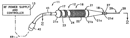
More in particular, the apparatus 11 of device 12 for
use therein for the ablation of tissue as shown in Figure 1
includes the hand held device 12 and a radio frequency power
supply and controller 13 as shown in block form.
The device 12 consists of a handle or housing 16 which
is to size so that it is adapted to be grasped by the human
hand or at least by two fingers of the human hand. The
handle or housing 16 is formed of a suitable material such
as a plastic which is molded into a desired shape as for
example, generally cylindrical as shown in Figure 1 and is
provided with proximal and distal extremities 17 and 18.
The handle 16 is provided with an outer surface 21 with a
semi-hemispherical portion 21a provided on the proximal
extremity, a cylindrical portion 21b extending from the
proximal extremity for a distance of approximately 2-1/2",
a tapered or conical portion 21c having a length of
approximately 1-1/2" and a smaller diameter cylindrical
portion 21d having a length of approximately 1/2". The
handle 16 can be of a suitable diameter such as 1/2". It
should be appreciated that if desired rather than it being
circular in cross section, the handle 16 can be rectangular
in cross section. A portion of the surface 21b is provided
with a plurality of circumferentially extending annular
grooves 22 spaced apart longitudinally of the central axis
of the handle 16 to facilitate gripping of the handle by the
fingers of a human hand. A pair of spaced apart annular
recesses 23 and 24 is provided on which identification
labels (not shown) can be placed.
A sharpened needle 26 is provided which has proximal
and distal extremities 27 and 28. It is formed of a
suitable conductive material such as stainless steel which
is capable of delivering radio frequency energy. Means is
provided for mounting the needle in the handle or housing 16
so that it is static or nondeployable. As shown in Figures
1 and 2, it is mounted on the distal extremity 18 by being
CA 02336279 2000-12-28
WO 00/66014
-5-
PCTNS00/11384
molded directly into the plastic handle or housing 16. The
proximal extremity 27 of the needle 26 is mounted in a
carrier 31 formed of a suitable material such as plastic
which is mounted within the handle 16 as shown in Figure 2.
A printed circuit board 32 is mounted on the carrier 3i
immediately adjacent the proximal extremity 27 of the needle
26.
Conductive means 36 is carried by the handle and is
connected to the needle and is adapted to be coupled to the
radio frequency power supply and controller 13 for supplying
radio frequency energy to the needle 26. Typically this
conductive means takes the form of a single conductor 37
hereinafter described which is coupled to the needle 26 by
suitable means such as solder and which extends proximally
through the housing and to a flexible cable 41 secured to
the proximal extremity of the handle 16. The flexible cable
41 carries a male adapter 42 which is adapted to be coupled
to a female adapter (not shown) to a cable 44 to the radio
frequency power supply and controller 13.
Means is carried by the handle or housing 16 and is
adapted to be coupled to the radio frequency power supply
and controller 13 for sensing the application of radio
frequency energy as it is supplied by the needle 26 to the
tissue in the human body for controlling the application of
radio frequency energy to the tissue and consists of at
least one device for sensing temperature and/or impedance.
Thus as shown there are provided first and second
thermocouples 46 and 47. In accordance with the present
invention, the first thermocouple 46 as shown in Figure 4 is
mounted in the distal extremity 18 of the handle 16 and is
provided for sensing the temperature of the tissue in the
immediate vicinity of the thermocouple 46 adjacent to an
intermediate portion of the needle 26 where it enters the
handle 16, as for example approximately 20 millimeters from
the end of the needle 26. First and second conductors 48
CA 02336279 2000-12-28
WO 00/66014 PCT/US00/11384
-6
and 49 are provided which are connected to the thermocouple
46. If desired, the conductors 48 and 49 alternatively can
be secured to the needle 26 by a shrink tube (not shown)
secured to the handle 16 The other or second thermocouple 47
is mounted in the distal extremity 28 of the needle 26 and
as shown can be supported by an epoxy 51 provided in a bore
52 in the needle 26 extending longitudinally of the needle
26. The epoxy 51 in addition to holding the thermocouple 47
seals oft the lumen or bore 52. The thermocouple 47 senses
the temperature of the tissue in the immediate vicinity of
the distal extremity 28 of the needle 26. First and second
conductors 53 and 54 connected to the thermocouple 47 extend
proximally within the bore 52 of the needle 26 from the
thermocouple 47. The conductors 48 and 49 and the
conductors 53 and 54 extend proximally to the printed
circuit board 32 as shown in Figure 2 and terminate in three
contacts 61, 62 and 63 provided on the printed circuit board
32 in which contact 62 is a common contact to which are
bonded conductors 66, 67 and 68 which extend into the cable
41. Another contact 71 is provided on the printed circuit
board 32 which is connected to the needle 26 by the
conductor 37 which is also connected to a conductor 72
extending into the cable 41. These conductors 66, 67, 68
and 72 are connected into the radio frequency power supply
and controller 13 and are utilized for supplying radio
frequency energy to the needle electrode 26 and for the
control of the radio frequency power supply and controller
13 in accordance with the parameters, i.e. temperatures,
being sensed by at least one thermocouple and preferably
both thermocouples 46 and 47.
Since the needle 26 is a static or nondeployable
needle, the needle 26 upon manufacture of the device 12 can
be selected to be of a suitable length projecting distally
from the distal extremity 18 of the handle 16. Thus a
needle having a length ranging from 15 to 30 mm and
CA 02336279 2000-12-28
WO 00/66014 PCT/US00/11384
preferably approximately 20 mm can be readily provided. The
needle can be of a certain size as for example 23-gauge.
It should be appreciated that insulation can be
extended on the needle so that a desired active length for
the needle is provided for supplying radio frequency energy
to the tissue.
Operation and use of the apparatus and the device for
use therewith may now be briefly described as follows.
Assuming that the device 12 has been connected to the radio
frequency power supply and controller 13, the physician
doing the desired tissue ablation procedure grasps the
handle 16 of the device by the fingers of a hand or in the
palm of the hand and with a straight needle 26 as shown in
Figure 1, the physician can utilize the handle to cause the
needle to penetrate the tissue it is desired to ablate. The
needle 26 is positioned so that the insulation engaging the
proximal end of the needle 26 is well past the mucosal
layers of the tissue, after which the radio frequency power
supply and controller 13 can be turned on. This ensures
that the mucosal layer will remain undamaged and will not be
thermally ablated.
The needle 26 can be utilized as a unipolar device
with a grounding pad (not shown) being provided on the
patient as for example on the back of the patient to
complete the circuit for the radio frequency energy from the
radio frequency power supply and the return to the radio
frequency power supply 13. For example with a straight
needle, the turbinates can be readily treated with the
device 12. The treatment can be carried out for an
appropriate length of time from 20 seconds to 5 minutes with
the radio frequency energy being applied at the desired
frequency, as for example a frequency of 580 kilohertz and
a power level ranging from 5 to 50 watts . The shorter times
are desirable where the size of the anatomical feature to be
treated is small (such as the uvula) or where the tissue is
CA 02336279 2000-12-28
WO 00/66014 PCTNS00/11384
_g_
highly hydrated or perfused. This helps to preserve
anatomical tissue in the region to be ablated, as for
example anatomical features which are then in cross-section
(i.e., mucosal membranes). The thermocouples 46 and 47 can
be utilized for automatically terminating the application of
radio frequency power when a certain temperature in the
tissue has been reached as sensed by either one or both of
the thermocouples 46 and 47. The delivery of radio
frequency energy to the needle 26 is terminated before the
needle 26 is withdrawn from the tissue to avoid surface
layer thermal damage. After the procedure has been
completed, the physician can withdraw the device 12 and can
further proceed with the procedure by inserting the needle
26 of the device 12 into another location using the same
procedure. The foregoing steps can be repeated as necessary
to complete the desired ablation of the tissue being
treated.
In the case of some smaller anatomical features, the
physician may use lower power levels to obtain a lesion of
sufficient size without premature desiccation of the tissue
surrounding the active electrode (needle). This lower rate
of energy delivery is an important aspect of the present
invention because it yields larger lesions and greater
volume per penetration than would occur if the power
settings were higher. In that case, rapid heating can
result in loss of current delivery due to tissue
desiccation: The reason the lower power settings result in
larger lesions is that the hydrated tissue exhibits thermal
conductivity at a fairly inefficient level, but is
nonetheless somewhat thermally conductive. If the power
setting is appropriate, the tissue is able to conduct the
energy outwardly in the form of heat and the tissue
immediately adjacent to the active electrode will be kept
below the temperature of vaporization of the fluid within
the tissue. When vaporization occurs, there is a
CA 02336279 2000-12-28
WO 00/66014 PCT/US00/11384
_g_
fluctuation of the ohmic impedance to current low and the
tissue rapidly desiccates, resulting in interruption of the
circuit. This loss of current flow due to overheating of
the tissue adjacent to the electrode needle can be an
advantage in that it is a safety aspect of the present
invention. For instance, in the event of inadvertent
setting of the power at a high level, the rapid desiccation
of the thin layer of cells in contact with the active
electrode will break the circuit and act as a "biologic
switch", cutting off current flow and preventing extensive
tissue damage. Only by setting the power at lower levels,
as for example 1 watt, can larger lesions be attained.
Typically, power settings up to 15 watts are used with the
type of device described in the present invention.
Situations where power levels as high as 50 or 100 watts can
occur where an electrode with a significantly larger surface
area is used or where the tissue is highly perfused and the
circulatory (blood flow) rate is high, resulting in
efficient cooling of the tissue being treated.
For performing other tissue ablation procedures where
a curved or bent needle 26 is desired, the needle 26 can be
formed of a malleable material and can be bent in a suitable
manner to the desired configuration to match the anatomy, as
for example the treatment of tonsils, adenoids and sinus
tissue. A straight needle can be utilized for treating the
uvula.
After the device has been used, it can be disposed of
because the device has been designed for a one time use even
though the device is manufactured in such a way that it is
sterilizable. However, sterilizing the same may be
undesirable when it is difficult to ensure that sufficient
sterilization has been accomplished and particularly if
blood has coagulated on the needle 26 causing a protein
buildup which may not be removed during the sterilization
procedure. It should be appreciated as hereinbefore
CA 02336279 2000-12-28
WO 00/66014 PCT/US00/11384
-10_
explained that the device can still be further simplified by
using only one thermocouple. It is possible to utilize only
one thermocouple by estimating the temperature gradient
which normally occurs between the first and second
thermocouples. By utilizing only the first thermocouple 46
it is possible to go to a solid wire for the needle 26
rather than a needle which has a lumen or bore therein.
In order to still further reduce the cost of the
device utilized in connection with the present apparatus,
another embodiment of the device is shown Figure 5 in which
the cable and connector are removably mounted on the device
as part of the device so that they can be disconnected and
only a part of the device disposed of after use. Thus as
shown in Figure 5 there is provided a device 81 which
consists of a handle 82 sized to fit into a human hand and
which has generally the same configuration as the handle 16,
but which is generally rectangular in cross-section rather
than circular. It is provided with proximal and distal
extremities 83 and 84. The proximal extremity 83 of the
housing has mounted therein the cable 41 hereinbefore
described in the embodiment shown in Figures 1-4. The
handle 82 is fabricated in two parts 82a and 82b with the
part 82a forming the proximal extremity 83 and the part 82b
forming the distal extremity 84. The printed circuit board
32 forming a part of the previous embodiment is also
included in the present embodiment with the associated
wiring (not shown) and is mounted in the reusable connector
portion 82a of the handle 82. A connector assembly 86 is
mounted in the two parts 82a and 82b and typically as shown
can consist of a female connector 87 mounted in the reusable
connector portion 82a and a male connector 88 mounted in the
disposable portion 82b.
A needle 91 is mounted in the distal extremity 84 in
the manner hereinbefore described in connection with the
embodiment shown in Figure 1. In the embodiment shown, the
CA 02336279 2000-12-28
WO 00/66014 PCT/US00/11384
-11-
needle 91 is inclined at an angle with respect to the
central axis of the handle 82 as for example at an angle of
45°. An insulating sleeve 92 is provided on the needle and
has a length so that the exposed end of the needle 91
extends for a suitable distance as for example 15 to 30 mm
and preferably approximately 20 mm. The insulating sleeve
92 is provided with a thread 93 on its exterior surface 94.
The threads 93 can be relatively coarse, as for example a
quarter pitch, so that a protective sleeve 96 with internal
threads 97 in a bore 98 matching the threaded exterior
surface 93 can be threaded onto and threaded off of the
insulating sleeve 92 with four to five turns of the
protective sleeve 96. The protective sleeve 96 can be
formed of a suitable material such as plastic. The
protective sleeve 96 has a length so that it will extend
over the length of the insulating sleeve 92 and still
provide adequate space for the needle 91 extending distally
from the insulating sleeve 92.
It can be seen that by providing a threaded protective
sleeve 96, the sleeve 96 can be rotated for removal of the
same. This threaded arrangement is preferable to one which
is mounted by a slip fit because a slip fit requires
movement of the sleeve towards and away from the needle
during pushing and pulling of the sleeve, making it possible
for the physician using the same to inadvertently be
punctured by the needle.
The device shown in Figure 5 can be used in a manner
very similar to that hereinbefore described with respect to
the previous embodiment. After the device has been used,
the portion 82b can be separated from the portion 82a and
only the portion 82b disposed of after use. The remaining
portion 82a with the cable 41 can be retained for future
reuse. This part 82a can be readily sterilized if necessary
and carries the carrier components which comprise the major
expense in fabricating the handle 81. Thus it can be seen
CA 02336279 2000-12-28
WO 00/66014 PCT/US00/11384
-12
that such a construction makes it possible to further reduce
the cost of the device utilized in the apparatus of the
present invention.
Still another embodiment incorporating the device of
the present invention is shown in Figure 6 which is slightly
more expensive than that shown in Figure 5, but however
retains as a separable part the connector and cable forming
a part of the handle. Thus as shown in Figure 6 there is
provided a device 101 which is also sized to fit into the
human hand but typically is larger so that it is adapted to
be held in the palm of the hand while a finger or fingers
are utilized for operating the device. The handle 102 as
shown is rectangular in cross section and is provided with
proximal and distal extremities 103 and 104 with the
proximal extremity 103 comprising the reusable part and the
distal extremity 104 comprising the disposable part. The
handle or housing 102 is formed of a suitable material such
as plastic with lower and upper parts 106 and 107 which are
fastened together in a suitable manner such as by an
adhesive or by ultrasonic bonding. A connector assembly 111
is provided for connecting the wires or conductors utilized
in the device and consists of a male connector 112 provided
in a distal extremity 104 and a female connector 113
provided in the proximal extremity or reusable portion 103.
The male connector assembly is provided with a rectangular
framework 116 formed of a suitable material such as plastic
to prevent accidental contact with the pins 117 forming a
part of the male connector assembly 112. The female
connector assembly 113 is connected to the cable 41
connected to the reusable proximal part 103.
A retractable needle 121 is carried by the handle or
housing 102 and is mounted on a slider 122 movable in slots
123 within the handle 102 from a distal extremity where the
needle is in an extended position extending beyond a
cylindrical insulation sleeve 124 forming a part of the
CA 02336279 2000-12-28
WO 00/66014 PCTNS00/11384
-13
handle 102 and a retracted position in which the needle is
completely retracted within the insulation sleeve 124.
Movement of the slider 122 is under the control of a
circular knob 126 slidably mounted on the exterior of the
handle or housing 102 and adapted to be grasped by a finger
of the hand and particularly the thumb of the hand holding
the device 101. The knob is provided with a centrally
disposed recess 127 adapted to be engaged by the thumb of
the holding hand. The circular knob 126 is provided with a
depending stem 128 which extends through a slot 129 in the
top cover 107. The slot 126 extends longitudinally of the
top cover 107 along the central axis of the handle or
housing 102. A printed circuit board 32 of the type
hereinbefore described is mounted within the handle 102 and
is provided with folded wires or conductors (not shown)
which permit the slider 122 to move between extended and
retracted positions while still continuing to receive
information from the thermocouples and also to supply radio
frequency energy to the needle 121.
Operation and use of the device 101 shown in Figure 6
is very similar to that hereinbefore described. However, in
many respects it is more user friendly than the other
embodiments of the device herein disclosed. For example by
providing a retractable needle 121, it is possible for the
physician to position the needle in the desired position
merely by engaging the knob 126 by the thumb of the hand
while the same hand is holding the device to advance the
needle 121 into the tissue to be treated. After the
application of radio frequency energy in the manner
hereinbefore described, the needle 121 can be retracted back
into the handle 102 without danger of the physician being
pricked by the needle. The major portion of the device can
still be saved by separating the proximal portion 103 which
carries the cable 41 from the distal portion 104 so that the
distal portion can thereafter be disposed of after a one-
CA 02336279 2000-12-28
WO 00/66014 PCT/US00/11384
-14
time use.
Another embodiment of a device incorporating the
present invention is shown in Figures 7-10. As shown
therein, there is provided a device 141 which is provided
with a handle 142 which is adapted to be grasped between the
fingers of a human hand for use of the device. The handle
142 is formed with a two-part housing 143 having a main
housing 146 and a connector housing 147. The main housing
146 is provided with mating top and bottom casings 146a and
146b and similarly, the connector housing 147 is provided
with mating top and bottom covers 147a and 147b. The
housing 143 can be formed of a suitable material such as
plastic with the top and bottom casings 146a and 146b being
fastened together in a suitable manner such as by ultrasonic
bonding. Similarly, the top cover 147a and the bottom cover
147b can be fastened together in a similar manner. The
handle 142 is provided with proximal and distal extremities
151 and 152.
An edge card 156 is mounted within the main housing
146. The edge card 156 is in the form of a printed circuit
board and has a plurality of edge mounted contacts on one
edge of the same, preferably the edge facing proximally of
the handle 142. Such a printed circuit card in addition to
carrying the desired circuitry also includes in the present
embodiment a fuse circuit which is embedded in a printed
circuit board for controlling or limiting the use of the
device as for example for only permitting two uses of the
device.
A suitable card edge connector 161 such as a 2 by 10
connector is mounted in the connector housing 147 and is
adapted to fractionally engage and electrically contact the
edge mounted contacts carried by the edge card 156. As
shown particularly in Figure 8, the edge card 156 is mounted
in the proximal end 151 of the main housing 146 whereas the
card edge connector 161 is mounted in the distal end 152 of
CA 02336279 2000-12-28
WO 00/66014 PCT/US00/11384
-15
the connector housing 147 so that the connector and edge
card can be mated when the main housing 146 and the
connector housing 147 are mated or interconnected. The card
edge connector 161 is connected to another printed circuit
card 162 in a conventional manner and is connected by
electrical conductors (not shown) to a flexible cable 163
extending into the connector housing 147 and extending
proximally therefrom. The distal extremity of the cable 163
and the printed circuit board 162 are encased by a custom
overmold 166 formed of a suitable plastic insulating
material to encapsulate the electrical conductors connecting
the cable 163 to the connector 161. The overmold 166 also
has a portion 166a which extends proximally from the
connector housing 147 to provide a strain relief for the
cable 163.
In the main housing 146, the proximal extremity of an
elongate flex circuit 171 is secured to and connected to the
circuitry carried by the edge card 156. The distal
extremity of the elongate flex circuit 171 is secured
physically and electrically to a PC board 172.
A needle 176 has its proximal extremity secured to the
underside of the PC board 172 by a metal clasp 177 to firmly
hold the needle. The needle 176 is provided with a fixed
insulating sleeve 178 so that an exposed sharpened distal
extremity 176a is provided on the needle. The needle 176
can carry one or more thermocouples of the type hereinbefore
described in connection with the previous embodiments. The
needle and thermocouples carried thereby are electrically
connected on the PC board 172. Electrical connections
extend from the PC board 172 through the flex circuit 171 to
the fuse circuit carried by the edge card 156 and thence
through the cable 163 to the radiofrequency power supply and
controller of the type hereinbefore described.
As shown in Figures 8 and 9 of the drawings, the top
and bottom casings 146a and 146b are provided with support
CA 02336279 2000-12-28
WO OOI66014 PCT/US00/11384
-16
means for supporting the edge card 156 and the PC board 162
in a firm position within the main housing. Such means
consists of pairs of support pedestals in which one pair
consists of mating support pedestals 181 and 182 and the
another pair consists of support pedestals 183 and 184
spaced from the first pair. One of the pedestals of each
pair is provided with a U-shaped recess 186 which is sized
to accommodate the edge card 156 for supporting the edge
card 156 in a firm position so that it can be readily
engaged and disengaged by the card edge connector 161
carried by the connector housing 147. Similar support means
is provided for the PC board 172 and consists of mating
support pedestals 187 and 188 supporting one end of the PC
board 172 and support pedestals 191 and 192 supporting the
other end of the PC board 172 and the proximal end of the
needle 176 secured thereto. A cross member 193 forms a part
of the lower casing 146b and extends transversely of the
bottom casing 146b and serves to support the flex circuit
171 in a region intermediate the PC board 172 and the edge
card 156.
The needle 176 is formed of a suitable material such
as a nickel-titanium alloy and has a suitable length as for
example approximately 4" and a suitable diameter as for
example 0.0026". The insulating sleeve 178 provided on the
needle 176 extends to near the tip 176a of the needle 176
but leaving a portion exposed as for example l5mm from the
sharp needle tip. Typically one of the thermocouples is
carried by the tip whereas the other thermocouple is carried
at the distal extremity of the insulation layer 176 as
hereinbefore described.
The main housing 146 has a generally tapered
appearance. It has a taper which extends down into a
circular necked-down region 201 after which distally there
is provided a spherical bulge 202. This necked-down region
201 and the spherical bulge 202 provide a region to
CA 02336279 2000-12-28
WO 00166014 PCT/US00/11384
-17
facilitate gripping of the handle 142 between two fingers of
the hand as for example the thumb and forefinger. After the
bulge 202, the main housing 146 is provided with a tapered
inclined region 203 and thence distally a tapered generally
cylindrical region 204 of a smaller diameter which encloses
the needle as it extends from the printed circuit board 172
as shown particularly in Figure 8. In this configuration,
the cylindrical region 204 extends at an angle of
approximately 45° with respect to the main portion of the
main housing 146. This inclination is provided to provide
high visibility to the physician using the device as
hereinafter described for viewing the cavity areas where the
device is being used.
A bracket 206 is provided within the top casing 146a
and is formed integral therewith. The bracket 206 is
provided with a hole 207 therein through which the needle
176 extends. A hole 208 is provided in the top casing 146a
in the vicinity of the bracket 206 and permits viewing of
the proximal extremity of the needle 176.
Cooperative attachment means is provided for removably
securing the connector housing 147 to the main housing 146
and consists of side latches or detents 211 and 212. Each
of the side latches or detents consists of a raised
protrusion 213 serving as a button adapted to be engaged by
a finger of the hand. The raised protrusion 213 is formed
integral with the bottom casing 146b. An elongate slot 214
(see Figure 9) is provided in the proximal extremity of the
lower casing 146b and extends in both directions from the
protrusion 213 so that the portion 215 of the bottom casing
146b immediately underlying the raised protrusion 213 can be
pressed inwardly to cause deflection of that part of the
housing and to cause inward movement of a protrusion 216
carried thereby which is triangular in cross section. The
protrusion 216 is provided with a vertical face 217 which
extends generally parallel to the direction of movement of
CA 02336279 2000-12-28
WO 00/66014 PCTNS00/11384
-1s
the portion 215 as it is depressed by the raised protrusion
213 (see Figure 10) and at an angle with respect to an
inclined surface 218. This triangular-shaped protrusion 216
is adapted to be received by a recess 221 provided in the
bottom cover 147b of the connector housing 147. Thus it can
be seen by grasping the handle 142 and depressing both of
the buttons or raised protrusion 213 with two fingers of the
hand and pushing inwardly on the same, the connector housing
147 can be detached from the main housing 146 and the card
edge connector 161 separated from the card edge connector
161. Similarly, the connector housing 147 can be attached
to the main housing and the card edge connector 161 by
relative movement between the connector housing 147 and the
main housing 146 which causes the connector housing to cam
over the inclined surface 218 while depressing the same to
provide clearance until the protrusions 216 can snap into
the recesses 221 provided on opposite sides of the bottom
cover 147b.
Operation and use of the device 141 shown in Figures
7 through 10 is very similar to that hereinbefore described
with the previously disclosed embodiments. The device 141
by the use of the cable 163 is connected to the
radiofrequency power supply and controller 13 hereinbefore
described. The physician desiring to do the tissue ablation
procedure grasps the handle 142 of the device 141 by placing
the forefinger and a thumb in the necked-down region 201
just proximal of the spherical bulge 202. The physician
while holding the device 141 in one hand can readily view
the needle 176 and by way of example can advance the needle
into the turbinate areas of the nasal passages of the
patient. Typically the needle is inserted into the
turbinate with its sharpened distal extremity 176a
penetrating the tissue so that the exposed area is disposed
within the tissue and the insulating layer 178 penetrates at
least slightly below the surface of the tissue being
CA 02336279 2000-12-28
WO 00/66014 PCTNS00/11384
-19
penetrated. Radiofrequency energy can then be applied to
accomplish the desired ablation procedure. After the
ablation procedure has been completed, the device 141 can be
removed. If desired, the device can be utilized to
penetrate other areas of the tissue to be treated if that is
desired.
The device 141 particularly lends itself to such
procedures because it is a low cost device. It has a number
of unique features which contribute to this cost containment
goal. The utilization of an edge card in conjunction with
a card edge connector simplifies the connection between the
main housing 146 and the connector housing. If desired, the
main housing 146 can be discarded after a one time use after
separating it from the connector housing 147 as hereinbefore
described. This makes it possible to save the cost of
replacing the connector housing 147 and its relatively
expensive cabling 163. The use of the flex circuit between
the edge card 156 and the PC board 172 also greatly
simplifies the construction and assembly of the device.
Another embodiment of a device incorporating the
present invention is shown in the device 241 in Figure 11.
As shown therein, this device 241 is comprised of a main
housing 242 and a connector housing 243 which are
constructed in a manner similar to the main housing 146 and
the connector housing 147 with differences in the outer and
inner configurations. The main housing 242 has the
connector housing 243 connected thereto in a manner as
hereinbefore described in conjunction with the device 141
and can be separated along a parting line 246 to provide
separation between a card edge connector (not shown)
provided in the connector housing 243 and connected to an
edge card (not shown) in the main housing 242. Raised
protrusions 249 are provided on opposite sides to facilitate
the connection and disconnection of the two housings 242 and
243 and correspond to the protrusions 213 provided in the
CA 02336279 2000-12-28
WO 00/G6014 PCT/US00/11384
-20
device 141.
A retractable needle 251 is slidably mounted in the
main housing 242 for movement between extended and retracted
positions and which in the retracted position is enclosed
within the main housing 242. The retractable needle is
provided with a sharpened tip 251a which is exposed for a
suitable distance as for example l5mm with the remaining
part being covered by an insulating layer 252. The
retractable needle 251 is mounted on a PC board (not shown)
similar to the PC board 172 which is carried by a slider
(not shown) corresponding to the slider 122 shown in Figure
6 which is connected to an oval-shaped knob 256 through an
extension (not shown) that travels in a slot such as slot
123 in Figure 6. The knob 256 is provided with an upwardly
facing oval-shaped recess 257 provided on the knob 256 and
which is adapted to be engaged by a finger of the hand as
for example the forefinger while the remainder of the hand
is being utilized for holding the device 241. The device
241 is provided with a tab 261 which covers the slot (not
shown) in the main housing so that it remains invisible as
the knob 256 is advanced and retracted during advancement
and retraction of the retractable needle 251.
The connector housing 243 is provided with a cable 266
of the same type as cable 163 hereinbefore described in
conjunction with the embodiment shown in Figures 7 through
10.
The device 241 has a main housing 242 and a connector
housing 243 sized is such a manner so that the device can be
readily held in the palm of a human hand. The main housing
242 is provided with an arcuate transversely disposed recess
271 which can be readily be grasped by the foref roger of the
hand holding the device while at the same time permitting
the thumb of the same hand to be inserted into the recess
257 to translate the knob 256 longitudinally of the main
housing for causing extension and retraction of the
CA 02336279 2000-12-28
WO 00/66014 PCT/US00/11384
-21-
retractable needle 251. The device 241 can be utilized for
ablation procedures of the type hereinbefore described as
for example for deployment of the needle 251 into the uvula,
soft palate and into the tongue regions of the oral cavity.
If desired, the retractable needle 251 can be formed of a
nickel-titanium alloy so that it can be provided with a
memory which returns to a straight shape even after it has
been bent. For the device as shown in Figure 11 it may be
desirable to provide a needle 251 which can be bent. If
that is the case, it is desirable that a material other than
a nickel-titanium alloy such as stainless steel be utilized
so that this makes it possible for the physician using the
device to preshape the curvature of the needle before
commencing the procedure or after commencement of the
procedure in more difficult to reach regions.
From the foregoing it can be seen that there has been
provided a device which can be utilized for ablation of
tissue in connection with a radio frequency power supply and
controller. The devices are small and adapted to be held by
the human hand and are designed in such a manner so that the
entire device or only a portion of the device can be
disposed of after a one-time use. Static or retractable
needles can be provided. The construction has been kept so
that it is relatively simple to minimize the cost of
construction and inexpensive materials have been utilized
where possible.