Note: Descriptions are shown in the official language in which they were submitted.
CA 02336940 2009-10-08
1
PROPORTIONAL PRESSURE ASSIST VENTILATION CONTROLLED
BY A DIAPHRAGM ELECTROMYOGRAPHIC SIGNAL
BACKGROUND OF THE INVENTION
1. Field of the invention:
The present invention relates to a neuro-ventilatory efficiency computation
device
for monitoring/controlling a level of ventilatory assist to a patient.
The present invention also relates to a device for monitoring/adjusting the
level of
positive end expiratory pressure produced by a pressure assist device in
relation to a
signal representative of inspiratory effort in view of minimizing the level of
pre-inspiratory
effort.
2. Brief description of the prior art:
Prior art algorithms used to create closed-loop ventilator systems are based
on
variables such as tidal volume, respiratory rate, inspiratory flow, end-tidal
carbon dioxide
levels and/or rate of rise in pressure. However, none of these parameters can
provide a
reliable measure of the respiratory neural drive because they are affected by
changes in
neuro-mechanical or neuro-ventilatory efficiency.
Neuro-ventilatory efficiency is a term used to express the amount of neural
drive
(breathing effort) needed to obtain a given tidal lung volume. In brief,
neural drive is
converted into mechanical tension, a process which is influenced by the muscle
length,
temperature, electrolyte imbalance, etc. The role of inspiratory flow in the
link between
neural drive and mechanical tension has previously been suggested, however the
proposed influence could not be demonstrated for mean inspiratory flow rates
up to 1.4
liters/second. The mechanical tension is then translated into pressure, a
process which
is affected by the shape of the diaphragm dome. Finally the pressure expands
the
alveoli and causes air to flow, and the translation of pressure to volume
depends on the
elasto-viscous behaviour of the respiratory system. Consequently, there are
many
1408478.1
CA 02336940 2009-10-08
2
factors that may influence the tidal volume output obtained for a given
increase in neural
drive (inspiratory effort).
Evaluation of respiratory drive by measurements such as the rate of rise in
pressure or lung volume is not reliable when, for example, the muscle length
or the
respiratory system impedance are affected by changes in the neuro-ventilatory
efficiency. In a patient, airway resistance and elastance can change from one
minute to
another and muscle length is continuously altered.
OBJECTS AND SUMMARY OF THE INVENTION
An object of the present invention is therefore to eliminate the drawbacks of
the
prior art.
Another object of the present invention is to provide a closed loop system
using:
(a) the intensity of the diaphragm electromyogram (EMG) for a given
inspiratory volume;
(b) the inspiratory volume for a given EMG intensity; or
(c) a combination of (a) and (b);
in view of controlling the level of gas flow, gas volume or gas pressure
delivered by a
mechanical (lung) ventilator; the closed loop ventilator system enables for
automatic or
manual adjustment of the level of inspiratory support in proportion to changes
in the
neuro-ventilatory efficiency such that the neural drive remains stable at a
desired target
level. An alarm can also be used to detect changes in neuroventilatory
efficiency in view
of performing manual adjustments.
Another object of the present invention is to provide a closed-loop system
responsive to the intensity of the diaphragm EMG measured for example
immediately
before the onset of inspiratory flow to quantify pre-inspiratory breathing
effort in view of
automatically or manually adjusting a level of extrinsic positive end
expiratory pressure
(PEEP) applied to a patient in proportion to changes in EMG intensity of pre-
inspiratory
efforts. In this manner, the pre-ventilatory intensity of the diaphragm EMG
can be
maintained at a desired, minimum level such that the pre-inspiratory neural
drive
remains stable at a desired target minimal level. Determination of the
duration from the
onset of EMG to the onset of respiratory flow can also be used for
quantitative
1408478.1
CA 02336940 2009-10-08
3
evaluation of the intrinsic PEEP, and to guide adjustment of the trigger
sensitivity of the
ventilator systems.
Different from pressure and ventilatory related indexes, the intensity of the
EMG
represents the temporal (mean MU (motor unit) rate coding) and spatial (MU
recruitment) summation of action potentials and can be obtained at the level
of the
sarcolemma muscle. The intensity of the EMG is therefore not affected by
changes in
the muscle's neuroventilatory coupling. The use of crural diaphragm EMG rests
on the
assumption that neural drive to the crural diaphragm is representative for the
total
respiratory drive. It is also based on the condition that neuromuscular
transmission and
innervation of the crural diaphragm are normal. For breathing with increased
demand
this assumption is well founded. Hence, the intensity of the EMG needed to
produce a
given inspiratory volume should express the efficiency relation between neural
drive and
volume output.
More specifically, according to the present invention, there is provided a
neuro-
ventilatory efficiency computation device for monitoring/controlling a level
of ventilatory
assist to a patient comprising: means for receiving an EMG signal intensity
representative of inspiratory effort of the patient; means for receiving a
lung volume
value representative of a lung volume of the patient; means for determining
from the
received EMG signal intensity and lung volume value at least one of the two
following
relations: an EMG signal intensity for a given lung volume value, the received
lung
volume value then being the given lung volume value, and a lung volume value
for a
given EMG signal intensity, the received EMG signal intensity then being the
given EMG
signal intensity; and means for increasing or decreasing the ventilatory
assist level
depending on whether the at least one relation has increased or decreased by
at least a
given percentage.
The present invention also relates to a neuro-ventilatory efficiency
computation
device for monitoring/controlling a level of ventilatory assist to a patient
comprising: a
first input for receiving an EMG signal intensity representative of
inspiratory effort of the
patient; a second input for receiving a lung volume value representative of a
lung
volume of the patient; connected to the first and second inputs a caiculator
of at least
one of the two following relations: an EMG signal intensity for a given lung
volume value,
1408478.1
CA 02336940 2009-10-08
4
the lung volume value received on the second input then being the given lung
volume
value; and a lung volume value for a given EMG signal intensity, the EMG
signal
intensity received on the first input then being the given EMG signal
intensity; and a
controller for increasing or decreasing the ventilatory assist level depending
on whether
the at least one relation has increased or decreased by at least a given
percentage.
The present invention is further concerned with a device for
monitoring/adjusting
the level of positive end expiratory pressure produced by a pressure assist
device in
relation to a signal representative of inspiratory effort in view of
minimizing the level of
pre-inspiratory effort, comprising: a) means for receiving a signal
representative of
inspiratory flow; b) means for calculating from the inspiratory flow signal an
onset time
for inspiration; c) means for receiving a signal representative of inspiratory
effort having
an amplitude; d) means for calculating a signal representative of pre-
inspiratory effort in
response to the onset time and the signal representative of inspiratory
effort; and e)
means for increasing or decreasing the level of positive end expiratory
pressure in
relation to the signal representative of pre-inspiratory effort.
The present invention still further relates to a controller for
monitoring/adjusting
the level of positive end expiratory pressure produced by a pressure assist
device in
relation to a signal representative of inspiratory effort in view of
minimizing the level of
pre-inspiratory effort, comprising: a) a first input for receiving a signal
representative of
inspiratory flow having an onset time for inspiration; b) a second input for
receiving a
signal representative of inspiratory effort having an amplitude; c) a computer
device
responsive to the onset time and the signal representative of inspiratory
effort to
compute a signal representative of pre-inspiratory effort; and d) an
adder/subtractor for
adding a preset increment to or subtracting a preset decrement from the level
of positive
end expiratory pressure in relation to the signal representative of pre-
inspiratory effort.
The objects, advantages and other features of the present invention will
become
more apparent upon reading of the following non restrictive description of an
illustrative
embodiment thereof,
1408478,1
CA 02336940 2001-01-10
WO 99/62580 PCT/CA99/00529
given by way of example only with reference to the accompanying
drawings.
II,
BRIEF DESCRIPTION OF THE DRAWINGS
5
In the appended drawings:
Figure 1 is a schematic representation of a set-up of an
EMG analysis system;
Figure 2 is a section of oesophageal catheter on which
an array of electrodes of the EMG analysis system of Figure 1 is
mounted;
Figure 3 illustrates a section of oesophageai catheter on
which a second embodiment of the array of electrodes is mounted;
Figure 4 is a graph showing a set of EMG signals of the
diaphragm (EMGdi signals) detected by pairs of successive electrodes of
the array of Figure 2;
Figuire 5 is a flow chart showing a method for conducting
a double subtraction technique of the EMGdi signals;
FigLire 6 is a graph showing the distribution of correlation
coefficients calculated for determining the position of the center of the
CA 02336940 2001-01-10
WO 99/62580 PCT/CA99/00529
6
depolarizing region of the diaphragm along the array of electrodes of
Figure 2;
Figure 7 is a schematic diagram illustrating in the time
domain a double subtraction technique for improving the signal-to-noise
ratio and to reduce ain electrode-position-induced filter effect along the
array of electrodes of Figure 2;
Figure 8a is a graph showing the power density
spectrum of electrode motion artifacts, the power density spectrum of
ECG, and the power density spectrum of EMGdi signals;
Figure 8b is a graph showing an example of transfer
function for a filter to be used for filtering out the electrode motion
artifacts, ECG, and thie 50 or 60 Hz disturbance from electrical mains;
Figuire 9 is a schematic diagram illustrating in the
frequency domain stalbilization by the double subtraction technique of the
center frequency upon displacement of the center of the depolarizing
region of the diaphragm along the array of electrodes of Figure 2;
Figure 10 is a schematic block diagram of a system
according to the inverition for controlling inspiratory assist by means of an
EMGdi signal obtained with the above mentioned double subtraction
technique and a measurement of the volume of air breathed by the
patient by a commercially available system;
- -- ---- --------
CA 02336940 2001-01-10
WO 99/62580 PCT/CA99/00529
7
Figure 11 is a schematic block diagram of a system
according to the inventidn ( a) capable to determine the time delay from
the onset of EMG to tlhe onset of inspiratory flow and ( b) using the level
of pre-inspiratory effort obtained through the EMGdi signal intensity
(common noise levell subtracted) during a predetermined time period
immediately preceding the onset of inspiratory flow to indicate the
presence of "intrinsic PEEP" and to adjust the level of applied "extrinsic
PEEP" and/or ventilator trigger sensitivity such that the level of pre-
inspiratory effort is suppressed, i.e the EMGdi signal intensity (common
noise level subtracted) during the above mentioned predetermined time
period is close to zero;
Figure 12a is an exemplary graph of a patient's
inspiratory flow versus time for quiet breathing in COPD (Chronic
Obstructive Pulmonary Disease); and
Figure 12b is an exemplary graph of a patient's EMG
RMS intensity versus time for quiet breathing in COPD.
DETAILED DESCRIPTION OF THE PREFERRED EMBODIMENT
Althiough the preferred embodiment of the present
invention will be described in relation to a double subtracted EMGdi
signal, it should be kept in mind that the concept of the present invention
can be used with any respiratory muscle signal.
CA 02336940 2001-01-10
WO 99/62580 PCT/CA99/00529
8
To rneasure EMG activity of the diaphragm 11 (EMGdi)
of a human patient 14, an array of electrodes such as 12 (Figures 1 anc!
2) are mounted on the free end section 15 of an oesophageal catheter
13, with a constant inter-electrode distance d (Figure 2). As shown in
Figure 1, the catheter 13 is introduced into the patient's oesophagus
through one nostril or the mouth until the array of electrodes 12 is situated
at the level of the gastroesophageal junction. The diaphragm 11 and/or
the oesophagus slightly move during breathing of the patient 14 whereby
the array of electrodes 12 also slightly moves about the diaphragm 11.
As will be explained in the following description, automatic compensation
for this displacement is provided for.
According to a preferred embodiment, an electrode 12
is mounted on the free end section 15 of the catheter 13 by winding
stainless steel wire (not shown) around that catheter 13. The wound
stainless steel wire presents a rough surface smoothed out by solder,
which in turn is electroplated with nickel, copper and then gold or silver.
Of course, it is within the scope of the present invention to use other
electrode structures. Also, the electrodes 12 can possibly be applied to
a nasogastric feeding tube (not shown) which is routinely introduced in
intensive-care unit (l(.U) patients.
Electric wires (not shown) interconnect each pair of
successive electrodes such as 1-7 (Figure 2) with a respective one of a
group of differential amplifiers 16. Obviously, these electric wires follow
the catheter 13 from the respective electrodes 12 to the corresponding
amplifiers 16, and are preferably integrated to the catheter 13.
Preferably, the electric wires transmitting the EMGdi signals collected by
CA 02336940 2001-01-10
WO 99/62580 PCT/CA99/00529
9
the various pairs 1-7 of electrodes 12 are shielded to reduce the influence
of external noise, in particular disturbance from the 50 or 60 Hz current
and voltage of the electrical mains.
The group of differential amplifiers 16 amplifies (first
subtraction step of a so-called double subtraction technique) and band-
pass filters each EMC.Idi signal. This first subtraction step may also be
carried out in the personnal computer 19 when the amplifiers 16 are
single-ended or equivalently designed amplifiers (monopolar readings).
In thie example illustrated in Figures 1 and 2, the free
end section 15 of the catheter 13 is provided with an array of eight
electrodes 12 defining seven pairs 1, 2, 3, 4, 5, 6 and 7 of successive
electrodes 12 respectively collecting seven different EMGdi signals.
Although it has been ifound that EMG activity of the diaphragm (EMGdi)
can be measured accurately with an oesophageal catheter 13 provided
on the free end section 15 thereof with an array of eight electrodes 12, a
different number and:/or configuration of pairs of electrodes 12 can be
contemplated depending on the patient's anatomy and movement of the
diaphragm. Also, the pairs 1-7 do not need to be pairs of successive
electrodes; as an exarnple Figure 3 illustrates an array of nine electrodes
to form seven overlapping pairs of electrodes 1-7.
A major problem in recording EMGdi signals is to
maintain the noise level as low and as constant as possible. Since the
electric wires transmifiting the EMGdi signals from the electrodes 12 to the
differential amplifiers '16 act as an antenna, it is crucial, as indicated in
the
foregoing description, to shield these electric wires to thereby protect the
CA 02336940 2001-01-10
WO 99/62580 PCTICA99/00529
EMGdi signals from additional artifactual noise. Also, the package
enclosing the differential amplifiers 16 is preferably made as small as
possible (miniaturized) and is positioned in close proximity to the patient
to decrease as much as possible the distance between the electrodes 12
and the amplifiers 16.
5
The amplified EMGdi signals are sampled by a personal
computer 19 through respective isolation amplifiers of a unit 18, to form
signal segments of fixed duration. Unit 18 supplies electric power to the
various electronic cornponents of the differential and isolation amplifiers
10 while ensuring adequate isolation of the patient's body from such power
supply. The unit 18 also incorporates bandpass filters included in the
respective EMGdi signal channels to eliminate the effects of aliasing. The
successive EMGdi signal segments are then digitally processed into the
personal computer 19 after analog-to-digital conversion thereof. This
analog-to-digital conversion is conveniently carried out by an analog-to-
digital converter implemented in the personal computer 19. The personal
computer 19 includes a monitor 40 and a keyboard 31.
It is believed to be within the capacity of those of
ordinary skill in the art to construct suitable differential amplifiers 16 and
an adequate isolation amplifiers and power supply unit 18. Accordingly,
the amplifiers 16 and the unit 18 will not be further described in the
present specification.
An example of the seven EMGdi signals collected by the
pairs 1-7 of successive electrodes 12 (Figures 1 and 2) and supplied to
the computer 19 is illustrated in Figure 4.
CA 02336940 2001-01-10
WO 99/62580 PCT/CA99/00529
11
As the diaphragm is generally perpendicular to the
longitudinal axis of the oesophageal catheter 13 equipped with an array
of electrodes 12, only a portion of the electrodes 12 are situated in the
vicinity of the diaphiragm. It is therefore important to determine the
position of the diaphragm with respect to the oesophageal electrode
array.
The portion of the crural diaphragm 11.which forms the
muscular tunnel through which the oesophageal catheter 13 is passed is
referred to the "diaphragm depolarizing region" (DDR). The thickness of
the DDR is 20-30 mm. It can be assumed that, within the DDR, the
distribution of active muscle fibers has a center from which the majority
of the EMGdi signals originate, i.e. the "diaphragm depolarizing region
center' (DDR center). Therefore, EMGdi signals detected on opposite
sides of the DDR center will be reversed in polarity with no phase shift;
in other words, EMGdi signals obtained along the electrode array are
reversing in polarity at the DDR center.
Moving centrally from the boundaries of the DDR,
EMGdi power spectrums progressively attenuate and enhance in
frequency. Reversal of signal polarity on either side of the electrode pair
4 with the most attenuated power spectrum confirms the position from
which the EMGdi signals originate, the DDR center.
Referring to Figure 5, the first task of the computer 19
is to determine the position of the center of the DDR along the array of
electrodes 12. The center of the DDR is repeatedly determined at
predetermined time intervals.
CA 02336940 2001-01-10
WO 99/62580 PCT/CA99/00529
12
For iEhat purpose, filtering step 505 removes from each
EMGdi signal the motion artifacts, the electrocardiogram (ECG)
component, and the disturbance from the electrical mains. Motion
artifacts are induced by motion of the electrodes 12. More generally,
motion artifacts are defined as a iow frequency fluctuation of the EMGdi
signals' DC level induced by mechanical alterations of the electrode metal
to electrolyte interface i.e. changes in electrode contact area and/or
changes in pressure ithat the tissue exerts on the electrode.
In step 501, the filtered EMGdi signals from step 505 are
cross-correlated in pairs. As well known to those of ordinary skill in the
art, cross-correlatiori is a statistical determination of the phase
relationship between iwo signals and essentially calculates the similarity
between two signals in terms of a correlation coefficient r (step 502). A
negative correlation coefficient r indicates that the cross-correlated
signals are of opposite polarities.
FigLire 6 shows curves of the value of the correlation
coefficient r versus tlhe midpoint between the pairs of electrodes from
which the correlated EMGdi signals originate. In this example, the inter-
electrode distance is 10 mm. Curves are drawn for distances between
the correlated pairs of electrodes 12 of 5 mm (curve 20), 10 mm (curve
21), 15 mm (curve 22) and 20 mm (curve 23). One can appreciate from
Figure 5 that negative correlation coefficients r are obtained when EMGdi
signals from respective electrode pairs situated on opposite sides of the
electrode pair 4 are cross-correlated. It therefore appears that the
change in polarity occurs in the region of electrode pair 4, which is
confirmed by the curves of Figure 4. Accordingly, it can be assumed that
CA 02336940 2001-01-10
WO 99/62580 PCT/CA99/00529
13
the center of the DE)R is situated substantially midway between the
electrodes 12 forminci pair 4.
For example, the center of the DDR can be precisely
determined by interpolation (step 503 of Figure 5) using a square law
based fit of the three most negative correlation coefficients of curve 21
obtained by successive cross-correlation of the EMGdi signal segments
from each electrode pair to the EMGdi signal segments from the second
next electrode pair. Association of the center of the DDR to a pair of
electrodes 12 provides a "reference position" from which to obtain EMGdi
signal segments withiin the DDR. Such control is essential in overcoming
the artifactual influence of perpendicuiar bipolar electrode filtering on the
EMGdi power spectrum.
It has been experimentally demonstrated that EMGdi
signals recorded in the oesophagus are satisfactory as long as they are
obtained from electrode pairs (with an inter-electrode distance situated
between 5 and 20 mnn) positioned at a distance situated between 5 and
30 mm on the opposite sides of the DDR center (the inter-pair distance
being therefore situat(Bd between 5 and 30 mm). Although EMGdi signals
obtained.from these positions offers a clear improvement in acceptance
rate, the signal-to-noise ratio during quiet breathing still tends to remain
unsatisfactorily low. 7fhe EMGdi signal obtained from one electrode pair
(for example channel 0 in Figure 7) situated in between the two electrode
pairs used to produce the double subtracted signal, can be added to this
double subtracted signal either before as a raw signal or after when RMS
or equivalent EMGd!i signal measure has been computed, in order to
minimize loss of signal.
CA 02336940 2001-01-10
WO 99/62580 PCT/CA99/00529
14
For example, in Figure 4, the EMGdi signals originating
from the electrode pairs 3 and 5 situated respectively 10 mm below and
mm above the DIDR are strongly inversely correlated at zero time
delay. In contrast to the inversely correlated EMGdi signals, the noise
components for electrode pairs 3 and 5 are likely to be positively
5 correlated. Hence, as illustrated in Figure 7, subtraction of the EMGdi
signals 24 and 25 frorn electrode pairs 3 and 5 will result into an addition
of the corresponding EMGdi signals (signal 26 of Figure 6) and into a
subtraction, that is an elimination of the common noise components. This
technique will be refeirred to as "the double subtraction technique" (step
10 504 of Figure 5). Again, the EMGdi signal obtained from one electrode
pair (for example channel 0 in Figure 7) situated in between the two
electrode pairs used to produce the double subtracted signal, can be
added to this double subtracted signal either before as a raw signal or
after when RMS or equivalent EMGdi signal measure has been
computed, in order to minimize loss of signal.
Subtraction step 504 (second subtraction step of the
double subtraction technique) can be carried out either in the time
domain, or after conversion of signals 24 and 25 in the frequency domain.
Double subtraction technique can be performed by subtracting other
combinations of signals, for example by subtracting the EMGdi signal
segments from electrode pair 2 from the EMGdi signal segments from
electrode pair 5 (Figure 4), by subtracting signal segments from electrode
pair 6 from the signal segments from electrode pair 3 and by adding these
differences, etc. What is important is to subtract two signals of opposite
polarities obtained in the vicinity of the muscle. More than two signal
pairs of opposite polarities can be used in the double subtraction. Again,
CA 02336940 2001-01-10
WO 99/62580 PCT/CA99/00529
the EMGdi signal obtained from one electrode pair (for example channel
0 in Figure 7) situated in between the two electrode pairs used to produce
the double subtracted signal, can be added to this double subtracted
signal either before as a raw signal or after when RMS or equivalent
EMGdi signal measure has been computed, in order to minimize loss of
5 signal.
The double subtraction technique is carried out in step
504 on the pair of EMGdi signals (for example the signals from electrode
pairs 3 and 5 shown in Figure 4) identified in step 503, after appropriate
10 filtering of these EMGdi signals in step 505. Still again, the EMGdi signal
obtained from one electrode pair (for example channel 0 in Figure 7)
situated in between ttie two electrode pairs used to produce the double
subtracted signal, can be added to this double subtracted signal either
before as a raw signal or after when RMS or equivalent EMGdi signal
15 measure has been cc-mputed, in order to minimize loss of signal.
The graph of Figure 8a shows the power density
spectrum of the above defined electrode motion artifacts, the power
density spectrum of IECG, and the power density spectrum of EMGdi
signals. The graph of Figure 8b shows an example of transfer function
for a filter (the dashed line showing the optimal transfer function, and the
solid line the transfer function implemented by the inventors) to be used
in step 505 for filtering out the electrode motion artifacts, ECG, and the
50 or 60 Hz disturbance from the electrical mains. Processing of the
EMGdi signals by the computer 19 to follow as closely as possible the
optimal transfer function of Figure 8b will conduct adequately filtering step
505.
- -------------- ------ - -
CA 02336940 2001-01-10
WO 99/62580 PCT/CA99/00529
16
Therefore, double-subtracted signal segments 509 are
obtained at the output of step 504 by subtracting the EMGdi signal
segments from the pair of electrodes 12 in optimal location above the
diaphragm from the EMGdi signal segments from the pair of electrodes
12 in optimal location below the diaphragm. More than two signal pairs
of opposite polarities can be used in the double subtraction. Again, the
EMGdi signal obtained from one electrode pair (for example channel 0 in
Figure 7) situated in between the two electrode pairs used to produce the
double subtracted signal, can be added to this double subtracted signal
either before as a raw signal or after when RMS or equivalent EMGdi
signal measure has been computed, in order to minimize loss of signal.
Referring back to Figure 5, step 506 calculates the RMS
(root-mean-square) or equivalent or similar value 510 of the double-
subtracted signal segments 509 produced in step 504. The increase in
intensity obtained with the double subtraction technique is associated with
a twofold increase in RMS values. RMS values obtained with the double
subtraction technique are closely and linearly related to the original
signals. It should be kept in mind that the RMS value can be replaced by
any other value representative of the strength of the double-subtracted
signal segments 509.
The digital RMS signal segment value 510 calculated by
the computer 19 in step 506 is finally digital-to-analog converted to an on-
line analog RMS value 508 (step 507) in view of controlling a lung
ventilator 54 (Figure 10). It should be mentioned that it is within the
scope of the present invention to supply a digital value 508.
CA 02336940 2001-01-10
WO 99/62580 PCT/CA99/00529
17
The double subtraction technique compensates for the
changes in signal strength and frequency caused by movement of the
diaphragm 11 (FigurE: 1) and/or the oesophagus during breathing of the
patient 14 causing movement of the array of electrodes 12 with respect
to the diaphragm 11. Referring to Figure 9, off center of the array of
electrodes 12 (electrode-position-induced filter effect) causes a variation
of center frequency values due to filtering (see curves 27 and 28) for the
EMGdi signals from ttie`electrode pairs 3 and 5. The double subtraction
technique eliminates such variation of center frequency values as
indicated by curve 29 as well as variation of signal strength. Therefore,
the reciprocal influence of the position of the DDR center on the EMGdi
signal frequency ccintent is eliminated by the double subtraction
technique.
It has been found that the double subtraction technique
may improve the sigrial-to-noise ratio by more than 2 dB and reduce an
electrode-position-induced filter effect. Double subtraction technique is
also responsible for a relative increase in acceptance rate by more than
30%.
Noise of non diaphragmatic origin or artifactual signals
are strongly correlated at zero time delay and equal in polarity between
all pairs of electrodes 12. Hence, this noise of non diaphragmatic origin
or artifactual signals appear as a common mode signal for all electrode
pairs and therefore, are substantially reduced by the double subtraction
technique.
CA 02336940 2001-01-10
WO 99/62580 PCT/CA99/00529
18
In the following description, it should be considered that
the flow and volume of air breathed by the patient can be measured by
any commercially available system.
Neuro-ventilatory efficien cy:
The neuro-ventilatory efficiency is obtained by relating
the diaphragm EMGdi signal intensity to changes in lung volume, or by
relating the lung volume to changes in diaphragm EMGdi signal intensity.
Since the relationship between the diaphragm. EMGdi signal intensity and
the lung volume is not linear, this non-linearity is minimized by
expressing:
- the intensity of the diiaphragm EMGdi signal for a given volume change
from end-expiratory lung volume, for example the EMGdi signal intensity
obtained during 400 ml inspiration starting from end-expiratory lung
volume (in the present disclosure, intensity is intended to encompass the
mean, peak, median and total RMS intensity of the diaphragm EMGdi
signal); or
- the lung volume obtained at a given diaphragm EMGdi signal intensity.
A relatively small tidal lung volume is suitable because the relationship
between diaphragm E:MGdi signal intensity and lung volume is relatively
linear at this low range. Secondly, the use of a fixed, given tidal volume
or diaphragm EMGdi signal intensity will protect against the non-linear
influences and allows for a reliable estimation of relative changes in
neuro-ventilatory efficiency.
CA 02336940 2001-01-10
WO 99/62580 PCT/CA99/00529
19
In this manner, a ventilatory efficiency index expressing:
- the EMGdi signal iritensity for a given inspiratory lung volume starting
from the end-expiratory lung volume; or
- the lung volume for a given diaphragm EMGdi signal intensity;
is calculated. If the EMGdi signal intensity for the above mentioned given
inspiratory lung volume or the lung volume for the above mentioned given
diaphragm EMGdi signal intensity is changing, the above indicated index
will also change and this change can be expressed in percentage (%).
For example, using the diaphragm EMGdi signal intensity for the above
mentioned fixed, given inspiratory lung volume, an increased EMGdi
signal intensity for the above mentioned given inspiratory lung volume will
increase the irrdex biut will express a reduction in the neuro-ventilatory
efficiency, and a decreased EMGdi signal intensity for that given
inspiratory lung volume will r-educe the index but will express an
improvement of the neuro-ventilatory efficiency.
I n the following description, an example using the
EMGdi signal intensity for a fixed, given inspiratory lung volume will be
given. However, it is within the scope of the present invention to use the
lung volume for a fixed, given diaphragm EMGdi signal intensity.
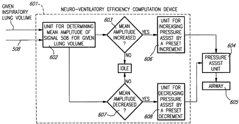
Referring now to Figure 10 a preferred, practical
embodiment is described. A neuro-ventilatory efficiency computation
device 601 receives the signal 508 of Figure 5 as well as the given, fixed
inspiratory lung volume. Device 601 comprises a unit 602 for determining
CA 02336940 2001-01-10
WO 99/62580 PCT/CA99/00529
the intensity of the signal 508 for the given inspiratory lung volume.
Although it is not illustrated, it is within the scope of the present
invention
to calculate, in unit 602, the peak, mean, median or any other intensity
measure of signal 508 for the given inspiratory lung volume. If the
intensity of signal 508 for the given inspiratory lung volume has increased
5 at least by a given percentage (step 603), i.e. the neuro-ventilatory
efficiency index has increased at least by said given percentage, the
pressure, flow, or volume assist unit 604 is controlled by a unit 606 in
view of increasing the magnitude of the pressure assist to the patient by
a preset increment until the intensity of the signal 508 for the given
10 inspiratory lung volume is restored to a predetermined, preset value.
Still referring to Figure 10, if the intensity for the given
inspiratory lung volurne has decreased at least by a given percentage
(step 607), i.e. the n+euro-ventiBatory efficiency index has decreased at
15 least by said given percentage, the pressure assist unit 604 is controlled
by the unit 608 in view of decreasing the magnitude of the pressure assist
by a preset incremerit until the intensity of the signal 508 for the given
inspiratory lung volurne is restored to the predetermined, preset value.
Although it is not illustrated, it is within the scope of the present
invention
20 to calculate, in unit 602, the peak, mean, median or any other intensity
measure of signal 508 for the given inspiratory lung volume, instead of
the intensity of this signal. Also, the signals at the outputs of the units
606 and 608 can be used to generate an alarm or to manually adjust the
pressure, flow or volume assist to the patient.
The response time is adjustable. The time base used
to calculate trends in the EMG intensity for a given volume or vice versa
CA 02336940 2001-01-10
WO 99/62580 I'CT/CA99/00529
21
and used for the corrections is relatively slow (minutes) and the levels of
applied support can be limited within a safe range. Again, an alarm can
be generated or the pressure assist can be manually or automatically
adjusted.
The pressure, flow, or volume assist unit 604 can be any
device which can be controlled to generate any airway pressure of
adjustable magnitude, for example any source of compressed gas; or a
flow or volume pump. Of course, airway 605 refers to or, to the least,
includes the patient's respiratory airway.
In this manner, the pressure assist unit 604 provides a
pressure, flow, or volume assist that is adjusted in proportion to changed
in neuro-ventilatory efficiency which is the EMGdi signal intensity at a
given lung volume or vice versa. The pressure, flow, or volume assist unit
continuoulsy operates to maintain a tracheal pressure, flow or volume that
is adjusted in proportion to changes in neuro-ventilatory efficiency which
is the EMGdi signal intensity at a given lung volume or vice versa.
Pre-ins irp ato ry bre<<thing effort:
A common problem with mechanically ventilated patients
is that the patients' inspiratory effort will not immediately cause an
inspiratory airflow so called "intrinsic PEEP" or "auto PEEP" which leads
to a decrease in the neuro-ventilatory efficiency. The effect of "intrinsic
PEEP" can be counteracted by the application of an "extrinsic PEEP" .
However, there are no easy applicable techniques to determine when the
applied level of "extrinsic PEEP" is adequate. The level of pre-inspiratory
CA 02336940 2001-01-10
WO 99/62580 PCT/CA99/00529
22
effort obtained through the EMGdi signal intensity (common noise level
subtracted) during 'for example a 100 milliseconds (ms) period
immediately precedirig the onset of inspiratory flow can be used to
indicate the presence of "intrinsic PEEP", and the level of applied
"extrinsic PEEP" can be adjusted such that the level of pre-inspiratory
effort is suppressed i.e the EMGdi signal intensity (common noise level
subtracted) during the above mentioned 100 ms period before onset of
inspiratory flow is close to zero. A feedback loop can then be used to
maintain the level of pre-inspiratory effort suppressed by adjusting as
explained above the level of "extrinsic PEEP".
Just a word to mention that the above mentioned period
of 100 ms can be replaced by a longer or shorter time period immediately
preceding the onset of inspiratory flow or by the neuro-ventilatory delay
800 (Figure 12b), i.e. the time period between the onset of EMG 801
(Figure 12b) and the onset of inspiratory flow 802 (Figure 12a).
Figure 11 of the appended drawings illustrates a
preferred, practical ernbodiment 700.
In tl7ie embodiment 700, an integrator 713 is responsive
to the RMS EMG sigrial 508 to continuously calculate the EMG intensity
for the above mentioned 100 ms period or neuro-ventitatory delay 800.
Embodiment 700 also comprises an inspiratory flow
detector 702 responsive to the patient's inspiratory flow 703 measured,
as indicated in the foregoing description, through any commercially
CA 02336940 2001-01-10
WO 99/62580 PCT/CA99/00529
23
available system, to produce an output signal 705 representative of EMG
activity.
The embodiment 700 of Figure 11 also comp(ses a
neuro-ventilatory delay calculator 704 responsive to ( a) the detection of
a RMS EMG signal intensity higher than the common noise level (5%),
and ( b) the detection of the onset of inspiratory flow by the detector 702
to calculate the neuro-ventilatory delay 800 (Figure 12b).
A detector 714 is responsive to the EMG intensity
calculated by the integrator 713 to detect the level of EMG intensity 803
(Figure 12b) at the onset of inspiratory flow 802 (Figure 12a) to trigger an
alarm 716 when the level of the EMG intensity 803 at the onset of
inspiratory flow 802 is higher than a given limit (detector 715). Upon
triggering of the alarrn 716, the level of applied "extrinsic PEEP" is either
automatically or mariualiy increased (device 708).
The detector 714 is responsive to the EMG intensity
calculated by the integrator 713 to detect the level of EMG intensity 803
(Figure 12b) at the onset of inspiratory flow 802 (Figure 12a) to trigger an
alarm 720 when the level of the EMG intensity 803 at the onset of
inspiratory flow 802 is lower than a given limit (detector 719). Upon
triggering of the alarrn 720, the level of applied "extrinsic PEEP" is either
automatically or manually decreased (device 711).
It should be mentioned that feedback from the neuro-
ventilatory delay or pre-inspiratory EMG activity can also be used to
adjust the sensitivity of the ventilators trigger functions.
CA 02336940 2001-01-10
WO 99/62580 PCT/CA99/00529
24
Again, the time base used for these corrections is
preferably relatively slow (minutes) and the levels of "extrinsic PEEP" can
be limited within a safe range.
The pressure assist unit 604 can be any device which
can be controlled to generate any airway flow and/or pressure of
adjustable magnitude, for example any source of compressed gas, or a
flow or volume pump.
In this manner, the delay from the beginning of the
mechanically ventilated patients' inspiratory effort to the onset of the
inspiratory assist will be minimized.
Although the present invention has been described
hereinabove with reference to preferred embodiments thereof, these
embodiments can be modified at will, within the scope of the appended
claims, without departing from the spirit and nature of the subject
invention.