Note: Descriptions are shown in the official language in which they were submitted.
CA 02337132 2001-01-15
WO 00/40281 PCT/US00/00190
TISSUE MAPPING INJECTION DEVICE
BACKGROUND
1. Technical Field
The present disclosure relates generally to a surgical instrument for
injecting a fluid into tissue and, more particularly to a surgical instrument
for
injecting an imaging radio label material into breast tissue for the detection
of' breast
carcinoma.
2. Background of Relateci Art
Breast carcinonia is the most conunon cancer and the second leading
cause of cancer-related death in women living in the United States. The
incidence of
breast cancer is increasing by about three percent per year. Recent studies
show that
one in eight women in the United States will develop breast cancer. Early
detection
lowers mortality and prolongs life expectancy of those having breast cancer.
Presently, standard screening tests for early detection of breast cancer
include breast self-examinatioii, breast examination by a physician, and
mammography. In general, physical examination alone will detect, at best,
orily sixty
to eighty percent of breast masses, whereas mammography will detect eighty t:o
nine zy
percent of breast masses in women not having dense breasts. In women having
dense
breasts, mammography has a false-negative rate of twenty-five to forty-five
percent,
and has a positive predictive value of only thirty percent. Only one in every
f'our to
six biopsies performed to confirm or rule out malignancy of suspicious lesions
detected during mammograms will be malignant. Thus, the majority of biopsies
prove to be unnecessary, i.e., the lesion is benign. Considering that the
economic
cost as well as the physical anii psychological stress of undergoing a biopsy
is high,
the need for a noninvasive and accurate technique to better discriminate
between
CA 02337132 2007-03-26
beni,-,n and nlall~~nant nlanlnlo-raphic abnormalities wfiich require biopsy
is clearly
present.
One stiich technique being developed for noninvasively and aecurately
discriniinatirig between nialianant and benign mammooraphic abnoi-malities is
Lymphatic Breast Mapping (''LBM"). During an LBM procedure, a quantity of'
radioactive tracer or dye is injected into and around a tumor. Because oi'the
tracer's
biochemistry, the tumor will collect more of the tracer than does normal
healthy tissue.
Thus. when the radioactive tracer decays and emits gamma rays. a Iiibher
number of'
these (tamma rays will originate from tumor sites than ii=om equal volumes of
healthy
tissue. T'he tracer disti-ibution and gamma ray emission can be identitied
using a
scintillation camera to enable doctors to identify the presence or absence
ofcancer.
Aecordin~ly, a need exists for a surgical instrument for injecting a
radioactive tracer into body tissue at precise locations adjacent a tiimor.
SUMMARY
In accordance with the present disclosure, a tissue mappinU injection
device is disclosed that is capable of injecting an imagin, radio label
material or dve
into the bodv at a location encompassing target tissue.
In accordance with an embodiment of the present invention there is
pi-ovided a suroical instrument for injecting a fluid into tissue comprisin~~:
a housin'~:
?0 a hollow elonbated body portion having a lon~itudinal axis extending
distally from the
housing and having an opening.; at a distal end thereof: an actuator assembly
including a
plunger slidably positioned within the housing, the plunger defining a fluid
delivery
channeL= aixl at least one needle having an injection tip operative{y
connected to the
plunger. the at least one needle defining a fluid injection channel which
communicates
2~ \\ ith the fluid delivery channel, the needle being formed from shape
memory material
and being movable in response to movement o1'the plunger between a
substantially
strai2ht deforrned configuration when positioned within the elon'~ated bod\
portion to a
relased curvecl contiV~uration when positioned externally of the elongated
bodv portion.
whcrein in the relaxed configuration, the injection tip extends out\vardly
trom the
)O elon-ated body portion through the distal opening.
In preferred embodiments an engagement member is coupled to or
monolithically formed with the plunger and is positioned to be engaged by the
thumb of
8 stn=~~eon. The plun(yer has a tirst end which extends
-~-
CA 02337132 2001-01-15
WO 00/40281 PCT/US00/00190
distally from one end of the housing in a direction opposite to the elongated
body
portion. The plunger defines a fluid delivery channel and includes a dis:al
end
adapted to receive a fluid delivery_hose.
A connector rod is coupled to and extends from the plunger through the
elongated body portion. The connector rod also defines a fluid delivery
channel
which communicates with the plunger delivery channel. The needles are
connected to
the distal end of the connector rod and are formed from a shape memory
material.
Each of the needles defines an injection delivery channel which communicates
with
the fluid delivery channel of the connector rod. In a relaxed state, the
needles curve
outwardly at a predetermineci angle relative to the longitudinal axis of the
elongated
body portion. In one embodliment, four needles are secured to the distal end
of the
connector rod. Each of the :needles is substantially identically shaped in its
relaxed
state.
In use, when the plunger is in the retracted position, the needles are
positioned wiihin elongated body portion and are deformed by the body portion
to a
substantially straight configuration. When the plunger is moved to the
advanced
position, the needles are moved distally out of the distal end of the
elongated body
portion. The needles are no longer deformed by the elongated body portion and
thus,
return to the relaxed state curving outwardly from the longitudinal axis of
the body
portion. Since each of the needles is similarly shaped, the tips of the
needles lie in a
common plane and extend into four quadrants surrounding a target tissue. Each
of
the needles is spaced approximately 90 from adjacent needles. Fluid can be
injected
into the tissue surrounding the target tissue via the delivery channels in the
plunger
and the injection channel fonned in the needles.
In an alternate embodiment, eight needles are secured to the distal end
of the connector rod. The eight needles form two sets of four needles, wherein
each
-3-
CA 02337132 2001-01-15
WO 00/40281 PCT/US00/00190
needle has a substantially identical configuration in the relaxed state as the
other
needles in that set of needles. When the needles are advanced out of the
distal end of
the elongated body portion, the tips of the first set of needles lie in a
first plane and
the tips of the second set of' needles lie in a second plane spaced from the
first plane.
Each of the needles of each set of needles extends into one of the four
quadrants
surrounding a target tissue and is spaced approximately ninety degrees from
adjacent
needles.
BRIEF DESCRIPTION OF THE DRAWINGS
Various preferred embodiments of the injection device for Lymphatic
Breast Mapping are described herein with reference to the drawings, wherein:
FIG. 1 is a perspective view of one embodiment of the injection device
in a non-deployed condition;
FIG. 2 is a perspective view with parts separated of the injection device
shown in FIG. 1;
FIG. 3 is a side view of the injection device shown in FIG. l. with parts
removed in a non-deployed condition;
FIG. 4 is an i:nlarged view of the indicated area of detail shown in
FIG. 4;
FIG. 5 is a side view of the injection device shown in FIG. l, with parts
removed and in a deployed condition;
FIG. 6 is a perspective view of the distal end of the injection device
shown in FIG. 1 in the deployed condition;
FIG. 6A is an alternate embodiment of the distal end of the injection
device shown in FIG. 1 in the deployed condition;
-4-
_
CA 02337132 2001-01-15
WO 00/40281 PC1'/US00/00190
FIG. 7 is a cannula suitable for use with the injections device shown in
FIGS. 1 and 6A;
FIG. 8 is a sicle cross-sectional view of the injection device shown in
FIG. 1 in a non-deployed condition passing through the cannula shown in FIG. 7
with
the cannula extending partially into body tissue; and
FIG. 9 is a sidle cross-sectional view of the injection device shown in
FIG. 1 in a deployed condition passing through the cannula shown in FIG. 7
with the
cannula extending partially irito body tissue.
DETAILED DESCRIPTION OF PREFERRED EMBODIMENTS
Preferred embodiments of the presently disclosed injection device will
now be described in detail with reference to the drawings, in which like
refe.rence
numerals designate identical or corresponding elements in each of the several
views.
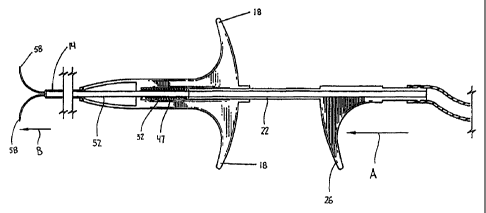
FIGS. 1-4 illustrate the injection device shown generally as 10.
Briefly, injection device 10 iricludes a housing 12, an elongated body portion
14, and
an actuator assembly 16. Housing 12 has a pair of radially extending fingers
18
configured to be engaged by the fingers of a surgeon. Elongated body portion
14 is
fixedly secured to one end 2CI of housing 12 and extends distally therefrom.
Actuator
assembly 16 includes a plunger 22 which is slidably positioned within housing
12 and
extends distally from the other end 24 of housing 12 in a direction opposite
to body
portion 14. An engagement imember 26 is secured to plunger 22 at a location to
be
grasped by the thumb of a surgeon while the surgeon's fingers grip radially
extending
fingers 18. Alternately, engagement member 26 can be monolithically formed
with
plunger 22.
Referring to FI:G. 2, housing 12 includes a pair of molded housing half-
sections 12a and 12b which are secured together via known techniques, e.g.,
-5-
CA 02337132 2001-01-15
WO 00/40281 PCT/USOO/00190
adhesives, ultrasonic welding, screws, etc., to form the housing. End 20 of
housing
12 includes a slot 28 configured and dimensioned to receive an annular flange
30
formed at the proximal end of body portion 14. Housing 12 also includes a
cylindrical bore 32 and a voiid 34. Cylindrical bore 32 is dimensioned to
slidably
receive plunger 22 (FIG. 1). A shoulder 36 is formed at one end of cylindrical
bore
32 to limit the extent of longitudinal movement of plunger 22 along bore 32
within
housing 12. Void 34 reduces the amount of material required to manufacture the
housing and, thus reduces the cost of manufacturing the housing.
Plunger 22 of actuator assembly 16 is preferably formed from molded
half-sections 22a and 22b which are secured together using known techniques,
e.g.,
adhesives, ultrasonic welding, screws, etc. Plunger 22 defines a fluid
delivery
channel 38. A first end 40 of plunger 22 includes an annular rib 42 to
facilitate
attachment of a fluid supply line 44 (FIG. 3) to the plunger. A second end 46
of
plunger 22 has a slot 48 fomied therein dimensioned to receive a flange 50
formed at
a proximal end of connector rod 52 to secure connector rod 52 in a
longitudinally
fixed position with respect to plunger 22. The second end 46 of plunger 22
also
includes an annular flange 45 dimensioned to engage a biasing member 47
positioned
in the forward end of cylindrical bore 32. Biasing member 47, which is
preferably a
coil spring, is positioned between annular flange 45 of plunger 22 and
shoulder 36 of
housing 12 to urge the plunger to a retracted position. The proximal end of
cylindrical bore 32 also includes a shoulder 49 to retain plunger 22 within
cylindrical
bore 32.
Referring also to FIGS. 3 and 4, connector rod 52 has a longitudinal
axis which is coaxial with the: longitudinal axis of plunger 22 and elongated
body
portion 14. Connector rod 52 extends from end 46 of plunger 22 through
elongated
body 14 and defines a fluid delivery channel 38' (See FIGS. 3 and 4) which
-6-
__
CA 02337132 2001-01-15
WO 00/40281 PCT/US00/00190
communicates with fluid delivery channel 38. A plurality of hollow needles 54
are
secured to the distal end of connector rod 52. Each of the needles defines an
injection chaiuiel 56 in fluid communication with delivery channel 38'. Each
of
needles 54 is constructed from a shape memory material and includes a
sharpened tip
58 having an outlet orifice 59. Preferably, the shape memory material is
Nitinol
although other shape memorY materials may be used. In the relaxed state, each
needle curves outwardly sucli that a tangent extending from needle tip 58
forms an
angle of about ninety (90) degrees with respect to the longitudinal axis of
the
elongated body 14. Alternately, other needle configurations are envisioned,
e.g.,
needle tip may extend outwairdly at an angle of between about 10 degrees to
about
150 degrees. The needles 54 are secured to connector rod 52 such that when
they are
deployed from within elongated body 14, the needles extend away from each
other
into four planar quadrants suirrounding target tissue. Preferably, the needles
are
positioned at ninety degree iritervals about the longitudinal axis of the
elongated body
portion 14, although different spacings are envisioned.
Referring to F]IGS. 3 and 4, when plunger 22 is in its retracted
position, connector rod 52 and needles 54 are positioned within elongated body
14.
In this position, the inner wall of elongated body 14 urges the needles from a
normally curved configuration to a substantially straight configuration.
Referring to F]:G. 5, when engagement member 26 is moved towards
housing 12 in the direction in.dicated by arrow "A", plunger 22 is moved
towards the
distal end of cylindrical bore 32 against the bias of spring 47. Longitudinal
advancement of plunger 22 within cylindrical bore 32 causes corresponding
longitudinal advancement of c:onnector rod 52 within elongated body portion
14. As
connector rod 52 is advanced, needles 54 are advanced in the direction
indicated by
arrow "B" in FIG. 5 from a position within elongated body portion 14 to a
position
-7-
CA 02337132 2001-01-15
WO 00/40281 PCT/US00/00190
extending outwardly from the distal end of elongated body portion 14. As
needles 54
exit the distal end of body portion 14, the needles return to a relaxed state
wherein
the needle tip 58 is pointed in a direction substantially perpendicular to the
longitudinal axis of the elongated body portion 14. In the relaxed state, each
of
needle tips 58 lies in the same vertical plane. See FIG. 6.
FIG. 6A illustrates an alternate embodiment of the injection device. In
the embodiment shown in FIG. 6A, the injection device has eight needles. In
the
relaxed state, four of the needles 54 extend away from each other into four
planar
quadrants surrounding target tissue and have tips 58 which lie in a first
vertical plane
and, four of the needles 54' extend away from each other into four pl"anar
quadrants
surrounding target tissue and have tips 58' which lie in a second vertical
plane spaced
from the first vertical plane. By providing additional needles, radioactive
tracer or
dye can be injected about the entire location of the target tissue.
Referring to FIGS. 7-9, during performance of a lymphatic breast
mapping procedure, a cannula 80 (FIG. 7) is inserted into tissue via known
techniques
adjacent the location of the target tissue 82. Next, the elongated body
portion 14 of
injection device 10 is inserted through cannula 80 in the direction indicated
by arrow
"C" in FIG. 8 to a position iri which the distal end of elongated body portion
14 is
located adjacent to the distal end 84 of cannula 80. Finally, actuator
assembly 16 is
actuated in the manner discussed above to advance connector rod 52 and needles
54 in
the direction indicated by arrciws "D" and "E", respectively, in FIG. 9, into
or
adjacent the target tissue. A radioactive tracer or dye 90 can now be injected
in and
about the location of the target tissue 82 via fluid supply line 44, fluid
delivery
channels 38 and 38' and injection channels 56.
It will be understood that various modifications may be made to the
embodiments disclosed herein. For example, although the injection device has
been
-8-
.
CA 02337132 2001-01-15
WO 00/40281 PCT/US00/00190
disclosed as having four neeciles which extend into four quadrants about the
target
tissue, a greater or lesser nuinber of needles may be provided. Moreover, the
configuration of the needles :in the relaxed state may be different than that
disclosed.
For example, the need;e can have a configuration in which the needle tip
extends
outwardly at an angle of sixty (60) degrees with respect to the base of the
needle.
Therefore, the above descripition should not be construed as limiting, but
merely as
exemplifications of preferred embodiments. Those skilled in the art will
envision
other modifications within the scope and spirit of the claims appended
thereto.
-9-