Note: Descriptions are shown in the official language in which they were submitted.
CA 02338199 2001-01-19
WO 00/10034 PCT/CA99/00751
APPLICATION OF SCATTER AND ATTENUATION
CORRECTION TO EMISSION TOMOGRAPHY
IMAGES USING INFERRED ANATOMY FROM ATLAS
Technical Field
The present invention relates to emission tomography and in particular to a
method and apparatus for applving scatter and attenuation correction to
emission tomography
images using anatomy inferred from an atlas.
1o Background Art
Single Photon Emission Computed Tomography (SPECT) and Positron
Emission Tomography (PET) are nuclear medicine diagnostic imaging techniques
used to
measure the three-dimensional distribution of a radiopharmaceutical within the
body. Brain
SPECT and PET imaging techniques are primarily used to measure regional
cerebral blood
flow in a patient injected with a radiopharmaceutical to assist in the
evaluation of stroke and
the diagnosis of dementias such as Alzheimer's disease.
Although SPECT and PET are useful imaging techniques, their poor
quantitative accuracy has been an obstacle in the ability to achieve increased
diagnostic
reliability. Quantitative accuracy of brain SPECT and PET imaging has however
been
improved significantly through the application of scatter and attenuation
correction to SPECT
and PET brain images. To be sufficiently accurate, the application of scatter
and attenuation
correction to SPECT and PET brain images must be guided by the distribution of
density
within the llead. Unfortunately, the distribution of density within the head
cannot be obtained
froni SPECT and PET brain scans and therefore, separate measurements are
required.
Transmission imaging has been used to measure the distribution of density
within the head to allow scatter and attenuation correction to be applied to
SPECT and PET
brain images. Unfortunately, the hardware necessary for making transmission
measurements
is complex, unreliable and requires extensive maintenance. Also, the need to
make
transmissiori imaging measurements in addition to the SPECT or PET brain
images, increases
the time required to complete the overall imaging procedure. SPECT and PET
imaging
procedures are themselves lengthy and require a patient to remain motionless
to ensure
accurate brain images. For sick and elderly patients, this is a difficult
task. Adding to the
length of the imaging procedure increases the likelihood that patients will
not remain
CA 02338199 2007-07-26
-2-
motionless. Movement of a patient during the transmission imaging procedure
results in
inaccurate measurements of the distribution of density within the head. This
of course
provides an inaccurate guide for the application of scatter and attenuation
correction to
SPECT and PET brain images. Accordingly, improved methods to increase the
diagnostic
reliability of emission tomography images are desired.
It is therefore an object of the present invention to provide a novel method
and
apparatus for applying scatter and attenuation correction to emission
tomography images.
Disclosure Of The Invention
Broadly stated, the present invention provides a method and apparatus for
applying scatter and attenuation correction to emission tomography images
which estimates
or "infers" the distribution of density within a region of interest of a
patient under observation
using a three-dimensional computer model of the region of interest. It has
been found that
scatter and attenuation correction guided by a computer model of the region of
interest under
observation produces results similar to those when using transmission images
of the actual
region of interest as the guide to the application of scatter and attenuation
correction.
Accordingly, in one aspect of the present invention there is provided a
computerized method of correcting emission tomography images of a region of
interest of a
subject under observation comprising the steps of:
aligning a three-dimensional computer model in the form of a two-component
atlas representing the density distribution within said region of interest
with said emission
tomography images, said computer model being created from image data of other
subjects
thereby to avoid the need to image said subject under observation to create
said computer
model; and
applying scatter and attenuation correction to said emission tomography
images using said aligned computer model as a guide.
In a preferred embodiment, the computer model is in the form of a two-
component atlas. During the aligning step, a functional component of the atlas
is firstly
aligned with the emission tomography images to generate a set of spatial
transformation
parameters. Following this, an anatomical component of the atlas is aligned
with the
CA 02338199 2007-07-26
-3-
emission tomography images using the set of spatial transformation parameters.
The atlas may be selected from a database of atlases based on degree of
registration with the emission tomography images. Alternatively, multiple
atlases maybe
combined to yield a resultant atlas which better registers with the emission
tomography
images.
According to another aspect of the present invention there is provided a
computerized emission tomography image correcting method comprising:
capturing emission tomography images of a region of interest of a subject; and
correcting scatter and attention in the emission tomography images using a
three-dimensional computer model in the form of a two-component atlas
approximating the
density distribution within the region of interest as a guide, said computer
model being
created from image data of other subjects thereby to avoid the need to image
said subject to
create said computer model.
According to yet another aspect of the present invention there is provided an
emission tomography image processing system comprising:
memory storing emission tomography images of a region of interest of a
subject under observation, said memory also storing at least one three-
dimensional computer
model of said region of interest, said computer model being in the form of a
two-component
atlas and representing the density distribution within said region of
interest, said computer
model being created from image data of other subjects thereby to avoid the
need to image
said subject under observation to create said computer model; and
a processor for registering said computer model with said emission
tomography images and for applying scatter and attenuation correction to said
emission
tomography images using said registered computer model as a guide.
According to still yet another aspect of the present invention there is
provided
an emission tomography imaging system comprising:
an imaging apparatus for taking emission tomography images of a region of
interest of a subject under observation to form a three-dimensional image of
said region of
interest;
memory to store said emission tomography images, said memory also storing
CA 02338199 2007-07-26
-4-
at least one three-dimensional computer model of said region of interest, said
computer
model being in the form of a two-component atlas and representing the density
distribution
within said region of interest, said computer model being created from image
data of other
subjects thereby to avoid the need to image said subject under observation to
create said
computer model; and
a processor for aligning said computer model with said emission tomography
images and for applying scatter and attenuation correction to said emission
tomography
images using said aligned computer model as a guide.
According to still yet another aspect of the present invention there is
provided
a computer readable medium including a computer program for applying scatter
and
attenuation correction to emission tomography images of a region of interest
of a subject
under observation, said computer program comprising:
computer program code for aligning a three-dimensional computer model
representing the density distribution within said region of interest with said
emission
tomography images, said computer modes being created from image data of other
subjects
thereby to avoid the need to image said subject under observation to create
said computer
model; and
computer program code for applying scatter and attenuation corrections to said
emission tomography images using said aligned computer model as a guide,
wherein said
computer program code for aligning comprises:
computer program code for aligning a functional component of said
computer model simulating one of a SPECT and PET scan of said region of
interest and for
generating a set of spatial transformation parameters; and
computer program code for aligning an anatomical component of said
computer model simulating a transmission scan of said region of interest using
said set of
spatial transformation parameters.
According to still yet another aspect of the present invention there is
provided
a computerized method of correcting emission tomography images of a region of
interest of a
subject under observation comprising the steps of:
aligning a three-dimensional computer model in the form of a two-component
CA 02338199 2007-07-26
-4a-
atlas representing the density distribution within said region of interest
with
said emission tomography images; and
applying scatter and attenuation correction to said emission tomography
images using said aligned computer model as a guide.
According to still yet another aspect of the present invention there is
provided
an emission tomography image processing system comprising:
memory storing emission tomography images of a region of interest of a
subject, said memory also storing at least one three-dimensional computer
model of said
region of interest, said computer model being a two-component atlas
representing the density
distribution within said region of interest; and
a processor for registering said computer model with said emission
tomography images and for applying scatter and attenuation correction to said
emission
tomography images using said registered computer model as a guide.
According to still yet another aspect of the present invention there is
provided
a computerized emission tomography imaging method comprising the steps of:
obtaining emission tomography images of a region of interest of a subject
under observation;
aligning, using a computer, a three-dimensional computer model in the form
of a two-component atlas representing the density distribution within said
region of interest
with said emission tomography images without requiring said subject to be
imaged to create
said computer model; and
applying, using said computer, scatter and attenuation correction to said
emission tomography images using said aligned computer model as a guide.
The present invention provides advantages in that by using a three-
dimensional computer model of the region of interest of a subject under
observation that
approximates its density as a guide for the application of scatter and
attenuation correction to
emission tomography images, the need for transmission imaging is obviated.
Therefore, in
the case of SPECT and PET imaging, the imaging procedures do not need to be
lengthened.
Also, since the distribution of density in the region of interest under
observation is
approximated by a three-dimensional computer model, additional hardware is not
required to
CA 02338199 2007-07-26
-4b-
create the guide for the application of scatter and attenuation correction.
This makes the
present invention significantly less expensive and more flexible than
transmission imaging
systems. In addition, since a three-dimensional computer model of the region
of interest
under observation is used as the guide for the application of scatter and
attenuation
correction, scatter and attenuation correction can be applied retrospectively
to existing
databases which include significant numbers of emission tomography images for
which no
transmission imaging measurements were acquired.
Brief Description Of The Drawings
An embodiment of the present invention will now be described more fully
with reference to the accompanying drawings in which:
Figure la shows a two-dimensional emission tomography brain image without
the application of scatter and attenuation correction;
Figure lb shows the two-dimensional image of Figure 1 a with the application
of scatter and attenuation correction;
Figure 1 c is a two-dimensional transmission brain image;
Figures ld and le are two-dimensional emission tomography brain images with
appurtenant anatomy derived from transmission images applied thereto;
CA 02338199 2001-01-19
WO 00/10034 PCT/CA99/00751
-5-
Figure 2 is a block diagram showing a method for applying scatter and
attenuation correction to emission tomography images in accordance with the
present
invention;
Figure 3a shows a two-dimensional emission tomography brain image of a
head phantom with scatter and attenuation correction;
Figure 3b is a two-dimensional transmission brain image;
Figures 3c and 3d are two-dimensional emission tomography brain images
with appurtenant anatomy derived from transmission images applied thereto;
Figures 3e and 3f are two-dimensional emission tomography brain images
with inferred anatomy derived from an atlas applied thereto;
Figures 4 and 5 show two-dimensional brain images and a graph comparing
uniform appurtenant anatomy, uniform inferred anatomy and brain contour;
Figure 6 shows two-dimensional brain images with non-uniform appurtenant
and non-uniform inferred anatomy applied thereto;
Figures 7 and 8 show quantitative evaluation of head phantom SPECT
reconstructions;
Figure 9 shows two-dimensionl SPECT brain images;
Figure 10 shows SPECT brain images superimposed onto transmission
reconstruction and inferred anatomy;
Figure 11 shows profiles taken through recoiistructions of a SPECT brain
image;
Figure 12 is a graph showing a comparison of regional cerebral blood flow
from reconstructed SPECT images guided by inferred anatomy and transmission
scans; and
Figure 13 is a graph showing the correlation between the reconstructed SPECT
images of Figure 12.
Best Mode for Carrying Out The Invention
During emission tomography imaging such as for example SPECT and PET, a
patient is injected with a radiopharmaceutical. Two-dimensional images or
projections of a
region of interest (ROI) of a patient are taken along an arbitrary axis. The
projections are
digitized and conveyed to a computer for storage in a three-dimensional array
in computer
memory to reconstruct a three-dimensional image of the region of interest.
Once the three-
CA 02338199 2001-01-19
WO 00/10034 PCT/CA99/00751
-6-
dimensional image is reconstructed, two-dimensional slices of the region of
interest can be
viewed along virtually any arbitrary axis using conventional software. Figure
la shows a
two-dimensional emission tomography brain image.
During the imaging procedure, the emissions from the radiopharmaceutical are
scattered and/or attenuated by different density tissue, air cavities and/or
bones in the region
of interest under observation. As can be seen in Figure I a, scattering and
attenuation of
radiopharmaceuticals affects quantitative image quality. As a result, acquired
emission
tomography images are often unreliable. Applying scatter and attenuation
correction to
emission tomography images, using transmission images of the same region of
interest taken
during the saine imaging procedure, is known but suffers from the
disadvantages discussed
previously. Figure 1 b shows the two-dimensional image of Figure 1 a with
scatter and
attenuation correction applied using transmission images taken of the same
anatomy. Figure
Ic shows an example of a transmission image and Figures ld and le show
appurtenant
anatomy (sa(Tittal and transverse respectively) derived from the transmission
images applied
to emission tomography brain images.
ln the present invention, a three-dimensional computer model or "atlas" of the
region of interest, which provides accurate density distribution of the region
of interest, is
stored in a database in computer memory and is used as a guide for the
application of scatter
and attenuation correction to emission tomography images. In the present
embodiment, the
atlas includes two components, namely a functional componeiit simulating a
SPECT OR PET
scan of the region of interest and an anatomical component simulating a
transmission scan of
the region of interest. The atlas can be created from existing transmission
images or x-ray CT
scans of similar regions of interest from other patients and averaged to form
a suitable
computer model or atlas. Multiple models for each region of interest and
models for different
regions of interest can be stored in the database and accessed individually
during scatter and
attenuation correction of emission tomography images. Since a computer model
of the region
of interest is used, additional hardware and procedure time is not required to
apply scatter and
attenuation correction to emission tomography images. An example of the
application of
scatter and attenuation correction to emission tomography brain images in
accordance with
the present invention will now be described.
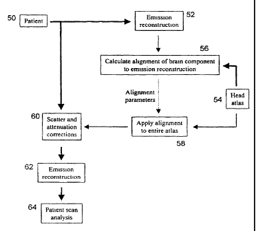
Referring now to Figure 2, a block diagram illustrating the present method for
applying scatter and attenuation correction to emission tomography brain
images is shown.
CA 02338199 2001-01-19
WO 00/10034 PCT/CA99/00751
-7-
Initially, emission tomography brain images of a patient (block 50) are
acquired in a known
manner. The acquired brain images are in the form of two-dimensional
projections of the
radiopharmaceutical distribution in the brain.
Initially, a preliminary reconstruction of the acquired brain images is
performed to digitize and assemble the two-dimensional projections into a
three-dimensional
array in computer memory to create a three-dimensional image of the brain
(block 52). An
atlas of the head (the region of interest in this case) is then downloaded
fi=om the database into
a datafile (block 54) and the functional component of the head atlas is
identified. Once
identified, the functional component of the head atlas, in this embodiment the
brain
component, is copied to a three-dimensional array. Following this, an
alignment procedure to
register spatially the brain component of the head atlas with the three-
dimensional brain
image is performed (block 56). In the present embodiment, a simplex algorithm
is used to
register spatially, the brain component of the head atlas with the brain image
as described in
"Numerical Recipes ln C" by Press et al, 2"d edition, New York, New York,
Canlbridge
University Press, 1992. Those of skill in tiie art will however appreciate
that other
registration algoritl-uns can be used to align the brain component of the head
atlas to the tllree-
dimensional brain image. During this alignment procedure, a set of 3D spatial
transformation
parameters representing the three-dimensional transformations, including but
not limited to
rotation, shifting and scaling, that are necessary to register the brain
component of the head
atlas with the three-dimensional brain iniage, is calculated and is stored in
the computer
memory as a matrix.
Once the set of 3D transformation parameters is calculated and stored, the 3D
spatial transformation parameters in the set are applied to the anatomical
component of the
head atlas to register it with the three-dimensional brain image (block 58).
With the
anatomical component of the head atlas aligned with the three-dimensional
brain image, the
atlas can be used as a density distribution guide to the application of
scatter and attenuation
correction to the three-dimensional brain image.
With an accurate density distribution guide established, scatter correction is
applied to the three-dimensional brain image followed by attenuation
correction in a known
manner (block 60). Once scatter and attenuation correction has been applied to
the three-
dimensional brain image, the brain image is reconstructed into a three-
dimensional array in
computer memory to complete the image correction process (block 62). The
corrected three-
CA 02338199 2001-01-19
WO 00/10034 PCT/CA99/00751
-8-
dimensional brain image can then be analyzed (block 64). Appendix A includes
pseudocode
representing the above-identified process.
Figures 3a to 3f show a comparison of two-dimensional brain images of a head
phantom with anatomy derived from transmission images (appurtenant anatomy)
and
anatomy derived from a head atlas (inferred anatomy) applied.
The present method and apparatus was tested using an anthropomorphic head
phantom modeling soft tissue, hard tissue and air cavities within a skull and
including a two-
compartment brain reservoir. The two compartments of the reservoir were
separately filled
with two water solutions of Tc-99m, having a specific activity ratio of 4:1.
Fan-beam SPECT
was acquired followed by a Tc-99m transmission scan sixty hours later. The
reconstructed
transmission image is referred to as appurtenant anatomy.
Five scatter and attenuation correction schemes were evaluated based on non-
unifonn appurtenant anatomy, uniform appurtenant anatomy, non-uniform inferred
anatoniy,
uniform inferred anatomy, and uniform brain contour. For uniform scatter and
attenuation
correction, the infer-red and appurtenant anatomies were segmented and
assigned uniform
attenuation coefficients of soft tissue (0.15cm"' for Tc-99m). A SPECT
reconstruction of the
head phantom was similarly processed to facilitate scatter and attenuation
correction guided
by brain contour. Scatter correction was based on a non-stationary
deconvolution scatter
subtraction as described in the paper authored by Stodilka et al entitled "The
Relative
Contributions of Scatter and Attenuation Correction Toward Brain SPECT
Quantification",
Phys Med Biol, 1998. Attenuation correction/reconstruction was subsequently
performed by
ordered-subsets expectation-maximization as set out in the paper authored by
Hudson HM et
al entitled "Accelerated Image Reconstruction using Ordered Subsets of
Projection Data",
IEEE Trans Med Iniaging 13 601-609, 1994. Although particular examples of
scatter
correction, attenuation correction and reconstruction algorithms are
described, those of skill
in the art will appreciate that other scatter correction, attenuation
correction and
reconstruction methods can be used.
Uniform scatter and attenuation correction should be guided by the contour of
the attenuating medium if an acceptable level of accuracy for objective
diagnosis is to be
expected. Figures 4 and 5 show that uniform appurtenant anatomy is better
approximated by
uniform inferred anatomy than by brain contour. As can be seen in Figure 6,
non-uniform
appurtenant anatomy and non-uniform inferred anatomy are similar.
CA 02338199 2001-01-19
WO 00/10034 PCT/CA99/00751
-9-
Head phantom SPECT reconstructions were quantitatively evaluated by four
metrics, namely bias, uniformity, contrast-recovery, and relative
quantification (see Figures 7
and 8). As will be appreciated, scatter aiid attenuation correction guided by
inferred anatomy
provides quantitative accuracy that is distinctively superior to scatter and
attenuation
correction guided by brain contour.
Application of scatter and attenuation correction to emission tomography
images of anatomy other than the head can also be performed. For example,
inferred
anatomy can also be used to apply scatter and attenuation correction to
cardiac images.
During construction of the cardiac atlas, an anatomical component of the
cardiac atlas
representing the anatomical features of the thorax is created and includes:
soft-tissues such as the heart, liver, muscle, and fat;
very low-density soft-tissues such as the lungs; and
high-density tissues such as bone and cartilage in the ribs and spine.
A functional component of the cardiac atlas is also created and simulates a
SPECT or PET cardiac image. Appropriate data to construct the cardiac atlas
can be obtained
in a variety of ways including, but not limited to, imaging a phantom or human
subject by X-
rav CT, MRI, or gamma-camera transmission coniputed tomography; or computer
simulation.
Also, the cardiac atlas can be constructed by amalgamating a plurality of
patient scans.
The procedure of using inferred cardiac anatomy to apply scatter and
attenuation correction to cardiac images remains the same as with brain
imaging described
above. First, a preliminary reconstruction of the patient's cardiac SPECT data
is performed.
The functional component of the cardiac atlas is then registered to the
preliminary
reconstruction. The registration includes a spatial transformation that may
include shifting,
rotation, scaling, and/or non-linear operations such as warping. The
registration procedure
calculates a matrix representing the spatial transformation that maps atlas
space into patient-
specific space. Once the matrix has been calculated, the matrix is applied to
the anatomical
component of the cardiac atlas, thus inferring anatomy in the chest.
Although the above methods describe the use of a single generic atlas of the
anatomy under observation, those of skill in the art will appreciate that
custom atlases can be
developed and stored in the database. For example, disease specific atlases
such as an
Alzheimer's disease atlas or a stroke atlas can be developed and used when
correcting
CA 02338199 2001-01-19
WO 00/10034 PCT/CA99/00751
-10-
emission tomography images of patients suffering from those diseases. The
disease specific
atlases may be tracer or lesion specific to allow for local concavities in
radiopharmaceutical
uptake. Patients with severe Alzheimer's or Pick's disease do not have normal
cerebral blood
flow. Areas with flow deficits can limit the accuracy of the registration of
the emissioii
tomography images with the functional component of the head atlas if the head
atlas assumes
normal blood flow and hence uniform radiopharmaceutcial uptake. Atlases can
also be
developed to take into account physical traits such as for example, an
exceptionally large
nasal sinus. During scatter and attenuation correction, the operator can
select the appropriate
atlas to use. Alternatively, the atlas can be selected automatically by
computer software. In
this case, the software, performs a preliminary reconstruction using each
custom atlas and
registers the atlas to the preliminary reconstruction to measure the accuracy
of the
registration. The atlas witli the highest registration accuracy is then
selected. If desired, the
software can use fuzzy logic, theoretical calculations or other criteria to
combine two or more
atlases to yield a single resultant atlas, which provides a better degree of
registration.
EXAMPLE
Example 1: Inferred Anatomv and Brain ImaQine
The following example is described for the purposes of illustration and is not
intended to limit the scope of the present invention.
Inferring Anatomy From A Head Atlas
A head atlas was prepared as follows. A Zubal three-dimensional digitized
MRI head phantom [Zubal et al 1994] was segmented to produce two data sets,
namely a
SPECT atlas simulating a SPECT scan of the phantom, and an anatomical atlas
simulating a
transmission scan of the phantom. The SPECT atlas consisted of voxels
containing gray-
matter and white-matter, to which 9""'Tc-HMPAO relative uptakes of 4 and I had
been
assigned respectively. The anatomical atlas consisted of voxels containing
hard-tissue, soft-
tissue and nasal sinus, to which the correspondent 140keV narrow-beam
attenuation
coefficients of 0.25, 0.15, and 0.075 cm' were assigned.
CA 02338199 2001-01-19
WO 00/10034 PCT/CA99/00751
-11-
A patient SPECT scan was then reconstructed without scatter and attenuation
correction. Facial activity was removed to yield a data set referred to as the
preliminary
patient reconstruction. The SPECT atlas was then registered to the preliminary
patient
reconstruction and the spatial transformation was recorded. This
trailsformation was then
applied to the anatomical atlas, thus inferring the location of the patient's
soft-tissue, hard-
tissue and air cavities (see Figure 9).
A general-purpose radiological analysis program (Hermes, Nuclear
Diagnostics, Stockholm, Sweden) was used to perform the unimodality
registration. The
large variation in liead orientation necessitated that a manual registration
be first performed.
This was followed by an automated refinement. The cost function of the
automated
registration was defined as the sum of absolute count differences [Hoh et al
1993]. A global
minimum was sought by a simplex search [Nelder and Mead 1965] within a
parameter space
consisting of rotating, shifting, and linear scaling in x, y, and z directions
[Holman et al 1991,
Slonika et al 1995].
Sequential Transmission And Emission Imaging
Ten dementia patients (5 females and 5 males, with a mean age of 64.3 years)
were analyzed. For each patient, a transmission scan was first acquired.
Patients became
very relaxed during the quiet transmission scan, and were then injected with
740 MBq of
99niTc-HMPAO. The SPECT procedure was started approximately 5 minutes post-
injection.
The SPECT system, which has transmission capabilities, is described in detail
[Kemp et al
1995]. It consists of a General Electric 400AC/T gamma camera (General
Electric,
Milwaukee, WI) with a 409.6 mm diameter circular field-of-view. Projections
were acquired
through a fan-beam collimator (Nuclear Fields, Des Plaines, IL) having a 600
mm focal
length and 1.5 mm flat-to-flat hexagonal hole width. The transmission
component includes a
franie mounted onto the camera's collimator that holds a tantalum-collimated
'Tc line
source along the focal line of the fan-beam collimator. Collimation of both
the line source
and camera minimizes scatter. As a result, the transmission system effectively
measures
narrow-beam attenuation coefficients [Tsui et al 1989, Kemp et al 1995]. SPECT
and
transmission scans were acquired with a 20% energy window, centered on the
""'Tc
photopeak of 140 keV. The scans consisted of 128 projections, equally spaced
over 360 .
Each circular projection was acquired into a 128 x 128 pixel square matrix (1
pixel = 3.2
CA 02338199 2001-01-19
WO 00/10034 PCT/CA99/00751
-12-
mm). Both transmission and SPECT scans were 10 seconds per projection and
count rates
were approximately 70 and 1.5 kcounts per second, respectively. All scans were
corrected for
uniformity using 100 million count flood images, and transmission scans were
normalized to
50 million count blank images. Radii of rotation varied among the patients;
the smallest
being 170 mm and the largest being 205 mm. Prior to reconstruction, all scans
were rebinned
to object-plane parallel-hole geometry via two-dimensional cubic
interpolation.
Scatter And Attenuation Correction And Reconstruction
The SPECT data were reconstructed using a maximum-likelihood estimator
with an unregularized 32-level ordered subset [Hudson and Larkin 1994]
implementation of
the expectation maximization algorithnl [Shepp and Vardi 1982, Lange and
Carson 1984]
(OSEM). The four projections that were used per sub-iteration were equally
spaced about
360 . Attenuation was modelled in the matched projector/backprojector pair,
and a scatter
estimate [Stodilka et al 1998b] was added as an a priori background following
forward
projection [Lange and Carson 1984, Bowsher et al 1996, Kadrmas et al 1998].
Both scatter
and attenuation modelling incorporated the narrow-beam attenuation
coefficients from
transmission imaging or inferred anatomy. Detector response was not included.
Four
iterations of OSEM were used, following initialization with a uniform [Nunez
and Llacer-
1990] support prior derived from transmission reconstruction or inferred
anatomy.
Reconstructions were then post-filtered [Nunez and Llacer 1990] using a three-
dimensional
Butterworth filter with an order of 8 and cutoff at 0.42 cm-'. The
transmission data were also
reconstructed by the emission OSEM algorithm, following blank scan
normalization and log-
transformation.
Line source collimation, coupled with limitations in detector count rate
capability and patient compliance resulted in less than ideal transmission
statistics. To reduce
the effects of transmission imaging noise propagation into the SPECT
reconstruction [Xu et
al 1991, Tung and Gullberg 1994], the transmission reconstructions were
segmented as
follows. Soft-tissue in the reconstructed transmission volumes was forced to
have uniform
density. First, a large region of interest (ROI) was drawn around soft-tissue
regions, and
mean and variance estimates were calculated. Then, all voxels having count
densities within
2 standard deviations of this mean were assigned to 0.15cm-'. Thus, the
transmission
CA 02338199 2001-01-19
WO 00/10034 PCT/CA99/00751
-13-
reconstructions were characterized by noiseless soft-tissue, yet featured hard-
tissue and air
cavities.
Template-Based OuantiGcation
Previous work has identified that a major confound to reproducible
quantification, originates from manual and threshold-dependent placement of
anatomical
ROIs onto SPECT scans [Msaki et al 1998]. To reduce this subjective source of
error, a
normal template [Msaki et al 1998] onto which twelve bilateral volumetric ROIs
are
demarcated [Karbe et al 1994] was used. The ability to store the template and
its ROIs
ensures reproducibility of the analysis. This quantification procedure also
introduces
standardization to the analysis, which facilitates the exchange of data among
different
institutions [Evans et al 1988]. All reconstructed scans were registered to
the normal
template, herein referred.to as "spatial normalization". Prior to spatial
normalization, voxels
previously identified as facial activity were set to zero. Following
superposition of the
template ROls onto each scan, the cortical rCBF for each ROI was normalized to
cerebellar
rCBF [Karbe et al 1994] and corrected for blood flow-dependent tracer reflux
[Lassen et al
1988]. Analysis of the absolute concentration of radiopharmaceutical was not
performed
since currently, absolute rCBF quantification is seldom used in SPECT [Bakker
and Pauwels
1997].
Ouantitative Error Analvsis
A sample of inferred anatomy and transmission reconstruction is illustrated in
Figure 10. In Figure 11, profiles through the SPECT reconstructions guided by
transmission
scans and inferred anatomy are presented. The profiles have not been
normalized to
cerebellar count density.
The means and standard errors of regional cerebral blood flow from SPECT
scans guided by inferred anatomy and transmission scans were compared ROI-by-
ROI (see
Figure 12). Statistical analysis was also performed ROI-by-ROI using repeated
analysis of
variance to determine where there were significant differences between the
inferred anatomy
versus transmission-guided reconstruction methods. Pooled-ROI and ROI-
dependent
correlation coefficients were calculated between inferred anatomy
reconstruction and
transniission-guided SPECT reconstructions. Figure 13 shows the correlation
between the
CA 02338199 2001-01-19
WO 00/10034 PCT/CA99/00751
-14-
two reconstruction and quantification methods by pooling all ROIs and patients
together.
The p value for significance was set to 0.05 for all tests.
Inferred Anatomy Error Propagation Analysis
Four sources of error were identified that contribute to discrepancies in ROI
quantification:
(1) inferred anatomy-guided scatter correction;
(2) inferred anatomy-guided attenuation correction;
(3) inferred anatomy-guided spatial normalization, which is equivalent to ROI
misplacement; and
(4) patient motion between transmission and SPECT scans.
The first two are inherent limitations to the principle of inferred anatomy,
whereas the last two represent artifactual exaggerations of errors in the
context of this
example. The first three sources of error, namely scatter, attenuation, and
ROI misplacement
were measured.
The propagation of ROI quantification to differences between inferred
anatomy scatter correction and transmission-guided scatter correction were
analyzed by
performing:
(1) inferred anatomy-derived scatter correction;
(2) transmission-derived attenuation correction; and
(3) applying the spatial normalization calculated to be optimal for
registering
the transmission-derived SPECT reconstruction to the quantification template.
A similar analysis for evaluating the effects of inferred anatomy-derived
attenuation correction was carried out by performing:
(1) transmission -derived scatter correction;
(2) inferred-anatomy derived attenuation correction; and
(3) applying the spatial normalization calculated to be optimal for
registering
the transmission-derived SPECT reconstruction to the quantification template.
CA 02338199 2001-01-19
WO 00/10034 PCT/CA99/00751
-15-
The effects of ROI misplacement were quantified by performing transmission-
derived scatter and attenuation correction, and then applying the spatial
transformation
calculated to be optimal for registering the inferred anatomy-derived SPECT
reconstruction
to the quantification template. Thus, the full propagation analysis resulted
in three
reconstructions, each of which was quantitatively compared (via the above-
described
template-based quantification procedure) with the "gold-standard" transmission-
derived
SPECT reconstruction and spatial normalization. This procedure was performed
for the ten
patients, and the results averaged. However, once the errors due to scatter,
attenuation, and
ROI misplacement were separately quantified, their totals were found to be
less than the
errors caused by the full inferred anatomy protocol. This additional source of
error is termed
"unaccountable" in Table 2, and is believed to be caused by patient motion.
Qualitative Analysis
A sample comparison of inferred anatomy and transmission reconstruction is
shown in Figure 10 for mid-sagittal and cortical-axial slices. Good
reproduction of soft-tissue
and hard-tissue at the cortical level is noted. Some discrepancy is seen near
the vertex;
however, this region is seldom included in quantitative analysis.
Discrepancies near the nasal
sinus are most marked. Fortunately, these structures mostly involve low-
density areas such
as air, and to a much lesser degree, soft-tissues and cartilage, which do not
scatter and
attenuate photons as much as higher density structures.
Mid-brain profiles, shown in Figure 11, compare a SPECT reconstruction
guided by transmission imaging with the same SPECT reconstruction guided by
inferred
anatomy. The profile was taken along the longest axis of the head, which is
most sensitive to
mis-registered transmission maps following scatter and attenuation correction
[Huang et al
1979].
Quantitative Error Analysis
Table 1 below shows the results of the repeated analysis of variance and
correlation analysis comparing transmission reconstruction and quantitative
and inferred
anatomy-guided reconstruction and quantification. Left frontal and central
sulcus ROls
showed the highest probability of a true difference, reaching statistical
significance with
p=0.001 and 0.002, respectively. These ROIs also had the highest correlation
coefficients
CA 02338199 2001-01-19
WO 00/10034 PCT/CA99/00751
-16-
relating transmission reconstruction and quantification and inferred anatomy-
guided SPECT
reconstruction and quantification. This increased correlation may be an
artifact of the
increased differences in the ROl means. Interestingly, the left frontal and
central sulcus ROIs
were also found to have marked rCBF deficits, suggesting that inferred anatomy
may have
difficulties near regions with substantially reduced radiopharmaceutical
uptake.
Figure 12 shows the means and standard errors for transmission reconstruction
and inferred aiiatomy in each of the 12 ROIs, averaged across the entire
population. The
mean absolute difference for all ROIs across the whole population was 7.5%.
Correlation for
all ROIs and all patients was found to be high: r'=0.92 as illustrated in
Figure 13.
TABLE 1
Region ANOVA Paired sample Paired sample
Significance correlation Significance
Left frontal 0.001 0.965 <0.001
Right frontal 0.088 0.953 <0.001
Left central sulcus 0.002 0.957 <0.001
Right central sulcus 0.234 0.937 <0.001
Left parietal 0.092 0.772 0.009
Right parietal 0.473 0.862 0.001
Left temporal 0.957 0.939 <0.001
Right temporal 0.397 0.835 0.003
Left occipital 0.271 0.835 0.003
Right occipital 0.073 0.858 0.001
Left cerebellar 0.545 0.938 <0.001
Right cerebellar 0.603 0.925 <0.001
Error Propagation Analysis
The results from the propagation analysis are presented in Table 2 below.
These results are presented as errors relative to the "gold-standard"
transmission
reconstruction-guided reconstruction and spatial normalization. Three sources
of error due to
inferring anatomy were analyzed namely, errors in scatter distribution
estimates, errors due to
misguided attenuation compensation, and errors due to region-of-interest
misplacement. The
error components, averaged across ten patients, are shown for each of the
twelve bilateral
regions-of-interest. The table summarizes the percentage that each of these
error sources
contributed to the total quantitative differences between SPECT
reconstructions guided by
transmission scans or inferred anatomy. The fourth numerical column indicates
the
percentage of total differences that could not be accounted for by inferring
anatomy.
CA 02338199 2001-01-19
WO 00/10034 PCT/CA99/00751
-17-
On average, it was found that errors in scatter distribution estimates results
in
approximately 10.0% of the total quantitative error; attenuation correction:
36.6%, and ROI
misplacement: 27.0%. The relative contributions of inferred anatomy-derived
scatter and
attenuation correction to the total error seem credible. Compensating for
attenuation is of
considerably greater consequence than removing scattered photons [Rosenthal et
al 1995].
Approximately 26.5% of the total discrepancy between inferred anatomy and
transmission
imaging could not be accounted for in the error propagation analysis of
scatter, attenuation,
and ROI niisplacement but is belived to be as a result of patient motion
during data
acquisition.
TABLE 2
Region Scatter Attenuation Region Unaccountable
correction % correction % mi ylacement % error %
Left frontal 6.1 44.5 28.6 20.8
Right frontal 12.0 49.9 29.4 8.7
Left central sulcus 5.7 40.4 17.9 36.0
Riglit centi-al sulcus 6.3 34.3 16.0 43.4
Left parietal 7.9 25.7 30.3 36.1
Riglit pai-ietal 10.0 34.1 25.9 30.0
Left temporal 10.0 38.3 20.0 31.8
Right temporal 13.8 27.0 30.7 28.5
Left occipital 13.2 23.2 27.6 36.0
Right occipital 8.3 22.5 29.0 40.2
Left cerebellar 13.5 48.7 31.9 5.8
Right ecrebellar 12.5 50.2 36.4 0.8
Average __v 10.0 36.6 27.0 26.5
Oualitative and Quantitative Comparisons
Comparing inferred anatomy with transmission reconstructions indicated good
reproduction of soft-tissue and hard-tissue in cortical areas for all ten
patients. However, in
many scans, a discrepancy was indicated near the sinus cavity, as shown in
Figure 10.
Despite this, inferred anatomy is more robust and accurate in providing
estimates of
underlying tissue distribution than fitting ellipses to photopeak emission
data. The technique
of fitting ellipses depends on adequate facial activity since the contours of
the brain and head
differ so considerably at the level of the cerebellum. Facial activity should
not form the basis
for estimating underlying tissue distributions since uptake varies with
radiopharmaceutical
and time between injection and scanning [Leveille et al 1992, Van Dyck et al
1996], making
CA 02338199 2001-01-19
WO 00/10034 PCT/CA99/00751
-1s-
it an unreliable dependency. Parentlietically, the qualitative similarities
demonstrated
between inferred anatomy and transmission reconstruction indicate confidence
in accurately
guiding scatter and attenuation correction. However, it is important to note
that similar
shape is a sufficient, but not necessary, prerequisite for accurate scatter
and attenuation
correction [Welch et al 1997, Natterer 1993].
Slight truncation effects are noticed on the transmission images for three of
the
ten scans. Truncation occurred for kyphotic patients with broad shoulders or
with short
necks. This limitation was generally restricted to transaxial slices below the
level of the
cerebellum or very near its base, where the gamma camera's circular field-of-
view proved to
be ineonvenient. The truncation only involved nasal cartilage, and is
therefore not expected
to significantly impact results, as is demonstrated in the quantitative
accuracy exhibited at the
cerebellar level (see Figure 12).
Inferred anatomy had difficulties in regions witll marked rCBF deficit, such
as
the left frontal lobe. Although frontal lobes generally exhibit high
variability in HMPAO
uptake [Deutsch et al 1997], only the left frontal lobe had statistically
significant error. This
suggests that regional quantitative errors incurred by inferred anatomy are
associated witl1
rCBF deficits. However, previous wol-k demonstrates that they may also be
sensitive to
spatial region. Achieving good quantitative accuracy in extended sources is
often difficult
and this is particularly true with peripheral ROIs, where reconstructed
activity is most
sensitive to misregistration of the attenuating medium [Huang et al 1979]. For
example, in
the brain a mismatch between emission and transmission data no greater than
5mm can
produce a 10% error in a 10 mm thick peripheral cortical ROI [Andersson et al
1995b, Huang
et al 1979]. In general, extended sources, such as the brain, are affected by
nlisregistration
more than compact sources, such as the heart [Andersson et al 1995b, McCord et
al 1992,
Ter-Pogossian 1992, Matsunari et al 1998]. Since the brain is elliptical, it
is expected that
regions along the periphery of the long axis of the head will be more
sensitive to attenuation
map misregistration than those along the short axis.
Although a preferred embodiment of the present invention has been described,
those of skill in the art will appreciate that variations and modifications
may be made to the
CA 02338199 2001-01-19
WO 00/10034 PCT/CA99/00751
-19-
present invention without departing from the spirit and scope thereof as
defined by the
appended claims.
CA 02338199 2001-01-19
WO 00/10034 PCT/CA99/00751
-20-
APPENDIX A
emis proj = //initialize 3D array. Projections are 2D, but we have
//many projections.
emis_proj_sc = //init 3D array. Store projections here after scatter
//correction.
emis_proj_sc_ac = //init 3D array. Store projections here after scatter and
//attenuation correction.
emis reco sc ac = //init 3D array. The reconstructed 3D brain scan.
tx_proj = //init 3D array. Transmission projections of anatomy.
anatomy = //init 3D array. The 3D distribution of anatomy goes
//here.
emis_proj = do_patient_emission_scan;
// first step is to acquire the patient emission scan.
// The acquire data is in the form of projections of the
radiopharmaceutical distribution.
// These projections require scatter and attenuation
correction.
// In order to perform these corrections, an estimate of
// anatomy must be obtained. Current, two methods of
obtaining an anatomy estimate are possible:
if (infer==1)
anatomy = infer_anatomy(emis_proj); This is our method. See the
// function, below. This is what
we are patenting.
elseif (infer==O)
{
Alternatively, we acquire transmission projections of
// the patient.
This is very similar to x-ray CT.
tx_proj = do_patient_transmission_scan;
CA 02338199 2001-01-19
WO 00/10034 PCT/CA99/00751
-21-
APPENDIX A
anatomy = reconstruct(tx_proj); // These projections are
reconstructed into a 3D
distribution of anatomy. Note
that this reconstruction is
almost identical to the
// emission reconstruction;
// however, transmission
// reconstructions do not
require scatter or attenuation
correction.
}
After we have our emission projections and an anatomy
estimate, we sequentially apply scatter correction and
attenuation correction to the emission data.
emis_proj_sc = scatter_correction(emis_proj,anatomy);
// Perform the scatter correction on the
emission data.
The scatter correction requires knowing the
anatomy.
emis_proj_sc_ac = attenuation_correction(emis_proj_sc,anatomy);
Perform the attenuation correction on the
emission data that has been scatter corrected.
The attenuation correction requires knowing the
anatomy.
emis_reco_sc_ac = reconstruct(emis_proj_sc_ac);
After correcting the emission data for scatter
and attenuation, perform the emission
reconstruction.
Stop
CA 02338199 2001-01-19
WO 00/10034 PCT/CA99/00751
-22-
APPENDIX A
function aligned_head_atlas = infer_anatomy(emis_proj);
{
emis reco init 3D array. This will hold a preliminary emission
// reconstruction that has NOT been corrected for scatter
// or attenuation.
head atlas = // init 3D array. This will hold the full head
// atlas (brain, skull, soft tissue). The head
// atlas is in its original orientation.
head atlas brain init 3D array. This will hold the brain
component of the head atlas. This brain
// component will be in its original
orientation.
alignment par = // a set of parameters that represent the 3D
transformations (including, but not limited to:
// rotation, shifting, scaling) necessary to align
head_atlas_brain to emis_reco. These
transformations will be applied to head_atlas.
aligned_head_atlas = // init 3D array. The alignment parameters
// are applied to head_atlas. This variable
is the function's output.
emis reco = reconstruct(emis proj) // Perform a preliminary
// reconstruction of the emission
// data.
CA 02338199 2001-01-19
WO 00/10034 PCT/CA99/00751
-23-
APPENDIX A
head atlas = load 3D data(atlas filename); Load the head
// atlas.
head_atlas_brain = extract(head_atlas,brain); Identify and
// extract the brain
from the head
// atlas.
alignment_par = find_optimal_alignment(emis_reco,head_atlas_brain);
// Calculate the 3D transformation parameters
to align head_atlas_brain to emis_reco.
// This procedure calculates the optimal set
// of transformation parameters. In medical
// science, this alignment is known as
"registration".
aligned_head_atlas = apply_alignment(head_atlas,alignment_par)t
Apply the alignment to head atlas.
aligned head atlas
represents an anatomy estimate that is
used for guiding the scatter and
attenuation corrections. (Note that the
// variable aligned_head_atlas is passed back
as anatomy to the main program.) Also note
// that, although the set of transformation
// parameters that was calculated represents
// the optimal set for aligning
head_atlas_brain to emis_reco, these same
parameters are now applied to head_atlas.
}
CA 02338199 2001-01-19
WO 00/10034 PCT/CA99/00751
-24-
REFERENCES
1. Acton PD and Friston KJ (1998) Statistical parametric mapping in functional
neuroimaging: beyond PET and fMR1 activation studies Eur J Nucl Med 25 663-667
2. Andersson JLR, Sundin A and Valind S (1995a) A Method for Coregistration of
PET and
MR Brain Images JNucl Med 36 1307-1315
3. Andersson JLR, Vagnhammer BE and Schneider H (1995b) Accurate Attenuation
Correction Despite Movement during PET Imaging JNucl Med 36 670-678
4. Andersson JLR (1995c) A Rapid and Accurate Method to Realign PET Scans
Utilizing
Image Edge Information J Nucl Med 36 657-669
5. Ardekani B, Braun M and Hutton B (1993) Improved quantification with the
use of
anatomical infonnation in PET image reconstruction. In: Uemura K, Lassen NA,
Jones T,
Kanno I, eds. Ouantification of Brairi Function, tracer kinetics and image
analYsis in
brain PET. International congress series no 1030 351-359
6. Ashare AB and Chakraborty DP (1994) Artificial Neural Networks: Better Than
the Real
Thing? J Nucl Med 35 2048-2049
7. Bacharach SL and Buvat I(1995) Attenuation correction in cardiac positron
emission
tomography and single-photon emission computed tomography JNucl Cardiol 2 246-
25 5
8. Bailey DL (1998) Transmission scanning in emission tomography Eur J Nucl
Med 25
774-787
9. Bakker D and Pauwels EKJ (1997) Stroke: the role of fiinctional imaging Eur
J Nucl Mecl
24 2-5
10. Bergstrom M, Litton J, Eriksson L, Bohm C and Blomkvist G (1982)
Determination of
Object Contour from Projections for Attenuation Correction in Cranial Positron
Emission
Tomography J Comp Assist Tomo 6 365-372
11. Bergstrom M, Eriksson L, Bohm C, Blomqvist G and Litton J (1983)
Correction for
Scatter Radiation in a Ring Detector Positron Camera by Integral
Transformation of the
Proj ection J Comput Assist Tomo 7 42-50
12. Bailey DL, Hutton BF and Walker PJ (1987) Improved SPECT using
simultaneous
emission and transmission tomography. J Nucl Med 28 844-851
13. Bowsher JE, Johnson VA, Turkington TG, Jaszczak RJ, Floyd CE and Coleman
RE
(1996) Bayesian Reconstruction and use of Anatomical A Priori Information for
Emission
Tomography IEEE Trans Med Imag 15 673-686
14. Budinger TF and Gullberg GT (1977) Transverse section reconstruction of
gamma-ray
emittiilg radionuclides in patients. In: Ter-Pogossian MM, Phelps ME, Brownell
GL, eds
Reconstruction tomography in diagnostic radiology and nuclear inedicine
Baltimore:
University Park Press 315-342
15. Chabriat H, Lavasseur M, Vidailhet M, Loc'h C, Maziere B, Bourguignon MH,
Bonnet
AM, Zilbovicius M, Raynaud C, Agid Y, Syrota A and Samson Y (1992) In-Vivo
SPECT
Imaging of D2 Receptor with Iodine-Iodolisuride: Results in Supranuclear Palsy
J Nucl
Med 33 1481-1485
CA 02338199 2001-01-19
WO 00/10034 PCT/CA99/00751
-25-
16. Chan KH, Johnson KA, Becker JA, Satlin A, Mendelson J, Garada B, and
Holman BL
(1994) A Neural Network Classifier for Cerebral Perfusion Imaging J Nucl Med
35 771-
774
17. Claus JJ, van Harskamp F, Breteler MMB, Krenning EP, van der Cammen TJM,
Hofman
A and Hasan D (1994) Assessment of cerebral perfusion with single-photon
emission
tomography in normal subjects and in patients with Alzheimer's disease:
effects of region
of interest selection. Eur J Nucl Med 21 1044-1051
18. DeFigueiredo RJP, Shankle WR, Maccato A, Dick MB, Mundkur P, Mena I and
Cotman
CW (1995) Neural-network-based classification of cognitively normal, demented,
Alzheimer disease and vascular dementia from single photon emission with
computed
tomography image data from brain Proc Natl Acad Sci USA 92 5530-5534
19. Deutsch G, Mountz JM, Katholi CR, Liu HG and Harrell LE (1997) Regional
Stability of
Cerebral Blood Flow Measured by Repeated Technetium-99m-HMPAO SPECT:
Implications for the Study of State-Dependent Change JNucl Med 38 6-13
20. Eberl S, Kanno I, Fulton RR, Ryan A, Hutton BF and Fuiham MJ (1996)
Automated
Interstudy Iinage Registration Technique for SPECT and PET JNucl Med 37 137-
145
21. Evans AC, Beil C, Marrett S, Thompson CJ and Hakim A (1988) Anatomical-
Functional
Correlation Using an Adjustable MRI-Based Region of Interest Atlas with
Positron
Emission Tomography J Cereb Blood Flow Metab 8 513-530
22. Floyd CE, Jaszczak RJ, Greer KL and Coleman RE (1985) Deconvolution of
Compton
Scatter in SPECT JNucl Med 26 406-408
23. Frey EC, Tsui BMW and Perry JR (1992) Simultaneous acquisition of emission
and
transnlission data for improved T1-201 cardiac SPECT imaging using a Tc-99m
transmission source. J Nucl Med 33 2238-2245
24. Hoh CK, Dahlbom M, Harris G, Choi Y, Hawkins RA, Phelps ME and Maddahi J
(1993)
Automated iterative three-dimensional registration of positron emission
tomography
images J Nucl Med 34 2009-2018
25. Hollinger EF, Loncaric S, Yu D-C, Ali A and Chang W (1998) Using Fast
Sequential
Asymmetric Fanbeam Transmission CT for Attenuation Correction of Cardiac SPECT
Imaging JNucl Med 39 1335-1344
26. Holman BL, Zimmerman RE, Jollnson KA, Carvalho PA, Schwartz RB, Loeffler
JS,
Alexander E, Pelizzari CA and Chen GTY (1991) Computer-Assisted
Superimposition of
Magnetic Resonance and High-Resolution Technetium-99m-HMPAO and Thallium-201
SPECT Images of the Brain JNucl Med 32 1478-1484
27. Hosoba M, Wani H, Toyama H, Murata H and Tanaka E (1986) Automated Body
Contour Detection in SPECT: Effects on Quantitative Studies J Nucl Med 27 1184-
1191
28. Huang S-C, Hoffman EJ, Phelps ME and Kuhl DE (1979) Quantitation in
Positron
Emission Computed Tomography: 2. Effects of Inaccurate Attenuation Correction
J
Comp Assist Tonzo 3 804-814
29. Huang S-C, Carson RE, Phelps ME, Hoffman EJ, Schelbert HR, and Kuhl DE
(1981) A
Boundary Method for Attenuation Correction in Positron Computed Tomography J
Nucl
Med 22 627-637
CA 02338199 2001-01-19
WO 00/10034 PCT/CA99/00751
-26-
30. Hudson HM and Larkin RS (1994) Accelerated Image Reconstruction using
Ordered
Subsets of Projection Data IEEE Trans Med Imag 13 601-609
31. lida H, Narita Y, Kado H, Kashikura A, Sugawara S, Shoji Y, Kinoshita T,
Ogawa T and
Eberl S (1998) Effects of Scatter and Attenuation Correction on Quantitative
Assessment
of Regional Cerebral Blood Flow with SPECT JNucl Med 39 181-189
32. Jaszczak RJ, Chang L-T, Stein NA and Moore FE (1979) Whole-body Single-
photon
Emission Computed Tomography using Dual, Large-field-of-view Scintillation
Cameras
Phvs Med Biol 24 1123-1143
33. Juni J (1994) Taking Brain SPECT Seriously: Reflections on Recent Clinical
Reports in
The Journal of Nuclear Medicine JNucl Med 35 1891-1895
34. Kadrmas DJ, Frey EC and Tsui BMW (1997) Analysis of the Reconstructibility
and
Noise Properties of Scatter Photons in 9v"'Tc SPECT Phys Med Biol 42 2493-2516
35. Kadrmas DJ, Frey EC, Karimi SS and Tsui BMW (1998) Fast implementations of
reconstruction-based scatter compensation in fully 3D SPECT image
reconstniction Phys
Med Biol 43 857-873
36. Kaplan MS, Miyaoka RS, Kohlmyer SK, Haynor DR, Harrison RL and Lewellen TK
(1996) Scatter and Attenuation Correction for In-111 Imaging Using Energy
Spectrum
Fitting Med Phys 23 1277-1285
37. Karbe H, Kertesz A, Davis J, Kemp BJ, Prato FS and Nicholson RL (1994)
Quantification
of functional deficit in Alzheimer's disease using a computer-assisted mapping
program
for 99niTc-HMPAO SPECT Neuroradiology 36 1-6
38. Kemp BJ, Prato FS, Nicholson RL and Reese L (1995) Transmission Computed
Tomography Imaging of the Head with a SPECT System and a Collimated Line
Source J
Nucl Med 36 328-335
39. Lange K and Carson R (1984) EM Reconstruction Algorithms for Emission and
Transmission Tomography J Conzp Assist Tonzo 8 306-316
40. Lassen NA, Anderson AR, Friberg L and Paulson OB (1988) The Retention of
99mTc-
d,I-HM-PAO in the human brain after intracarotid bolus injection: a kinetic
analysis J
Cereb Blood Flow Metab 8 S 13-S22
41. Leveille J, Demonceau G and Walovitch RC (1992) Intrasubject comparison
between
technetium-99m-ECD and technetiunl-99m-HMPAO in healthy human subjects J Nucl
Med 33 480-484
42. Liclio R, Glick SJ, Xia W, Pan T-S, Penney BC and King MA (1999)
Attenuation
Compensation in 99mTc SPECT Brain Imaging: A Comparison of the Use of
Attenuation
Maps Derived from Transmission Versus Emission Data in Normal Scans J Nucl Med
40
456-463
43. Mangin JF, Frouin V, Bloch I, Bendriem B and Lopez-Krahe J (1994) Fast
nonsupervised
three-dimensional registration of PET and MR images of the brain J Cereb Blood
Flow
Metab 14 749-762
CA 02338199 2001-01-19
WO 00/10034 PCT/CA99/00751
-27-
44. Malko JA, Van Heertum RL, Gullberg GT and Kowalsky WP (1986) SPECT liver
imaging using an iterative attenuation correction and an external flood source
J Nucl Med
27 701-705
45. Matsunari I, Boning G, Ziegler SI, Kosa I, Nekolla SG, Ficaro EP and
Schwaiger M
(1998) Effects of Misalignment Between Transmission and Emission Scans on
Attenuation-Corrected Cardiac SPECT JNucl Med 39 411-416
46. Mclntosh AR, Bookstein FL, Haxby JV and Grady CL (1996) Spatial Pattern
Analysis of
Functional Brain Images Using Partial Least Squares Neuroimage 3 143-157
47. McCord ME, Bacharach SL, Bonow RO, Dilsizian V, Cuocolo A and Freedman N
(1992)
Misalignment Between PET Transmission and Emission Scans: Its Effects on
Myocardial
Imaging JNucl Med 33 1209-1214
48. Meikle SR, Dalllbom M and Cherry SR (1993) Attenuation Correction Using
Count-
Limited Transmission Data in Positron Emission Tomography JNucl Med 34 143-150
49. Meikle SR, Hutton BF and Bailey DL (1994) A Transmission-Dependent Method
for
Scatter Con=ection in SPECT J Nucl Med 35 360-367
50. Mielke R, Pietrzyk U, Jacobs A, Fink GR, Ichimiya A, Kessler J, Herholz K
and Heiss
WD (1994) HMPAO SPET and FDG PET in Alzheimer's disease and vascular dementia:
comparison of perfusion and metabolic pattern Eur= JNucl Med 21 1052-1060
51. Minoshima S, Berger KL, Lee KS and Mintun MA (1992) An Automated Method
for
Rotational Correction and Centering of Three-Dimensional Functional Brain
Images J
Nucl Med 33 1579-1585
52. Minoshima S, Koeppe RA, Frey KA and Kuhl DE (1994) Anatomic
Standardization:
Linear Scaling and Nonlinear Warping of Functional Brain Images J Nucl Med 35
1528-
1537
53. Msaki P, Prato FS, Stodilka RZ, Davis J, Kemp BJ, Kertesz A and Nicholson
RL (1998)
The Merits of Fully-Automated rCBF Determination in Eliminating Subjective
Errors in
Quantitative SPECT [Abstract] JNucl Med 39 106P
54. Natterer F (1993) Determination of tissue attenuation in emission
tomography of optically
dense media Inverse Problems 9 731-736
55. Nelder JA and Mead R (1965) A Simplex method for function minimization
Comput J 7
308-313
56. Nunez J and Llacer J (1990) A Fast Bayesian Reconstruction Algorithm for
Emission
Tomography with Entropy Prior Converging to Feasible Images IEEE Trans Med
Iniag 9
159-171
57. Panin VY, Zeng GL and Gullberg GT (1998) Reconstructions of Truncated
Projections
Using an Optimal Basis Expansion Derived from the Cross-Correlation of a
"Knowledge
Set" of a priori Cross-Sections IEEE Trans Nucl Sci 45 2119-2125
58. Panin VY, Zeng GL and Gullberg GT (1999) Registration Problem in
Attenuation Map
Estimation using only Emission Data with Data Consistency Conditions
[Abstract] J Nucl
Med 40 294P
CA 02338199 2001-01-19
WO 00/10034 PCT/CA99/00751
-28-
59. Pellizari CA, Chen GTY, Spelbring DR, Weichselbaum RR and Chen C-T (1989)
Accurate Three-Dimensional Registration of CT, PET, and/or MR Images of the
Brain J
Comp Assist Tomo 13 20-26
60. Patterson JC, Early TS, Martin A, Walker MZ, Russell JM and Villanueva-
Meyer H
(1997) SPECT Iinage Analysis Using Statistical Parametric Mapping: Comparison
of
Technetium-99m-HMPAO and Technetium-99m-ECD JNucl Med 38 1721-1725
61. Phillips RL, London ED, Linlcs JM and Cascella NG (1990) Program for PET
Image
Alignment: Effects on Calculated Differences in Cerebral Metabolic Rates for
Glucose.J
Nucl Med 31 2052-2057
62. Pietrzyk U, Herholz K, Fink G, Jacobs A, Mielke R, Slansky I, Wurker M and
Heiss W-D
(1994) An Interactive Technique for Three-Dimensional Image Registration:
Validation
for PET, SPECT, MRI and CT Brain Studies J Nucl Med 35 2011-2018
63. Rosenthal MS, Cullom J, Hawkins W, Moore SC, Tsui BMW and Yester M (1995)
Quantitative SPECT Imaging: A Review and Recommendations by the Focus
Committee
of the Society of Nuclear Medicine Computer and Instrumentation Council JNucl
Med 36
1489-1513
64. Seibyl JP, Woods SW, Zoghbi SS, Baldwin RM, Dey HM, Goddard AW, Zea-Ponce
Y,
Zubal G, Germine M, Smith EO, Heninger GR, Chamey DS, Kung HF, Alavi A, Hoffer
PB and Innis RB (1992) Dynamic SPECT Imaging of Dopamine D2 Receptors in Human
Subjects with Iodine-123-IBZM JNucl Med 33 1964-1971
65. Shepp LA and Vardi Y (1982) Maximum likelihood reconstruction for emission
tomography IEEE Trans Med Imag 1 113-122
66. Slomka PJ, Hurwitz G, Stephenson JA and Cradduck TD (1995) Automated
alignment
and sizing of myocardial stress and rest scans to three-dimensional normal
templates
using an image registration algorithnl: a method for reproducible
quantification J Nucl
Med36 11 15-1 122
67. Stodilka RZ, Kemp BJ, Prato FS and Nicholson RL (1998a) Importance of Bone
Attenuation in Brain SPECT Quantification J Nucl Med 39 190-197
68. Stodilka RZ, Kemp BJ, Msaki P, Prato FS and Nicholson RL (1998b) The
Relative
Contributions of Scatter and Attenuation Corrections toward improved brain
SPECT
Quantification Phys Med Biol 43 2991-3008
69. Stodilka RZ, Kemp BJ, Msaki P, Prato FS and Nicholson RL (1998c)
Retrospective
Scatter and Attenuation Correction by Inferring Anatomy from a Head Atlas IEEE
Medical Imaging Conference [Abstract]
70. Stokking R, van Isselt .IW, van Rijk PP, de Klerk JMH, Huiskes WLC,
Mertens IJR,
Buskens E and Viergever MA (1999) Integrated Visualization of Functional and
Anatomic Brain Data: A Validation Study JNucl Med 40 311-316
71. Sugiura M, Kawashima R, Sadato N, Senda M, Kanno I, Oda K, Sato K,
Yonekura Y and
Fukuda H (1999) Anatomic Validation of Spatial Normalization Methods for PET
JNucl
Med 40 317-322
CA 02338199 2001-01-19
WO 00/10034 PCT/CA99/00751
-29-
72. Talairach J, Tournoux P and Rayport M, eds (1988) Co planai- stereotaxic
atlas of the
human br=ain. Three-dimensional proportional system: an appi-oach to cerebi-al
imaging
New York: Thieme Inc 1-122
73. Tan P, Bailey DL, Meikle SR, Eberl S, Fulton RR and Hutton BF (1993) A
scanning line
source for simultaneous emission and transmission measurements in SPECT J Nucl
Med
34 1752-1760
74. Ter-Pogossian MM (1992) Misalignment Between PET Transmission and Emission
Scans: Effect on Myocardial Imaging JNucl Med 33 1214-1215
75. Tsao J, Stundzia A and Ichise M (1998) Fully Automated Establishment of
Stereotaxic
Image Orientation in Six Degrees of Freedom for Technetium-99m-ECD Brain SPECT
J
1Vucl jUed 39 503-508
76. Tsui BMW, Gullberg GT, Edgerton ER, Ballard JG, Perry JR, McCartney WH and
Berg J
(1989) Correction of Nonuniform Attenuation in Cardiac SPECT Imaging J Nucl
Med 30
497-507
77. Tung C-H and Gullberg GT (1994) A simulation of emission and transmission
noise
propagation in cardiac SPECT imaging with nonuniform attenuation correction
Med Phys
21 1565-1576
78. Tung C-H, Gullberg GT, Zeng GL, Christian PE, Datz FL and Morgan HT (1992)
Nonuniform attenuation correction using simultaneous transnlission and
emission
converging tomography IEEE Ti-ans Nucl Sci 39 1134-1143
79. Van Dyck CH, Lin H, Smith EO, Wisniewski G, Cellar J, Robinson R, Narayan
M,
Bennett A, Delaney RC, Bronen RA and Hoffer PB (1996) Comparison of Technetium-
99m-HMPAO and Technetium-99m-ECD Cerebral SPECT lmages in Alzheimer's
Disease JNucl Med 37 1749-1755
80. Webb S, Flower MA, Ott Rl and Leach MO (1983) A comparison of attenuation
correction methods for quantitative single photon emission computed tomography
Phys
Med Biol 28 1045-1056
81. Welch A, Clack R, Natterer F, and Gullberg GT (1997) Toward accurate
attenuation
con-ection in SPECT witliout transmission measurements IEEE Ti-ans Med Iniag
16 532-
541
82. Woods K and Bowyer KW (1997) Generating ROC Curves for Artificial Neural
Networks IEEE Trans Med Imag 16 329-337
83. Woods RP, Cherry SR and Mazziotta JC (1992) Rapid Automated Algorithm for
Aligning and Reslicing PET Images J Comp Assist Tomo 16 620-633
84. Woods RP, Mazziotta JC and Cherry SR (1993) MRI-PET Registration with
Automated
Algorithm J Comp Assist Tomo 17 536-546
85. Xu EZ, Mullani NA, Gould KL and Anderson WL (1991) A Segmented Attenuation
Correction for PET J Nucl Med 32 161-165
86. Zubal IG, Spencer SS, Imam K, Seibyl J, Smith EO, Wisniewski G and Hoffer
PB (1995)
Difference Images Calculated from Ictal and Interictal Technetium-99m-HMPAO
SPECT
Scans of Epilepsy JNucl Med 36 684-689
CA 02338199 2001-01-19
WO 00/10034 PCT/CA99/00751
-30-
87. Zubal IG, Harrell CR, Smith EO, Rattner Z, Gindi GR and Hoffer PB (1994)
Computerized three-dimensional segmented human anatomy Med Phvs 21 299-302