Note: Descriptions are shown in the official language in which they were submitted.
CA 02338543 2008-06-23
METHOD OF DIRECT IDENTIFICATION OF COMPOUND EFFICACY AT
A G PROTEIN-COUPLED ORPHAN RECEPTOR
FIELD OF THE INVENTION
The invention disclosed in this patent document relates to transmembrane
receptors,
more particularly to endogenous, constitutively active G protein-coupled
receptors for which
the endogenous ligand is unknown, and most particularly to the use of such
receptors for the
direct identification of candidate compounds via screening as agonists,
partial agonists or
inverse agonists to such receptors.
CA 02338543 2001-01-23
WO 00/06597 - PCT/US99/17425
-2-
BACKGROUND OF THE INVENTION
A. G protein-coupled receptors
G protein-coupled receptors share a common structural motif. All these
receptors have
seven sequences of between 22 to 24 hydrophobic amino acids that form seven
alpha helices, each
of which spans the membrane. The transmembrane helices are joined by strands
of amino acids
having a larger loop between the fourth and fifth transmembrane helix on the
extracellular side of
the membrane. Another larger loop, composed primarily of hydrophilic amino
acids, joins
transmembrane helices five and six on the intracellular side of the membrane.
The carboxy
terniinus of the receptor lies intracellularly with the amino terminus in the
extracellular space. It
is thought that the loop joining helices five and six, as well as the carboxy
terminus, interact with
the G protein. Currently, Gq, Gs, Gi, and Go are G proteins that have been
identified. The general
structure of G protein-coupled receptors is shown in Figure 1.
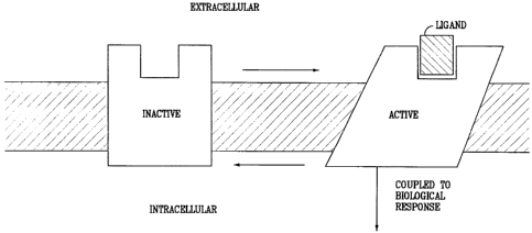
Under physiological conditions, G protein-coupled receptors exist in the cell
membrane
in equilibrium between two different states or conformations: an "inactive"
state and an "active"
state. As shown schematically in Figure 2, a receptor in an inactive state is
unable to link to the
intracellular transduction pathway to produce a biological response. Changing
the receptor
conformation to the active state allows linkage to the transduction pathway
and produces a
biological response.
A receptor may be stabilized in an active state by an endogenous ligand or an
exogenous
agonist ligand. Recent discoveries such as, including but not exclusively
limited to, modifications
to the amino acid sequence of the receptor provide means other than ligands to
stabilize the active
SUBSTITUTE SHEET (RULE26)
CA 02338543 2001-01-23
WO 00/06597 PCT/US99/17425
-3-
state conformation. These means effectively stabilize the receptor in an
active state by simulating
the effect of a ligand binding to the receptor. Stabilization by such ligand-
independent means is
termed "constitutive receptor activation." A receptor for which the endogenous
ligand is unknown
or not identified is referred to as an "orphan receptor."
B. Traditional Compound Screening
Generally, the use of an orphan receptor for screening purposes to identify
compounds that
modulate a biological response associated with such receptor has not been
possible. This is
because the traditional "dogma" regarding screening of compounds mandates that
the ligand for
the receptor be known, whereby compounds that competitively bind with the
receptor, i.e., by
interfering or blocking the binding of the natural ligand with the receptor,
are selected. By
defmition, then, this approach has no applicability with respect to orphan
receptors. Thus, by
adhering to this dogmatic approach to the discovery of therapeutics, the art,
in essence, has taught
and has been taught to forsake the use of orphan receptors unless and until
the natural ligand for
the receptor is discovered. The pursuit of an endogenous ligand for an orphan
receptor can take
several years and cost millions of dollars.
Furthermore, and given that there are an estimated 2,000 G protein-coupled
receptors in
the human genome, the majority of which being orphan receptors, the
traditional dogma castigates
a creative approach to the discovery of therapeutics to these receptors.
C. Exemplary Orphan Receptors: GPR3, GPR4, GPR6, GPR12, GPR21,
GHSR, OGR1 and AL022171
GPR3 is a 330 amino acid G protein coupled receptor for which the endogenous
ligand
is unknown. (Marchese, A. et al. (1994) Genomics 23:609; see also, Iismaa,
T.P. et al (1994)
Genomics 24:391; see Figure 1 for reported nucleic acid and amino acid
sequence.) GPR3 is
constitutively active in its endogenous form. (Eggerick, D. et al. (1995)
Biochem. J. 389:837).
SUB~STITUTE SHEET (RU(.E26)
CA 02338543 2001-01-23
WO 00/06597 PCT-/US99/17425
-4-
GPR12 is a 334 amino acid homolog of GPR3; the endogenous ligand for GPR12 is
unknown
(Song, Z. -H., et al (1995) Genomics, 28:347; see Figure 1 for reported amino
acid sequence).
GPR6 is a 362 amino acid homolog of GPR3; the endogenous ligand for GPR6 is
unknown (Song,
Z.-H. et al, supra.; see Figure 1 for reported amino acid sequence). GPR6
transcripts are reported
to be abundant in the human putamen and to a lesser extent in the frontal
cortex, hippocampus, and
hypothalamus (Heiber, M. et al. DNA and Cell Biology (1995)14(1): 25; see
Figure 1 for reported
nucleic acid and amino acid sequences for GPR6). GPR4 has also been identified
as an orphan
GPCR (Heiber, M. et al, 14 DNA Cell Biol. 25 (1995)). OGR1, an orphan GPCR, is
reported to
have a high level of homology with GPR4 (Xu, Y. and Casey, G., 35 Genomics 397
(1996)).
GPR21 is a 349 amino acid G protein coupled receptor for which the endogenous
ligand is
unknown (see GenBank Accession # U66580 for nucleic acid and deduced amino
acid sequence).
GPR21 has been reported to be located at chromosome 9q33. O'Dowd B. et al.,
187 Gene 75
(1997). AL022171 is a human DNA sequence from clone 384F21 on chromosome 1q24.
AL022171 has been identified to contain an open reading frame of 1,086 bp
encoding for a 361
amino acid protein. (see GenBank Accession number AL022171). AL022171 is 68%
homologous
to GPR21(see Figure 5B). GHSR is also identified as an orphan GPCR (Howard,
A.D. et al, 273
Science 974 (1996)).
SUIVIMARY OF THE INVENTION
Disclosed herein are methods for screening of candidate compounds against
endogenous, constitutively activated G protein-coupled orphan receptors
(GPCRs) for the
direct identification of candidate compounds as agonists, inverse agonists or
partial agonists
SUBSTITUTE SHEET (RULE26)
CA 02338543 2006-10-24
-5-
to such receptors. For such screening purposes, it is preferred that an
endogenous,
constitutively activated orphan GPCR:G protein - fusion protein be utilized.
Various embodiments of this invention provide a method for directly
identifying a
candidate compound as a compound selected from the group consisting of an
inverse
agonist, a partial agonist and an agonist, to an endogenous, constitutively
active G protein-
coupled orphan receptor, comprising the steps of: (a) contacting a candidate
compound
with a G protein-coupled receptor (GPCR) fusion protein, said GPCR fusion
protein
comprising an endogenous, constitutively active G protein-coupled orphan
receptor and a G
protein; and (b) determining, by measurement of the compound efficacy at said
contacted
receptor, whether said compound is an inverse agonist, a partial agonist or an
agonist of said
receptor. The method may further comprise modulating the G protein-coupled
orphan
receptor by contacting said receptor in vitro with the identified compound.
The method
may also further comprise preparing a pharmaceutical composition by combining
the
identified compound with at least one pharmaceutically acceptable carrier.
CA 02338543 2006-10-24
-5a-
BRIEF DESCRIPTION OF THE DRAVYINGS
Figure 1 shows a generalized structiue of a G protein-coupled receptor with
the numbers
assigned to the transmembrane helixes, the intracellular loops, and the
extracellular loops.
Figure 2 schematically shows the two states, active and inactive, for a
typical G protein
coupled receptor and the linkage of the active state to the second messenger
transduction pathway.
Figure 3 is computerized representation of a "dot-blot" showing the
distribution of the
orphan receptor GPR4 across a variety of human tissues (see Appendix A for
grid-code).
Figure 4 is a diagram showing enhanced binding of ["S]GTPyS to membranes
prepared
from 293T cells transfected with the orphan receptor GPR3 compared to those
transfected with
control vector alone at 75 gJwell membrane protein. The radiolabeled
concentration of
['SS]GTPyS was held constant at 1.2 nM and the GDP concentration was held
constant at l M.
The assay was performed on 96-well format in Wallac scintistrips.
Figure 5A shows the amino acid alignment of orphan receptors GPR3, GPR6, and
GPR12. Figure 5B shows the amino acid alignment of orphan receptors GPR21 and
A1022171
(Consensus 41 indicates matching residues).
Figure 6A is a diagram showing that the orphan receptors GPR3, GPR6, and GPR
12 are
confirmed to be constitutively active by their enhanced ability to induce
expression of P-
gaiactosidase from a CRE driven reporter system in VIP cells. Figure 6B and 6C
are diagrams of
orphan receptors GPR21 and AL022171, respectively, that have also been
confumed to be
CA 02338543 2001-01-23
WO 00/06597 - PCT/US99/17425
-6-
constitutively active by their enhanced ability to induce expression of the
luciferase gene from a
CRE driven reporter system in both 293 and 293T cells.
Figures 7A, 7B and 7C show the relative distribution of the expression of the
GPR3 (A),
GPR6 (B), and GPR12 (C) orphan receptors across several normal human tissues
as determined
by RT-PCR. Abbreviations: Ocx = occipital cortex; Hypoth = hypothalamus; Tex =
temporal
cortex; Fcx = frontal cortex.
Figures 8A and 8B show GPR3 receptor expression in norrnal (A) and epileptic
(B)
human brain tissue as examined by RT-PCR.
Figure 9A is a copy of an autoradiograph evidencing the results from in situ
hybridization
(normal rat) using GPR6 probe; Figure 9B is a reference map of the
corresponding region of the
rat brain.
Figure l0A is a copy of an autoradiograph evidencing the results from in situ
hybridization (Zucker rat - lean) using GPR6 probe; Figure lOB is a copy of an
autoradiograph
evidencing the results from in situ hybridization (Zucker rat - obese) using
GPR6 probe; Figure
lOC is a reference map of the corresponding region of the rat brain.
Figures 11A-F are copies of autoradiographs evidencing the results from in
situ
hybridization (normal rat) using GPR12 probe.
Figure 12 is a copy of an autoradiograph evidencing the results from in situ
hybridization
(normal rat) using GPR6 probe (12A), and orexin 1 receptor probe (12B) with
overlays for
detennination of co-localization of the two receptors (12C and 12D).
Figure 13 is a copy of an autoradiograph evidencing the results from in situ
hybridization
(normal rat) using GPR6 probe (13A), and melanocortin-3 receptor probe (13B)
with overlays for
deternlination of co-localization of the two receptors (13C and 13D).
SUBST1Tt1TE SHEET (RULE26)
CA 02338543 2001-01-23
WO 00/06597 PCT/US99/17425
-7-
Figure 14 provides results from co-localization experiment, evidencing that
GPR6 and
AGRP are co-localized within the arcuate. The arrow directs attention to to a
specific cell within
the arcuate, with the circle surrounding the cell; the "dots" are radiolabeled
GPR6, and beneath
those, in a darker shade, is AGRP.
Figure 15 provides graphic results of body weight over time from animals (n =
5)
receiving antisense oligonucleotides to GPR6 (star symbol at Day 5 indicates
day on which
animals received d-amphetamine sulfate injection; see Figure 16).
Figure 16 provides bar graph results from baseline locomotor activity and from
amphetamine-induced locomotive behavior in the animals of Figure 15.
Figure 17 provides bar-graph results from the direct identification of
candidate compounds
screened against GPR3 Fusion Protein (Figure 17A) and GPR6 Fusion Protein
(Figure 17B).
Figure 18A-L is a sequence diagram of the preferred vector pCMV, including
restriction enzyme site locations.
DETAILED DESCRIPTION
The scientific literature that has evolved around receptors has adopted a
number of terms
to refer to ligands having various effects on receptors. For clarity and
consistency, the following
definitions will be used throughout this patent document. To the extent that
these defmitions
conflict with other defmitions for these terms, the following definitions
shall control:
AGONISTS shall mean materials (e.g., ligands, candidate compounds) that
activate the
intracellular response when they bind to the receptor, or enhance GTP binding
to membranes.
AIVIINO ACID ABBREVIATIONS used herein are set out in Table 1:
SUBSTITUTE SHEET (RULE26)
CA 02338543 2001-01-23
WO 00/06597 PCT/US99/17425
-8-
TABLE 1
ALANINE ALA A
ARGININE ARG R
ASPARAGINE ASN N
ASPARTIC ACID ASP D
CYSTEINE CYS C
GLUTAMIC ACID GLU E
GLUTANIINE GLN Q
GLYCINE GLY G
HISTIDINE HIS H
ISOLEUCINE ILE I
LEUCINE LEU L
LYSINE LYS K
METHIONINE MET M
PHENYLALANINE PHE F
PROLINE PRO p
SERINE SER S
THREONINE THR T
TRYPTOPHAN TRP W
TYROSINE TYR Y
VALINE VAL V
PARTIAL AGONISTS shall mean materials (e.g., ligands, candidate compounds)
which activate the intracellular response when they bind to the receptor to a
lesser
degree/extent than do agonists, or enhance GTP binding to membranes to a
lesser degree/extent
than do agonists
ANTAGONIST shall mean materials (e.g., ligands, candidate compounds) that
competitively bind to the receptor at the same site as the agonists but which
do not activate the
intracellular response initiated by the active form of the receptor, and can
thereby inhibit the
intracellular responses by agonists or partial agonists. ANTAGONISTS do not
diminish the
baseline intracellular response in the absence of an agonist or partial
agonist.
SUBSTITUTE SHEET (RULE26)
CA 02338543 2001-01-23
WO 00/06597 PCT/US99/17425
-9-
CANDIDATE COMPOUND shall mean a molecule (for example, and not limitation,
a chemical compound) which is amenable to a screening technique. Preferably,
the phrase
"candidate compound" does not include compounds which were publicly known to
be
compounds selected from the group consisting of inverse agonist, agonist or
antagonist to a
receptor, as previously deteimined by an indirect identification process
("indirectly identified
compound"); more preferably, not including an indirectly identified compound
which has
previously been determined to have therapeutic efficacy in at least one
mammal; and, most
preferably, not including an indirectly identified compound which has
previously been
determined to have therapeutic utility in humans.
COMPOSITION means a material comprising at least one component; a
"pharmaceutical composition" is an example of a composition.
COMPOUND EFFICACY shall mean a measurement of the ability of a compound
to inhibit or stimulate receptor functionality, as opposed to receptor binding
affinity. A most
preferred means of detecting compound efficacy is via measurement of GTP (via
[35S]GTPyS)
or cAMP, as further disclosed in the Example section of this patent document.
CONSTITUTIVELY ACTIVATED RECEPTOR (Constitutively Active Receptor)
shall mean a receptor subject to constitutive receptor activation. A
constitutively activated
receptor can be endogenous or non-endogenous.
CONSTITUTIVE RECEPTOR ACTIVATION shall mean stabilization of a
receptor in the active state by means other than binding of the receptor with
its endogenous
ligand or a chemical equivalent thereof.
CONTACT or CONTACTING shall mean bringing at least two moieties together,
whether in an in vitro system or an in vivo system.
SUBSTIIIJT'E SHEET (RULE261
CA 02338543 2001-01-23
WO 00/06597 PCT/US99/17425
-10-
DIRECTLY IDENTIFYING or DIRECTLY IDENTIFIED, in relationship to the
phrase "candidate compound", shall mean the screening of a candidate compound
against a
constitutively activated receptor, preferably a constitutively activated
orphan receptor, and most
preferably against a constitutively activated G protein-coupled cell surface
orphan receptor, and
assessing the compound efficacy of such compound. This phrase is, under no
circumstances, to
be interpreted or understood to be encompassed by or to encompass the phrase
"indirectly
identifying" or "indirectly identified."
ENDOGENOUS shall mean a material that a mammal naturally produces.
ENDOGENOUS in reference to, for example and not limitation, the term
"receptor," shall
mean that which is naturally produced by a mammal (for example, and not
limitation, a
human) or a virus. By contrast, the term NON-ENDOGENOUS in this context shall
mean
that which is not naturally produced by a mammal (for example, and not
limitation, a human)
or a virus. For example, and not limitation, a receptor which is not
constitutively active in its
endogenous form, but when manipulated becomes constitutively active, is most
preferably
referred to herein as a "non-endogenous, constitutively activated receptor."
Both terms can be
utilized to describe both "in vivo" and "in vitro" systems. For example, and
not limitation, in a
screening approach, the endogenous or non-endogenous receptor may be in
reference to an in
vitro screening system. As a further example and not limitation, where the
genome of a
mammal has been manipulated to include a non-endogenous constitutively
activated receptor,
screening of a candidate compound by means of an in vivo system is viable.
G PROTEIN COUPLED RECEPTOR FUSION PROTEIN and GPCR FUSION
PROTEIN, in the context of the invention disclosed herein, each mean a non-
endogenous
protein comprising an endogenous, constitutively activated orphan GPCR fused
to at least one
SUDSTtTUTE SHEEt' (RUlE26)
CA 02338543 2001-01-23
WO 00/06597 - PC'P/US99/17425
-11-
G protein, most preferably, the alpha (a) subunit of such G protein (this
being the subunit that
binds GTP), with the G protein preferably being of the same type as the G
protein that
naturally couples with endogenous orphan GPCR. For example, and not
limitation, in an
endogenous state, the G protein "Gsa" is the predominate G protein that
couples with GPR6
such that a GPCR Fusion Protein based upon GPR6 would be a non-endogenous
protein
comprising GPR6 fused to Gsa. The G protein can be fused directly to the c-
terminus of the
endogenous, constitutively active orphan GPCR, or there may be spacers between
the two.
INDIRECTLY IDENTIFYING or INDIRECTLY IDENTIFIED means the
traditional approach to the drug discovery process involving identification of
an endogenous
ligand specific for an endogenous receptor, screening of candidate compounds
against the
receptor for determination of those which interfere and/or compete with the
ligand-receptor
interaction, and assessing the efficacy of the compound for affecting at least
one second
messenger pathway associated with the activated receptor.
INHIBIT or INHIBITING, in relationship to the term "response" shall mean that
a
response is decreased or prevented in the presence of a compound as opposed to
in the absence
of the compound.
INVERSE AGONISTS shall mean materials (e.g., ligand, candidate compound)
which bind to either the endogenous form of the receptor or to the
constitutively activated form
of the receptor, and which inhibit the baseline intracellular response
initiated by the active form
of the receptor below the normal base level of activity which is observed in
the absence of
agonists or partial agonists, or decrease GTP binding to membranes.
Preferably, the baseline
intracellular response is inhibited in the presence of the inverse agonist by
at least 30%, more
SUBSTITUTE SHEET (RULE26)
CA 02338543 2001-01-23
WO 00/06597 PC'i'/US99/17425
-12-
preferably by at least 50%, and most preferably by at least 75%, as compared
with the baseline
response in the absence of the inverse agonist.
LIGAND shall mean an endogenous, naturally occurring molecule specific for an
endogenous, naturally occurring receptor.
ORPHAN RECEPTOR shall mean an endogenous receptor for which the
endogenous ligand specific for that receptor has not been identified or is not
known.
PHARMACEUTICAL COMPOSITION shall mean a composition comprising at
least one active ingredient, whereby the composition is amenable to
investigation for a
specified, efficacious outcome in a mammal (for example, and not limitation, a
human). Those
of ordinary skill in the art will understand and appreciate the techniques
appropriate for
determining whether an active ingredient has a desired efficacious outcome is
based upon the
needs of the artisan.
NON-ORPHAN RECEPTOR shall mean an endogenous naturally occun-ing
molecule specific for an endogenous naturally occurring ligand wherein the
binding of a ligand
to a receptor activates an intracellular signaling pathway.
STIMULATE or STIMULATING, in relationship to the tenn "response" shall mean
that a response is increased in the presence of a compound as opposed to in
the absence of the
compound.
The order of the following sections is set forth for presentational efficiency
and is not
intended, nor should be construed, as a limitation on the disclosure or the
claims to follow.
A. Introduction
The traditional study of receptors has always proceeded from the a priori
assumption
(historically based) that the endogenous ligand must first be identified
before discovery could
SUBSTMM SHEET (RUt.E2S )
CA 02338543 2001-01-23
WO 00/06597 - PCT/US99/17425
-13-
proceed to find antagonists and other molecules that could affect the
receptor. Even in cases where
an antagonist might have been known first, the search immediately extended to
looking for the
endogenous ligand. This mode of thinking has persisted in receptor research
even after the
discovery of constitutively activated receptors. What has not been heretofore
recognized is that
it is the active state of the receptor that is most useful for discovering
agonists, partial agonists, and
inverse agonists of the receptor. For those diseases which result from an
overly active receptor,
what is desired in a therapeutic drug is a compound which acts to diminish the
active state of a
receptor, not necessarily a drug which is an antagonist to the endogenous
ligand. This is because
a compound (drug) which reduces the activity of the active receptor state need
not bind at the same
site as the endogenous ligand. Thus, as taught by a method of this invention,
any search for
therapeutic compounds should start by screening compounds against the ligand-
independent active
state. The search, then, is for an inverse agonist to the active state
receptor.
Screening candidate compounds against the endogenous, constitutively activated
orphan
receptors, for example, and not limited to, the endogenous, constitutively
active GPCRs set forth
herein, GPR3, GPR4, GPR6, GPR12, GPR2 1, GHSR, OGR1, RE2 and AL022171, allows
for the
direct identification of candidate compounds which act at these orphancell
surface receptors,
without requiring any prior knowledge or use of the receptor's endogenous
ligand. By
determining areas within the body where such receptors are expressed and/or
over-expressed, it
is possible to determine related disease/disorder states which are associated
with the expression
and/or over-expression of these receptors; such an approach is disclosed in
this patent document.
B. Disease/Disorder Identification and/or Selection
SUBSTIT(!TE SHEET (RULE26)
CA 02338543 2001-01-23
WO 00/06597 - PCT/US99/17425
-14-
As will be set forth in greater detail below, most preferably inverse agonists
to
endogenous, constitutively activated orphan receptors, e.g., such as those set
forth herein (GPR3,
GPR4, GPR6, GPR12, GPR21, GHSR, OGRl, RE2 and AL022171) can be identified by
the
methodologies of this invention. Such inverse agonists are ideal candidates as
lead compounds
in drug discovery programs for treating diseases related to these receptors.
Indeed, an antagonist
to such a receptor (even if the ligand were known) may be ineffective given
that the receptor is
activated even in the absence of ligand-receptor binding. Because of the
ability to directly identify
inverse agonists to these receptors, thereby allowing for the development of
pharmaceutical
compositions, a search, for diseases and disorders associated with these
receptors is possible. For
example, scanning both diseased and nonnal tissue samples for the presence of
these orphan
receptors now becomes more than an academic exercise or one which might be
pursued along the
path of identifying an endogenous ligand. Tissue scans can be conducted across
a broad range of
healthy and diseased tissues. Such tissue scans provide a preferred first step
in associating a
specific receptor with a disease and/or a disorder.
Preferably, the DNA sequence of the endogenous, constitutively activated GPCR
is used
to make a probe for RT-PCR identification of the expression of the receptor in
tissue samples. The
presence of a receptor in a diseased tissue, or the presence of the receptor
at elevated
concentrations in diseased tissue compared to normal tissue, can be utilized
to identify a
correlation with that disease. Receptors can equally well be localized to
regions of organs by this
technique. Based on the known functions of the specific tissues to which the
receptor is localized,
the putative functional role of the receptor can be deduced.
C. Homology Identification
SUBSTITUTE SHEET (RULE26)
CA 02338543 2001-01-23
WO 00/06597 - PC'F/US99/17425
-15-
The identification and association of an orphan receptor with diseases and/or
disorders can
be beneficially enhanced via identification of additional receptors having
homology with the
original orphan receptor. This approach was utilized in the identification of
both GPR6 and
GPR1 2, based upon their sequence homology with GPR3, and in the
identification of AL022171,
having sequence homology to GPR21. GPR3 was previously identified as a
constitutively
activated orphan receptor (see Eggerick, supra). What was not known, prior to
this invention, was
that GPR6, GPR12, GPR21 and AL022171 are also constitutively active in their
endogenous
states. Using known computerized databases (e.g., dbEST), GPR6, GPR12, GPR21
and
AL022171 were identified.
This highlights certain unique benefits of the invention disclosed herein:
because the
dogma in drug screening relies upon knowledge and identification of a
receptor's endogenous
ligand, the art had no motivation to explore whether or not GPR3 homologs were
constitutively
active in their endogenous forms (other than for, at best, academic
curiosity). However, with the
power of the present invention to directly identify inverse agonists to such
receptors, coupled with
the ability to locate the distribution of such receptors in tissue samples,
the present invention
dramatically transcends such idle curiosity and provides a means for
alleviating diseases and
disorders which impact the human condition.
D. Screening of Candidate Compounds
1. Generic GPCR screening assay techniques
When a G protein receptor becomes constitutively active, it binds to a G
protein (eg., Gq,
Gs, Gi, Go) and stimulates the binding of GTP to the G protein. The G protein
then acts as a
GTPase and slowly hydrolyzes the GTP to GDP, whereby the receptor, under
normal conditions,
becomes deactivated. However, constitutively activated receptors continue to
exchange GDP to
SUBSTtTUTE SHEET (RULE26)
CA 02338543 2001-01-23
WO 00/06597 - PCT/US99/17425
-16-
GTP. A non-hydrolyzable analog of GTP, [35S]GTPyS, can be used to monitor
enhanced binding
to membranes which express constitutively activated receptors. It is reported
that [35S]GTP~S can
be used to monitor G protein coupling to membranes in the absence and presence
of ligand. An
example of this monitoring, among other examples well-known and available to
those in the art,
was reported by Traynor and Nahorski in 1995. The preferred use of this assay
system is for initial
screening of candidate compounds because the system is generically applicable
to all G protein-
coupled receptors regardless of the particular G protein that interacts with
the intracellular domain
of the receptor. It is in the context of the use of a GTP assay system that a
GPCR Fusion Protein
is preferably utilized.
B 2. Specific GPCR screening assay techniques
Once candidate compounds are identified using the "generic" G protein-coupled
receptor assay (i.e. an assay to select compounds that are agonists, partial
agonists, or inverse
agonists), further screening to confirm that the compounds have interacted at
the receptor site
is preferred. For example, a compound identified by the "generic" assay may
not bind to the
receptor, but may instead merely "uncouple" the G protein from the
intracellular domain.
In the case of GPR3, GPR4, GPR6, GPR12, GPR21, GHSR, OGRI, RE2 and AL022171,
it has
been determined that these receptors couple the G protein Gs. Gs stimulates
the enzyme
adenylyl cyclase (Gi, on the other hand, inhibits this enzyme). Adenylyl
cyclase catalyzes
the conversion of ATP to cAMP; thus, because these receptors are activated in
their
endogenous forms, increased levels of cAMP are associated therewith (on the
other hand,
endogenously activated receptors which couple the Gi protein are associated
with decreased
levels of cAMP). See, generally, "Indirect Mechanisms of Synaptic
Transmission," Chpt. 8,
From Neuron To Brain (31 Ed.) Nichols, J.G. et al eds. Sinauer Associates,
Inc. (1992). Thus,
SUBSI1TUTE SHEET (RULE26)
CA 02338543 2001-01-23
WO 00/06597 PCT/US99/17425
-17-
assays that detect cAMP can be utilized to determine if a candidate compound
is an inverse
agonist to the receptor (i. e., such a compound which contacts the receptor
would decrease the
levels of cAMP relative to the uncontacted receptor). A variety of approaches
known in the
art for measuring cAMP can be utilized; a most preferred approach relies upon
the use of anti-
cAMP antibodies. Another type of assay that can be utilized is a whole cell
second
messenger=reporter system assay. Promoters on genes drive the expression of
the proteins that
a particular gene encodes. Cyclic AMP drives gene expression by promoting the
binding of a
cAMP-responsive DNA binding protein or transcription factor (CREB) which then
binds to the
promoter at specific sites called cAMP response elements and drives the
expression of the gene.
Reporter systems can be constructed which have a promoter containing multiple
cAMP response
elements before the reporter gene, e.g., 0-galactosidase or luciferase. Thus,
an activated Gs
receptor such as GPR3 causes the accumulation of cAMP which then activates the
gene and
expression of the reporter protein. The reporter protein such as (3-
galactosidase or luciferase can
then be detected using standard biochemical assays (see, for example, Chen et
al. 1995). A cAMP
assay is particularly preferred.
The foregoing specific assay approach can, of course, be utilized to initially
directly
identify candidate compounds, rather than by using the generic assay approach.
Such a
selection is primarily a matter of choice of the artisan. With respect to
GPR6, use of a
modified, commercially available cAMP assay was initially utilized for the
direct
identification of inverse agonists.
C 3. GPCR Fusion Protein
The use of an endogenous, constitutively activated orphan GPCR for use in
screening of
candidate compounds for the direct identification of inverse agonists,
agonists and partial agonists
SUBSTITUTE SHEEi' (RULE26)
CA 02338543 2001-01-23
WO 00/06597 - PCT/US99/17425
-18-
provides a unique challenge in that, by definition, the endogenous receptor is
active even in the
absence of an endogenous ligand bound thereto. Thus, in order to differentiate
between, e.g., the
endogenous receptor in the presence of a candidate compound and the endogenous
receptor in the
absence of that compound, with an aim of such a differentiation to allow for
an understanding as
to whether such compound may be an inverse agonist, agonist, partial agonist
or have no affect
on such a receptor, it is preferred that an approach be utilized that can
enhance such differentiation.
A preferred approach is the use of a GPCR Fusion Protein.
Generally, once it is detennined that an endogenous orphan GPCR is
constitutively active,
using the assay techniques set forth above (as well as others), it is possible
to detemline the
predominant G protein that couples with the endogenous GPCR. Coupling of the G
protein to the
GPCR provides a signaling pathway that can be assessed. Because it is most
preferred that
screening take place by use of a manunalian expression system, such a system
will be expected
to have endogenous G protein therein. Thus, by definition, in such a system,
the endogenous,
constitutively active orphan GPCR will continuously signal. In this regard, it
is preferred that this
signal be enhanced such that in the presence of, e.g., an inverse agonist to
the receptor, it is more
likely that one will be able to more readily differentiate, particularly in
the context of screening,
between the receptor when it is or is not contacted with the inverse agonist.
The GPCR Fusion Protein is intended to enhance the efficacy of G protein
coupling with
the endogenous GPCR. The GPCR Fusion Protein appears to be important for
screening with an
endogenous, constitutively activated GPCR because such an approach increases
the signal that is
most preferably utilized in such screening techniques. Facilitating a
significan t "signal to noise"
ratio is important for the screening of candidate compounds as disclosed
herein.
SUBSTITUY'E SHEET (RULE26)
CA 02338543 2001-01-23
WO 00/06597 - PCT/US99/17425
-19-
The construction of a construct useful for expression of a GPCR Fusion Protein
is within
the purview of those having ordinary skill in the art. Commercially available
expression vectors
and systems offer a variety of approaches that can fit the particular needs of
an investigator. One
important criterion for such a GPCR Fusion Protein construct is that the
endogenous GPCR
sequence and the G protein sequence both be in-fiame (preferably, the sequence
for the
endogenous GPCR is upstream of the G protein sequence) and that the "stop"
codon of the GPCR
must be deleted or replaced such that upon expression of the GPCR, the G
protein can also be
expressed. The GPCR can be linked directly to the G protein, or there can be
spacer residues
between the two (preferably no more than about 12, although this number can be
readily
ascertained by one of ordinary skill in the art). We have evaluated both
approaches, and in terms
of measurement of the activity of the GPCR, the results are substantially the
same; however, there
is a preference (based upon convenience) of use of a spacer in that some
restriction sites that are
not used will, effectively, upon expression, become a spacer. Most preferably,
the G protein that
couples to the endogenous GPCR will have been identified prior to the creation
of the GPCR
Fusion Protein construct. Because there are only a few G proteins that have
been identified, it is
preferred that a construct comprising the sequence of the G protein (i.e., a
universal G protein
construct) be available for insertion of an endogenous GPCR sequence therein;
this provides for
efficiency in the context of large-scale screening of a variety of different
endogenous GPCRs
having different sequences.
E. Medicinal Chemistry
Generally, but not always, direct identification of candidate compounds is
preferably
conducted in conjunction with compounds generated via combinatorial chemistry
techniques,
whereby thousands of compounds are randomly prepared for such analysis.
Generally, the
SUBSTfIT1TE SHEET (RULE26)
CA 02338543 2001-01-23
WO 00/06597 - PCT/US99/17425
-20-
results of such screening will be compounds having unique core structures;
thereafter, these
compounds are preferably subjected to additional chemical modification around
a preferred
core structure(s) to further enhance the medicinal properties thereof. In this
way, inverse
agonists, agonists and/or partial agonists that are directly identified can be
beneficially
improved upon prior to development of pharmaceutical compositions comprising
such
compounds. Generally, it is preferred that the binding affinity of a directly
identified
compound selected for further refinement into a pharmaceutical composition
have a binding
affinity for the receptor of less than 100nM, although this is generally a
preference selection
based upon the particular needs of the artisan. Such techniques are known to
those in the art
and will not be addressed in detail in this patent document.
F. Pharmaceutical Compositions
Candidate compounds selected for further development can be formulated into
pharmaceutical compositions using techniques well known to those in the art.
Suitable
pharmaceutically-acceptable carriers are available to those in the art; for
example, see Remington's
Pharmaceutical Sciences, 16`h Edition, 1980, Mack Publishing Co., (Oslo et
al., eds.).
EXAMPLES
The following examples are presented for purposes of elucidation, and not
limitation,
of the present invention. While specific nucleic acid and amino acid sequences
are disclosed
herein, those of ordinary skill in the art are credited with the ability to
make minor
modifications to these sequences while achieving the same or substantially
similar results
reported below. It is intended that equivalent, endogenous, constitutively
activated human
orphan receptor sequences having eighty-five percent (85%) homology, more
preferably
SUBSTITUTE SHEET {RULE2S)
CA 02338543 2001-01-23
WO 00/06597 - PCT/US99/17425 _
-21-
having ninety percent (90%) homology, and most preferably having grater than
ninety-five
percent (95%) homology to GPR3, GPR4, GPR6, GPR12, GPR21, GHSR, OGR1, RE2 and
AL022171 fall within the scope of any claims appended hereto.
Example 1
Preparation of In Situ Probes
In situ probes for GPR3, GPR6, and GPR12 were prepared. The following PCR
protocol was utilized for all three probes: the reaction condition utilized
was 1X rTth DNA
polymerase buffer II, 1.5 mM Mg(OAc)2, 0.2 mM each of the 4 nucleotides, 0.228
g rat
genomic DNA, 0.25 M of each primer (see below) and 1 unit of rTth DNA
polymerase
(Perkin Elmer) in 50 l reaction volume. The cycle condition was 30 cycles of
94 C for I
min, 55 C for 1 min and 72 C for 45 sec with a Perkin Elmer Cetus 2400
thermal cycler.
1. Rat GPR3 in situ probe
Because the full length cDNA sequence for rat GPR3 is not data-base available,
the
DNA fragment for the in situ probe was obtained by PCR using a 3' degenerate
oligonucleotide based on the published human and mouse GPR3 sequences in the
middle of
the transmembrane domain 3, and a 5' degenerate oligonucleotide near the
beginning of the
5' extracellular domain. The sequences of the oligonucleotides utilized were
as follows:
5'-GGAGGATCCATGGCCTGGTTCTCAGC-3' (SEQ.ID.NO.:1; 5' oligo)
5'-CACAAGCTTAGRCCRTCC MG RCA RTTCCA-3' (SEQ.ID.NO.: 2; 3' oligo)
where R=A or G, and M=A or C.
SU6STITUTE SHEET (RULF.26)
CA 02338543 2001-01-23
WO 00/06597 - PCT/US99/17425
-22-
A 537 bp PCR fragment containing nucleotide 24 through to the middle of
transmembrane
3 was digested with Bam HI and Hind III and was subcloned into a Bam HI-Hind
III site of
pBluescript.
2. Rat GPR 6 in situ probe
The in situ probe DNA fragment of rat GPR6 was obtained by PCR based on the
published rat GPR6 cDNA sequences. The sequences of the oligonucleotides
utilized were as
follows:
5'-GGAGAAGCTTCTGGCGGCGATGAACGCTAG-3' (SEQ.ID.NO.: 3; 5' oligo)
5'-ACAGGATCCAGGTGGCTGCTAGCAAGAG-3' (SEQ.ID.NO.: 4; 3' oligo)
A 608 bp PCR fragment containing nucleotide -10 through to the middle of
transmembrane
domain 4 was digested with Bam Hi and Hind III and was subcloned into Bam HI-
Hind III
site of pBluescript.
3. Rat GPR12 in situ probe
The in situ probe DNA fragment of rat GPR12 was obtained by PCR based on the
published rat GPR12 cDNA sequences. The sequences of the oligonucleotides
utilized were
as follows:
5'-CTTAAGCTTAAAATGAACGAAGACCCGAAG-3' (SEQ.ID.NO.: 5; 5' oligo)
5'-GGAGGATCCCCAGAGCATCACTAGCAT-3' (SEQ.ID.NO.: 6; 3' oligo)
A 516 bp PCR fragment containing nucleotide -5 through to the middle of
transmembrane
domain 4 was digested with Bam HI and Hind III and subcloned into a Bam HI-
Hind III site
of pBluescript.
In situ probe sequences generated were as follows:
SUBSTITUTE SHEET (RULE2fi)
CA 02338543 2001-01-23
WO 00/06597 - PCT/US99/17425
-23-
Rat GPR3 probe:
GGAGGATCCATGGCCTGGTTCTCAGCCGGCTCAGGCAGTGTGAATGTGAGCAT
AGACCCAGCAGAGGAACCTACAGGCCCAGCTACACTGCTGCCCTCTCCCAGGG
CCTGGGATGTGGTGCTGTGCATCTCAGGCACCCTGGTGTCCTGCGAGAATGCT
CTGGTGATGGCCATCATTGTGGGCACGCCTGCCTTCCGCGCCCCCATGTTCCTG
CTGGTGGGCAGCTTGGCCGTAGCAGACCTGCTGGCAGGCCTGGGCCTGGTCCT
GCACTTCGCTGCTGACTTCTGTATTGGCTCACCAGAGATGAGCTTGGTGCTGGT
TGGCGTGCTAGCAACGGCCTTTACTGCCAGCATCGGCAGCCTGCTGGCCATCA
CCGTTGACCGCTACCTTTCCCTGTACAACGCCCTCACCTACTACTCAGAGACAA
CAGTAACTCGAACCTACGTGATGCTGGCCTTGGTGTGGGTGGGTGCCCTGGGC
CTGGGGCTGGTTCCCGTGCTGGCCTGGAACTGCCGGGACGGTCTAAGCTT
(SEQ.ID.NO.: 7)
Rat GPR6 probe:
AAGCTTCTGGCGGCGATGAACGCTAGCGCCGCCGCGCTCAACGAGTCCCAGGTGGTGGCAGTAGCG
GCCGAGGGAGCGGCAGCTGCGGCTACAGCAGCAGGGACACCGGACACCAGCGAATGGGGACCTCCG
GCAGCATCCGCGGCGCTGGGAGGCGGCGGAGGACCTAACGGGTCACTGGAGCTGTCTTCGCAGCTG
CCCGCAGGACCCTCAGGACTTCTGCTTTCGGCAGTGAATCCCTGGGATGTGCTGCTGTGCGTGTCGGG
GACTGTGATCGCAGGCGAAAATGCGCTGGTGGTGGCGCTCATCGCATCCACTCCCGCGCTGCGCACG
CCCATGTTTGTGCTCGTGGGTAGTCTGGCCACTGCTGACCTGCTGGCGGGCTGTGGCCTCATCCTACA
CTTCGTGTTCCAGTACGTGGTGCCCTCGGAGACTGTGAGCCTGCTCATGGTGGGCTTCCTGGTGGCGT
CCTTCGCCGCCTCAGTCAGCAGCCTGCTCGCTATCACAGTGGACCGTTACCTGTCCCTTTACAACGCG
SUBSTITUTE SHEET (RUt.E26)
CA 02338543 2001-01-23
WO 00/06597 - PCT/US99/17425
-24-
CTCACCTACTACTCGCGCCGGACCCTGTTGGGCGTGCACCTCTTGCTAGCAGCCACCTGGATCC
(SEQ.ID.NO.: 8)
Rat GPR12 probe:
AAGCTTAAAATGAACGAAGACCCGAAGGTCAATTTAAGCGGGCTGCCTCGGGACTGTATAGAAGCT
GGTACTCCGGAGAACATCTCAGCCGCTGTCCCCTCCCAGGGCTCTGTTGTGGAGTCAGAACCCGAGC
TCGTTGTCAACCCCTGGGACATTGTCTTGTGCAGCTCAGGAACCCTCATCTGCTGTGAAAATGCCGTC
GTGGTCCTTATCATCTTCCACAGCCCCAGCCTGCGAGCACCCATGTTCCTGCTGATAGGCAGCCTGGC
TCTTGCAGACCTGCTGGCTGGTCTGGGACTCATCATCAATTTTGTTTTTGCCTACCTGCTTCAGTCAGA
AGCCACCAAGCTGGTCACAATTGGACTCATTGTCGCCTCTTTCTCTGCCTCTGTCTGCAGTTTGCTGG
CTATCACTGTGGACCGCTACCTCTCGCTGTATTACGCCCTGACGTACCACTCCGAGAGGACCGTCACC
TTTACCTATGTCATGCTAGTGATGCTCTGGGGATCC (SEQ.ID.NO.: 9)
Example 2
Receptor Expression
1. cDNA and Vectors
With respect to GPR3 and GPR6, expression vectors comprising cDNA were
generously supplied by Brian O'Dowd (University of Toronto). The vector for
GPR3 cDNA
was pcDNA3; the vector for GPR6 was pRcCMV (the coding region for GPR6 was
subcloned
into pCMV vector at a Hind III-Xbal site). GPR12 cDNA was prepared using the
following
protocol: Human GPR12 cDNA was obtained by PCR using human genomic DNA and a
5'
primer from the 5' untranslated region with a Hind III restriction site, and a
3' primer from
the 3' untranslated region containing a Bam HI site'. Primers had the
following sequences:
5'-CTTAAGCTTGTGGCATTTGGTACT-3' (SEQ.ID.NO.: 10; 5' oligo)
SUBSTI111TE SHEET (RULE26)
CA 02338543 2001-01-23
WO 00/06597 PCT/US99/17425
-25-
5'-TCTGGATCCTTGGCCAGGCAGTGGAAGT-3 (SEQ.ID.NO.: 11; 3' oligo)
PCR was performed using rTth polymerase (Perkin Elmer) with the buffer system
provided
by the manufacturers, 0.25 M of each primer, 0.2 M of each of the four
nucleotides and 0.2
g of genomic DNA as template. The cycle condition was 30 cycles of 94 C for I
min, 57
C for 1 min and 72 C for 1.5 min. The 1.2 kb PCR fragment was digested with
Hind III and
Bam HI, and subcloned into Hind III-Bam HI site of pCMV expression vector. The
resulting
cDNA clones were fully sequenced and consistent with published sequences.
With respect to GPR2 1, PCR was performed using genomic DNA as template and
rTth
polymerase (Perkin Elmer) with the buffer system provided by the manufacturer,
0.25 M of
each primer, and 0.2 mM of each of the four nucleotides. The cycle condition
was 30 cycles
of 94 C for 1 min, 62 C for 1 min and 72 C for 1 min and 20 sec. The 5' PCR
primer was
kinased with the sequence:
5'-GAGAATTCACTCCTGAGCTCAAGATGAACT-3' (SEQ.ID.NO.:12)
and the 3' primer contained a BamHI site with the sequence:
5'-CGGGATCCCCGTAACTGAGCCACTTCAGAT-3' (SEQ.ID.NO.:13).
The resulting 1.1 kb PCR fragment was digested with BamHI and cloned into
EcoRV-BamHI
site of pCMV expression vector. Nucleic acid (SEQ.ID.NO.:14) and amino acid
(SEQ.ID.NO.:15) sequences for human GPR21 were thereafter determined.
With respect to AL022171, PCR was performed using genomic DNA as template and
rTth polymerase (Perkin Elmer) with the buffer system provided by the
manufacturer, 0.25
M of each primer, and 0.2 mM of each of the four nucleotides. The cycle
condition was 30
cycles of 94 C for 1 min, 54 C for 1 min and 72 C for 1 min and 20 sec. The
5' primer
contains an HindIII site with the following sequence:
SUBSTITUTE SHEE'T (RULE26)
CA 02338543 2001-01-23
WO 00/06597 PC'F/US99/17425
-26-
5'-AGGAAGCTTTAAATTTCCAAGCCATGAATG-3' (SEQ.ID.NO.:16)
and the 3' primer contained a EcoRI site with the following sequence:
5'-ACCGAATTCAGATTACATTTGATTTACTATG-3' (SEQ.ID.NO.:17). The resulting 1.15
kb PCR fragment was digested with HindI1I and EcoRI and cloned into HindlIl-
EcoRI site
ofpCMV expression vector. Nucleic acid (SEQ.ID.NO.:18) and amino acid
(SEQ.ID.NO.:19)
sequences for human AL022171 were thereafter determined and verified.
With respect to GPR4 (GenBank accession number L36148), expression vectors
comprising the cDNA was generously supplied by Brian O'Dowd (University of
Toronto).
The vector for GPR4 cDNA was pcDNA3 and this subcloned into pCMV vector at a
Hind III-
Xbal site (the 5' untranslated region between HindIII and an ApaI site was
trimmed by
conducting digestion/self ligation).
With respect to RE2 (GenBank accession number AF091890), PCR was performed
using human brain cDNA as template and rTth polymerase (Perkin Elmer) with the
buffer
system provided by the manufacturer, 0.25 M of each primer, and 0.2 mM of
each of the four
nucleotides. The cycle condition was 30 cycles of 94 C for 1 min, 62 C for 1
min and 72 C
for 1 min and 30 sec. The 5' PCR primer contained an EcoRl site with the
sequence
5' -AGCGAATTCTGCCCACCCCACGCCGAGGTGCT-3' (SEQ. ID. No. 20)
and the 3' primer contained a BamHl site with the sequence
5'-TGCGGATCCGCCAGCTCTTGAGCCTGCACA-3' (SEQ. ID. NO.: 21). The 1.36 kb PCR
fragment that resulted after two rounds of PCR was then digested with EcoRI
and BamHI and
cloned into EcoRI-BamHI site of pCMV. Nucleic acid (SEQ. ID. NO. 22) and amino
acid
sequence (SEQ. ID. NO. 23) was thereafter determined.
SUBSITTUTE SHEET (RULE26)
CA 02338543 2001-01-23
WO 00/06597 PCT/US99/17425
-27-
With respect to OGR1 (GenBank accession number U48405), PCR was performed
using human genomic DNA as template and rTth polymerase (Perkin Elmer) with
the buffer
system provided by the manufacturer, 0.25 M of each primer, and 0.2 mM of
each of the four
nucleotides. The cycle condition was 30 cycles of 94 C for 1 min, 62 C for
lmin and 72 C
for 1 min and 20 sec. The 5' PCR primer contained a HindIII site with the
sequence
5'-GGAAGCTTCAGGCCCAAAGATGGGGAACAT-3' (SEQ. ID. No. 24)
and the 3' primer contain a BamHI site with the sequence
'-GTGGATCCACCCGCGGAGGACCCAGGCTAG-3' (SEQ.ID.NO.25). The resulting 1.14
kb PCR fragment was digested with HindIII and BamHI and cloned into HindIII-
BamHI site
pCMV. Nucleic acid (SEQ. ID. NO. 26) and amino acid sequence (SEQ. ID. NO. 27)
was
thereafter determined.
With respect to GHSR, PCR was performed using hippocampus cDNA as template
and TaqPlus Precision polymerase (Stratagene) with the buffer system provided
by the
manufacturer, 0.25 M of each primer, and 0.2 mM of each 4 nucleotides. The
cycle
condition was 30 cycles of 94 C for 1 min, 68 C for lmin and 72 C for 1 min
and 10 sec. For
first round PCR, the 5' PCR primer sequence:
5'-ATGTGGAACGCGACGCCCAGCG-3' (SEQ.ID.N0.40)
and the 3' primer sequence:
5'-TCATGTATTAATACTAGATTCT-3' (SEQ.ID.NO.41).
Two microliters of the first round PCR was used as a template for the second
round PCR
where the 5' primer was kinased with sequence:
5'-TACCATGTGGAACGCGACGCCCAGCGAAGAGCCGGGGT-3' (SEQ.ID.NO.:42) and
the 3' primer contains an EcoRl site with the sequence:
SUBSTITUTE SHEET (RULE26)
CA 02338543 2001-01-23
WO 00/06597 PCT/US99/17425
-28-
5'-CGGAATTCATGTATTAATACTAGATTCTGTCCA~'iGCCCG-3' (SEQ.ID.NO.:43). The
1.1 kb PCR fragment was digested with EcoRI and cloned into blunt-EcoRI site
of CMVp
expression vector. Nucleic acid (SEQ.ID.NO.:44) and amino acid (SEQ.ID.NO.:45)
sequences for human GHSR were thereafter determined.
2. Transfection procedure
On day one, 1 X 10" 293 or 293T cells per 150mm plate were plated out. On day
two, two
reaction tubes were prepared (the proportions to follow for each tube are per
plate): tube A was
prepared by mixing between 8-20 g DNA (e.g., pCMV vector; pCMV vector with
receptor
cDNA; pCMV with GPCR Fusion Protein, supra) in 1-2m1 serum free DMEM (Irvine
Scientific,
Irvine, CA); tube B was prepared by mixing 50-120 1 lipofectamine (Gibco BRL)
in 1-2m1 serum
free DMEM. Tubes A and B were then admixed by inversions (several times),
followed by
incubation at room temperature for 30-45min. The admixture is referred to as
the "transfection
mixture". Plated cells were washed with 1XPBS, followed by addition of 10-12m1
serum free
DMEM. 2.4m1 of the transfection mixture was then added to the cells, followed
by incubation
for 4hrs at 37 C/5% COZ. The transfection mixture was then removed by
aspiration, followed by
the addition of 25ml of DMEM/10% Fetal Bovine Serum. Cells were then incubated
at 37 C/5%
COz.
For GPCR Fusion Protein, preferred amounts to the above are as follows:12 g
DNA; 2m1
sernun free DMEM; 60 l lipofectamine; 293 cells 9 and an addition of 12m1
serum free DMEM).
Example 3
Tissue Distribution of GPCR
For some orphan receptors, it will be apparent to those in the art that there
is an
understanding of the distribution of such receptors within, e.g., a human, or
associated with a
SUBSTtTUTE SHEET (RULE26)
CA 02338543 2001-01-23
WO 00/06597 PCT/US99/17425
-29-
disease state. However, for many orphan receptors, such information is not
known, or will not be
known. It is therefore preferred that some understanding of where such
receptors may be
distributed be understood; this allows for the ability to gain a predictive
opportunity to associate
a particular receptor with a disease state or disorder associated with the
particular tissue where the
receptor may be preferentially expressed. Using a commercially available mRNA
dot-blot format,
the distribution of endogenous, constitutively active GPCRs in various tissue
types was assessed.
Preferably, the entire coding region of the receptor is used to generate a
radiolabeled
probe using a Prime-It IIT"" Random Primer Labeling Kit (Stratagene, #300385),
according
to the manufacturer's instructions. As an example, this approach was utilized
for GPR4.
Human RNA Master BlotT"' kit (Clontech, #7770-1) was hybridized with this
probe
and washed under stringent conditions, in accordance with manufacturer
instructions. The
blot was exposed to Kodak BioMaxTM Autoradiography film overnight, at -80 C.
Results are
presented in Figure 3. Based upon these results, it is noted that GPR4 appears
to be expressed
throughout a variety of fetal tissue types (row G), as well as non-fetal heart
(C 1), and non-fetal
lung (F 1). This approach can be readily utilized for other receptors.
Example 4
GTP MEMBRANE BINDING SCINTILLATION PROXIMITY ASSAY
When a G protein-coupled receptor is in its active state, either as a result
of ligand binding
or constitutive activation, the receptor binds to a G protein (in the case of
GPR3, GPR4, GPR6,
GPR12, GPR21, GHSR, OGR1, RE2 and AL022171, Gs) and stimulates the binding of
GTP to
the G protein. The trimeric G protein-receptor complex acts as a GTPase and
slowly hydrolyzes
the GTP to GDP, at which point the receptor normally is deactivated.
Constitutively activated
receptors continue to exchange GDP for GTP. The non-hydrolyzable GTP analog,
[35S]GTPyS,
can be utilized to demonstrate enhanced binding of [35S]GTPyS to membranes
expressing
SUBSTIME SHEET (RULE26)
CA 02338543 2001-01-23
WO 00/06597 - PC'F/US99/17425
-30-
constitutively activated receptors. The advantage of using [35S]GTPyS binding
to measure
constitutive activation is that: (a) it is generically applicable to all G
protein-coupled receptors; (b)
it is proximal at the membrane surface making it less likely to pick-up
molecules which affect the
intracellular cascade.
The assay utilizes the ability of G protein coupled receptors to stimulate
[35S]GTPyS
binding to membranes expressing the relevant receptors. The assay can,
therefore, be used in
the direct identification method to screen candidate compounds to known,
orphan and
constitutively activated G protein coupled receptors. The assay is generic and
has application
to drug discovery at all G protein coupled receptors.
The [35S]GTPyS assay was incubated in 20 mM HEPES, pH 7.4, binding buffer with
12
nM [35S]GTPyS and 75 g membrane protein [e.g., 293T cells expressing GPR3]
and 1 M GDP
for 1 hour. Wheatgerm agglutinin beads (25 l; Amersham) were then added and
the mixture was
incubated for another 30 minutes at room temperature. The tubes were then
centrifuged at 1500
X g for 5 minutes at room temperature and then counted in a scintillation
counter.
Referring to Figure 4, GPR3 receptor was determined to have increased activity
as
compared to control; this heightened activity is not the result of autocrine
stimulation in that the
data were obtained from membrane preparations, as opposed to whole cell
preparations.
Example 5
Receptor Homology Determination
Following confirmation that GPR3 is a constitutively activated receptor, a
homology
search of the available G protein-coupled data banks (GeneBank), using the
commercially
available program, DNA Star, identified two highly homologous receptors, GPR6
and GPRl2 (see
Figure 5A); both of these receptors are orphan receptors. While the sequence
of these receptors
was previously "known" (i.e., they were available on the databases), it was
not known that these
SUBSTttUTE SHLE7 (RUi.E26)
CA 02338543 2001-01-23
WO 00/06597 - PCT/US99/17425
-31-
two receptors are constitutively activated in their endogenous forms (see
Example 6, Figure 7).
Furthermore, heretofore there would be no reason to search for such receptors
for use in a drug
discovery program in that the ligands therefore are not known or have not been
identified. As
such, the dogma approach to drug discovery would at best find the homology
between GPR3,
GPR6 and GPR12 of minor interest or, more likely, irrelevant.
Example 6
Analysis of Homologous Receptors For Constitutive Activation
Although a variety of cells are available to the art for the expression of
proteins, it is
most preferred that mammalian cells be utilized. The primary reason for this
is predicated
upon practicalities, i.e., utilization of, e.g., yeast cells for the
expression of a GPCR, while
possible, introduces into the protocol a non-mammalian cell which may not
(indeed, in the
case of yeast, does not) include the receptor-coupling, genetic-mechanism and
secretary
pathways that have evolved for mammalian systems - thus, results obtained in
non-
mammalian cells, while of potential use, are not as preferred as that obtained
from mammalian
cells. Of the mammalian cells, COS-7, 293 and 293T cells are particularly
preferred, although
the specific mammalian cell utilized can be predicated upon the particular
needs of the artisan.
1. Analysis of GPR3, GPR6 and GPR12
To generate a 13-galactosidase reporter containing multiple Ga14 binding
sites, a Bgl II/
HindIIl fragment was removed from the somatostatin promoter-containing plasmid
1.4(5xGa1)CAT (Leonard, J. et al (1992) PNAS USA 89:6247-6251) and cloned into
p B gal-Basic
(Promega). The Bgl II/ HindIII fragment contains a variant of the minimal
somatostatin promoter
(from -71 bp to +50 bp relative to the transcription start site) in which the
core 4bp of the cAMP
Response Element (-46 to -43) were replaced with 5 copies of the recognition
sequence for the
yeast transcription factor Ga14. When this reporter is co-transfected with an
expression plasmid
SUMTrrUTE SHEET (RULE26)
CA 02338543 2001-01-23
WO 00/06597 - PCT/US99/17425
-32-
encoding a Ga14-CREB fusion protein, it is highly responsive to agents that
increase the cAMP
signaling pathway.
VIP2.OZc is a cell line that has been stably transfected with the reporter
gene A-
galactosidase under the control of a cAMP responsive VIP promoter (Konig et
al. Mol. Cell.Neuro.
1991, 2, 331-337). The cell line was used here to indirectly measure the
accumulation of
intracellular cAMP. Approximately 2 million cells were plated in 6 cm plate
the day before
transfection. DNA (5 g), for each receptor, was niixed with 2.5 ml serum-free
DMEM
containing 200 g/ml DEAE dextran and 100 M chloroquine, and added to a
rinsed cell
monolayer. After incubation for 90 min in a COZ incubator, the transfection
medium was
removed. The cells were washed with serum-free medium and supplemented with
fresh complete
medium. Twenty four hours after transfection, the cells were replated into 96-
well plate at a
density of 50 - 100 K per well and the f3-galactosidase activity was assayed
48 to 72 hours after
transfection.
The assay buffer contained 100 mM sodium phosphate, 2 mM MgSO4, 0.1 mM MnC12,
pH 8Ø The cells were washed with PBS, and 25 l /well of hypotonic lysis
buffer consisting of
0.1 X assay buffer was added. Ten minutes later, 100 l of assay buffer
containing 0.5% Triton
X- 100 and 40 mM B-mercaptoethanol was added to each well and incubation at
room temperature
continued for 10 minutes. The substrate solution containing 5 mg/ml
chlorophenol red- 13-D-
galactopyranoside (CPRG) in assay buffer was added at 25 l/well and the plate
was incubated
at 37 C for 30 minutes before absorbance at 595 nm was measured with a plate
reader.
GPR3, GPR6 and GPR12 were assayed using the foregoing system, and it was
determined
that both GPR6 and GPR12 are constitutively active. See Figure 6A.
2. Analysis of GPR21 and AL022171
SUBST~TUi'E SHEET (RULE26)
CA 02338543 2001-01-23
WO 00/06597 PC"T/US99/17425
-33-
293 and 293T cells were plated-out on 96 well plates at a density of 2 x 10⁴
cells per
well and were transfected, using Lipofectamine Reagent (BRL), the following
day according
to manufacturer instructions. A DNA/lipid mixture was prepared for each 6-well
transfection
as follows: 260ng of plasmid DNA in l 00 1 of DMEM were gently mixed with 2 1
of lipid
in 100 1 of DMEM (the 260ng of plasmid DNA consisted of 200ng of a 8xCRE-Luc
reporter
plasmid, 50ng of pCMV comprising endogenous receptor or non-endogenous
receptor or
pCMV alone, and l Ong of a GPRS expression plasmid (GPRS in pcDNA3
(Invitrogen)). The
8XCRE-Luc reporter plasmid was prepared as follows: vector SRIF-(3-gal was
obtained by
cloning the rat somatostatin promoter (-71/+51) at Bg1V-HindIll site in the
pogal-Basic
Vector (Clontech). Eight (8) copies of cAMP response element were obtained by
PCR from
an adenovirus template AdpCF126CCRE8 (see 7 Human Gene Therapy 1883 (1996))
and
cloned into the SRIF-p-gal vector at the Kpn-Bg1V site, resulting in the 8xCRE-
(3-gal reporter
vector. The 8xCRE-Luc reporter plasmid was generated by replacing the beta-
galactosidase
gene in the 8xCRE-p-gal reporter vector with the luciferase gene obtained from
the pGL3-
basic vector (Promega) at the HindIII-BamHI site. Following 30 min. incubation
at room
temperature, the DNA/lipid mixture was diluted with 400 l of DMEM and 100 1
of the
diluted mixture was added to each well. 100 l of DMEM with 10% FCS were added
to each
well after a 4hr incubation in a cell culture incubator. The following day the
transfected cells
were changed with 200 l/well of DMEM with 10% FCS. Eight (8) hours later, the
wells
were changed to 100 l /well of DMEM without phenol red, after one wash with
PBS.
Luciferase activity were measured the next day using the LucLiteTM reporter
gene assay kit
(Packard) following manufacturer instructions and read on a 1450 MicroBetaTM
scintillation
and luminescence counter (Wallac). Results are summarized in Figures 6B and
6C.
SUBSTIME SHEET (RULE26)
CA 02338543 2001-01-23
WO 00/06597 PCT/US99/17425
-34-
GPR21 and AL022171 were assayed using the foregoing system, and based upon
these
results, it was determined that both GPR21 and AL022171 are constitutively
active in their
endogenous forms. See Figure 6B and 6C.
3. Analysis of GPR4, RE2, OGR1 and GHSR
Using the protocols defmed herein, GPR4, RE2, OGRI and GHSR were analyzed and
determined to be constitutively active in their endogenous forms (data not
shown).
Example 7
Tissue Distribution of GPR3, GPR6 and GPR12
Tissue samples were examined for expression of these orphan receptors by
comparative
RT-PCR, using the following primers:
GPR3:
5'-CTGGTCCTGCACTTTGCTGC-3' (SEQ. ID. NO.: 28)
5'-AGCATCACATAGGTCCGTGTCAC-3' (SEQ.ID.NO.: 29)
These primers amplify a 194bp fragment.
GPR6:
5'-ACCAGAAAGGGTGTGGGTACACTG-3' (SEQ. ID. NO.: 30)
5'-GGAACGAAAGGGCACTTTGG-3' (SEQ. ID. NO.: 31)
These primers amplify a 249bp fragment.
GPR12:
5'-GCTGCCTCGGGATTATTTAG-3' (SEQ. ID. NO.: 32)
5'-GCCTATTAGCAGGAACATGGGTG-3' (SEQ. ID. NO.: 33)
These primers amplify a 220bp fragment.
SUBSTtTUTE SHEET (RULE26)
CA 02338543 2001-01-23
WO 00/06597 PC'F/US99117425 _
-35-
These amplicons were designed to be non-overlapping, i.e., there is no
sequence similarity
between them, and to have similar Tm's, such that each primer pair amplifies
its respective target
at the same optimal annealing temperature. This diminishes the chance that an
amplicon from one
primer pair will act as an annealing target for the other primers in the
multiplex reaction, therefore
reducing the chance of interference with other primer pairs.
Total RNA was extracted from tissue samples (human) using TRIzo1T"" Reagent
(Gibco/BRL), following manufacturer instructions. cDNA was generated using 2mg
total RNA
and a First-StrandT'" cDNA synthesis kit (Pharmacia). The cDNA samples were
then diluted 1:3
in H20 and comparative PCR was performed as described (Jensen, J. et al.
(1996) J. Biol. Chem.
271:187490) in the presence of [32P]dCTP. All reactions included the SP 1-
specific primers, which
amplify a 300bp fragment, to serve as an internal control. Using the primers
outlined above, under
defmed PCR conditions (1 cycle: 95 C, 5min; 23 cycles: 95 C, 30sec, 58 C,
30sec, 72 C, lmin;
1 cycle: 72 C,10min) gave consistently reliable and quantitatively accurate
results. It was further
determined that the selected primer pairs did not interfere with each other
when multiplexed. PCR
products were visualized by denaturing gel electrophoresis (7M urea, 5%
polyacrylamide (Long
RangerT"" Solution, AT Biochemical, 0.6 XTBE) and subsequent autoradiography.
Figures 7A, 7B, and 7C show the distribution of GPR3, GPR6 and GPR12 across
human
tissues. This information allows for assessing disease states that are
associated with such tissue,
as well as determining specific regions within such tissue where such
expression predominates,
thus allowing for correlating such receptor expression with particular disease
states. This, in turn,
then allows for direct identification of compounds that impact such receptors,
without the need to
understand or know the endogenous ligand for such receptor. Further screening
reveals that GPR3
suBST-TUTE sHEET (RuLE2s)
CA 02338543 2001-01-23
WO 00/06597 PCT/US99/17425
-36-
is expressed in much higher levels in human epilepsy tissue samples (tissue
source: temporal
cortex), as compared with controls, as evidenced by RT-PCR analysis (Figure
8).
Example 8A
Functional Analysis - GPR6 (In Situ Analysis)
The distribution of GPR6 in the hypothalamus suggested possible involvement in
feeding
behavior. Accordingly, a functional analysis of this receptor was undertaken.
In situ analysis was
conducted as follows:
1. Probe Design
GPR6 probe was produced from a 450bp HindIII-ScaI fragment of the GPR6
receptor
cloned into the HindIIl-Smal site of pBluescriptSK+. Riboprobes were produced
using a T7
transcription system in a standard labeling reaction consisting of: 1 g of
linearized plasmid,
2 1 of 5x transcription buffer, 125 Ci of 35S-UTP, 150 M of GTP, CTP and ATP,
12.5mM
dithiothreitol, 20U of RNase inhibitor and 6U of appropriate polymerase. The
reaction was
incubated at 37 C for 90 min., labeled probe being separated from free
nucleotides over
Sephadex G-50 spin columns.
2. Tissue preparation
Dissected tissue was frozen in isopentane cooled to -42 C and subsequently
stored at
-80 C prior to sectioning on a cryostat maintained at -20 C. Slide-mounted
tissue sections
were then stored at -80 C.
3. In Situ Hybridization Protocol
Tissue sections were removed from the -80 C freezer and incubated with a 1
g/ml
solution of proteinase-K to permeabilize the tissue and inactivate endogenous
RNase. After
this treatment, sections were incubated in succession in water (1 min), 0.1 M
triethanolamine
(pH 8.0; 1 min), and 0.25% acetic anhydride in 0.1 M triethanolamine (10 min).
The tissue
SUBSTITUTE SHEET (RULE261
CA 02338543 2001-01-23
WO 00/06597 - PCT/US99/17425
-37-
was then washed in 2 x SSC (0.3 mM NaCI, 0.03 nM Na citrate, pH 7.2; 5 min)
and
dehydrated through graded concentrations of ethanol. Sections were then
hybridized with 1.5
x 106 dpm of [35S]UTP-labeled cRNA probes in 20 l of a hybridization buffer
containing
75% formamide,10% dextran sulfate, 3 x SSC, 50 mM sodium phosphate buffer (pH
7.4), 1
x Denhart's solution, 0.1 mg/ml yeast tRNA, and 0.1 mg/mi sheared salmon sperm
DNA.
Tissue sections were covered with coverslips that were sealed with rubber
cement. The slides
were incubated overnight at 50 C. On the following day, the rubber cement was
removed, the
coverslips were soaked-off with 2 x SSC, and the tissue sections were washed
for 10 min in
fresh 2 x SSC solution. Single stranded probe not hybridized with endogenous
mRNAs was
removed by incubating the sections for 30 min in 200 pg/mi solution of RNase-A
at 37 C.
The tissue was then washed in increasingly stringent SSC solutions (2, 1 and
0.5 x SSC; 10
min each), followed by a 1 hr wash in 0.5 x SSC at 60 C. After this final
wash, the tissue
sections were dehydrated using graded concentrations of ethanol, air dried and
prepared for
detection by x-ray autoradiography on Kodak XAR-5 film.
4. Analysis
Utilizing the above protocol on normal male rats (Sprague-Dawley, Charles
River),
it was determined that GPR6 is expressed in the following areas of the brain:
hypothalamus,
hippocampus, nucleus accumbens, caudate and cerebral cortex. See Figure 9A for
a
representative tissue section (GPR6 receptor is presented in the dark areas;
Figure 9B provides
a reference map of the rat brain.)
Given the high levels of expression of GPR6 in areas of the brain associated
with
feeding, an in situ analysis was conducted using the above protocol on both
lean and obese
male Zucker rats (Charles River). As those in the art appreciate, the Zucker
animals are
SUBSTITUTE SHEET (RULEZfi)
CA 02338543 2001-01-23
WO 00/06597 PCT/US99717425
-38-
genetically bred to result in animals that exhibit a lean or obese phenotype.
Figure 10A
provides a representative tissue section of GPR6 receptor expression in the
lean Zucker
animals; Figure l OB provides a representative tissue section of GPR6 receptor
expression in
the obese Zucker animals; Figure lOC is a reference map of this section of the
rat brain.
These results support the position that the endogenous, constitutively
activated orphan
receptor GPR6 is relatively overexpressed in a model of obesity.
Example 8B
Functional Analysis - GPR12 (In Situ Analysis)
In situ analysis for the GPR12 receptor was conducted as follows:
1. Probe Design
GPR12 probe was produced from a 515bp (NT5 - NT520) HindIII-BamHI fragment
ofthe rat GPR12 receptor cloned into the Hindlll-BamH1 site ofpBluescriptSK+.
Riboprobes
were produced using a T3/T7 transcription system in a standard labeling
reaction consisting
of: 1 g of linearized plasmid, 2 l of 5x transcription buffer, 125 Ci of 35S-
UTP, 150 M of
GTP, CTP and ATP, 12.5mM dithiothreitol, 20U of Rnase inhibitor and 6U of
appropriate
polymerase. The reaction was incubated at 37 C for 90 min., labeled probe
being separated
from free nucleotides over Sephadex G-50 spin colunms.
2. Tissue preparation
Dissected tissue was frozen in isopentane cooled to -42 C and subsequently
stored at
-80 C prior to sectioning on a cryostat maintained at -20 C. Slide-mounted
tissue sections
were then stored at -80 C.
3. In Situ Hybridization Protocol
Tissue sections were removed from the -80 C freezer and incubated with a I
g/ml
solution of proteinase-K to permeabilize the tissue and inactivate endogenous
RNase. After
SUBSTlTilrE SHEET (RULE26j
CA 02338543 2001-01-23
WO 00/06597 PCT/US99/17425
-39-
this treatment, sections are incubated in succession in water (1 min), 0.1 M
triethanolamine
(pH 8.0; 1 min), and 0.25% acetic anhydride in 0.1 M triethanolamine (10 min).
The tissue
was then washed in 2 x SSC (0.3 mM NaCI, 0.03 nM Na citrate, pH 7.2; 5 min)
and
dehydrated through graded concentrations of ethanol. Sections were then
hybridized with 1.5
x 106 dpm of [35S]UTP-labeled cRNA probes in 20 l of a hybridization buffer
containing
75% formamide, 10% dextran sulfate, 3 x SSC, 50 mM sodium phosphate buffer
(pH'7.4), 1
x Denhart's solution, 0.1 mg/ml yeast tRNA, and 0.1 mg/mi sheared salmon sperm
DNA.
Tissue sections were covered with coverslips that were sealed with rubber
cement. The slides
were incubated overnight at 50 C. On the following day, the rubber cement was
removed, the
coverslips were soaked-off with 2 x SSC, and the tissue sections were washed
for 10 min in
fresh 2 x SSC solution. Single stranded probe not hybridized with endogenous
mRNAs was
removed by incubating the sections for 30 min in 200 g/mi solution of RNase-A
at 37 C.
The tissue was then washed in increasingly stringent SSC solutions (2, 1 and
0.5 x SSC; 10
min each), followed by a l hr wash in 0.5 x SSC at 60 C. After this final
wash, the tissue
sections were dehydrated using graded concentrations of ethanol, air dried and
prepared for
detection by x-ray autoradiography on Kodak XAR-5 film.
4. Analysis
Utilizing the above protocol on normal male rats (Sprague-Dawley, Charles
River),
it was determined that GPR12 is expressed in the following areas of the brain:
hippocampus
(particularly in regions CA3, CA4and the dentate gyrus; outer layers of the
cerebral cortex; and
the amygdala - all of these regions are well known in the art as associated
with regions
important for learning and memory); and thalamic relay nuclei, including the
lateral geniculate
nucleus, the medial geniculate nucleus and the lateral thelamic nucleus
(regions related to
SUBST~TUTE SHEET (RULE26-1
CA 02338543 2001-01-23
WO 00/06597 PCT/US99117425
-40-
lateral relay functions, e.g., vision and hearing). See Figures 11 A-F for
representative tissue
sections (GPR12 receptor is presented in the dark areas).
Example 8C
Functional Analysis - Co-Localization of GPR6 With Feeding-Behavior Receptors
(In Situ Analysis)
The human orexin receptor OX, R, previously an orphan GPCR (a.k.a.,
"HFGAN72"), has
been localized in the lateral hypothalamic region of the brain and has been
hypothesized to be
involved in regulation of feeding behavior. Sakurai, T. et al 92(4) Cell 573
(1998). As noted in
Sakurai, "pharmacological intervention directed at the orexin receptors may
prove to be an
attractive avenue toward the discovery of novel therapeutics for diseases
involving disregulation
of energy homeostasis, such as obesity and diabetes mellitus." Id at 582. The
melanocortin-3
receptor (MC-3) has also been identified, Gantz, I. Et al, 268(11) J. Biol.
Chem. 8246 (1993), and
is similarly associated with energy homeostasis.
An understanding of the neural pathways involved in the regulation and
disregulation of
energy homeostasis is important for appreciation of hierarchical nuances that
are critical for
rational drug design. Merely affecting one receptor, particularly a receptor
that is "downstream"
of a more relevant receptor-pathway, may lead to a substantial expenditure of
time and resources
that ultimately results in the development of a pharmaceutical compound that
may have little, if
any, substantive impact on a particular disease state. For example, leptin,
while clearly involved
in some fashion with energy homeostasis, has not, to date, evidenced an
opportunity for the
development of a pharmaceutical product in the area of obesity. And while both
the OX, and MC-
3 receptors (as well as other melanocortin receptors) are also, in some
manner, involved with
energy homeostasis, development of pharmaceuticals based upon the traditional
receptor
"antagonist" approach may prove to be more frustrating than fruitful if, for
example, these
SUBSTITUTE SHEET (RULE26)
CA 02338543 2001-01-23
WO 00/06597 PCT/US99117425
-41-
receptors are not constitutively active in their endogenous forms, and, within
the energy
homeostasis pathway, there is a receptor that is constitutively active in its
endogenous state.
Indeed, the endogenous, constitutively active receptor would, by definition,
continually signal
whereas the endogenous, non-constitutively active receptors would require
ligand-binding for such
signaling. Thus, in the case of GPR6, which is not only constitutively active
in its endogenous
form, but also appears to be significantly up-regulated in an animal model of
obesity, GPR6
would, in essence, "trump" other energy homeostasis related receptors in that
even with complete
blockage via receptor antagonists to these receptors, GPR6 would continue
signaling. Thus, a
determination of whether these receptors (and others within the energy
homeostasis pathway) are
co-localized within discrete, neuronal regions, is useful in providing a more
refined receptor target
for drug development.
In situ hybridization studies were performed as described above for GPR6, OX,
R and MC-
3 receptors. For GPR6, the in situ probe utilized was as set forth in Example
7A. For OX,R and
MC-3, the probes were based upon the published rat sequences and were
approximately 950bp and
441 bp, respectively. Tissue preparation (normal rats) and in situ
hybridization were substantially
the same as set forth in Example 8A.
Results are presented in Figure 12 (GPR6 and OX,R) and Figure 13 (GPR6 and MC-
3),
where a red filter was used for GPR6 hybridization and a green filter was used
for OX, R(Figure
12B) and MC-3 (Figure 13B). Figures 12C and 12D (a magnified version of 12A)
are generated
by overlay of Figures 12A and 12B; co-localization is evidenced by areas
having an orange color
(from the combination of red and green). Thus, in Figure 12C, it can be seen
that GPR6 and OX, R
are co-localized in a sub-set of cells in the lateral arcuate and in the
ventromedial hypothalamic
nucleus, both of these regions being involved in the energy homeostasis
pathways. A similar
SUBSTITUTE SNEET (RULE26)
CA 02338543 2001-01-23
WO 00/06597 - PC'f/US99/17425
-42-
overlay-procedure for Figures 1 3A (GPR6) and 1 3B (MC-3 ) provides evidence
that these receptors '
are co-localized primarily in the lateral arcuate.
Information continues to develop within the art as to the neural pathways
associated with
feeding behavior. An important component of this pathway is the neuropeptide
agouti-related
peptide (AGRP) sometimes referred to as agouti-related transcript (ART). The
expression of
AGRP is largely restricted to the arcuate nucleus (see Flier, J. S. and
Maratos-Flier, E., and Figure
1 therein ). The cells that produce AGRP also produce neuropeptide Y (NPY).
Animal studies
have evidenced that administration of AGRP and administration of NPY leads to
increases in
feeding behavior and obesity. AGRP has also been shown to be an antagonist to
the melanocortin
4 (MC-4) receptor, and antagonism of the MC-4 receptor is also known to
increase feeding
behavior an obesity. Thus, AGRP appears to be involved in at least two
pathways associated with
feeding behavior. As set forth below, it has been discovered that the GPR6
receptor is co-localized
within cells that produce AGRP, and based upon the results set forth below in
Example 8, coupled
with the fact that GPR6 is an endogenous, constitutively activated GPCR, it is
apparent that GPR6
is in some manner a potential "regulator" of the system - when expression of
the GPR6 receptor
is reduced via the use of antisense protocols (Example 9) there was a
exceedingly rapid loss in
body weight of the animals tested, suggesting that GPR6 may regulate the
expression of AGRP.
Unlike the "overlay" approach above, the protocol set forth in Marks, D.L. et
al, 3 Mol.
& Cell. Neuro. 395 (1992) was utilized for assessment of co-localization. AGRP
(the AGRP
cRNA probe was synthesized from a 382bp fragment of AGRP cDNA cloned into
Bluescript SK
vector) was analyzed in conjunction with radiolabeled GPR6 and both were found
to be co-
localized in the arcuate (see Figure 14). Given the role that AGRP plays with
respect to
homeostasis, and further given hat GPR6 is constitutively active in its
endogenous state, the results
SUBST[TTF SHEET (RULE26)
)
CA 02338543 2001-01-23
WO 00/06597 - PCT/US99/17425 _
-43-
obtained from Example 9, infra, would be consistent with these data in that
the almost immediate,
significant loss of weight can be understood in the context of GPR6
influencing AGRP.
Example 9
Functional Analysis - GPR6 (In Vivo Analysis: GPR6 Antisense)
Based upon the results developed from Example 7, and while not wishing to be
bound by
any particular theory, it was hypothesized that reduction in the expression of
the GPR6 receptor
would lead to a reduction in, inter alia, feeding behavior, metabolism, body
weight, etc.; thus, by
decreasing expression of this receptor via use of an antisense
oligonucleotide, it was hypothesized
that such animals would evidence changes in funetional feeding behavior and/or
feeding-related
metabolism. Examination of this hypothesis was considered analogous to
utilization of an inverse
agonist to the receptor in that an inverse agonist would reduce the
constitutive activity ofthe GPR6
receptor, akin to reducing the expression of the receptor itself. It is noted
that such an approach
results in "knock-down", as opposed to "knock-out", of the receptor, i. e., in
general, it is accepted
that an antisense approach reduces expression of the target protein by
approximately 30%.
Sixteen adult male Sprague-Dawley rats (Harlan, San Diego) were used for this
study.
Animals were vivarium-acclimated for at least one week prior to use. Animals
were housed
(groups of two) in hanging plastic cages with food and water available ad lib.
Animals were
weighed and handled for at least one day prior to surgery (to establish
baseline weight) and
throughout the study (to assess the effects of the treatment). Daily food
intake for pairs of
animals in a cage was assessed by weighing the food in the feeding trough each
morning
before and after refilling. Groups included antisense (n = 5), missense (n =
4) and sterile
water (n = 5).
SUBSTtTUTE SHEET (RULE26)
CA 02338543 2001-01-23
WO 00/06597 PCT/US99/17425
-44-
Surgeries were performed under sodium pentobarbital anesthesia (60 mg/kg),
supplemented with halothane as necessary. Animals were stereotaxically
implanted with a
single cannula (brain infusion kit, Alza Pharmaceuticals) aimed at the lateral
ventricle
(bregma, AP -1.0, Lat -1.5, DV -3.8 from the surface of the brain). The inlet
of the cannula
was connected via flexible tubing to the outlet of an osmotic minipump (Model
2001, Alza
Pharmaceuticals), that was implanted subcutaneously between the shoulder
blades according
to instructions provided by the manufacturer.
Pumps contained antisense oligonucleotide 5'-GsCTAGCGTTCATCGCCGsC-3'
(SEQ.ID.NO.:34; antisense) (wherein the small "s" denotes a phosphorothioate
linkage) or
missense oligonucleotide 5'-CsTGGACTGTATCGCCCCsG-3' (SEQ.ID.NO.: 35;
missense), or
sterile water vehicle. Oligonucleotides were synthesized by Genset Corp and
diluted to 2 g/ l
in sterile water. Because the pumps utilized deliver 1 l/hour, animals
received 48 g/day of
antisense or missense oligonucleotides, or 24 1/day of sterile water. Pumps
were primed prior
to implant by incubation in sterile saline at 37 C for at least four hours
prior to implant.
Five days after surgery, animals were treated with d-amphetamine sulfate; six
days
after surgery, baseline and amphetamine-stimulated locomotor behavior were
examined; seven
days after surgery, animals were euthanized and brains rapidly removed and
frozen for
histological analysis.
Animals were taken from the vivarium to the testing room, placed into an open
field
enclosure (see below), and baseline activity assessed for 30 minutes. At the
end of 30
minutes, animals were briefly removed from the enclosure, injected with d-
amphetamine
sulfate (1.0 mg/kg s.c., diluted in sterile saline; National Institute on Drug
Abuse Drug Supply
Program), and immediately returned to the enclosure for 150 minutes. Locomotor
behavior
SUBSTITUTE SHEET (RULF26
)
CA 02338543 2001-01-23
WO 00/06597 PC'F/US99117425
-45-
was quantified at 10 minute intervals in order to follow the time-course of
baseline and
amphetamine-stimulated activity.
Baseline and amphetamine-stimulated locomotor behavior were assessed in a San
Diego Instruments Flex Field System, consisting of 16" x 16" x 15" clear
plexiglas open field
enclosures. Photocell arrays (16 in each dimension) which surrounded the open
fields were
interfaced with a personal computer for collection of data. One array at 2"
above the floor of
an enclosure detected locomotor activity, and a second at 5" detected rearing
behavior. The
computer provided a variety of measures of locomotor activity, including total
photocell beam
breaks, time active, time resting, distance traveled, total number of rears,
and time spent
rearing (data not shown). During testing, the testing room was dimly lit by an
overhead
incandescent bulb, with white noise to mask outside sounds.
Results are presented in Figures 15 and 16. In Figure 15, it is noted that
animals
receiving the antisense oligonucleotide (GPR6 "knock-down" animals) had
significantly
greater loss of weight as compared with either the missense oligonucleotide-
treated animals,
or the control-treated animals. With respect to locomotor activity, the
results of Figure 16
support the position that the base-line and amphetamine-treatment locomotor
activities were
substantially the same across all three groups.
Example 10
GPCR Fusion Protein Preparation
The design of the endogenous, constitutively activated GPCR-G protein fusion
construct was accomplished as follows: both the 5' and 3' ends of the rat G
protein Gsa (long
form; Itoh, H. et al., 83 PNAS 3776 (1986)) were engineered to include a
HindIII (5'-
AAGCTT-3') sequence thereon. Following confirmation of the correct sequence
(including
the flanking HindlIl sequences), the entire sequence was shuttled into
pcDNA3.1(-)
SUBSTITUTE SHEET (RULE26)
CA 02338543 2001-01-23
WO 00/06597 PCT/US99/17425
-46-
(Invitrogen, cat. no. V795-20) by subcloning using the HindIII restriction
site of that vector.
The correct orientation for the Gsa sequence was determined after subcloning
into
pcDNA3.1(-). The modified pcDNA3.1(-) containing the rat Gsa gene at HindIII
sequence
was then verified; this vector was now available as a "universal" Gsa protein
vector. The
pcDNA3.1 (-) vector contains a variety of well-known restriction sites
upstream of the HindIII
site, thus beneficially providing the ability to insert, upstream of the Gs
protein, the coding
sequence of an endogenous, constitutively active GPCR.. This same approach can
be utilized
to create other "universal" G protein vectors, and, of course, other
commercially available or
proprietary vectors known to the artisan can be utilized - the important
criteria is that the
sequence for the GPCR be upstream and in-frame with that of the G protein.
Both GPR3-Gsa Fusion Protein construct and GPR6-Gsa Fusion Protein construct
were then made as follows: primers were designed for both the GPR3 and GPR6.
For GPR3,
the primers were as follows:
5'-gatcTCTAGAATGATGTGGGGTGCAGGCAGCC-3' (SEQ. ID. NO. 36; sense)
5'-ctagGGTACCCGGACATCACTGGGGGAGCGGGATC-3' (SEQ. ID. NO. 37, antisense)
The sense and anti-sense primers included the restriction sites for Xbal and
KpnI, respectively.
For GPR6, the primers were as follows:
5'-gatcTCTAGAATGCAGGGTGCAAATCCGGCC-3' (SEQ. ID. NO. 38, sense)
5'-ctagGGTACCCGGACCTCGCTGGGAGACCTGGAAC-3' (SEQ.ID.NO. 39, antisense).
The sense and anti-sense primers also contained restriction sites for Xbal and
Kpnl, respectively.
These restriction sites are available upstream of the HindIII site in the
pcDNA3.1(-) vector.
PCR was then utilized to secure the respective receptor sequences for fusion
within the
Gsa universal vector disclosed above, using the following protocol for each:
100ng cDNA for
SUBSTITUTE SHEET (RULE26)
CA 02338543 2001-01-23
WO 00/06597 - PCT/US99/17425
-47-
GPR3 and GPR6 was added to separate tubes containing 2ul of each primer (sense
and anti-sense),
3uL of 10mM dNTPs, lOuL of lOXTaqPIusTM Precision buffer, luL of TaqPlusTM
Precision
polymerase (Stratagene: #600211), and 80uL of water. Reaction temperatures and
cycle times for
GPR3 were as follows: the initial denaturing step was done it 94 C for five
minutes, and a cycle
of 94 C for 30 seconds; 55 C for 30 seconds; 72 C for two minutes (repeated 30
times for GPR3).
A final extension time was done at 72 C for ten minutes. For GPR6, the initial
denaturing step
was done at 96 C for seven minutes, and a cycle of 96 C for 30seconds, 55 C
for 30 seconds, and
72 C for two minutes was repeated 30 times. A fmal extension time of ten
minutes at 72 C was
done for GPR6. Both PCR products for GPR3 and GPR6 were ran on a 1% agarose
gel and then
purified (data not shown). Each purified product was digested with XbaI and
KpnI (New England
Biolabs) and the desired inserts were isolated, purified and ligated into the
Gs universal vector at
the respective restriction site. The positive clones were isolated following
transformation and
determined by restriction enzyme digest; expression using 293 cells was
accomplished following
the protocol set forth infra. Each positive clone for GPR3:Gs - Fusion Protein
and GPR6:Gs -
Fusion Protein was sequenced and made available for the direct identification
of candidate
compounds.
GPCR Fusion Proteins were analyzed as above and verified to be constitutively
active
(data not shown).
Example 11
Protocol: Direct Identification of Inverse Agonists and Agonists Using
[35S]GTPyS
Although we have utilized endogenous, constitutively active GPCRs for the
direct
identification of candidate compounds as, e.g., inverse agonists, for reasons
that are not altogether
understood, intra-assay variation can become exacerbated. Preferably, then, a
GPCR Fusion
Protein, as disclosed above, is utilized. We have determined that when such a
protein is used,
SUBSTITUTE SHEET (RULE26)
CA 02338543 2001-01-23
WO 00/06597 - PCT/US99/17425
-48-
intra-assay variation appears to be substantially stabilized, whereby an
effective signal-to-noise
ratio is obtained. This has the beneficial result of allowing for a more
robust identification of
candidate compounds.
It is important to note that the following results have been obtained using an
orphan
receptor; as that data support, it is possible, using the techniques disclosed
herein, to directly
identify candidate compounds that modulate the orphan receptor as inverse
agonists, agonists and
partial agonists, directly from a primary screen; indeed, the methods
disclosed herein are sensitive
enough to allow for direct identification of both inverse agonist and agonist
modulators on the
same assay plate.
1. Membrane Preparation
Membranes comprising the endogenous, constitutively active orphan GPCR fusion
protein
of interest (see Examples 2 and 10) and for use in the direct identification
of candidate compounds
as inverse agonists, agonists or partial agonists were prepared as follows:
(a) Materials
Membrane Scrape Buffer was comprised of 20mM HEPES and 10mM EDTA, pH 7.4;
Membrane Wash Buffer was comprised of 20 mM HEPES and 0.1 mM EDTA, pH 7.4;
Binding
Buffer was comprised of 20mM HEPES, 100 mM NaCl, and 10 mM MgC12, pH 7.4
(b) Procedure
All materials were kept on ice throughout the procedure. Firstly, the media
was aspirated
from a confluent monolayer of cells, followed by rinse with lOml cold PBS,
followed by
aspiration. Thereafter, 5m1 of membrane Scrape Buffer was added to scrape
cells; this was
followed by transfer of cellular extract into 50m1 centrifuge tubes
(centrifuged at 20,000 rpm for
17 minutes at 4 C). Thereafter, the supernatant was aspirated and the pellet
was resuspended in
SUBST1TUTE SHEET (RULE26)
CA 02338543 2001-01-23
WO 00/06597 PCT/US99/17425
-49-
30ml Membrane Wash Buffer followed by centrifuge at 20,000 rpm for 17 minutes
at 4 C. The
supernatant was then aspirated and the pellet resuspended in Binding Buffer.
This was then
homogenized using a Brinkman polytronTM homogenizer (15-20 second bursts until
the all material
was in suspension). This is referred to herein as "Membrane Protein".
2. Bradford Protein Assay
Following the homogenization, protein concentration of the membranes was
determined using the Bradford Protein Assay (protein can be diluted to about
1.5mg/ml,
aliquoted and frozen (-80 C) for later use; when frozen, protocol for use is
as follows: on the
day of the assay, frozen Membrane Protein is thawed at room temperature,
followed by vortex
and then homogenized with a polytron at about 12 x 1,000 rpm for about 5-10
seconds; it is
noted that for multiple preparations, the homogenizor should be thoroughly
cleaned between
homoginezation of different preparations).
(a) Materials
Binding Buffer (as per above); Bradford Dye Reagent; Bradford Protein Standard
were
utilized, following manufacturer instructions (Biorad, cat. no. 500-0006).
(b) Procedure
Duplicate tubes were prepared, one including the membrane, and one as a
control
"blank". Each contained 800u1 Binding Buffer. Thereafter, l Oul of Bradford
Protein Standard
(1 mg/ml) was added to each tube, and l 0u1 of membrane Protein was then added
to just one
tube (not the blank). Thereafter, 200u1 of Bradford Dye Reagent was added to
each tube,
followed by vortex of each. After five (5) minutes, the tubes were re-vortexed
and the
SUBSTITUTE SHEE1' (RULE26)
CA 02338543 2001-01-23
WO 00/06597 PCT/US99%17425
-50-
material therein was transferred to cuvettes. The cuvettes were then read
using a CECIL 3041
spectrophotometer, at wavelength 595.
3. Direct Identification Assay
(a) Materials
GDP Buffer consisted of 37.5 ml Binding Buffer and 2mg GDP (Sigma, cat. no. G-
7127),
followed by a series of dilutions in Binding Buffer to obtain 0.2 uM GDP
(final concentration of
GDP in each well was 0.1 uM GDP); each well comprising a candidate compound,
had a final
volume of 200ul consisting of l 00ul GDP Buffer (final concentration, 0.1 uM
GDP), 50ul
Membrane Protein in Binding Buffer, and 50u1 [35S]GTPyS (0.6 nM) in Binding
Biiffer (2.5 ul
[35S]GTPyS per lOml Binding Buffer).
(b) Procedure
Candidate compounds (Tripos, Inc., St. Louis, MO) were received in 96-well
plates (these
can be frozen at -80 C). Membrane Protein (or membranes with expression vector
excluding the
GPCR Fusion Protein, as control), were homogenized briefly until in
suspension. Protein
concentration was then detemiined using the Bradford Protein Assay set forth
above. Membrane
Protein (and control) was then diluted to 0.25mg/ml in Binding Buffer (final
assay concentration,
12.5ug/well). Thereafter, 100 ul GDP Buffer was added to each well of a Wallac
ScintistripTM
(Wallac). A 5u1 pin-tool was then used to transfer 5 ul of a candidate
compound into such well
(i. e., 5ul in total assay volume of 200 ul is a 1:40 ratio such that the
final screening concentration
of the candidate compound is I OuM). Again, to avoid contamination, after each
transfer step the
pin tool was rinsed in three reservoirs comprising water (IX), ethanol (1X)
and water (2X) -
excess liquid should be shaken from the tool after each rinse and dried with
paper and kimwipes.
Thereafter, 50 ul of Membrane Protein is added to each well (a control well
comprising
SUBSTITUTE SHEET (RULE26)
CA 02338543 2001-01-23
WO 00/06597 - PCT/US99/17425 _
-51-
membranes without the GPCR Fusion Protein is also utilized), and pre-incubated
for 5-10 minutes
at room temperature (the plates were covered with foil in that the candidate
compounds obtained
from Tripos are light sensitive). Thereafter, 50 ul of [35SJGTPYS (0.6 nM) in
Binding Buffer was
added to each well, followed by incubation on a shaker for 60 minutes at room
temperature (again,
in this example, plates were covered with foil). The assay was then stopped by
spinning of the
plates at 4000 RPM for 15 minutes at 22 C. The plates were then aspirated with
an 8 channel
manifold and sealed with plate covers. The plates were then read on a Wallacc
1450 using setting
"Prot. #37" (as per manufacturer instructions).
Exemplary results are presented in Figure 17A (GPR3:Gs Fusion Protein) and
Figure 17B
(GPR6:Gs Fusion Protein) where each designation is a well comprising a
different candidate
compound, standard deviations based upon the mean results of each plate are in
dashed lines and
the vertical lines are the percent response. Note in Figure 17A well
designation C4 - this
compound was directly identified as an inverse agonist to the GPR3 receptor.
Note in Figure 17B
wells designated G7 and H9 - these compounds were directly identified as an
inverse agonist and
a agonist, respectively, to the GPR6 receptor. In both cases, these are orphan
receptors.
It is preferred that following such direct identification, IC50 (inverse
agonist) or EC50
(agonist) values be deternlined; those having ordina.ry skill in the art are
credited with utilizing IC50
and EC50 assay protocols of choice.
Example 12
Protocol: Confirmation Assay
After using an independent assay approach to provide a directly identified
candidate
compound as set forth above, it is preferred that a confumation assay then be
utilized. In this case,
the preferred confirmation assay is a cyclase-based assay.
SUBSTIIM SHEET (RULE26)
CA 02338543 2001-01-23
WO 00/06597 PCT/US99%17425 _
-52-
A modified Flash PlateTM Adenylyl Cyclase kit (New England Nuclear; Cat. No.
SMP004A) was utilized for confirmation of candidate compounds directly
identified as inverse
agonists and agonists to endogenous, constitutively activated orphan GPCRs in
accordance with
the following protocol.
Transfected cells were harvested approximately three days after transfection.
Membranes
were prepared by homogenization of suspended cells in buffer containing 20mM
HEPES, pH 7.4
and 10mM MgC12. Homogenization was performed on ice using a Brinkman
PolytronT^" for
approximately 10 seconds. The resulting homogenate was centrifuged at 49,000 X
g for 15
minutes at 4 C. The resulting pellet was then resuspended in buffer containing
20mM HEPES,
pH 7.4 and 0.1 mM EDTA, homogenized for 10 seconds, followed by centrifugation
at 49,000 X
g for 15 minutes at 4 C. The resulting pellet can be stored at -$0 C until
utilized. On the day of
direct identification screening, the membrane pellet is slowly thawed at room
temperature,
resuspended in buffer containing 20mM HEPES, pH 7.4 and 10mM MgCL2, to yield a
final
protein concentration of 0.60mg/ml (the resuspended membranes are placed on
ice until use).
cAMP standards and Detection Buffer (comprising 2 Ci of tracer [125I cAMP
(100 l] to
11 ml Detection Buffer) were prepared and maintained in accordance with the
manufacturer's
instructions. Assay Buffer was prepared fresh for screening and contained 20mM
HEPES, pH 7.4,
10mM MgC12i 20mM phospocreatine (Sigma), 0.1 units/ml creatine phosphokinase
(Sigma), 50
M GTP (Sigma), and 0.2 mM ATP (Sigma); Assay Buffer can be stored on ice until
utilized.
Candidate compounds identified as per above (iffrozen, thawed at room
temperature) were
added to plate wells (3 l/well; 12 M final assay concentration), together with
40 l Membrane
Protein (30 g/well) and 50 l of Assay Buffer. This admixture was then
incubated for 30 minutes
at room temperature, with gentle shaking.
SUSSTITUTE SHEET (RULE26)
CA 02338543 2006-10-24
-53-
Following the incubation,100 1 of Detection Buffer was added to each well.,
followed by
incubation for 2-24 hours. Plates were then counted in a Wallac MicroBetaTM
plate reader using
"Prot. 431 " (as per manufacturer instructions).
Although a variety of expression vectors are available to those in the art, it
is most
preferred that the vector utilized be pCMV. This vector has been deposited
with the American
Type Culture Collection (ATCC) on October 13, 1998 (10801 University Blvd.,
Manassas, VA
20110-2209 USA) under the provisions of the Budapest Treaty for the
Intemational Recognition
of the Deposit of Microorganisms for the Purpose of Patent Procedure. The
vector was tested by
the ATCC and determined to be viable. The ATCC has assigned the following
deposit number
to pCMV: ATCC #203351. A diagram ofpCMV (including restriction sites) is set
forth in Figure
18.
As those skilled in the
art will appreciate, numerous changes and modifications may be made to the
preferred
embodiments of the invention without departing from the spirit of the
invention. It is intended that
all such variations fall within the scope of the invention.
CA 02338543 2001-01-23
WO 00/06597 PCT/US99117425
-54-
Appendix A
Figure 3 Grid Code
A2 - amygdala; A3 - caudate nucleus; A4 - cerebellum; A5 - cerebral cortex; A6
- frontal cortex;
A7 - hippocampus; A8 - medulla oblongata
B 1- occipital cortex; B2 - putamen; B3 - substantia nigra; B4 - temporal
cortex; B5 - thalamus;
B6 - sub-thalamic nucleus; B7 - spinal cord
C 1- heart; C2 - aorta; 0 - skeletal muscle; C4 - colon; C5 - bladder; C6 -
uterus; C7 - prostate;
C8 - stomach
D1 - testis; D2 - ovary; D3 - pancreas; D4 - pituitary gland; D5 - adrenal
gland; D6 - thyroid;
D7 - salivary gland; D8 - mammary gland
E 1- kidney; E2 - liver; E3 - small intestine; E4 - spleen; E5 - thymus; E6 -
peripheral leukocyte;
E-8 lymph node; E9 - bone marrow
Fl - tonsil; F2 - lung; F3 - trachea; F4 - placenta
G1 - fetal brain; G2 - fetal heart; G3 - fetal kidney; G4 - fetal liver; G5 -
fetal spleen; G6 - fetal
thymus; G8 - fetal lu.ng
StIBSMWE SHEET. (RU
uEzs)
CA 02338543 2001-05-04
1
SEQUENCE LISTING
<110> Arena Pharmaceuticals, Iric.
<120> Endogenous Constitutively Activated
G-Protein-Coupled Orphan Receptors
<130> 63189-467
<140> Canadian entry of PCT/US99/17425
<141> 1999-07-30
<160> 45
<170> PatentIn Ver. 2.1
<210> 1
<211> 26
<212> DNA
<213> Artificial Sequence
<220>
<223> Description of Artificial Sequence: Synthetic
Sequence
<400> 1
ggaggatcca tggcctggtt ctcagc 26
<210> 2
<211> 29
<212> DNA
<213> Artificial Sequence
<220>
<223> Description of Artificial Sequence: Synthetic
Sequence
<400> 2
cacaagctta grccrtccmg rcarttcca 29
<210> 3
<211> 30
<212> DNA
<213> Artificial Sequence
<220>
<223> Description of Artificial Sequence: Synthetic
Sequence
<400> 3
ggagaagctt ctggcggcga tgaacgctag 30
<210> 4
<211> 28
CA 02338543 2001-05-04
2
<212> DNA
<213> Artificial Sequence
<220>
<223> Description of Artificial Sequence: Synthetic
Sequence
<400> 4
acaggatcca ggtggctgct agcaagag 28
<210> 5
<211> 30
<212> DNA
<213> Artificial Sequence
<220>
<223> Description of Artificial Sequence: Synthetic
Sequence
<400> 5
cttaagctta aaatgaacga agacccgaag 30
<210> 6
<211> 27
<212> DNA
<213> Artificial Sequence
<220>
<223> Description of Artificial Sequence: Synthetic
Sequence
<400> 6
ggaggatccc cagagcatca ctagcat 27
<210> 7
<211> 530
<212> DNA
<213> Artificial Sequence
<220>
<223> Description of Artificial Sequence: Synthetic
Sequence
<400> 7
ggaggatcca tggcctggtt ctcagccggc tcaggcagtg tgaatgtgag catagaccca 60
gcagaggaac ctacaggccc agctacactg ctgccctctc ccagggcctg ggatgtggtg 120
ctgtgcatct caggcaccct ggtgtcctgc gagaatgctc tggtgatggc catcattgtg 180
ggcacgcctg ccttccgcgc ccccatgttc ctgctggtgg gcagcttggc cgtagcagac 240
ctgctggcag gcctgggcct ggtcctgcac ttcgctgctg acttctgtat tggctcacca 300
gagatgagct tggtgctggt tggcgtgcta gcaacggcct ttactgccag catcggcagc 360
ctgctggcca tcaccgttga ccgctacctt tccctgtaca acgccctcac ctactactca 420
gagacaacag taactcgaac ctacgtgatg ctggccttgg tgtgggtggg tgccctgggc 480
ctggggctgg ttcccgtgct ggcctggaac tgccgggacg gtctaagctt 530
<210> 8
<211> 601
CA 02338543 2001-05-04
3
<212> DNA
<213> Artificial Sequence
<220>
<223> Description of Artificial Sequence: Synthetic
Sequence
<400> 8
aagcttctgg cggcgatgaa cgctagcgcc gccgcgctca acgagtccca ggtggtggca 60
gtagcggccg agggagcggc agctgcggct acagcagcag ggacaccgga caccagcgaa 120
tggggacctc cggcagcatc cgcggcgctg ggaggcggcg gaggacctaa cgggtcactg 180
gagctgtctt cgcagctgcc cgcaggaccc tcaggact.tc tgctttcggc agtgaatccc 240
tgggatgtgc tgctgtgcgt gtcggggact gtgatcgcag gcgaaaatgc gctggtggtg 300
gcgctcatcg catccactcc cgcgctgcgc acgcccat.gt ttgtgctcgt gggtagtctg 360
gccactgctg acctgctggc gggctgtggc ctcatcctac acttcgtgtt ccagtacgtg 420
gtgccctcgg agactgtgag cctgctcatg gtgggcttcc tggtggcgtc cttcgccgcc 480
tcagtcagca gcctgctcgc tatcacagtg gaccgttacc tgtcccttta caacgcgctc 540
acctactact cgcgccggac cctgttgggc gtgcacctct tgctagcagc cacctggatc 600
c 601
<210> 9
<211> 510
<212> DNA
<213> Artificial Sequence
<220>
<223> Description of Artificial Sequence: Synthetic
Sequence
<400> 9
aagcttaaaa tgaacgaaga cccgaaggtc aatttaagcg ggctgcctcg ggactgtata 60
gaagctggta ctccggagaa catctcagcc gctgtcccct cccagggctc tgttgtggag 120
tcagaacccg agctcgttgt caacccctgg gacattgtct tgtgcagctc aggaaccctc 180
atctgctgtg aaaatgccgt cgtggtcctt atcatcttcc acagccccag cctgcgagca 240
cccatgttcc tgctgatagg cagcctggct cttgcagacc tgctggctgg tctgggactc 300
atcatcaatt ttgtttttgc ctacctgctt cagtcagaag ccaccaagct ggtcacaatt 360
ggactcattg tcgcctcttt ctctgcctct gtctgcagtt tgctggctat cactgtggac 420
cgctacctct cgctgtatta cgccctgacg taccactccg agaggaccgt cacctttacc 480
tatgtcatgc tagtgatgct ctggggatcc 510
<210> 10
<211> 24
<212> DNA
<213> Artificial Sequence
<220>
<223> Description of Artificial Sequence: Synthetic
Sequence
<400> 10
cttaagcttg tggcatttgg tact 24
<210> 11
<211> 28
<212> DNA
<213> Artificial Sequence
CA 02338543 2001-05-04
4
<220>
<223> Description of Artificial Sequence: Synthetic
Sequence
<400> 11
tctggatcct tggccaggca gtggaagt 28
<210> 12
<211> 30
<212> DNA
<213> Artificial Sequence
<220>
<223> Description of Artificial Sequence: Synthetic
Sequence
<400> 12
gagaattcac tcctgagctc aagatgaact 30
<210> 13
<211> 30
<212> DNA
<213> Artificial Sequence
<220>
<223> Description of Artificial Sequence: Synthetic
Sequence
<400> 13
cgggatcccc gtaactgagc cacttcagat 30
<210> 14
<211> 1050
<212> DNA
<213> Artificial Sequence
<220>
<223> Description of Artificial Sequence: Synthetic
Sequence
<400> 14
atgaactcca ccttggatgg taatcagagc agccaccctt tttgcctctt ggcatttggc 60
tatttggaaa ctgtcaattt ttgccttttg gaagtattga ttattgtctt tctaactgta 120
ttgattattt ctggcaacat cattgtgatt tttgtatttc actgtgcacc tttgttgaac 180
catcacacta caagttattt tatccagact atggcatatg ctgacctttt tgttggggtg 240
agctgcgtgg tcccttcttt atcactcctc catcaccccc ttccagtaga ggagtccttg 300
acttgccaga tatttggttt tgtagtatca gttctgaaga gcgtctccat ggcttctctg 360
gcctgtatca gcattgatag atacattgcc attactaaac ctttaaccta taatactctg 420
gttacaccct ggagactacg cctgtgtatt ttcctgattt ggctatactc gaccctggtc 480
ttcctgcctt cctttttcca ctggggcaaa cctggatatc atggagatgt gtttcagtgg 540
tgtgcggagt cctggcacac cgactcctac ttcaccctgt tcatcgtgat gatgttatat 600
gccccagcag cccttattgt ctgcttcacc tatttcaaca tcttccgcat ctgccaacag 660
cacacaaagg atatcagcga aaggcaagcc cgcttcagca gccagagtgg ggagactggg 720
gaagtgcagg cctgtcctga taagcgctat gccatggtcc tgtttcgaat cactagtgta 780
ttttacatcc tctggttgcc atatatcatc tacttcttgt tggaaagctc cactggccac 840
agcaaccgct tcgcatcctt cttgaccacc tggcttgcta ttagtaacag tttctgcaac 900
tgtgtaattt atagtctctc caacagtgta ttccaaagag gactaaagcg cctctcaggg 960
CA 02338543 2001-05-04
gctatgtgta cttcttgtgc aagtcagact acagccaacg acccttacac agttagaagc 1020
aaaggccctc ttaatggatg tcatatctga 1050
<210> 15
<211> 349
<212> PRT
<213> Artificial Sequence
<220>
<223> Description of Artificial Sequence: Synthetic
Sequence
<400> 15
Met Asn Ser Thr Leu Asp Gly Asn Gln Ser Ser His Pro Phe Cys Leu
1 5 10 15
Leu Ala Phe Gly Tyr Leu Glu Thr Val Asn Phe Cys Leu Leu Glu Val
20 25 30
Leu Ile Ile Val Phe Leu Thr Val Leu Ile Ile Ser Gly Asn Ile Ile
35 40 45
Val Ile Phe Val Phe His Cys Ala Pro Leu Leu Asn His His Thr Thr
50 55 60
Ser Tyr Phe Ile Gln Thr Met Ala Tyr Ala Asp Leu Phe Val Gly Val
65 70 75 80
Ser Cys Val Val Pro Ser Leu Ser Leu Leu His His Pro Leu Pro Val
85 90 95
Glu Glu Ser Leu Thr Cys Gln Ile Phe Gly Phe Val Val Ser Val Leu
100 105 110
Lys Ser Val Ser Met Ala Ser Leu Ala Cys Ile Ser Ile Asp Arg Tyr
115 120 125
Ile Ala Ile Thr Lys Pro Leu Thr Tyr Asn Thr Leu Val Thr Pro Trp
130 135 140
Arg Leu Arg Leu Cys Ile Phe Leu Ile Trp Leu Tyr Ser Thr Leu Val
145 150 155 160
Phe Leu Pro Ser Phe Phe His Trp Gly Lys Pro Gly Tyr His Gly Asp
165 170 175
Val Phe Gln Trp Cys Ala Glu Ser Trp His Thr Asp Ser Tyr Phe Thr
180 185 190
Leu Phe Ile Val Met Met Leu Tyr Ala Pro Ala Ala Leu Ile Val Cys
195 200 205
Phe Thr Tyr Phe Asn Ile Phe Arg Ile Cys Gln Gln His Thr Lys Asp
210 215 220
Ile Ser Glu Arg Gln Ala Arg Phe Ser Ser Gl.n Ser Gly Glu Thr Gly
225 230 235 240
CA 02338543 2001-05-04
6
Glu Val Gln Ala Cys Pro Asp Lys Arg Tyr Ala Met Val Leu Phe Arg
245 250 255
Ile Thr Ser Val Phe Tyr Ile Leu Trp Leu Pro Tyr Ile Ile Tyr Phe
260 265 270
Leu Leu Glu Ser Ser Thr Gly His Ser Asn Arg Phe Ala Ser Phe Leu
275 280 285
Thr Thr Trp Leu Ala Ile Ser Asn Ser Phe Cys Asn Cys Val Ile Tyr
290 295 300
Ser Leu Ser Asn Ser Val Phe Gln Arg Gly Leu Lys Arg Leu Ser Gly
305 310 315 320
Ala Met Cys Thr Ser Cys Ala Ser Gln Thr Thr Ala Asn Asp Pro Tyr
325 330 335
Thr Val Arg Ser Lys Gly Pro Leu Asn Gly Cys His Ile
340 345
<210> 16
<211> 30
<212> DNA
<213> Artificial Sequence
<220>
<223> Description of Artificial Sequence: Synthetic
Sequence
<400> 16
aggaagcttt aaatttccaa gccatgaatg 30
<210> 17
<211> 31
<212> DNA
<213> Artificial Sequence
<220>
<223> Description of Artificial Sequence: Synthetic
Sequence
<400> 17
accgaattca gattacattt gatttactat g 31
<210> 18
<211> 1086
<212> DNA
<213> Artificial Sequence
<220>
<223> Description of Artificial Sequence: Synthetic
Sequence
<400> 18
atgaatgaat ccaggtggac tgaatggagg atcctgaaca tgagcagtgg cattgtgaat 60
gtgtccgagc gtcactcctg cccacttgga tttggccact acagtgtggt ggatgtctgc 120
CA 02338543 2001-05-04
7
atcttcgaga cagtggttat tgtgttgctg acatttctga tcattgctgg gaatctaaca 180
gttatctttg tctttcattg tgctccactg ttacatcatt atactaccag ctatttcatt 240
cagacgatgg catatgctga tcttttcgtt ggagttagct gcttggttcc tactctgtca 300
cttctccact actccacagg tgtccacgag tcattgactt gccaggtttt tggatatatc 360
atctcagttc taaaaagtgt ttctatggca tgtcttgctt gcatcagtgt ggatcgttat 420
cttgcaataa ccaagcctct ttcctacaat caactggtca ccccttgtcg cttgagaatt 480
tgcattattt tgatctggat ctactcctgc ctaattttct tgccttcctt ttttggctgg 540
gggaaacctg gttaccatgg tgacattttt gaatggtgtg ccacgtcttg gctcaccagt 600
gcctatttta ctggctttat tgtttgttta ctttatgctc ctgctgcctt tgttgtctgc 660
ttcacttact tccacatttt caaaatttgc cgtcagcaca ccaaagagat aaatgaccga 720
agagcccgat tccctagtca tgaggtagat tcttccagag agactggaca cagccctgac 780
cgtcgctacg ccatggtttt gtttaggata accagtgtat tttatatgct gtggctcccc 840
tatataattt actttcttct agaaagctcc cgggtcttgg acaatccaac tctgtccttc 900
ttaacaacct ggcttgcaat aagtaatagt ttttgtaact gtgtaatata cagcctctcc 960
aacagcgttt tccggctagg cctccgaaga ctgtctgaga caatgtgcac atcctgtatg 1020
tgtgtgaagg atcaggaagc acaagaaccc aaacctagga aacgggctaa ttcttgctcc 1080
atttga 1086
<210> 19
<211> 361
<212> PRT
<213> Artificial Sequence
<220>
<223> Description of Artificial Sequence: Synthetic
Sequence
<400> 19
Met Asn Glu Ser Arg Trp Thr Glu Trp Arg Ile Leu Asn Met Ser Ser
1 5 10 15
Gly Ile Val Asn Val Ser Glu Arg His Ser Cys Pro Leu Gly Phe Gly
20 25 30
His Tyr Ser Val Val Asp Val Cys Ile Phe Glu Thr Val Val Ile Val
35 40 45
Leu Leu Thr Phe Leu Ile Ile Ala Gly Asn Leu Thr Val Ile Phe Val
50 55 60
Phe His Cys Ala Pro Leu Leu His His Tyr Thr Thr Ser Tyr Phe Ile
65 70 75 80
Gln Thr Met. Ala Tyr Ala Asp Leu Phe Val Gly Val Ser Cys Leu Val
85 90 95
Pro Thr Leu Ser Leu Leu His Tyr Ser Thr Gly Val His Glu Ser Leu
100 105 110
Thr Cys Gln Val Phe Gly Tyr Ile Ile Ser Val Leu Lys Ser Val Ser
115 120 125
Met Ala Cys Leu Ala Cys Ile Ser Val Asp Arg Tyr Leu Ala Ile Thr
130 135 140
Lys Pro Leu Ser Tyr Asn Gln Leu Val Thr Pro Cys Arg Leu Arg Ile
145 150 155 160
CA 02338543 2001-05-04
8
Cys Ile Ile Leu Ile Trp Ile Tyr Ser Cys Leu Ile Phe Leu Pro Ser
165 170 175
Phe Phe Gly Trp Gly Lys Pro Gly Tyr His Gly Asp Ile Phe Glu Trp
180 185 190
Cys Ala Thr Ser Trp Leu Thr Ser Ala Tyr Phe Thr Gly Phe Ile Val
195 200 205
Cys Leu Leu Tyr Ala Pro Ala Ala Phe Val Val Cys Phe Thr Tyr Phe
210 215 220
His Ile Phe Lys Ile Cys Arg Gln His Thr Lys Glu Ile Asn Asp Arg
225 230 235 240
Arg Ala Arg Phe Pro Ser His Glu Val Asp Ser Ser Arg Glu Thr Gly
245 250 255
His Ser Pro Asp Arg Arg Tyr Ala Met Val Leu Phe Arg Ile Thr Ser
260 265 270
Val Phe Tyr Met Leu Trp Leu Pro Tyr Ile Ile Tyr Phe Leu Leu Glu
275 280 285
Ser Ser Arg Val Leu Asp Asn Pro Thr Leu Ser Phe Leu Thr Thr Trp
290 295 300
Leu Ala Ile Ser Asn Ser Phe Cys Asn Cys Val Ile Tyr Ser Leu Ser
305 310 315 320
Asn Ser Val Phe Arg Leu Gly Leu Arg Arg Leu Ser Glu Thr Met Cys
325 330 335
Thr Ser Cys Met Cys Val Lys Asp Gln Glu Ala Gln Glu Pro Lys Pro
340 345 350
Arg Lys Arg Ala Asn Ser Cys Ser Ile
355 360
<210> 20
<211> 32
<212> DNA
<213> Artificial Sequence
<220>
<223> Description of Artificial Sequence: Synthetic
Sequence
<400> 20
agcgaattct gcccacccca cgccgaggtg ct 32
<210> 21
<211> 30
<212> DNA
<213> Artificial Sequence
CA 02338543 2001-05-04
9
<220>
<223> Description of Artificial. Sequence: Synthetic
Sequence
<400> 21
tgcggatccg ccagctcttg agcctgcaca 30
<210> 22
<211> 1382
<212> DNA
<213> Artificial Sequence
<220>
<223> Description of Artificial Sequence: Synthetic
Sequence
<400> 22
ggccttatct ttccagtcgt ccagcatgct ctgcccaccc cacgccgagg tgcactgacc 60
atgagcctca actcctccct cagctgcagg aaggagctga gtaatctcac tgaggaggag 120
ggtggcgaag ggggcgtcat catcacccag ttcatcgcca tcattgtcat caccattttt 180
gtctgcctgg gaaacctggt catcgtggtc accttgtaca agaagtccta cctcctcacc 240
ctcagcaaca agttcgtctt cagcctgact ctgtccaact tcctgctgtc cgtgttggtg 300
ctgccttttg tggtgacgag ctccatccgc agggaatgga tctttggtgt agtgtggtgc 360
aacttctctg ccctcctcta cctgctgatc agctctgcca gcatgctaac cctcggggtc 420
attgccatcg accgctacta tgctgtcctg taccccatgg tgtaccccat gaagatcaca 480
gggaaccggg ctgtgatggc acttgtctac atctggcttc actcgctcat cggctgcctg 540
ccacccctgt ttggttggtc atccgtggag tttgacgagt tcaaatggat gtgtgtggct 600
gcttggcacc gggagcctgg ctacacggcc ttctggcaga tctggtgtgc cctcttcccc 660
tttctggtca tgctggtgtg ctatggcttc atcttccgcg tggccagggt caaggcacgc 720
aaggtgcact gtggcacagt cgtcatcgtg gaggaggatg ctcagaggac cgggaggaag 780
aactccagca cctccacctc ctcttcaggc agcaggagga atgcctttca gggtgtggtc 840
tactcggcca accagtgcaa agccctcatc accatcctgg tggtcctcgg tgccttcatg 900
gtcacctggg gcccctacat ggttgtcatc gcctctgagg ccctctgggg gaaaagctcc 960
gtctccccga gcctggagac ttgggccaca tggctgtcct ttgccagcgc tgtctgccac 1020
cccctgatct atggactctg gaacaagaca gttcgcaaag aactactggg catgtgcttt 1080
ggggaccggt attatcggga accatttgtg caacgacaga ggacttccag gctcttcagc 1140
atttccaaca ggatcacaga cctgggcctg tccccacacc tcactgcgct catggcaggt 1200
ggacagcccc tggggcacag cagcagcacg ggggacactg gcttcagctg ctcccaggac 1260
tcaggtaacc tgcgtgcttt ataagcctct cacctgtcgc gttttccctg tgttgcgttt 1320
cccccgtgtc gcgtttcccc tgtgcaggct caagagctgg cggaggggca tttcccacgg 1380
tg 1382
<210> 23
<211> 407
<212> PRT
<213> Artificial Sequence
<220>
<223> Description of Artificial Sequence: Synthetic
Sequence
<400> 23
Met Ser Leu Asn Ser Ser Leu Ser Cys Arg Lys Glu Leu Ser Asn Leu
1 5 10 15
Thr Glu Glu Glu Gly Gly Glu Gly Gly Val Ile Ile Thr Gln Phe Ile
20 25 30
CA 02338543 2001-05-04
Ala Ile Ile Val Ile Thr Ile Phe Val Cys Leu Gly Asn Leu Val Ile
35 40 45
Val Val Thr Leu Tyr Lys Lys Ser Tyr Leu Leu Thr Leu Ser Asn Lys
50 55 60
Phe Val Phe Ser Leu Thr Leu Ser Asn Phe Leu Leu Ser Val Leu Val
65 70 75 80
Leu Pro Phe Val Val Thr Ser Ser Ile Arg Arg Glu Trp Ile Phe Gly
85 90 95
Val Val Trp Cys Asn Phe Ser Ala Leu Leu Tyr Leu Leu Ile Ser Ser
100 105 110
Ala Ser Met Leu Thr Leu Gly Val Ile Ala Ile Asp Arg Tyr Tyr Ala
115 120 125
Val Leu Tyr Pro Met Val Tyr Pro Met Lys Ile Thr Gly Asn Arg Ala
130 135 140
Val Met Ala Leu Val Tyr Ile Trp Leu His Ser Leu Ile Gly Cys Leu
145 150 155 160
Pro Pro Leu Phe Gly Trp Ser Ser Val Glu Phe Asp Glu Phe Lys Trp
165 170 175
Met Cys Val Ala Ala Trp His Arg Glu Pro Gly Tyr Thr Ala Phe Trp
180 185 190
Gln Ile Trp Cys Ala Leu Phe Pro Phe Leu Val Met Leu Val Cys Tyr
195 200 205
Gly Phe Ile Phe Arg Val Ala Arg Val Lys Ala Arg Lys Val His Cys
210 215 220
Gly Thr Val Val Ile Val Glu Glu Asp Ala Gln Arg Thr Gly Arg Lys
225 230 235 240
Asn Ser Ser Thr Ser Thr Ser Ser Ser Gly Ser Arg Arg Asn Ala Phe
245 250 255
Gln Gly Val Val Tyr Ser Ala Asn Gln Cys Lys Ala Leu Ile Thr Ile
260 265 270
Leu Val Val Leu Gly Ala Phe Met Val Thr Trp Gly Pro Tyr Met Val
275 280 285
Val Ile Ala Ser Glu Ala Leu Trp Gly Lys Ser Ser Val Ser Pro Ser
290 295 300
Leu Glu Thr Trp Ala Thr Trp Leu Ser Phe Ala Ser Ala Val Cys His
305 310 315 320
Pro Leu Ile Tyr Gly Leu Trp Asn Lys Thr Val Arg Lys Glu Leu Leu
325 330 335
Gly Met Cys Phe Gly Asp Arg Tyr Tyr Arg Glu Pro Phe Val Gln Arg
340 345 350
Gln Arg Thr Ser Arg Leu Phe Ser Ile Ser Asn Arg Ile Thr Asp Leu
355 360 365
CA 02338543 2001-05-04
11
Gly Leu Ser Pro His Leu Thr Ala Leu Met Ala Gly Gly Gln Pro Leu
370 375 380
Gly His Ser Ser Ser Thr Gly Asp Thr Gly Phe Ser Cys Ser Gln Asp
385 390 395 400
Ser Gly Asn Leu Arg Ala Leu
405
<210> 24
<211> 30
<212> DNA
<213> Artificial Sequence
<220>
<223> Description of Artificial Sequence: Synthetic
Sequence
<400> 24
ggaagcttca ggcccaaaga tggggaacat 30
<210> 25
<211> 30
<212> DNA
<213> Artificial Sequence
<220>
<223> Description of Artificial Sequence: Synthetic
Sequence
<400> 25
gtggatccac ccgcggagga cccaggctag 30
<210> 26
<211> 1697
<212> DNA
<213> Artificial Sequence
<220>
<223> Description of Artificial Sequence: Synthetic
Sequence
<400> 26
actcccaaag tgctgggctt acaggtgtaa gccatcatgt ccagccgttc agatattcta 60
gttgaattgg agttggtggg ctagtacacc ttctaaatta aatgagtaaa ggatttagaa 120
tggtgcctga cacacagtag gtgctacatt catgttagct actattataa acctttcctg 180
cctctgactt tcagggtctt gcccaccacc agcgatgccc agcccttggt agagcttgaa 240
ccaccttcta taaacaggat ggcggtggag agacaggccc agtccctgag cccatgagga 300
gtgtggcccc ttcaggccca aagatgggga acatcactgc agacaactcc tcgatgagct 360
gtaccatcga ccataccatc caccagacgc tggccccggt ggtctatgtt accgtgctgg 420
tggtgggctt cccggccaac tgcctgtccc tctacttcgg ctacctgcag atcaaggccc 480
ggaacgagct gggcgtgtac ctgtgcaacc tgacggtggc cgacctcttc tacatctgct 540
cgctgccctt ctggctgcag tacgtgctgc agcacgacaa ctggtctcac ggcgacctgt 600
cctgccaggt gtgcggcatc ctcctgtacg agaacatcta catcagcgtg ggcttcctct 660
gctgcatctc cgtggaccgc tacctggctg tggcccatcc cttccgcttc caccagttcc 720
ggaccctgaa ggcggccgtc ggcgtcagcg tggtcatctg ggccaaggag ctgctgacca 780
gcatctactt cctgatgcac gaggaggtca tcgaggacga gaaccagcac cgcgtgtgct 840
cA 02338543 2001-05-04
12
ttgagcacta ccccatccag gcatggcagc gcgccatcaa ctactaccgc ttcctggtgg 900
gcttcctctt ccccatctgc ctgctgctgg cgtcctacca gggcatcctg cgcgccgtgc 960
gccggagcca cggcacccag aagagccgca aggaccagat ccagcggctg gtgctcagca 1020
ccgtggtcat cttcctggcc tgcttcctgc cctaccacgt gttgctgctg gtgcgcagcg 1080
tctgggaggc cagctgcgac ttcgccaagg gcgttttcaa cgcctaccac ttctccctcc 1140
tgctcaccag cttcaactgc gtcgccgacc ccgtgctcta ctgcttcgtc agcgagacca 1200
cccaccggga cctggcccgc ctccgcgggg cctgcctggc cttcctcacc tgctccagga 1260
ccggccgggc cagggaggcc tacccgctgg gtgcccccga ggcctccggg aaaagcgggg 1320
cccagggtga ggagcccgag ctgttgacca agctccaccc ggccttccag acccctaact 1380
cgccagggtc gggcgggttc cccacgggca ggttggccta gcctgggtcc tccgcgggtg 1440
gctccacgtg aggcctgagc cttcagccca cgggcctcag ggcctgccgc ctcctgcttc 1500
cctcgctgcg gaggcaggga agcccctgta actccggaag cctgctctcg cttgctgagc 1560
ccgctgggac cgccgagggt gggaataagc cccggttggc tcgtgggaat aagccgtgtc 1620
ctctgccgcg gctgcgatgt ggccacgctg gggctgctgg tcgggggaaa acagtgaact 1680
gcgtcccctg gcctgct 1697
<210> 27
<211> 365
<212> PRT
<213> Artificial Sequence
<220>
<223> Description of Artificial Sequence: Synthetic
Sequence
<400> 27
Met Gly Asn Ile Thr Ala Asp Asn Ser Ser Met Ser Cys Thr Ile Asp
1 5 10 15
His Thr Ile His Gln Thr Leu Ala Pro Val Val Tyr Val Thr Val Leu
20 25 30
Val Val Gly Phe Pro Ala Asn Cys Leu Ser Leu Tyr Phe Gly Tyr Leu
35 40 45
Gln Ile Lys Ala Arg Asn Glu Leu Gly Val Tyr Leu Cys Asn Leu Thr
50 55 60
Val Ala Asp Leu Phe Tyr Ile Cys Ser Leu Pro Phe Trp Leu Gln Tyr
65 70 75 80
Val Leu Gln His Asp Asn Trp Ser His Gly Asp Leu Ser Cys Gln Val
85 90 95
Cys Gly Ile Leu Leu Tyr Glu Asn Ile Tyr Ile Ser Val Gly Phe Leu
100 105 110
Cys Cys Ile Ser Val Asp Arg Tyr Leu Ala Val Ala His Pro Phe Arg
115 120 125
Phe His Gln Phe Arg Thr Leu Lys Ala Ala Val Gly Val Ser Val Val
130 135 140
Ile Trp Ala Lys Glu Leu Leu Thr Ser Ile Tyr Phe Leu Met His Glu
145 150 155 160
Glu Val Ile Glu Asp Glu Asn Gln His Arg Val Cys Phe Glu His Tyr
165 170 175
CA 02338543 2001-05-04
13
Pro Ile Gln Ala Trp Gln Arg Ala Ile Asn Tyr Tyr Arg Phe Leu Val
180 185 190
Gly Phe Leu Phe Pro Ile Cys Leu Leu Leu Ala Ser Tyr Gln Gly Ile
195 200 205
Leu Arg Ala Val Arg Arg Ser His Gly Thr Gl.n Lys Ser Arg Lys Asp
210 215 220
Gln Ile Gln Arg Leu Val Leu Ser Thr Val Val Ile Phe Leu Ala Cys
225 230 235 240
Phe Leu Pro Tyr His Val Leu Leu Leu Val Arg Ser Val Trp Glu Ala
245 250 255
Ser Cys Asp Phe Ala Lys Gly Val Phe Asn Ala Tyr His Phe Ser Leu
260 265 270
Leu Leu Thr Ser Phe Asn Cys Val Ala Asp Pro Val Leu Tyr Cys Phe
275 280 285
Val Ser Glu Thr Thr His Arg Asp Leu Ala Arg Leu Arg Gly Ala Cys
290 295 300
Leu Ala Phe Leu Thr Cys Ser Arg Thr Gly Arg Ala Arg Glu Ala Tyr
305 310 315 320
Pro Leu Gly Ala Pro Glu Ala Ser Gly Lys Ser Gly Ala Gln Gly Glu
325 330 335
Glu Pro Glu Leu Leu Thr Lys Leu His Pro Ala Phe Gln Thr Pro Asn
340 345 350
Ser Pro Gly Ser Gly Gly Phe Pro Thr Gly Arg Leu Ala
355 360 365
<210> 28
<211> 20
<212> DNA
<213> Artificial Sequence
<220>
<223> Description of Artificial Sequence: Synthetic
Sequence
<400> 28
ctggtcctgc actttgctgc 20
<210> 29
<211> 23
<212> DNA
<213> Artificial Sequence
<220>
<223> Description of Artificial Sequence: Synthetic
Sequence
CA 02338543 2001-05-04
14
<400> 29
agcatcacat aggtccgtgt cac 23
<210> 30
<211> 24
<212> DNA
<213> Artificial Sequence
<220>
<223> Description of Artificial Sequence: Synthetic
Sequence
<400> 30
accagaaagg gtgtgggtac actg 24
<210> 31
<211> 20
<212> DNA
<213> Artificial Sequence
<220>
<223> Description of Artificial Sequence: Synthetic
Sequence
<400> 31
ggaacgaaag ggcactttgg 20
<210> 32
<211> 20
<212> DNA
<213> Artificial Sequence
<220>
<223> Description of Artificial Sequence: Synthetic
Sequence
<400> 32
gctgcctcgg gattatttag 20
<210> 33
<211> 23
<212> DNA
<213> Artificial Sequence
<220>
<223> Description of Artificial Sequence: Synthetic
Sequence
<400> 33
gcctattagc aggaacatgg gtg 23
<210> 34
<211> 18
<212> DNA
<213> Artificial Sequence
CA 02338543 2001-05-04
<220>
<223> Description of Artificial Sequence: Synthetic
Sequence
<400> 34
gctagcgttc atcgccgc 18
<210> 35
<211> 18
<212> DNA
<213> Artificial Sequence
<220>
<223> Description of Artificial Sequence: Synthetic
Sequence
<400> 35
ctggactgta tcgccccg 18
<210> 36
<211> 32
<212> DNA
<213> Artificial Sequence
<220>
<223> Description of Artificial Sequence: Synthetic
Sequence
<400> 36
gatctctaga atgatgtggg gtgcaggcag cc 32
<210> 37
<211> 35
<212> DNA
<213> Artificial Sequence
<220>
<223> Description of Artificial Sequence: Synthetic
Sequence
<400> 37
ctagggtacc cggacatcac tgggggagcg ggatc 35
<210> 38
<211> 31
<212> DNA
<213> Artificial Sequence
<220>
<223> Description of Artificial Sequence: Synthetic
Sequence
<400> 38
gatctctaga atgcagggtg caaatccggc c 31
CA 02338543 2001-05-04
16
<210> 39
<211> 35
<212> DNA
<213> Artificial Sequence
<220>
<223> Description of Artificial Sequence: Synthetic
Sequence
<400> 39
ctagggtacc cggacctcgc tgggagacct ggaac 35
<210> 40
<211> 22
<212> DNA
<213> Artificial Sequence
<220>
<223> Description of Artificial Sequence: Synthetic
Sequence
<400> 40
atgtggaacg cgacgcccag cg 22
<210> 41
<211> 22
<212> DNA
<213> Artificial Sequence
<220>
<223> Description of Artificial Sequence: Synthetic
Sequence
<400> 41
tcatgtatta atactagatt ct 22
<210> 42
<211> 38
<212> DNA
<213> Artificial Sequence
<220>
<223> Description of Artificial Sequence: Synthetic
Sequence
<400> 42
taccatgtgg aacgcgacgc ccagcgaaga gccggggt 38
<210> 43
<211> 39
<212> DNA
<213> Artificial Sequence
<220>
<223> Description of Artificial Sequence: Synthetic
Sequence
CA 02338543 2001-05-04
17
<400> 43
cggaattcat gtattaatac tagattctgt ccaggcccg 39
<210> 44
<211> 1101
<212> DNA
<213> Artificial Sequence
<220>
<223> Description of Artificial Sequence: Synthetic
Sequence
<400> 44
atgtggaacg cgacgcccag cgaagagccg gggttcaacc tcacactggc cgacctggac 60
tgggatgctt cccccggcaa cgactcgctg ggcgacgagc tgctgcagct cttccccgcg 120
ccgctgctgg cgggcgtcac agccacctgc gtggcactct tcgtggtggg tatcgctggc 180
aacctgctca ccatgctggt ggtgtcgcgc ttccgcgagc tgcgcaccac caccaacctc 240
tacctgtcca gcatggcctt ctccgatctg ctcatcttcc tctgcatgcc cctggacctc 300
gttcgcctct ggcagtaccg gccctggaac ttcggcgacc tcctctgcaa actcttccaa 360
ttcgtcagtg agagctgcac ctacgccacg gtgctcacca tcacagcgct gagcgtcgag 420
cgctacttcg ccatctgctt cccactccgg gccaaggtgg tggtcaccaa ggggcgggtg 480
aagctggtca tcttcgtcat ctgggccgtg gccttctgca gcgccgggcc catcttcgtg 540
ctagtcgggg tggagcacga gaacggcacc gacccttggg acaccaacga gtgccgcccc 600
accgagtttg cggtgcgctc tggactgctc acggtcatgg tgtgggtgtc cagcatcttc 660
ttcttccttc ctgtcttctg tctcacggtc ctctacagtc tcatcggcag gaagctgtgg 720
cggaggaggc gcggcgatgc tgtcgtgggt gcctcgctca gggaccagaa ccacaagcaa 780
accgtgaaaa tgctggctgt agtggtgttt gccttcatcc tctgctggct ccccttccac 840
gtagggcgat atttattttc caaatccttt gagcctggct ccttggagat tgctcagatc 900
agccagtact gcaacctcgt gtcctttgtc ctcttctacc tcagtgctgc catcaacccc 960
attctgtaca acatcatgtc caagaagtac cgggtggcag tgttcagact tctgggattc 1020
gaacccttct cccagagaaa gctctccact ctgaaagatg aaagttctcg ggcctggaca 1080
gaatctagta ttaatacatg a 1101
<210> 45
<211> 366
<212> PRT
<213> Artificial Sequence
<220>
<223> Description of Artificial Sequence: Synthetic
Sequence
<400> 45
Met Trp Asn Ala Thr Pro Ser Glu Glu Pro Gly Phe Asn Leu Thr Leu
1 5 10 15
Ala Asp Leu Asp Trp Asp Ala Ser Pro Gly Asn Asp Ser Leu Gly Asp
20 25 30
Glu Leu Leu Gln Leu Phe Pro Ala Pro Leu Leu Ala Gly Val Thr Ala
35 40 45
Thr Cys Val Ala Leu Phe Val Val Gly Ile Ala Gly Asn Leu Leu Thr
50 55 60
Met Leu Val Val Ser Arg Phe Arg Glu Leu Arg Thr Thr Thr Asn Leu
65 70 75 80
CA 02338543 2001-05-04
18
Tyr Leu Ser Ser Met Ala Phe Ser Asp Leu Leu Ile Phe Leu Cys Met
85 90 95
Pro Leu Asp Leu Val Arg Leu Trp Gln Tyr Arg Pro Trp Asn Phe Gly
100 105 110
Asp Leu Leu Cys Lys Leu Phe Gln Phe Val Ser Glu Ser Cys Thr Tyr
115 120 125
Ala Thr Val Leu Thr Ile Thr Ala Leu Ser Val Glu Arg Tyr Phe Ala
130 135 140
Ile Cys Phe Pro Leu Arg Ala Lys Val Val Val Thr Lys Gly Arg Val
145 150 155 160
Lys Leu Val Ile Phe Val Ile Trp Ala Val Ala Phe Cys Ser Ala Gly
165 170 175
Pro Ile Phe Val Leu Val Gly Val Glu His Glu Asn Gly Thr Asp Pro
180 185 190
Trp Asp Thr Asn Glu Cys Arg Pro Thr Glu Phe Ala Val Arg Ser Gly
195 200 205
Leu Leu Thr Val Met Val Trp Val Ser Ser Ile Phe Phe Phe Leu Pro
210 215 220
Val Phe Cys Leu Thr Val Leu Tyr Ser Leu Ile Gly Arg Lys Leu Trp
225 230 235 240
Arg Arg Arg Arg Gly Asp Ala Val Val Gly Ala Ser Leu Arg Asp Gln
245 250 255
Asn His Lys Gln Thr Val Lys Met Leu Ala Val Val Val Phe Ala Phe
260 265 270
Ile Leu Cys Trp Leu Pro Phe His Val Gly Arg Tyr Leu Phe Ser Lys
275 280 285
Ser Phe Glu Pro Gly Ser Leu Glu Ile Ala Gln Ile Ser Gln Tyr Cys
290 295 300
Asn Leu Val Ser Phe Val Leu Phe Tyr Leu Ser Ala Ala Ile Asn Pro
305 310 315 320
Ile Leu Tyr Asn Ile Met Ser Lys Lys Tyr Arg Val Ala Val Phe Arg
325 330 335
Leu Leu Gly Phe Glu Pro Phe Ser Gln Arg Lys Leu Ser Thr Leu Lys
340 345 350
Asp Glu Ser Ser Arg Ala Trp Thr Glu Ser Ser Ile Asn Thr
355 360 365