Note: Descriptions are shown in the official language in which they were submitted.
CA 02338735 2001-O1-22
WO 00104831 PCT/US99/16472
SYNTHETIC STRUCTURAL IMAGING AND VOLUME
ESTIMATION OF BIOLOGICAL TISSUE ORGANS
BACKGROUND
1. Technical Field
This disclosure relates to medical imaging, and in particular to a system
and method for imaging and determining body organs.
2. Description of the Related Art
The noninvasive visualization of the internal anatomy of organ systems,
and the supporting vascular network, provide invaluable medical diagnostic
information of the patient. There have been considerable studies over the past
ten
years investigating volume visualization techniques for representing
anatomical
structures, using direct volume rendering and surface-fitting algorithms.
These
volume visualization techniques have been applied to various imaging
modalities, such
as ultrasound (US), magnetic resonance imaging (MRI), and computer tomography
(CT).
In the field of diagnostic ultrasound, the use of conventional 2-D
images requires the operator to try to mentally reconstruct and visualize the
3-D
properties of the anatomy and related pathology. However, the ability to
"think
3-D" varies considerably among clinicians and depends on their experience and
innate
ability in spatial perception. It is sometimes very difficult for the
radiologist to
develop a 3-D "picture" from the 2-D slices, to detect some lesions even with
multiple views, and to visualize the supporting vascular system.
Three-dimensional ultrasound (3-D US) images are derived from two-
dimensional contigous slices from conventional ultrasound scans. The tissue
volume
is spatially sampled, digitally stored and simultaneously displayed in a
multiplanar
CA 02338735 2001-O1-22
WO 00/04831 PCT/US99/16472
array format to provide any three perpendicular anatomic planes desired, with
rotation, thresholding and dissection (electronic scalpel), as needed, in
order to
optimally view the structures of interest. By maintaining the entire volume of
data,
analysis can be performed off-line, after the patient has left the clinic.
This allows
the multiplanar images to be reviewed in many arbitrary planes and with
various
processing options. For example, analysis can obtain specific region-of
interest
statistics and their variation with time, merge information from multiple
modalities,
and allow for motion description and compensation.
3-D US imaging has been very effective in Ob/Gyn studies. It has
been successfully used for detecting congenital abnormalities in fetal surface
features
(gestational ages 10 to 39 weeks), such as cleft lip and cleft palate, and for
the early
detection of chromosomal anomalies in the first trimester, distinguishing
between
cystic hygroma colli and physiologic nuchal translucency. 3D US has also
permitted
measurement of fetal organ volume for assessment of fetal growth and fetal
abnormalities. For the first time, 3-D US permits the possibility of measuring
the
fetal lung volume and relating it to gestational age and fetal weight. 3-D
multiplanar
US can also be effective in identifying and assessing the standard cardiac
planes from
14 weeks to term; clinical tests have shown that a 3-D measure of cardiac
volume can
be used to improve screening for fetal cardiac anomalies, with best results
between 22
and 27 weeks gestation.
3-D US has also been effective in improving visualization of vessels
and tumors in the prostate gland and breast, for visualizing the cardiac
chambers,
uterine anatomy, carotid artery and endoluminal structures. In recent clinical
prostate
studies, 3-D US gave a greater confidence level in identifying permanent
transperineal
radioactive seed implants, for the goal of real-time optimization of prostate
brachytherapy. In breast studies, intraoperative and 3-D US are very effective
in
-2-
CA 02338735 2001-O1-22
WO 00/04831 PCT/US99/16472
detecting and localizing areas of free silicone from ruptured breast implants
when the
ruptured implants are surgically removed. 3-D US gives a more accurate spatial
localization of lesions for biopsy, compared to conventional 2-D US, and
provides an
accurate assessment of vascular structures and their related pathologies. In a
broad
range of clinical studies, 3-D endoluminal US provided unique information
about
spatial relationships of anatomic structures, such as the size and shape of
the vascular
lumen and the distribution, location and type of plaque, that could not be
obtained
with conventional 2-D imaging. 3-D US can present a more accurate distribution
of
tumor along the ureter, and its relationship adjacent structures, and provide
a measure
of the tumor volume. it can also greatly facilitate the visualization and
staging of
colorectal masses.
Some major limitations to 3-D ultrasound imaging are (1) the
considerable number of "looks" or "slices" that are required for image
reconstruction
(typically several hundred slices in magnetic resonance imaging (MRI) and
computed
tomography (CT) and about 64 slices in ultrasound), (2) the long data
acquisition time
required for imaging, (3) the accuracy required for mufti-plane anatomical
registration
(for example, resolutions less than about 0.5 mm), and (4) the requirements
for large
memory storage and rapid computation. There is a need for improved imaging
technology to address such limitations; for example, a shortened acquisition
time
greatly minimizes the effects of target movement, such as fetal motion, and
results in
less exposure time with the accompanying less risk of bioeffects from normal
biological activity or from sudden movements.
Other imaging techniques have been used for improved detection and
classification of objects. For example, Synthetic Structural Imaging (SSI)
techniques
use low frequency transmissions for the successful detection and
classification in both
radar and sonar, of aircraft and of acoustic mines and submarines,
respectively.
-3-
CA 02338735 2001-O1-22
WO 00/04831 PCTNS99/16472
The SSI concept has been demonstrated to provide acoustical target
identification and structural feature estimation in sonar applications. Test
results have
indicated that the acoustic transient response is uniquely characteristic of
target
identity, with features strongly related to simple geometrical shape features.
It has
been shown that such signatures may be used for a narrow bandwidth to provide
pictorial information of sufficient quality as well as volume estimates of
sufficient
accuracy for substantially accurate target identification.
SSI employs ramp response analysis, which was developed in radar
studies of airwing identification, and has also been successfully applied to
imaging
underwater (scaled) targets. Similar to conventional high frequency imaging,
the SSI
method is direction-dependent, but is considerably more robust. It has less
resolution
than conventional techniques but its correlation to shape is much stronger.
SSI
technology has been shown in previous experimental studies to be very
promising for
specific radar and sonar applications.
The application of SSI techniques to biological mediums may provide
an estimate of the volume of biological organs, tumors and other structures.
To date,
there has been little progress in estimating the primary tissue classifiers of
volume,
size and shape in a clinical environment and relating them to tissue
pathology. In
addition, the discrimination of normal tissue from abnormal tissue has not
been
successfully accomplished using SSI.
SUMMARY
A novel, non-invasive, acoustic measurement and imaging system and
method is disclosed which uses SSI techniques to provide unique information
concerning the size and shape of biological tissue structures for
classification and
visualization of normal and abnormal tissues, organs, tumors, etc.
-4-
CA 02338735 2001-O1-22
WO 00/04831 PCT/US99/16472
The SSI system includes a processor and memory for generating low
frequency ultrasound signals to be applied to a biological structure to
generate a
synthetic structural image of the structure. The SSI system analyzes the low
frequency ramp response of the tissue structure which is used to generate a
graphic
representation of the tissue structure as well as to estimate the volume of
the tissue
structure and to classify the tissue structure as to type and condition of the
tissue
using a set of stored tissue data. The classifier may include a neural network
and/or
a nearest neighbor rule processor.
The disclosed system and method utilize low frequency ultrasound
transmissions for detection and classification, in which the amplitude and
phase
information as a function of tissue type, target direction and frequency are
stored as
an acoustic database. The system exploits a correlation between target shape
and low
frequency signature features.
Low frequency imaging in combination with high frequency imaging
requires considerably fewer imaging planes or "slices" than conventional
methods to
realize real-time 3-D imaging of tissue structures. The system and method
provide a
unique measure of biological tissue volume as well as material composition,
which
may be used as inputs to a classifier for tissue classification. Predetermined
tissue-
specific signal waveforms, a priori information concerning the general
properties and
anatomical location of biological "targets" of interest, and temporal and
frequency
processing are used to minimize possible ambiguities and artifacts. The
disclosed
system and method may be integrated with existing high-end radiology or
cardiology
imaging systems, including the use of color flow imaging techniques.
BRIEF DESCRIPTION OF THE DRAWINGS
-5-
CA 02338735 2001-O1-22
WO 00/04831 PCT/US99/16472
The features of the disclosed tissue imaging system and method will
become more readily apparent and may be better understood by referring to the
following detailed description of illustrative embodiments of the present
invention,
taken in conjunction with the accompanying drawings, in which:
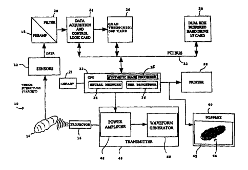
FIG. 1 is a block diagram of the tissue imaging system;
FIG. 2 is a graph of a backscattered response; and
FIG. 3 is a flowchart of the method of operation of the tissue imaging
system.
DESCRIPTION OF THE PREFERRED EMBODIMENTS
Referring now in specific detail to the drawings, with like reference
numerals identifying similar or identical elements, as shown in FIG. 1, the
present
disclosure describes a tissue imaging system and method for determining the
size and
shape of biological structures for classification and visualization of normal
and
abnormal tissues, organs, biological structures, etc.
For clarity of explanation, the illustrative embodiments of the disclosed
tissue imaging system and method are presented as having individual functional
blocks, which may include functional blocks labelled as "processor" and
"processing
unit" . The functions represented by these blocks may be provided through the
use of
either shared or dedicated hardware, including, but not limited to, hardware
capable
of executing software. For example, the functions of the processor and
processing
unit presented herein may be provided by a shared processor or by a plurality
of
individual processors. Moreover, the use of the functional blocks with
accompanying
labels herein is not to be construed to refer exclusively to hardware capable
of
executing software. Illustrative embodiments may include digital signal
processor
(DSP) hardware, such as the AT&T DSP16 or DSP32C, read-only memory (ROM)
-6-
CA 02338735 2001-O1-22
WO 00/04831 PCT/US99/16472
for storing software performing the operations discussed below, and random
access
memory (RAM) for storing DSP results. Very large scale integration (VLSI)
hardware embodiments, as well as custom VLSI circuitry in combination with a
general purpose DSP circuit, may also be provided. Any and all of these
embodiments may be deemed to fall within the meaning of the labels for the
functional blocks as used herein.
In the illustrative embodiment of FIG. 1, the system 10 processes input
data signals provided by a plurality of sensors 12 which respond to biological
tissue
14 under test in response to low frequency ultrasonic signal insonification;
for
example, frequencies in the range of about 10 kHz to about 1.0 MHz, with an
ultrasonic transmitted waveform produced by a transmitter 46 and an ultrasonic
generation device such as a projector 16 both known in the art. The projector
16 is
capable of applying a broad range of carefully controlled ultrasound signals
from the
transmitter 46 to the tissue 14 under test to generate corresponding ramp
response
signatures. The ramp signatures are detected by the sensors 12 which generate
corresponding receive input data signals, which are then analyzed to determine
the
characteristics of the tissue 14, such as size, shape, composition, volume,
and normal
or abnormal condition.
The receive input data signals are processed by pre-amplifiers 18 and
then filtered by filters 20. The filtered data signals are then processed by a
processing unit which includes a central processing unit (CPU) 22 operating in
conjunction with a data acquisition and control logic card 24 and a DSP card
26. The
CPU 22 and other components of the system 10 may be controlled by an
application
program written, for example, in the C + + programming language to implement
the
features and methods described herein.
CA 02338735 2001-O1-22
WO 00/04831 PCT/US99/16472
The memory may be a hard drive 28 and/or RAM, and the optional
RAM on the DSP card 26 may be used for faster memory access and processing.
The hard drive 28 and cards 24, 26 communicate with the CPU 22 using a bus 32,
such as a PCI bus operating the PCI protocol. The CPU 22 may include a neural
network 34 and/or a nearest neighbor rule (NNR) processor 36 for
classification, as
described in greater detail below.
After processing the input data signals, the system 10 generates a
processed signal for output by an output device such as a printer 38, or
optionally a
display 40, an audio system, or other types of output devices known in the
art. The
output device may output alpha-numeric text messages indicating the condition
of the
tested tissue 14, and/or may output a classification message indicating the
degree to
which the tissue 14 under test is within or outside predetermine normal tissue
conditions, for example, a percentage compared to 100 % normal may be
generated
and output. The output device may also generate video or graphic
representations of
the tissue 14 based on the processing of the tissue ramp signatures. For
example, the
display 40 includes a screen 42 for displaying a graphic representation 44 to
a
clinician.
The CPU 22 sends control signals to a transmitter 46, including a
programmable waveform generator 48 for generating signal waveforms, and
including
a power amplifier 50 for amplifying such signal waveforms, which are sent to
the
projector 16 for generating the ultrasound applied to the tissue structure 14.
In an illustrative embodiment, the projector 16 is a piezoceramic
projector, comprised of one or more transducer piezoceramic elements,
calibrated for
insonifying the tissue structure 14, such as the internal organs of a patient,
in the
frequency range of about 10 kHz to about 100 kHz. The projector 16 may be
either
the F30 or F41 transducer, available from the Underwater Sound Reference
Division
_g_
CA 02338735 2001-O1-22
WO 00/04831 PCT/US99/16472
(USRD) of the Naval Research Laboratory (NRL). Echo returns from the tissue
structures are received by the sensors 12, which may be four calibrated
wideband
sensors, such as the B&K Model 8103, oriented to provide four distinct target
aspects
in orthogonal planes.
In an alternative embodiment, the sensors I2 and the projector 16 may
be incorporated as a single device, including three custom piezoceramic
transducers
designed and fabricated to both insonify a tissue structure 14, such as a
breast tumor,
and receive the backscattered returns in the frequency range of about 100 kHz
to
about 800 kHz. A class of highly crystalline and oriented thermoplastic
polymers,
such as polyethylene teraphthalate, may also be used for producing a broadband
frequency response from about 10 Khz to about 1 Mhz. The outputs of the
sensors
12 and transducers are sent over coaxial cables to individual pre-amplifiers
18 and
anti-aliasing filters 20, and then to a data acquisition card 24 operatively
connected to
the CPU 22, which may be embodied as a personal computer or a workstation.
The pre-amplifiers 18 may be separate and independent low noise,
wideband programmable gain amplifiers, such as the AD 601 which is commonly
used in medical ultrasound, preceded by a low noise JFET, to provide an input
dynamic range of about 80 Db while minimizing noise and distortion. The pre-
amplifiers 18 may be configured on a computer card or board which plugs into
the
system 10, and which allows the user to change gain settings through software
controls without degrading the frequency response as the gain is increased.
The filter
20 may be anti-aliasing filters, such as TTE Inc. delay equalized elliptic
filters,
subsequent to the pre-amplifiers 18 to provide a relatively high rolloff rate
of about
84 dB/octave to provide aliasing protection. In the illustrative embodiment,
the
receiver pre-amplifiers 18 have an input dynamic range of about 72 Db, while
minimizing noise and distortion.
-9-
CA 02338735 2001-O1-22
WO 00/04831 PCT/US99/16472
The output of the anti-aliasing filters 20 is sent to the data acquisition
and control logic card 24 or board, which may include, for example, four
differential
input S/H amplifiers and 12-bit, 10 MHz analog-to-digital converters (ADC),
such as
the Burr-Brown ADS802 or Analog Devices AD9042, operating as a data
acquisition
subsystem with a throughput data rate of about 80 Mbytes per second. Each
channel
of the ADCs may have its own programmable gain amplifier with sufficient gain
to
provide the full voltage range of the ADC and a common mode rejection ratio of
about 100 dB.
The data acquisition subsystem, including the CPU 22, the cards 24-28,
and operating and control software, may be incorporated in a "FALCON" computer
system, available from Sonoran Microsystems, Inc., or incorporated in an "HT-
600"
computer system, available from Hi-Techniques, Inc.
The CPU 22 may be an "INTEL"-based "PENTIUM" microprocessor,
and the DSP card 26 may be a quad TMS220C6201. The hard drive 28 may include
one or more Seagate 18.2 GB fast SCSI hard drives for total storage.
The data acquisition and control logic card 24 formats the data to be in
standard personal computer file formats, such as ASCII data formats, to allow
the
data to be replayed in the laboratory using modified system software and/or
using
commercial third-party analysis software, such as application programs
including S-
PLUS or MAPLE. Real-time performance is achieved through the use of multiple
COTS DSP boards for the DSP card 26. The DSP card 26 is used to acquire the
data, to pack and pass the data to the CPU 22 for storage on the hard drive
28, and to
simultaneously band-pass the data, low-pass filter and decimate the band-
passed data,
and to perform various processing operations such as data normalization, fast
Fourier
transform (FFT) analysis and parameter estimation.
- 10-
CA 02338735 2001-O1-22
WO 00/04831 PCT/US99/16472
The system 10 determines a three-dimensional image of biological
organs requiring a minimal number of "looks"; for example, at most three
slices.
The system 10 also generates a diagnostically useful estimate of organ volume
and
tumor size, and provides an assessment of biological tissue composition by
classification of the tissue 14 using the neural network 34 and/or the NNR
processor
36. In performing the classification, predetermined 3-D STIC images of normal
tissue structures such as biological organs without tumors, as well as
estimates of
organ volume, are used as the basis of the classification; for example, to
train the
neural network 34 and/or to be processed by the NNR processor 36 to compare
the
current tissue data with the stored tissue data in the library 21.
The DSP card 26 may use 3-D STIC ultrasound data acquisition, signal
processing, and image reconstruction techniques derived from in vivo and in
vitro
analysis of predetermined and identified tissue to generate the tissue
database stored in
the hard drive 28.
Synthetic structural imaging (SSI), utilizing the low frequency ramp
response signature, offers a unique, effective technique for presenting three-
dimensional medical data to the clinician. By applying SSI techniques with
advanced
signal and image processing methods, the system 10 obtains clinically
meaningful
measurements of the size and shape of biological organs, as well as their
composition
and condition.
The system 10 applies low frequency signal insonification (i.e. a ramp
signature) matched to the spatial frequencies of the anatomical structure such
as tissue
14, in which the lower frequencies provide unique information as to overall
dimension
and approximate shape of the tissue 14. The system 10 then reconstructs 3-D
images
of tissue phantoms with no more than 3 distinct "looks" or insonifying planes
and
with a data acquisition time approaching real-time operation.
-11-
CA 02338735 2001-O1-22
WO 00/04831 PCT/US99/164'72
Estimation of the volume of target tissue, such as organs and tumors, is
performed by determining the volume of the target tissue as a unique spatially-
invariant classification parameter, derived from processed low frequency echo
returns.
In use, the system 10 measures the critical dimensions of various
organs, particularly the breast, prostate, uterus, and testes, for the purpose
of
detecting pathologies in advance of performing a biopsy. The system 10 may
also be
applied to provide unique anatomical information of the eye, of fetal head
growth and
heart ventricles, and of tumors and other lesions.
In another embodiment, the system 10 combines SSI techniques using
low frequency imaging with known high frequency imaging (for example, using
conventional ultrasound frequencies in the 2 to 12 MHz region), so that far
fewer
imaging planes or "slices" are required to produce meaningful 3-D diagnostic
images.
In the illustrative embodiment, a maximum of three slices are used as compared
to at
least 64 slices with previous technology. The unique information provided by
SSI,
together with reduced data acquisition time, facilitates and significantly
improves
clinical interpretation for a broad range of tissue studies, particularly in
echo-
cardiography, in intra-operation procedures, in analyzing specific body organs
such as
the prostate and kidney, and in ophthalmology.
The ramp response signature is the basis for low frequency
characterization, which has the property that the derived physical optics
approximation of the target's ramp response R(t) is directly proportional to
the target
cross-sectional area A(r) along the direction of propagation of the incident
field, and
may be expressed as:
- 12-
CA 02338735 2001-O1-22
WO 00/04831 PCT/US99/16472
R(t) = 1 A(r)
'~ c 2 =ct/2
in which c is the velocity of propagation in the medium, and r is the radial
distance.
Thus, the ramp response provides a unique low frequency measure of target
shape,
orientation and material.
The classical acoustic target backscattered response versus kA is shown
in FIG. 2 in terms of the Rayleigh, Resonance, and Optical regions, with the
Ka
range depicted for SSI operation, in which k is the wavenumber; i.e.
frequency,
which is 2~/A, A is the wavelength and A is the target radius. From previous
experiments with radar and sonar tests, valid estimates of ramp responses may
be
obtained for the insonification frequencies lying in the upper Rayleigh region
and low
resonance region of the target's scattering characteristics, i.e. in the
region 52 shown
in FIG. 2 from about .8 Ka to about 30 Ka. A variety of information may be
derived
from time and frequency domain analysis of region 52. Using time domain
analysis,
synthetic image generation as well as the determination of target parameters,
such as
area, volume, length, diameter, and aspect of the target; i.e. orientation in
3-D space,
may be performed. Using frequency domain analysis, feature vectors may be
generated such as Doppler characteristics, the aspect of the target, the
spectrum
shape, and the probability of misclassification. Natural resonances of the
target may
also be determined from frequency domain analysis, which facilitate the
determination
of the type of target, such as liver tissue as opposed to bone tissue.
Low frequency imaging is characterized by a narrow bandwidth and
low absorption loss while high frequency imaging is characterized by a wide
bandwidth and high absorption loss. Accordingly, high frequency imaging tends
to be
applied to shorter tissue depths for characterization. The high frequencies
-13-
CA 02338735 2001-O1-22
WO 00/04831 PCTNS99/16472
characterize the fine detail of the target while the lower frequencies provide
information as to overall dimension and approximate shape. Higher frequencies
may
be used to sharpen the image, but images may be difficult to attain without
low
frequency information.
In electromagnetic applications, the physical optics approximation
provides estimates of the waveform-target size and shape for an illuminated
portion of
the target, which is significant if the target is a perfect conductor, is
smooth, and has
dimensions as large as a few wavelengths. Test results show that the ramp
response
may be approximated by examining a target's electromagnetic response over the
frequency range corresponding to wavelengths starting with half the size of
the target
and increasing to about ten times its dimension.
Ramp responses have also been found to be applicable to ultrasound
imaging. The imaging technique used by the system 10 employs low frequency
ultrasound signals for target size and shape, and such imaging is enhanced by
providing additional information on structural discontinuities utilizing high
frequency,
short-pulse data.
As shown in FIG. 2, ultrasound having a low frequency ramp response
may be applied to the tissue 14. Such ramp responses may be represented by
receive
echo signals which vary over time, and which may be discontinuous. As
described
herein, low frequencies may be used for detection and classification of the
tissue 14.
Although there may be a many-to-one correspondence between a ramp response
feature and the possible structural discontinuities that may produce it, this
ambiguity
may be resolved by employing short, high frequency pulses using the impulse
response. Accordingly, the ramp response from a low frequency pulse may be
distinguished by using high frequencies short pulses. The target impulse
response is
sensitive to the curvature in the cross-sectional area and thus sensitive to
boundary
- 14-
CA 02338735 2001-O1-22
WO 00/04831 PCTNS99/16472
discontinuities and scattering centers. Therefore, by including high
frequencies to
define target scattering centers, the number of low frequencies required to
image the
target may be significantly reduced. This suggests that the optimum target
response is
a weighted sum of the ramp, step, and impulse responses.
In another embodiment, the low frequencies which generate the ramp
response are used as a feature vector for pattern classification by the neural
network
34 and/or the nearest neighbor rule (NNR) processor 36. As inputs to the
neural
network 34, the ramp responses may be processed as an input feature vector to
generate a neural network output which classifies the ramp response relative
to a
training set of tissue data. The nearest neighbor rule, as implemented by the
NNR
processor 36, is generally a robust decision rule ideally suited for
discriminating low
frequency data. Radar test results have shown that over 90 % reliability of
classification may be achieved with about four frequencies of the ramp
response,
utilizing amplitude information and vertical polarization data; fewer
frequencies are
needed by employing phase information, since phase is a sensitive measure of
changes
in target shape. In acoustic applications, since the particle velocity is
"rotational",
only amplitude and phase modulation data is required.
Heretofore, low frequency insonification has not been widely used for
biological analysis and diagnosis. One low frequency diagnosis technique using
frequencies in the range of about 10 to about 1000 Hz is capable of imaging
abnormal
regional elasticity in tissue, referred to as sonoelasticity, by mechanically
vibrating
tissue at low frequency which modulates an ultrasound carrier frequency. The
resulting Doppler displacements are a function of tissue stiffness; i.e.
Young's
modulus, and may then be displayed with a Doppler flow mapping imaging system,
including color flow mapping, known in the art.
-15-
CA 02338735 2001-O1-22
WO 00/04831 PCT/US99/16472
Preliminary indications are that "stiff' or "hard" tissue vibrates less
than soft tissue, depending upon the degree of hardness, and that vibrational
frequencies between about 100 to about 300 Hz are useful for discrimination.
The low frequencies required for applying SSI techniques are generally
higher than that used for sonoelasticity or elastography. SSI provides
estimates of
classification parameters which have not heretofore been derivable otherwise,
which,
in turn, are used by the system 10 to derive feature patterns describing
benign and
malignant disease.
The acoustic echo signature is generally rich with information from
IO distributed tissue structures, and by using SSI techniques for acoustic and
elastic
scattering in real biological media, such tissue structures may be detected
and
classified with substantial accuracy.
The system processing takes into consideration the frequency
dependence of tissue attenuation, the response of tissue shear wave
generation, and
the impact of inter-connective tissue and adjacent structures, veins and
arteries, as
well as the effect of wide-beam insonification.
Ultrasound attenuation increases with increasing frequency and the
depth of tissue penetration. For significant variance in response due to
attenuation
relative to the total dynamic range of the ramp response, the variance affects
the
relationship between the ramp response and organ geometry. The frequency range
required for applying SSI to meet organs of interest is about IO to about 100
Khz.
For one-way longitudinal absorption of about 1.0 Db/cm-Mhz, the two-way
absorption incurred at a depth of about 10 cm. is about 0.2 to about 2.0 Db
over the
frequency band.
Such a variance may be compensated in the transmitted ultrasound
signal by having a dynamic range of about 48 dB. Based on the dimensions of
actual
- 16-
CA 02338735 2001-O1-22
WO 00/04831 PCT/US99/16472
human tissue organs considered, the SSI frequencies employed may also generate
vibrational shear modes. In practice, some cross-coupling of modes between
shear
and compression may occur, so the system 10 evaluates such shear waves.
Typically,
the shear wave attenuation coefficient has been measured to be about 104 times
the
longitudinal wave attenuation coefficient.
Theoretically, the biological ultrasound ramp response is affected by
tissue/organ attachments such as blood vessels, cartilage and neighboring
anatomical
structures. However, at the spatial frequencies required for the synthetic
structural
imaging of much larger organs, the associated vessels are generally
acoustically
transparent.
Since normal SSI operation employs a wide beamwidth, several organs
may be insonified at one time. In order to resolve the echo ramp signature
into
components related to individual organs, the system 10 uses a priori
information
concerning the general spatial properties of the organs, a selection of the
aspect of the
target, and time gating. High frequency 2-D image data may be used to resolve
any
ambiguities in identification and classification. The system 10 is then
capable of
obtaining the size and shape of biological organs and tumors, and determining
their
composition.
Prior to use, the system 10 is configured to use in vivo and in vitro
measurements of the low frequency ramp response of tissue, including organs
and
tissue-mimicking breast tumors, to derive empirical 3-D images and measures of
volume and material composition, which are stored in a library database 21 in
the
hard drive 28. Such data from tissue organs may be naturally corrupted by
tissue
speckle, system noise, and artifacts, which may be introduced by tissue/organ
attachments and adjacent structures. The system 10 may be integrated into
known
- 17-
CA 02338735 2001-O1-22
WO 00/04831 PCT/US99/16472
imaging systems and used to conduct in vivo tests of human subjects to enhance
and
refine the library database 21.
The system 10 uses the transmitted signals from the projector 16: for
empirically obtaining the ramp response of specific biological organs and
tumors; for
generating low frequency synthetic images of biological organs and tumors; for
estimating the volume of the organs and tumors; for deriving measures of
tissue
composition from the measured ramp response, such as density and elasticity;
for
assessing other SSI biological structural characteristics, such as.attenuation
and target
aspect; and for assessing the effects of shear waves and wide-beam
insonification.
From the empirical data collection, unique signal waveforms or
ultrasonic signatures are stored in the hard drive 28 corresponding to
specific tissue
organs and tumors of interest. The signal waveforms are designed such that,
when
transmitted, the signal waveform generates the tissue phantoms with a distinct
ramp
response signature for specific tissue organs, such as prostate, kidney, eye,
and breast
tumors. The signal waveform, including both amplitude and phase modulation
over
the required frequency band, may be adjusted to match the spatial frequencies
characterizing these structures.
For example, the system 10 may use signal waveforms with frequencies
from about 10 to about 100 kHz for organ visualization, and frequencies from
about
100 kHz to about 800 kHz for visualizing breast tumors from about 2 mm to
about 5
mm in size. For such organ detection and identification, the transmitted
signal
fundamental-to-harmonic ratio is about 48 dB, and the signals used may be
transmitted according to a predetermined complex function to provide
sufficient
dynamic range and minimal false echoes.
Prior to collecting data, all system components are calibrated to
establish, for example, the ultrasonic spatial and frequency responses,
including
-18-
CA 02338735 2001-O1-22
WO 00/04831 PCT/US99/16472
projector and hydrophonic spatial characteristics and responses; source level
and
spectral purity; receiver bandwidths; the gain of the pre-amplifier 18 and
input noise
level; any integral and differential non-linearities; any harmonic and IM
distortion;
any spurious-free dynamic range of the ADCs; any back-scatter data; and any
background transients naturally-occurring within the measured frequency band.
The
amplitude and phase differential between channels may then be measured and
compensated. In the system 10, the power levels employed for transmission may
be
compatible with SPTA requirements specified in AIUM/NEMA Standard 9-17-1981.
The DSP card 26 performs echo data processing in which an FFT of
the target's complex spectral response is appropriately weighted to construct
an FFT
approximation of the target's ramp response signature. The ramp response
derived
from the echo amplitude and phase data is further processed in order to
approximate
the target cross-sectional area function or "profile function" . The
empirically-derived
profile functions are modified based upon known geometrical constraints and
are used
as the input data for the image reconstruction techniques employed by the CPU
22.
Image reconstruction may use, for example, limiting-surfaces to
generate an isometric image of the target, in which the target generally
includes a few
simple shapes, such as shapes described by a circular or elliptical cross-
section.
Image reconstruction of such shapes generally requires few parameters for
contour
estimation. A generalized surface, such as an ellipse, is fitted to the set of
profile
functions, in which at least one such generalized surface is calculated for
each look
angle. An image is then generated by calculating an image surface which
encloses a
volume common to substantially all of the single-aspect angle limiting
surfaces.
Orthogonality between the look angles may be used to greatly simplify such
image
processing.
- 19-
CA 02338735 2001-O1-22
WO 00/04831 PCT/US99/16472
The three-dimensional reconstructed images are compared with gross
examination of actual tissue images in the library. The actual volume of each
tissue
phantom employed is compared with the volume measured by integrating the
empirically-derived profile functions at the various aspects. The volume error
is used
as a measure of image accuracy. In addition, the ramp response is examined to
derive information concerning the composition of the tissue.
As shown in FIG. 3, the system 10 operates using a method including
the steps of: starting the imaging of the tissue 14 in step 54 by, for
example,
initializing and calibrating the system 10; retrieving tissue signatures from
the
database in the hard drive 28 in step 56; generating low frequency ultrasound
in step
58 using the tissue signatures to be applied to the tissue 14; detecting
tissue structures
in step 60 using the sensors 12, with the tissue ramp signatures being
generated by the
tissue 14 in response to the low frequency ultrasound; and processing the
tissue ramp
signatures to determine tissue characteristics in step 62.
The step 62 of processing the tissue ramp signatures may include, for
example, any combination of steps 64-68. In an illustrative embodiment, the
system
10 classifies the tissue 14 in step 64 using a classifier such as the neural
network 34
and/or the NNR processor 36 which uses predetermined tissue data for
classifying the
tissue 14. A discussion of neural networks and NNR processors is found in U.S.
Patent Application Serial No. 09/167,868, the contents of which are
incorporated
herein by reference.
In addition, the system 10 determines the volume, aspect, shape, etc. of
the tissue 14 in step 66 using SSI techniques, signal processing techniques
such as
FFT processing, etc. , and the system 10 also determines a condition of the
tissue 14
in step 68 as being normal or abnormal.
-20-
CA 02338735 2001-O1-22
WO 00/04831 PCT/US99/16472
The system 10 may then output the tissue characteristics to a clinician.
The system 10 may also use such tissue characteristics to generate a graphic
representation 44 of the tissue 14 in step 70.
Using the system 10 and methods described herein, a clinician may
then non-invasively, acoustically measure and generate an image of tissue
structures
in a patient to provide unique information concerning the size and shape of
biological
structures for classification and visualization of normal and abnormal
tissues, organs,
biological structures, etc. with improved accuracy and diagnostic analysis.
While the disclosed tissue imaging system and method have been
particularly shown and described with reference to the preferred embodiments,
it is
understood by those skilled in the art that various modifications in form and
detail
may be made therein without departing from the scope and spirit of the
invention.
Accordingly, modifications such as those suggested above, but not limited
thereto, are
to be considered within the scope of the invention.
-21 -