Note: Descriptions are shown in the official language in which they were submitted.
CA 02339252 2001-02-01
WO 00/07514 PCT/US99/17176
TREATMENT OF PIGMENTED TISSUES
USING OPTICAL ENERGY
CA 02339252 2006-10-11
WO 00/07514 PCT/US99/17176
2
BACKGROUND OF THE INVENTION
The present invention is directed to a method and apparatus for treating
pigmented
tissues by selective photoactivation of pigments in such tissues using optical
energy and
more specifically two-photon excitation. This selective photoactivation may be
used to
effect photobleaching of such pigments or to effect photochemical conversion
of such
pigments into phototoxic products. Photobleaching reduces or eliminates
undesirable
pigmentation, for example that caused by pigments present in moles, freckles,
hair follicles
and tattoos. Photochemical conversion produces phototoxic products that
destroy
pigmented tissues, such as those pigmented tissues in pigmented tumors. The
present
invention is also directed to selective thermal destruction of pigmented
tissues using
related optical means.
Photobleaching is the transient or permanent reduction of pigmentation in
pigmented tissues upon optical illumination, typically occurring during
intense illumination
with visible or ultraviolet light. Photobleaching occurs when photoactive
pigments are
photochemically transformed from a highly colored state to a less highly
colored state (de-
pigmentation). For example, photobleaching may be used to reduce or eliminate
undesirable pigmentation present in moles and hair follicles or to destroy
dyes present in
tattoos. It is desired that treated tissues will exhibit localized de-
pigmentation without
side effects, such as irritation or cell necrosis. However, previous methods
for
photobleaching tissues using visible or ultraviolet light have produced
undesirable
collateral effects, including irritation of surrounding tissues and possible
scarring at the
treatment site.
In contrast to photobleaching, photochemical conversion of pigments into
phototoxic products involves stimulation of localized cell necrosis in treated
tissues. This
is also effected by optical illumination, typically occurring when intense
visible or
ultraviolet light is used to illuminated susceptible pigmented tissues. Such
localized
necrosis may be useful for selective destruction of diseased tissues, such as
those present
in tumors or benign skin lesions.
More specificaliy, an important subset of pigmented tissues are pigmented
tumors,
such as melanomas, which are life threatening and highly difficult to treat.
While
CA 02339252 2001-02-01
WO 00/07514 PCTIUS99/17176
3
melanomas can be treated if detected early using standard surgical, radiation
or
chemotherapeutic methods, these methods still do not have acceptable levels of
effectiveness and produce high levels of collateral damage to normal tissue.
Hence, even
if detected relatively early, the prognosis is usually poor.
Further, if a melanoma has metastasized beyond the primary tumor site, less
than
20% of patients will survive beyond five years. For such melanomas, there are
no
effective therapies. Patients diagnosed with such a metastatic melanoma will
survive on
average only 3-6 months after the diagnosis even with therapeutic
intervention.
Further exacerbating the difficulties in treating melanomas is the fact that
the
incidence of melanoma in Caucasians is increasing at a rate of 6% per year.
This is
currently the second fastest rate of increase in cancer occurrences -- second
only to
tobacco related cancers of the lung in women. Currently, the lifetime risk of
melanoma
in the U.S. is 1 in 75. Accordingly, new effective therapeutic modalities are
required to
treat both primary and metastatic pigmented tumors such as melanomas.
One possible approach for treating pigmented tissues involves the use of
melanins,
their precursors, and other endogenous or exogenous pigments.
More specifically, there are several pigments in humans that are collectively
known
as melanins. The function of melanins are to protect tissues from the
deleterious effects
of electromagnetic radiation (e.g. light). However, melanins and their
precursors can also
be converted to phototoxic products. For example, a melanin precursor (5-SCD)
has been
shown to photobind to DNA after exposure to 300 nm (uitraviolet light)
illumination.
Further, 5-SCD has been shown to be chemically unstable in the presence of
ultraviolet
(UV) illumination and oxygen, thereby suggesting that phototoxic products of
the (1)
Type I variety (phototoxic) or the (2) Type 11 variety (photocatalytic) may be
produced.
Additionally, many melanoma cells are amelanotic. These cells produce melanin
precursors but only small quantities of melanin. Phototoxic damage (induction
of single
strand breaks) to DNA by at least two precursors to melanin (5-SCD and DIHCA)
has
been demonstrated upon exposure to UV light. Amelanotic cells will be killed
by
photodynamic therapy (PDT) performed on such precursors to melanin (e.g., 5-
SCD,
DIHEA). Thus, melanomas can be killed by delivering energy via light.
However, utilization of such phototoxic reactions by illumination of melanin,
melanin precursors, or other endogenous pigments has not previously been
possible. The
UV/Near UV light required for photoactivation is unable to penetrate into
normal or
CA 02339252 2006-10-11
WO 00/07514 PCT/US99/17176
4
cancerous skin (i.e. beyond 2-3 mm.) More specifically, the poor penetration
of such iight
has produced little effect on patients whose skin tumors are larger than or at
a depth
greater than 3 mm. As a result, only 40-50% of patients whose tumors exceed 3
mm will
survive. Accordingly, the survival rate of melanoma patients with tumors whose
depth
is less than 1 mm is drastically better than those who have tumors which are
either located
at a depth of greater than 3 mm or extend to a depth greater than 3 mm.
Previous photodynamic methods using UV/Near UV light also produced
undesirable collateral effects that not only prohibited the photoconversion of
melanin and
prevented it from killing pigmented tissues but also was potentially dangerous
to the
patient. For example, UV light can create thymidine dimers which damage
genetic
material. DNA damage is a major and possibly the sole cause of skin cancers
like
melanomas. Melanin's absorbance of UV light is designed to prevent this from
happening.
However, UV light, chemotherapy, and ionizing radiation have recently been
shown to
increase the virulence oftumor cells. As a result, tumor cells when treated
with UV light
will have a greater mutation and error rate because the UV light can
inactivate
mechanisms designed to identify and correct genetic errors (in addition to
creating new
errors). Therefore, prior techniques were not only unable to effectively kill
pigmented
tissues by accessing endogenous pigments but also created side effects that
could be lethal.
In many instances, the effectiveness of various photodynamic processes have
been
found to be markedly increased by simultaneous photoactivation and localized
heating
(hyperthermia). Typically, by heating the treatment zone 2-10 C above normal
temperatures, the effectiveness of PDT is increased many fold. Such heating
alone,
however, has not been shown to produce a significant therapeutic effect. In
contrast, the
inventors of the present invention have conceived that more acute localized
heating (i.e.,
> 2-10 C temperature rise) of tissues and tissue components within the
treatment zone
may produce a therapeutic effect by causing thermal overload in the treated
tissues.
Therefore, the present invention seeks to provide a method for accessing
endogenous pigments in pigmented tissues so as to be able to selectively
photobleach said pigments.
Another aspect of the present invention seeks to provide a method for
accessing endogenous pigments in pigmented tissues so as to be able to
photochemically convert said pigments into phototoxic products.
CA 02339252 2006-10-11
WO 00/07514 PCT/US99/17176
Still further, the present invention seeks to provide a method that will
access said endogenous pigments in pigmented tissues without accessing
endogenous pigments in healthy tissues surrounding said pigmented tissues.
Another aspect of the present invention seeks to provide a method that will
5 augment the effectiveness of said photochemical conversion of said
endogenous
pigments in said pigmented tissues through the localized application of
hyperthermia in said pigmented tissues.
Further still, the present invention seeks to provide a method that will
photothermally destroy pigmented tissues without harming healthy tissues
surrounding said pigmented tissues.
SUMMARY OF THE INVENTION
The present invention is directed to a method and apparatus for treatment of a
particular volume of tissue or material containing an endogenous pigment. In
general,
typically, the present invention uses the unique properties of simultaneous
two-photon
excitation with endogenous pigment in a particular volume of tissue, such as a
tumor, to
selectively photoactivate the pigment.
This photoactivated pigment may thereby be photobleached or photochemically
converted into a phototoxic product. Such photoactivation results from the
simultaneous
two-photon excitation of the pigment. Preferably, the photons responsible for
photoactivation are provided by a laser which produces a beam of light
comprising a train
of one or more ultrashort pulses. This beam of light can be a focused beam of
light if the
location and extent of the particular volume of tissue to be treated is
precisely known.
The focused beam of light can then be scanned throughout the volume of the
tissue to
treat the entirety of the pigmented tissue. Alternatively, where the location
and extent of
the pigmented tissue in a volume of tissue is not precisely known, a non-
focused light
beam can be used.
In an alternative embodiment, an exogenous photodynamic agent can be added to
the particular volume of tissue. The exogenous agent can be photoactivated by
the
simultaneous two-photon excitation. Activation of the exogenous photodynamic
agent
augments the effectiveness of the endogenous pigment.
CA 02339252 2006-10-11
WO 00/07514 PCT/US99/17176
= 6
In a further alternate embodiment of the invention, the effectiveness of such
photoactivation is augmented through the localized application of hyperthermia
in the
pigmented tissues.
In an additional further alternative embodiment of the invention, the
particular
volume of tissue is treated with light to promote thermal overload of the
pigmented
tissues. Thermal overload heats and kills the pigmented tissues.
The invention in one broad aspect provides an apparatus for treating a
particular volume of tissue containing an endogenous pigment, wherein the
tissue
does not contain an exogenous pigment added to the tissue for the purpose of
increasing absorption of light energy applied to the tissue by the apparatus.
The
apparatus comprises: a single continuous wave or pulsed source of light and a
light
delivery apparatus for directing a beam of light at and into the particular
volume
of tissue, and directing the beam of light throughout the particular volume of
tissue, the light having a wavelength between approximately 800 nm and 1400 nm
and being selected to promote thermal overload of pigmented cells in the
particular
volume of tissue, wherein the thermal overload kills the pigmented cells.
CA 02339252 2001-02-01
WO 00/07514 PCT/US99/17176
7
BRIEF DESCRIPTION OF THE DRAWINGS
In describing the preferred embodiments, reference is made to the accompanying
drawings:
FIGURE 1 illustrates an example energy level diagram for simultaneous two-
photon excitation;
FIGURE 2 illustrates an example of absorption and scattering properties for
animal tissue covering the ultraviolet to infrared spectral region;
FIGURE 3 shows the general trends in optical absorption properties of animal
tissue for short wavelength and long wavelength light;
FIGURE 4 illustrates a comparison of optical activation in tissue when single-
photon and two-photon excitation methods are used;
FIGURE 5 illustrates an embodiment of the present invention for selective two-
photon photoactivation of inelanin, melanin-precursors or endogenous pigments
using
focused light;
FIGURE 6 illustrates an another embodiment for selective two-photon
photoactivation of inelanin, melanin-precursors, or endogenous pigments using
focused
light;
FIGURE 7 illustrates a further embodiment for selective two-photon
photoactivation of melanin, melanin-precursors, or endogenous pigments using
non-
focused light;
FIGURE 8 illustrate still another embodiment for selective two-photon
photoactivation of melanin, melanin-precursors, or endogenous pigments in a
subsurface
tissue using non-focused light;
FIGURE 9 illustrates an alternate embodiment for the present invention wherein
a focused light beam is used to thermally overload and kill pigmented tumor
cells; and
FIGURE 10 illustrates another alternate embodiment for the present invention
wherein a non-focused light beam is used to thermally overload and kill
pigmented tumor
cells.
DETAILED DESCRIPTION OF THE PRESENTLY PREFERRED
EMBODIMENT
The present invention is directed to a method and apparatus for treating
pigmented
tissues using light. Such treatment includes the following photochemical
outcomes of
CA 02339252 2001-02-01
WO 00/07514 PCT/US99/17176
8
therapeutic value: (1) the elimination of undesirable pigmentation in
pigmented tissues
through photobleaching, and (2) the permanent destruction of pigmented tissues
through
photochemical conversion of pigments into phototoxic products. More
specifically,
simultaneous two-photon excitation is used to photochemically convert
endogenous or
exogenous pigments into desired photoactive products, resulting in the desired
photobleaching or tissue destruction. Photobleaching is used to reduce or
efiminate
undesirable coloration of tissue, such as that in moles, freckles, hair
follicles and tattoos.
The production of phototoxic products may be used to preferentially kill
pigmented tumor
cells or other undesirable tissues while sparing normal cells. Significantly,
the methods
and apparatus in the present invention used for photobleaching and production
of
phototoxic products utilize equivalent photoactivation mechanisms, differing
substantially
only in the intended treatment target.
In the preferred embodiment, the present invention uses simultaneous two-
photon
excitation to photoactivate pigments in the pigmented tissues, yielding
photobleached or
phototoxic products.
In an alternate preferred embodiment, the present invention uses related
optical
means to selectively destroy pigmented tissues via photothermal means.
Simultaneous Two Photon Excitation
"Simultaneous two-photon excitation" is the non-linear optical excitation
occurring
as a result of the essentially simultaneous interaction of two photons
originating from a
single ultrashort laser pulse with one or more agents or pigments to produce
one or more
photoactivated agents or pigments. "Non-linear optical excitation" means those
excitation
processes involving the essentially simultaneous interaction of two photons
with one or
more agents or pigments. "Essentially simultaneous interaction" means those
excitation
processes occurring as a result of the interaction of one or more agents or
pigments with
photons provided by a single ultrashort laser pulse. Ultrashort means less
than
approximately 10 ns.
As shown in Figure 1, simultaneous two-photon excitation to an allowed energy
level 10 occurs when a photoactive agent is excited from a first allowed
electronic energy
level 16 upon absorption of a certain energy E, that is provided by the
simultaneous,
combined interaction of two photons 12 and 14 with the agent. If the energies
of both
photons 12 and 14 are identical, the excitation process is termed
"degenerate". The
CA 02339252 2006-10-11
WO 00/07514 PCT/US99/17176
9
simultaneous interaction of the two photons is frequently described as being
mediated by
a transient virtual state 20 with a lifetime on the order of 10 femtoseconds
(fs) or less. If
both photons do not interact with the agent during this lifetime, excitation
does not occur
and the agent fails to reach the excited state S. (18). Typically, intersystem
crossing, IX,
subsequently occurs to bring the excited agent to a long-lived activated state
T. from
which a photochemical reaction R can occur.
Simultaneous two-photon excitation may thereby be used to excite processes
that
normally occur upon absorption ofa single UV orvisible photon through the
simultaneous
absorption of two near-infrared photons.
An example of the simultaneous two-photon excitation process is the promotion
ofinelanin precursors from a ground electronic state to an excited electronic
state through
the simultaneous absorption of two photons at 600 nm, followed by binding of
the excited
melanin precursor to DNA (this is conventionally excited using a single photon
at 300
nm).
In this example, the probability of excitation is related to the product of
the instantaneous
or peak powers of the first of two photons 12 and the second of two photons
14. This
can be conceptualized in the form of a photochemical reaction,
MoleculeciROVrm STATE + 2 hv woõm - MoleculeExcrren sTni=r: (1 ~
which shows that a molecule in the ground state is promoted to an excited
state following
simultaneous absorption of two photons at 600 nm, hv 6.,,,,,. The reaction
rate R, is given
by R = k[MolecuieoRourD STATE] [hv wo .]2, where k is a rate constant and
where
[MoleculecROUNOST,,TC] and [hv,,.] symbolize concentrations of ground state
molecules
and excitation photons, respectively. Hence, due to the well known quadratic
dependence
on instantaneous photon irradiance, simultaneous two-photon excitation to an
allowed
energy level 10 is also referred to as a non-linear excitation process.
A more detailed explanation of simultaneous two-photon excitation and
other on-linear and linear process is described in Canadian Patent File No.
2,252,783 filed October 28, 1997 for "Method For Improved Selectivity In
Photoactivation Of Molecular Agents" assigned to the same assignee of the
present
application and which may be referred to for further details.
CA 02339252 2001-02-01
WO 00/07514 PCTIUS99/17176
Significance of absorbance and scattering properties in single-photon and
simultaneous
two-photon processes:
While the cross-section for simultaneous two-photon excitation may be
5 considerably lower than that observed with single-photon excitation, use of
the
simultaneous two-photon excitation in the present invention may be favorable
over single-
photon excitation under many conditions because of lower matrix absorption and
optical
scattering of longer wavelength optical radiation. For example, FIGURE 2 shows
the
absorption and scattering properties for various components of animal tissue,
such as
10 human dermis, covering the ultraviolet (UV) to near infrared (NIR) spectral
region.
Specifically, FIGURE 2 demonstrates how higher-energy photons 32 may
experience considerably greater tissue absorption than lower-energy photons
34. For
example, human skin strongly absorbs higher-energy photons 32 at 400 nm, but
is
relatively transparent to lower-energy photons 34 at 800 nm. This is a
consequence of the
natural absorbance of higher-energy photons 32 by blood, pigments, proteins,
and genetic
materials, among other natural components, of skin.
FIGURE 2 further demonstrates how higher-energy photons 42 may experience
considerably greater tissue scatter than lower-energy photons 44. Any
optically dense
medium, such as human skin, will strongly scatter higher-energy photons 42,
for example
at 400 nm, but will exhibit much lower scatter for lower-energy photons 44 at
800 nm.
These differences in optical properties have two important consequences.
First,
absorption of short-wavelength, higher-energy photons 32 by tissue can result
in
undesirable tissue damage upon exposure to UV or other high-energy light. In
contrast,
negligible effects may be experienced upon illumination with lower-energy
photons 34,
such as NIR light, even when the optical power of the NIR light is many-fold
higher than
CA 02339252 2001-02-01
WO 00/07514 PCT/US99/17176
11
that of the UV light. Secondly, the inherently high absorption and scatter of
higher-energy
photons 32 by tissue can result in very shallow tissue penetration depths,
while lower-
energy photons 34 generally have much greater penetration depths.
These important differences in absorption and penetration depth properties for
higher-energy and lower-energy light are shown schematically in FIGURE 3. When
UV
light 50, for example light at 400 nm, impinges on human tissue 52, the
majority of the
optical energy is immediately absorbed and scattered in the outermost layers
54, such as
the epidermis and dermis. Absorption may occur due to excitation of certain
molecules
in the celis of these outermost layers 54, such as those composing the genetic
material in
the cellular nucleus. This absorption of higher-energy light by cellular
constituents can
thereby initiate a variety of collateral photochemical changes 56 in these
cells. These
collateral photochemical changes 56 resulting from absorption of UV light 50
can include
irreversible genetic damage and induction of cancer.
In contrast, NIR light 58, for example at 800 nm, will not be appreciably
absorbed
or scattered by tissue 52 or its outermost layers 54. The overall depth of
penetration will
be much greater, and the extent of'collateral damage to cells will be
substantially lower.
Hence, if long-wavelength excitation light is used to replace the higher-
energy light used
for conventional single-photon excitation, it is possible to photoactivate
specific molecules
or pigments using relatively non-damaging, high penetration depth,
simultaneous two-
photon excitation.
Furthermore, the properties ofsimultaneous two-photon excitation have
additional
implications when coupled with the inherent non-damaging nature and low
absorption of
NIR light. For example, FIGURE 4 compares the extent of optically-induced
damage in
tissue when single-photon excitation 60 and simultaneous two-photon NIR
excitation 62
methods are used to illuminate a subcutaneous tumor 64.
CA 02339252 2001-02-01
WO 00/07514 PCT/US99/17176
12
Single-photon excitation 60 produces a photoactivation zone 66 that extends
substantially along the entire optical path and has no significant
biospecificity. Hence, in
addition to induction of the desired photoactivation in the tumor 64,
collateral damage can
occur throughout surrounding tissues, such as the dermis 68 and surrounding
healthy
tissue 70. If the single-photon excitation 60 is focussed, the photoactivation
zone 66 will
be slightly enhanced at the focus 72. This photoactivation zone 66, however,
might not
even extend into the tumor 64 if the UV or visible light is absorbed by the
epidermis,
dermis 68 or surrounding healthy tissue 70 prior to reaching the tumor 64.
This can occur
as a consequence of the inherently high absorptivity of tissue at short
wavelengths.
In contrast, use of NIR light for simultaneous two-photon excitation 62
produces
a sharply defined remote photoactivation zone 74 that is spatially localized
at the focus
76 as a consequence of the non-linear properties of this excitation method.
Such
localization of activation in such a focal zone is a unique property of non-
linear excitation
processes, such as two-photon excitation. Furthermore, because tissue does not
appreciably absorb NIR light, collateral damage to the surrounding dermis 68
and healthy
tissue 70 is minimized.
Therapeutic applications of simultaneous two-photon excitation:
The foregoing discussion suggests that the fundamental differences in the
absorption of UV and NIR light by tissue and cellular constituents, coupled
with the
special non-linear properties of simultaneous two-photon excitation, have
direct
applicability for improvements in various medical treatments, specifically in
the
modification or elimination of pigmented tissues.
CA 02339252 2001-02-01
WO 00/07514 PCT/US99/17176
13
Such simultaneous two-photon excitation enables improved localization in the
photoactivation of photoactive agents with significantly reduced potential for
collateral
tissue damage compared with that possible using conventional methods.
Where control of penetration is not critical, non-focussed NIR light may be
used
to stimulate simultaneous two-photon photoactivation of agents present in a
relatively
large illuminated area. In such a case, the extent of agent photoactivation is
controlled
by varying the location, intensity and duration of exposure of such agents to
the NIR
beam.
Where precise control of penetration depth or volume extent of therapeutic
application is more critical, focussed NIR light may be used to stimulate the
simultaneous
two-photon photoactivation process. In such a case, beam irradiance, exposure
duration,
and degree of focussing are used to control the extent of agent
photoactivation.
In both cases, high-irradiance NIR light may be used to achieve maximum
efficacy.
Furthermore, the high penetration depths achievable with NIR light combined
with the
inherent localization of photoactivation that is possible with focused
simultaneous two-
photon excitation provide a means for photoactivating agents in subsurface
tissues without
damaging overlying or underlying healthy tissues.
Simultaneous Two-Photon Treatment with Endogenous Pigments
The method of the present invention improves on the above-described advantages
through the use of simultaneous two-photon excitation to produce a therapeutic
outcome
based on photoactivation of endogenous pigments in order to treat pigmented
tissues.
"Endogenous" means pre-existing in a patient or target. "Pigments" means
naturally
occurring agents that absorb optical energy. Examples of such pigments include
melanin,
melanin precursors, carotenes, porphyrins (such as hemoglobin), various tattoo
dyes and
CA 02339252 2001-02-01
WO 00/07514 PCT/US99/17176
14
other optically active species. "Therapeutic outcome" means photobleaching or
photodynamic destruction of treated pigmented tissues resulting from the
natural
biological action of a photoactivated endogenous pigment. "Photobleaching" is
the
reduction or elimination of undesirable pigmentation, for example that caused
by
endogenous pigments present in moles, freckles, hair follicles and tattoos.
"Photodynamic
destruction" is localized tissue necrosis resulting from photochemical
production of
phototoxic products that destroy pigmented tissues, such as those pigmented
tissues in
pigmented tumors. Tissues suitable for treatment include pigmented tissues in
which a
specific therapeutic outcome is desired, such as moles, freckles, pigmented
tumors, benign
lesions, hair follicles and tattoos.
In a further embodiment of the present invention, a precursor to the
endogenous
pigments may be used. Examples of such precursors to pigments include 5-S-
cysteinyldopa (5-SCD) and 5,6-dihydroxyindole (DHl), dopa, dopa semiquinone,
leucodopachrome, dopachrome, eumalanins, pheomelanins, sepia melanins, and 5,6-
dihydroxyindole-2-carboxylic acid. Such precursors have both photoprotective
and
phototoxic abilities. A metabolic precursor to melanin is a biochemical (e.g.
5-SCD, DHI)
that is produced by the cell as part of the synthetic pathway that produces
melanin.
Melanin precursors, when activated by light, can generate photoxic products
that damage
cellular materials (e.g., DNA) killing the target cells. Melanin precursors
can be activated
by two-photon excitation, as explained supra.
As also explained supra, melanin, melanin precursors, and other endogenous
pigments are naturally occurring in human tissue, inclucling in tumors. Such
melanins,
melanin precursors, or other endogenous pigments can be converted to
phototoxic
products after exposure to light.
CA 02339252 2001-02-01
WO 00/07514 PCTIUS99/17176
The present invention uses the above-described simultaneous two-photon
excitation to specifically target melanin, melanin precursors, or other
endogenous
pigments in pigmented tissues (such as melanomas and other tumors). The
pigment is
converted to a phototoxic product by NIR light upon simultaneous two-photon
excitation.
5 The phototoxic product then causes damage to the pigmented tissues (by for
example
photobinding to cellular DNA or causing breaks in this DNA). This kills the
cells in the
pigmented tissues and, therefore, destroys it. Because simultaneous two-photon
excitation is used to specifically target the melaniii, melanin precursors, or
other
endogenous pigments only in the targeted tissue, any melanin, melanin
precursors, or
10 other endogenous pigments in the tissue surrounding the targeted tissue are
not converted
to phototoxic products.
More specifically, use of simultaneous two-photon excitation produces a
sharply
defined focal zone that is substantially localized in depth and cross-section.
This focal
zone can be localized to the targeted tissue (such as a tumor) to be killed or
a small zone
15 within or surrounding this tissue. As a result, photoactivation will only
occur in the focal
zone (i.e. in the tumor). Hence, any melanin, melanin precursors, or other
endogenous
pigment not in the targeted tissue, such as for example, in tissue surrounding
a tumor, will
not be photoactivated because it is outside the focal zone.
Additionally, as explained supra, the simultaneous two-photon excitation is
able
to penetrate deep into normal or cancerous tissue and photoactivate melanin or
other
endogenous pigments located deep within the tissue. As a result, tumors
located deep
within the body or large, deep tumors can be reached and destroyed.
Destruction of these
tumors can be done without activating melanin or other endogenous pigments
along the
path of the light or surrounding the tumor.
CA 02339252 2001-02-01
WO 00/07514 PCTIUS99/17176
16
In addition to photodynamic destruction of pigmented tissues, such as those in
pigmented tumors, the above-described unique features of simultaneous two-
photon
excitation may be used to achieve improved safety and specificity in the
photobleaching
of pigmented tissues, such as in moles, freckles, hair follicles and tattoos.
The pigments
present in such tissues can be activated by simultaneous two-photon
activation, as
explained supra, and upon activation may become photobleached. Thus, the
present
invention also uses simultaneous two-photon excitation to specifically target
endogenous
pigments in such pigmented tissues, thereby causing photobleaching and a
desired
reduction or elimination of apparent pigmentation.
It is a specific preferred embodiment of the present invention to employ the
output
of a NIR source, such as the mode-locked titanium: sapphire laser, to induce
simultaneous
two-photon photoactivation so as to photoactivate melariin, melanin
precursors, or other
endogenous piginents using light at a wavelength approximately twice that
necessary for
such conversion using conventional single-photon photoactivation. As explained
supra,
such NIR light exhibits improved penetration into tissue relative to that used
for
conventional single-photon photoactivation, and is less likely to produce
collateral damage
in tissues adjacent to the desired treatment target.
For the sake of simplicity and clarity, the following descriptions of
preferred
embodiments will focus on photodynamic destruction of' pigmented tumor
tissues, such
as those in melanomas. However, it is important to note that the methods and
apparatus
described are equally applicable to the photobleaching of pigmented tissues,
such as moles
or tattoos, differing substantially only in the intended treatment target. In
both classes of
treatment, it is the photoactivation ofthe pigment that is fundamentally
responsible for the
desired therapeutic outcome.
CA 02339252 2001-02-01
WO 00/07514 PCT/US99/17176
17
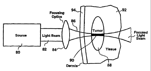
Accordingly, a preferred embodiment is shown in FIGURE 5. The source 80
produces a beam of light 82 consisting of a rapid series of high peak power
pulses of NIR
light. For example, standard commercially available mode-locked titanium-
sapphire lasers
are capable of outputting mode-locked pulses with durations <200 fs and pulse
energies
of about 1-20 nJ at pulse repetition frequencies in excess of 75 MHz. This
source
produces a quasi-continuous beam of light having a relatively low average
power (up to
several Watts) but high peak power (on the order of 100 kW) that is
continuously tunable
over a NIR wavelength band from approximately 690-1080 nm. The pulse train
from the
source 80 constitutes a beam of light 82 that is easily focussed using
standard optical
means, such as reflective or refractive optics 84. The focused beam 86 can
then be
directed into a tumor 88 or other localized treatment target.
Simultaneous two-photon photoactivation of the melanin, melanin precursors, or
other endogenous pigments will be substantially limited to the focal zone 90
of the focused
light beani 86 due to the high instantaneous irradiance level that is only
present at the
focus. Furthermore, regardless of whether melanin, melanin precursors, or
another
endogenous pigment is present in surrounding healthy tissue 92 or skin 94,
insignificant
collateral photoactivation, photodamage or conversion into a phototoxic
product will
occur outside the focal zone 90. This is a consequence of the non-linear
relationship
between instantaneous optical power and simultaneous two-photon excitation,
which
limits significant excitation to the focal zone 90. Even if melanin, melanin
precursors, or
another endogenous pigment is present outside of the focal zone 90, excitation
intensities
are below that necessary to produce significant photoactivation.
The apparatus of the present invention can also include, for example, a
focusing
apparatus for focusing the light throughout a range of focal lengths extending
from a
surface of the tissue to a depth substantially beyond the surface. The source
of light and
CA 02339252 2001-02-01
WO 00/07514 PCTIUS99/17176
18
focusing apparatus cooperate to promote simultaneous two-photon excitation of
the
pigment at controllable locations throughout the volume of tissue.
By scanning the location of the focus of the beam 86 throughout the volume of
the
tumor 88, complete photoactivation of the melanin, melanin precursors, or
other
endogenous pigments into a phototoxic product throughout the tumor 88 can be
effected.
This scanning action can be produced by changing the position of the focus 86
relative to
the tumor 88, or by moving the tumor 88 relative to a stationary focus 86
location. The
quality of the focal region 90 of the focused light beam 86 may be improved by
pre-
expanding the light beam 82, using a beam expander or other device, prior to
focusing
using standard optical means.
This scanning can be done, for example, by positioning a focus of a beam of
light
over a range of positions so that a focal plane of the light beam occurs at a
site located
between a surface of the tissue and a point substantially beyond the tissue
surface. As a
result, treating the particular volume of tissue may extend to penetrate deep
within the
tissue. This scanning can further include varying, while the beam of light is
extant, the
radial position of the focal plane within the tissue, thereby to photoactivate
the
endogenous pigment at a multiplicity of positions betweeri the tissue surface
and a position
located substantially beyond the tissue surface.
The simultaneous two-photon photoactivation embodiment of the present
invention has several variations for the treatment of topical tissues, as
shown in FIGURE
6 and in FIGURE 7. For example, the non-damaging nature of focused NIR light,
shown
in FIGURE 6, or of non-focused NIR light, shown in FIGURE 7, allows
photoactivation
of melanin or other endogenous pigments at topical locations without risk to
underlying
or surrounding tissues.
CA 02339252 2001-02-01
WO 00/07514 PCT/US99/17176
19
Focused simultaneous two-photon photoactivation of melanin or other
endogenous pigments for topical therapy, as shown in FIGURE 6, is effected
when a beam
of light 82 from a source 80 is focused 86 onto a tumor 88 or other localized
treatment
target using standard optical means, such as reflective or refractive optics
84. In this
manner, photoactivation of the melanin, melanin precursors, or other
endogenous
pigments into a phototoxic product occurs only at the focal zone 90. The
surrounding
healthy tissue 92 and skin 94 are unaffected in this process, even if they
also contain
melanin, melanin precursors, or another endogenous pigment, since
photoactivation is
substantially limited to the focal zone 90. As described previously, a
scanning action can
be used to effect photoactivation of the melanin, melanin precursor, or other
endogenous
pigment into a phototoxic product throughout the volume of the tumor 88.
Non-focused simultaneous two-photon photoactivation of melanin, melanin
precursors, or other endogenous pigments for topical therapy, as shown in
FIGURE 7,
is effected when a non-focused or expanded beam of light 96 from a source 80
is directed
onto a topical tumor 88 or other localized treatment target. This beam of
light 96 may
have a cross sectional area smaller than, equal to, or larger than that of the
tumor 88.
Since melanin, melanin precursors, or other endogenous pigments are present in
substantially higher levels in the tumor 88, the therapeutic action will be
substantially
limited to the volume of the tumor 88. Since the beam of light 96 is non-
damaging to
tissues that do not contain a significant concentration of pigment, damage to
surrounding
healthy tissue 92 and skin 94 is avoided. This embodiment may be particularly
useful
when the exact location, size and shape of the tumor 88 are not known, or when
it is
otherwise undesirable to carefully control the location of application of the
beam of light
96, since careful control of the location of the beam of light 96 is not
critical for successful
administration of this therapeutic regime. When non-focused light is used,
employment
CA 02339252 2001-02-01
WO 00/07514 PCT/US99/17176
of extremely high peak power excitation sources, such as Q-switched lasers or
regeneratively amplified mode-locked lasers, may be beneficial due to their
exceptionally
high peak radiant power (which is in the GW range) that will thereby afford a
high
instantaneous irradiance over a large area.
5 Afinal related variation ofthis preferred embodiment for simultaneous two-
photon
photoactivation is shown in FIGURE 8, where a non-focused or expanded beam of
light
96 from a source 80 is directed onto a tumor 88 or other localized treatment
target
located below the skin's surface. This beam of light 96 may have a cross
sectional area
smaller than, equal to, or larger than that of the tumor 88. Since melanin,
melanin
10 precursors, or other endogenous pigments are present in substantially
higher levels in a
tumor 88, the therapeutic action will be substantially limited to the volume
of the tumor
88. Since the beam of light 96 is non-damaging to tissues that do not contain
a significant
concentration of pigment, damage to surrounding healthy tissue 92 and skin 94
is avoided.
This embodiment may also be particularly useful when the exact location, size
and shape
15 of the tumor 88 are not known, or when it is otherwise undesirable to
carefully control the
location of application of the beam of light 96, since careful control of the
location of the
beam of light 96 is not critical for successful administration of this
therapeutic regime. As
in the previous non-focused embodiment, employment of extremely high peak
power
excitation sources may be beneficial due to their exceptionally high peak
radiant power
20 and potential high instantaneous irradiance over a large area.
Preferably, the simultaneous two-photon excitation will be produced by an
ultrashort pulsed NIR laser light having a wavelength of from approximately
450 nm to
1400 nm with a pulse width of from approximately 25 fs to 10 ns and a greater
than
approximately I kHz pulse repetition frequency. Such laser light can be
produced by a
mode-locked titanium:sapphire laser or related laser sources.
CA 02339252 2001-02-01
WO 00/07514 PCT/US99/17176
21
The extent and duration of excitation affected with such sources will be
controlled
by varying the location, irradiance and duration of application of the light.
The effectiveness of the therapeutic outcome may be markedly increased by
simultaneous photoactivation and localized heating (hyperthermia) of the
treatment site.
Such heating occurs as a secondary effect of illumination with laser light,
and may also be
controlled by varying the location, irradiance and duration of application of
the light, so
as to yield heating in the treatment zone of 2-10 C above normal temperatures.
For
example, application of light at intensities of 150-3000 mW/cm2 may be used to
produce
such desirable hyperthermia. Alternately, secondary thermal sources, such as
infrared
lamps or warm fluid baths, may be used to effect such desirable hyperthermia
at the
treatment site.
While the foregoing disclosure has primarily focused on example therapeutic
applications using two-photon excitation of agents with ultrashort pulsed NIR
light
produced by mode-locked titanium:sapphire lasers, the present invention is not
limited to
such excitation nor to such narrowly defined optical sources. In fact, aspects
of the
present invention are applicable when optical excitation is effected using
linear or other
non-linear methods. For example, various other optical sources are applicable,
alone or
in combination, such as continuous wave and pulsed lamps, diode light sources,
semiconductor lasers; other types of gas, dye, and solid-state continuous,
pulsed, or
mode-locked lasers, including: argon ion lasers; krypton ion lasers; helium-
neon lasers;
helium-cadmium lasers; ruby lasers; Nd:YAG, Nd:YLF, Nd:YAP, Nd:YVO4, Nd:Glass,
and Nd:CrGsGG lasers; Cr:LiSF lasers; Er:YAG lasers; F-center lasers; Ho:YAG
and
Ho:YLF lasers; copper vapor lasers; nitrogen lasers; optical parametric
oscillators,
amplifiers and generators; regeneratively amplified lasers; chirped-pulse
amplified lasers;
and sunlight.
CA 02339252 2001-02-01
WO 00/07514 PCT/US99/17176
22
In an alternative embodiment, an exogenous photodynamic agent can be added to
the patient to be activated in conjunction with the endogenous pigments.
"Exogenous"
agents are photoactive materials not pre-existent in a patient or other target
which are for
example administered for the purpose of increasing efficiency of conversion of
optical
energy into a therapeutic process. Examples of such exogenous agents include
Rose
Bengal, psoralen derivatives, indocyanine, Lutex, Sn(ET2) and various
porphyrin
derivatives, including porfimer sodium and benzoporphyrin derivative.
Preferably, the
targeted tissue is pretreated with the exogenous agent so that it retains a
therapeutic
concentration of the agent when the tissue is treated with light so as to
promote
simultaneous two-photon activation of the agent. Alternatively, the agent can
be added
at other times during the process. Upon administration and accumulation in
targeted
tissue, such agents can be used to efficiently interact with NIR light so as
to kill tissue by
Type I or Type II PDT mechanisms. Such killing can be used to augment or
supplement
killing of pigmented tissues using endogenous photoactive agents as described
supra.
Another alternate embodiment of the present invention is directed to the
thermal
destruction of melanomas and other pigmented lesions.
Melanomas are usually dramatically darker than surrounding healthy tissue. The
dark color associated with melanomas is caused by increased production of
melanin by
tumor cells. Melanin is a strong absorber of ultraviolet (UV) and visible
light, and
normally protects cells from the deleterious effects of solar UV radiation.
For example,
FIGURE 2 shows that -nelanin is highly absorptive at wavelengths shorter than
approximately 1000 nm. In contrast, hemoglobin has minimal absorbance above
450 nm.
The high concentration of melanin in most melanoma cells makes them capable of
strongly
and selectively absorbing light at wavelengths longer than 450 nm and shorter
than 1000
CA 02339252 2001-02-01
WO 00/07514 PCT/US99/17176
23
nm. Thus, illumination of melanoma cells with light at such wavelengths will
produce
much more heat in those cells as compared to cells in less pigmented.tissue.
Currently, laser illumination is used in cosmetic applications to remove
unwanted
hair. Laser hair removal is accomplished because there is more pigment in the
hair
follicles than in surrounding tissue. Therefore, when a laser illuminates the
pigmented hair
follicle, it absorbs much more of the light, causing localized heating. The
localized
hyperthermia thereby created in the bulb of the hair follicle kills the hair
follicle while
sparing surrounding tissue (which is not heated to a significant extent by the
laser
illumination).
The inventors of the present application have discovered a process to kill
pigmented tumor cells by thermally overloading them whereas the relatively
unpigmented
cells in healthy tissues surrounding the tumor are spared. Figs. 9 and 10
illustrate such an
alternate embodiment for the present invention wherein a focused light beam 86
(Fig. 9)
and a non-focused light beam 96 (Fig. 10), respectively, are used to kill
pigmented tumor
cells 98. Such pigmented tumor cells 98 may be located at the surface of
tissue 92 to be
treated, or may be located significantly below the surface. Illumination of
pigmented
tumor cells 98 may be effected using a continuous wave or pulsed laser source
operating
in either of two wavelength bands between approximately 450 and 800 nm and
between
approximately 800 and 1400 nm.
For wavelengths between 450 and 800 nm, direct linear excitation of melanin is
used to selectively promote thermal overload of pigmented tumor cells 98.
Light in this
band is preferred when pigmented tumor cells 98 are located at the surface of
tissue or at
depths of approximately 2 mm or less below the surface since such light is not
capable of
penetrating tissue to significantly greater depths. For such excitation, it is
preferred that
illumination be effected via application of one or more short pulses of light
having a pulse
CA 02339252 2001-02-01
WO 00/07514 PCT/US99/17176
24
duration of 10 ns (nanoseconds) or less, and more preferably of 10 ps
(picoseconds) or
less. Use of such short duration pulses reduces thermal loss to surrounding
tissues,
thereby improving efficiency in selective thermal overload of the pigmented
tumor cells
98. It is further preferred that the wavelength of this light be between
approximately 600
and 800 nm to afford improved specificity for excitation of melanin relative
to
hemoglobin. Moreover, it is further preferred that such light be produced by a
light
source such as a mode-locked titanium:sapphire laser, which is readily able to
deliver such
light pulses at such wavelengths. A focused light beam 86 is preferable where
the location
and extent of the lesion is precisely known, since improved control over the
extent of the
treatment zone is thereby possible. By scanning this focused light beam 86
throughout
the volume of the tumor, it is possible to treat the entirety of the pigmented
tumor cells
98. However, where the location and extent of the lesion is not precisely
known, or
where the lesion is exceptionally large, use of a non-focused light beam 96 is
preferred to
assure that treatment is effected in all of the pigmented tumor cells 98.
For wavelengths between 800 and 1400 nm, excitation of melanin via linear
mechanisms and non-linear two-photon mechanisms is used to selectively promote
thermal
overload of pigmented tumor cells 98. Light in this band is preferred when
pigmented
tumor cells 98 are located below the surface of tissue at depths of
approximately 2 mm
or greater since such light is capable of penetrating tissue to such depths.
For such
excitation, it is preferred that illumination be effected via application of
one or more short
pulses of light having a pulse duration of 10 ps or less, and more preferably
of 1 ps or less.
Use of such short duration pulses increases the efficiency of non-linear
excitation
mechanisms while simultaneously reducing thermal loss to surrounding tissues,
thereby
improving efficiency in selective thermal overload of the pigmented tumor
cells 98. A
focused light beam 86 is preferable where the location and extent of the
lesion is precisely
CA 02339252 2001-02-01
WO 00/07514 PCTIUS99/17176
known, since improved control over the extent of the treatment zone is thereby
possible.
Use of such a focused light beam 86 improves efficiency of non-linear
excitation
mechanisms, allowing relatively low energy light sources 80, such as mode-
locked
titanium:sapphire lasers, to be successfully used. By scanning this focused
light beam 86
5 throughout the volume of the tumor it is possible to treat the entirety of
the pigmented
tumor cells 98. However, where the location and extent of the lesion is not
precisely
known, or where the lesion is exceptionally large, use of a non-focused light
beam 96 is
preferred to assure that treatment is effected in all of the pigmented tumor
cells 98. Under
such illumination conditions, amplified or other higher energy light sources
80, such as the
10 regeneratively amplified mode-locked titanium:sapphire laser, are preferred
so as to
increase illumination intensities to levels sufficient to achieve efficient
non-linear
excitation.
It will be clear that the methods and apparatus described for this alternate
embodiment will be equally applicable to the treatment of other pigmented
blemishes, such
15 as for example moles, port wine stains, freckles, scars, and tattoos, and
for the reduction
or elimination of pigments in hair.
While the present invention has been illustrated and described as embodied in
general methods and apparatus for killing pigmented tumors by activation of
endogenous
pigments using optical radiation, it is not intended to be limited to the
details shown, since
20 it will be understood that various omissions, modifications, substitutions
and changes in
the forms and details of the method illustrated and in its operation can be
made by those
skilled in the art without departing in any way from the spirit of the present
invention.
This description has been offered for illustrative purposes only and is not
intended
to limit the invention of this application, which is defined in the claims
below.
CA 02339252 2001-02-01
WO 00/07514 PCT/US99/17176
26
What is claimed as new and desired to be protected by Letters Patent is set
forth
in the appended claims.