Note: Descriptions are shown in the official language in which they were submitted.
CA 02339491 2001-02-02
WO 00/07500 PCT/IB99/01363
1
AUTOMATIC/MANUAL LONGITUDINAL POSITION TRANSLATOR
AND ROTARY DRIVE SYSTEM FOR CATHETERS
CROSS-REFERENCE TO RELATED APPLICATIONS
This is related to U.S. Patent Application No.
09/047,064, filed May 7, 1998 and entitled "Combined Motor
Drive and Automatic Longitudinal Position Translator for
Ultrasonic Imaging System"; U.S. Patent Application No.
08/721,433 filed September 27, 1996 and entitled "Catheter
System and Drive Assembly Thereof"; U.S. Patent Application
No. 08/722,325 filed September 27, 1996 and entitled "Device
for Controlled Longitudinal Movement of an Operative Element
Within a Catheter Sheath and Method"; and U.S. Patent No.
5,361,768, issued November 8, 1994 and entitled "Automated
Longitudinal Position Translator for Ultrasonic Positioning
Probes, and Method of Using Same".
BACKGROUND OF THE INVENTION
The present invention relates generally to catheters
systems. In particular, the present invention is directed to
a catheter system that provides for the controlled
longitudinal movement of an elongate element--such as a
rotatable imaging core with an ultrasonic transducer or an
optical fiber imaging device at its distal end, or a drive
cable with an atherectomy cutter at its distal end--housed
within a sheath positioned within a patient.
Arteriosclerosis, also known as atherosclerosis, is
a common human ailment arising from the deposition of fatty-
like substances, referred to as atheromas or plaque, on the
walls of blood vessels. Such deposits occur in both
peripheral blood vessels which feed the limbs of the body and
the coronary vessels which feed the heart. When the deposits
accumulate in localized regions of a blood vessel, stenosis,
or narrowing of the vascular channel, occurs. Blood flow is
restricted and the person's health is at serious risk.
CA 02339491 2001-02-02
WO 00/07500 PCT/IB99/OI363
2
Numerous approaches for reducing and removing such
vascular deposits have been proposed, including balloon
angioplasty where a balloon-tipped catheter is used to dilate
a region of atheroma, and other devices that are pushed or
pulled along or through a deposit, such as atherectomy where a
blade or cutting bit is used to sever and remove the atheroma,
spark gap reduction in which an electrical spark burns through
the plaque, laser angioplasty where laser energy is used to
ablate at least a portion of the atheroma, and opening of
vessels through the use of stents.
Two major difficulties in using such devices are
maintaining a constant translational rate for the device and
obtaining images of and information on the region of the blood
vessel to be treated. Several imaging techniques have been
proposed. Catheters incorporating mechanical rotation of
ultrasonic transducers for imaging are disclosed in U.S.
Patent Nos. 4,794,931; 5,000,185; 5,049,130; and 5,024,234.
These catheters scan in a plane normal to the catheter axis.
Catheters employing phased array imaging systems are disclosed
in U.S. Patent Nos. 4,841,977 and 4,917,097. Catheters
employing fiber optic imaging components are also known.
Generally deposits extend some longitudinal distance
along the length of a vessel. To view different portions of
the deposit a physician typically moves a handle attached to a
proximal end of the imaging catheter along the vessel, for
example, by pushing or pulling the catheter.
Imaging using computer-assisted reconstruction
algorithms enables physicians to view a representation of the
patient's interior intravascular structures in two or three
dimensions (i.e., so-called three-dimensional or longitudinal
view reconstruction). In this connection, image
reconstruction algorithms typically employ data-averaging
techniques which assume that the intravascular structure
between an adjacent pair of data_samples will simply be an
average of each such data sample. Thus, the algorithms use
graphical "fill in" techniques to depict a selected section of
a patient's vascular system under investigation. Of course,
if data samples are not sufficiently closely spaced, then
v': VON' EPA-~1UE:VCHF~1' 04. : l t- ts- v : __ _ _ _ _
CA 02339491 2001-02-02
1 1-08-2000 . , . I B 009901363
WO 00107500 PCT~IB9910I363
3
lesions andJor other vessel abnormalities may in fact remain
undetected (i.e., since they might lie hetween a~pair of data
samples and thereby be ~tm,asked" by the image reconstruction
algorithms mentioned previously).
Even with the must skilled physician, it is
practically imposszb3e to manually exercise sufficiently slow
constant rate longitudinal translation of the ultrasound
imaging device (which thereby provides for a precisely known
_.. separatior._distance__betweenadjacent_data_ samples)-.- __In
ZO addition, w~.th manual trazislation, the physician must
manigulate the translation device while observing >rhe
conventional two-ditmensional sectional images. This division
of the phys~.cian~s attention and difficulty in providing a
sufficiently slow constant trans3ation rate caa result in some
diagnostic information being missed. To minimize the risk
that diagnostic information is missed, it is necessary to
lengthen the imaging scan time which may be stressful. to the
patient. Similarly, it is difficult for physicians to
manually control the translatior_al rate of atherectomy
catheters and other interventional devices that are
longitudinally advanced and retracted through blood vessel and
other body lumens.
U.s. Patent No. 5,485,$~f discloses an ultrasound
imaging transducer which is capable of being translated
longitudinally within a section of a patient s vascular system
at a precise constant rate through the use of a longitudinal
translation assembly. The longitudinal translation assernbZy
moves the entixe rotary drive assembly to provide tha desires
longitudinal movement of the transducer. Such an ability
3o enab7.es a series of precisely separated data samples to be
obtained thereby minimizing (if not eliminating) distorted
and/or inaccurate reconstructions of the ultrasonically
scanned vessel section (i.e., since a greater number o~ more
closely spaced data samples can reliably >re obtained). Also,
such aaZ assembly can be operated in a "hands-off" manner which
allows the physician to devote his or her attention entirely
to the real-time images with the assurance that all sections
tai the vessel are~displayed. while such a longitudinal
AMENDED SHEET
CA 02339491 2001-02-02
WO 00/07500 PCT/IB99/01363
4
translation assembly can work well, it is relatively large,
bulky and heavy; it is expensive; and it is cumbersome to set
up, in part because the rotary drive and longitudinal
translation assemblies are wrapped in separate sterile drapes
(plastic bags) for sterility.
One drawback with conventional catheter imaging
systems is the cost of replacing the disposable catheter
assembly. The catheter assembly is mounted to a draped
pullback assembly for use, used and then discarded after use.
However, the catheter assembly includes the electronics
necessary to send, receive and filter signals. These
electronic components are disposed of with the rest of the
catheter assembly which raises the cost of the procedure.
SUMMARY OF THE INVENTION
The present invention provides an automatic pullback
catheter system in which costly electronic signal processing
components can be removed from the disposable catheter
assembly and incorporated into the drivf~ assembly. This helps
reduce the cost of each use. With the present invention, the
catheter assembly need only include the sheath, the elongate
operative element within the sheath, the drive connector, and
the data/information connector, typically a coaxial electrical
connector. The drive and data/information connectors are
preferably combined into a combined connector. The resulting
structure is compact, simple to use, and reduces the cost of
the disposable catheter assembly.
The drive assembly includes a body to which a drive
chassis is mounted for movement along a longitudinal path by a
longitudinal driver. The longitudinal driver typically
includes a motor which rotates a longitudinal drive screw
selectively coupled to the drive chassis by a threaded clamp
or clutch. The drive assembly also includes a rotary driver
mounted to the drive chassis and_movable with the drive
3S chassis along the longitudinal path. The rotary driver
includes a rotary drive motor and a first combined connector
rotatable by the rotary drive motor.
CA 02339491 2001-02-02
WO 00/07500 PCT/1B99/01363
S
The catheter assembly includes a hollow sheath
housing an elongate operative element, typically a rotatable
imaging core or cable having an imaging element at its distal
end. The sheath includes a proximal portion removably mounted
to the body. The catheter assembly also includes a rotatable
and axially movable second combined connector connected to the
proximal end of the cable or other operative element. The
second combined connector is preferably housed within the
proximal portion of the sheath.
The first and second combined connectors are
preferably blind matable connectors to facilitate mounting the
catheter assembly to and dismounting the catheter assembly
from the drive assembly. The combined connectors provide for
the transfer of information/data from the operative element to
the drive assembly as well as for both the longitudinal
movement coupling of the two combined connectors and the
rotary movement coupling of the two combined connectors.
Therefore, rotating the first combined connector by the rotary
drive motor mounted to the drive dhassis rotates the second
combined connector thereby rotating the elongate operative
element. Likewise, actuating the longitudinal driver drives
the drive chassis along the longitudinal path which causes the
longitudinal movement of the operative element within the
sheath.
The drive chassis is preferably mounted to the body
along a linear bearing. Using a linear bearing helps to
ensure that the longitudinal movement of the drive chassis is
smooth, encounters little friction and is very stable. Using
a manually actuated threaded clamp to selectively secure the
drive chassis to the longitudinal drive screw permits the user
to decouple the drive chassis from the longitudinal drive
shaft when desired and manually move the drive chassis, and
thus the imaging element at the distal end of the imaging
core, to the desired longitudinal position.
Another advantage of the invention results when the
connection between the first and second combined connectors is
a blind matable connection. That is, the connectors need only
be properly aligned so pushing the two connectors together
CA 02339491 2001-02-02
WO 00/07500 PCT/IB99/01363
6
causes them to properly mate. Preferably, the
data/information connection is made using coaxial plug and
socket connectors which not only provide the desired
electrical, optical or other type of connection, but also
provides sufficient frictional engagement between the first
and second combined connectors to permit the longitudinal
movement of the elongate operative element (e. g. cable) within
the sheath. While the frictional engagement between the two
data/information connectors may be enough to provide an
acceptable rotary drive interface, it is preferred that the
first and second combined connectors include first and second
rotary drive connectors including rotary drive surfaces.
These rotary drive surfaces are preferably arranged to guide
the two connectors into proper rotary alignment when engaged.
The proximal portion of the cable is preferably much
stiffer than the remainder of the cable. This helps to ensure
a fluid-tight seal can be provided between the cable and the
proximal portion of the sheath. This is important when a
fluid or flush port is provided distally of such seal; the
seal helps to prevent the fluid from entering into the drive
assembly. In addition, this stiff, proximal portion can be
made sufficiently long so that when the rotary driver is in
the longitudinally pulled-back or proximal position, only the
stiff proximal portion of the cable is c=xterior of the sheath
and inside the drive assembly. The stiff portion of the cable
is stiff enough to be self-supporting and does not droop
within the drive assembly. Pushing the cable distally is also
facilitated by the use of a cable with a stiff proximal
portion.
Other features and advantages will appear from the
following description in which the preferred embodiment has
been set forth in detail in conjunction with the accompanying
drawings.
BRIEF DESCRIPTION OF THE DRAWINGS
Fig. 1 is a simplified schematic view of a catheter
system made according to the invention;
CA 02339491 2001-02-02
WO 00/07500 PCT/IB99/01363
7
Fig. 2 is a perspective view of a prototype of the
drive assembly and the proximal portion of the catheter
assembly of Fig. 1 with the drive assembly in a distal,
extended position and with the sides of the body of the drive
S assembly removed for convenient access;
Fig. 2A is similar to Fig. 2 with the drive assembly
in a proximal, pulled-back or retracted position;
Fig. 3 is a partial cross-sectional view of the
catheter assembly of Figs. 1 and 2 with the cable assembly in
a distal position, the bellows collapsed and the imaging
element at the tip of the sheath;
Fig. 3A is a view similar to Fig. 3 with the cable
assembly in the proximal position of Fig. 2A, the bellows
expanded and the imaging element pulled back away from the tip
1S of the sheath;
Fig. 4 is an isometric view of the cable assembly of
Fig. 3;
Fig. 5 is an exploded isometric view of the cable
assembly of Fig. 4;
Fig. 6 is an isometric view of the first combined
connector and bearing assembly of Fig. 1; and
Fig. 7 is an exploded isometric view of the first
combined connector and bearing assembly of Fig. 6.
2S DESCRIPTION OF THE PREFERRED EMBODIMENT
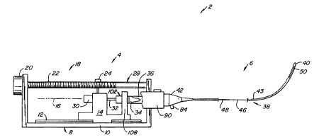
Fig. 1 illustrates, in schematic form, a catheter
system 2 including a drive assembly 4 to which a typically
disposable catheter assembly 6 is removably mounted.
Referring now also to Figs. 2 and 2A, drive assembly 4
includes a body 8 having a base 10 supporting a linear bearing
track 12. A drive chassis 14 is mounted for linear movement
along a longitudinal path 16 by a longitudinal driver 18.
Longitudinal driver 18 includes a longitudinal drive motor 20
which rotates a longitudinal drive screw 22 rotatably
3S supported at each end by body 8. Driver 18 also includes a
threaded clamp 24 having threads which match the threads on
drive screw 22. Clamp 24 is mounted to and moves with drive
chassis I4. Clamp 24 is normally biased into engagement with
CA 02339491 2001-02-02
WO 00/07500 PCT/IB99/01363
8
drive screw 22, but can be moved out of engagement with drive
shaft 22 by the user moving a drive clamp handle 26; doing so
disengages clamp 24 from drive screw 22 and permits the user
to move drive chassis 14 along longitudinal path 16.
Releasing drive clamp handle 26 allows clamp 24 to again
engage drive screw 22; this secures drive chassis 14 in
position along longitudinal path 16, subject to any subsequent
rotation of drive screw 22.
Drive assembly 4 also includes a rotary driver 28
mounted to and carried by drive chassis 14. Rotary driver 28
includes a rotary drive motor 30, mounted to drive chassis 14,
which rotates a drive shaft 32. As will be discussed in more
detail below, drive shaft 32 rotates a first combined
connector 34. First combined connector 34 moves with drive
chassis 14 along longitudinal path 16. First combined
connector 34 operably engages a second combined connector 36,
also discussed below, the second combined connector being a
part of catheter assembly 6.
Turning now to Figs. 3-5, catheter assembly 6 will
be described in more detail. Catheter assembly 6 includes a
hollow sheath 38 extending between a tip 40 at its distal end
and a hub 42 at its proximal end with a main portion 43 in
between. Sheath 38 houses a cable assembly 44, see Figs. a_
and 5. Cable assembly 44 includes second combined
connector 36 from which a cable 46 (the elongate operative
element) extends. Cable 46 includes a stiff initial
section 48 extending from second combined connector 36 and an
image element 50 at the distal end of cable 46. Stiff initial
section 48 is typically made by securing a length of hypotube
(a thin-walled stainless steel tube) over the proximal end of
the cable. Cable assembly 44 can be used for ultrasonic
imaging, laser imaging or other imaging purposes. Also, cable
assembly 44 could be used for purposes other than imaging such
as ablation, photodynamic therapy, delivery of therapeutic or
diagnostic fluids, delivery of devices such as stents, and for
other purposes. In the preferred embodiment, tip 40 is open
to permit fluid to be flushed through the interior of
sheath 44; in some situations tip 40 may be sealed. In some
~V. VON ~ EPA-MUEIyCHEN U4 : i i - u- _ ~~ : 1~ ~ m ~ .. __--~ -... ....- . .
_ _ _ _
"-CA 02339491 2001-' 02-02
11-08-2000 . , . . I B 009901363
wo ooro~50o ~Cras9mo363
9
cases image element 50 may be positioned past tip 40 and
external of sheath 39. .
second combined connector 3s includes a second
rotar~r drive connector 52 and a second electrical connector or
pl ug 54 (a data/infoz~maCion connector) . A drive jai.nt 56 has
three spring fingers SB which engage recesses 60 formed in
drive connector 52 to secure plug 54 in place. Plug 54 has
spring finger electrical. contacts &2 surrounding a center pin
64. Initial section 4H of cable 46 is secured ro the interior
I0 of a rotary shaft coupler 66, coupler o'6 being affi.~ced to a hub
portion 68 of drive joint 56. Appropriate electrical
Connections between drive cable 46 and plug 54 are made in a
conventional manner.
Hub 42 of sheath 38 includes a main cavity 70 which
Z5 houses second combined connector 36. Catheter assembly 6 also
includes a bearing washer 74 mounted between second combined
connector 36 and thG proximal end of main cavity 70.
Bellows 78, typically made of polyester shrink (?) or PTFE, is
fastened at ~.ts prc~xi.mal edge 88 to drive joint 56 and its
20 distal edge 82 to hub 42. Bellows 78 is used to he~.p prevent
contamination of the interior of sheath 38. Bearing
washer 74, used to secure the distal end of a bellows 78 to
hub 42, is made of a low friction polymer.
Hub 42 also includes a fluid or flush port B4 .
25 coupled to the interior of sheath 38 to permit the sheath
interior to be flushed with a saline solution. or other
suitable fluid. An. O-ring fluid seal 86 is used between
hub 42 and coupler 66 (in position of Fig. 2) or stiff initial
section 48 (in position of Fig. 2A? to prevent the flow of
30 fluid from flush port 84 back into cavity 70 and thus into the
interior of drive assembly 4.
Catheter assembly 6 is quickly and easily mounted to
drive assembly 4. To do so, the proximal. end of hub 42 is
inserted into a through-hole 98 ~o=-med in a proximal portion
35 mount 90 of body 8 of drive assembly 4 until latched into
place. This occurs by the engagement cf an upwardly-
extending, spring-biased latch pin, not shown, engaging a
recess 94 formed in hub 42; the latch pin can be removed from
AMENDED SHEET
V. VOV:EP~Hr::V U4 . it- o- .. ~ ~~-.~- .
_ ~ ' CA 02339491 2001-02-02
11-08-2000 ~ ~ ~ ~ I B 009901363
wo ao»7soo ~c'rr~s99~oi3s3
~e
recess 94 by she user pressing on a release button 9~.
Mounting catheter assembly 6 to drive assembly ~ preferably
occurs when rotary driver 28 is in the distal-most or
fu7.ly-extended position of Figs. 1 and 2. Upon mounting
S hub a2 into proximal portion member 90, second combined
connector 36 automatically blind matably cotuiects with first
Combined connector 34 to provide for longitudinal and
rotational driving connection and for electrical connection.
'I~rning now to Figs. 6 and 7, first combined
iQ____do~~ctor assembly-34--wild-be-dis-cussed_ -_First-_co~ined-._.. _.
connector assembly 34 includes a first rotazy drive
connector 96 configured for cvmplernentazy mating engagement
with second rotary drive connector 52. Assembly 34 also
includes a first electrical connector or socket 98 secured
15 bet~reen connector 96 and a second drive joint 100. Socket 9E
and plug 54 are configured to mate with one another in a
coaxial mating arrangement in a manner similar to
connector 22,.electrical connector 54 and drive joint 56 of
Fig. 5. First combined connector 34 is ceup3.ed to a bearing
2o assembly 102. Bearing assembly 102 includes first and second
housing portions 104. 106 which house drive shaft 32.
ylectrical wires IO? extend from socket 98, through housing
portion 104, and through a cut-out 109 in housing portion 106
for connection to appropriate circuitry, not shown. Searing
25 assembly ib2 is supported by the front end 108 of drive
chassis 14 engaging housing portion 104. Drive shaft 32 is
supported withzn housing portions 104, lab by a pair of
bearings 110, 17.2_ Drive shaft 32 passes freely through a
non-rotating ferrite 114 which is fixed within distal housing
30 portion 104, typically through a light press fit or through
the use of an adhesive. Non-rotating ferrite 114 is
positioned at a necked down portion 116 of drive shaft ~z . A
rotating ferrite 118 is mounted to drove shaft 32 to be
positioned adjacent to non-rotating ferrite 114_ Rotating
35 ferrite 118 is secured to drive shaft 32, such as through a
light press fit, and rotates with the drive shaft. Rotating
and non-rotating ferrites 118, 11~! are used to monitor the
rotation of drive shaf t 32 and, thus, of cable assembly 44.
AMENDED SHEET
CA 02339491 2001-02-02
WO 00/07500 PCT/IB99/01363
11
The hub portion 120 of drive joint 100 is housed
within the distal end of distal housing portion 104. The
distal end 122 of drive shaft 32 is keyed or otherwise fixed
to hub portion 120 so that rotating drive shaft 32 causes
first rotary drive connector 96 to rotate, thus rotating cable
assembly 44. The proximal end 124 of drive shaft 32 extends
beyond housing portion 106 and into driven engagement with
rotary drive motor 30. Thus, rotary motion of rotary
drive motor 30 causes drive shaft 32 to rotate, thus rotating
cable assembly 44. Longitudinal movement of drive chassis 14
through the engagement of clamp 24 with drive screw 22 causes
rotary driver 28 to move along longitudinal path 16. However,
the frictional connection between plug 54 and socket 98 causes
cable assembly 44, see Fig. 4, to be pulled along longitudinal
path 16 with the longitudinal movement of rotary driver 28.
The use of stiffened section 48 as the initial portion of
cable 46 provides the necessary support for the cable during
this longitudinal movement.
In use, an appropriate catheter assembly 6 is
selected to be used during a procedure. If rotary driver 28
is not in its distal-most position of Figs. 1 and 2, the user
will place rotary driver 28 at its distal-most position,
typically by grasping drive clamp handle 26, temporarily
disengaging clamp 24 from drive screw 22, and then manually
moving the rotary driver in the distal direction to its
distal-most position. Once in position, drive clamp handle 26
is released, permitting the reengagement of threaded clamp 24
with drive screw 22, thus securing rotary driver 28 in place.
Spring biased release button 92 is then depressed and the
proximal end of hub 42 is inserted into through-hole 88 formed
in proximal portion mount 90. Once hub 42 is partially
inserted, release button 92 is released and hub 42 is
continued to be inserted into mount 90 until the locking pin
attached to release button 92 engages recess 94 formed in
hub 42. When this occurs, hub 42 become: properly secured to
drive assembly 4.
During this insertion, first and second combined
connectors 34, 36 blind matably connect t:o one another. First
CA 02339491 2001-02-02
WO 00/07500 PCT/IB99/01363
12
and second rotary drive connectors 96, 52 have alignment/drive
surfaces 126, 128 which extend both circumferentially and
longitudinally so to provide the proper rotary orientation
upon engagement of the connectors. In addition, plug 54 and
socket 98 are axially aligned during this movement and become
electrically coupled to one another as the first and second
rotary drive connectors 96, 52 mate.
Tip 40 of catheter assembly 6 is then transluminally
positioned to the target area within the patient. With rotary
driver 28 in its distal-most position, which causes imaging
element SO to be in its distal-most position, rotary drive
motor 30 and longitudinal drive motor 20 can be simultaneously
or sequentially actuated to permit 360° images to be obtained
along a length of vessel or other cavity within a patient.
Excessive proximal movement is prevented when drive chassis 14
engages a limit switch 130 mounted to body 8.
The movements of imaging element SO are controlled
in a very stable manner through the use of motors 20, 30 to
provide superior imaging than could otherwise be obtained. If
desired, rotary driver 28 can be manually longitudinally
positioned through the disengagement of clamp 24, the manual
movement of rotary actuator 28 along longitudinal path 16 and
the reengagement of clamp 24 with drive screw 22. In the
preferred embodiment, the length of longitudinal travel will
be about 10 cm. Drive assembly 4 can be made to accommodate
other lengths of longitudinal travel as well.
Modification and variation can be made to the
disclosed embodiment without departing from the subject of the
invention as defined in the following claims. For example,
other types of rotary and longitudinal drivers could be used,
such as a longitudinal chain or belt drive in lieu of the
longitudinal drive screw 22. It is desirable that combined
connectors 34, 36 be engagable and disengagable by straight
longitudinal movement of the connectors into and out of
engagement. T.~owever, in appropriate circumstances, a type of
twist-lock blind matable connector may be used as well.
Alignment/drive surfaces 126, 128 are basically dual V-shaped
surfaces so that cable assembly 44 ends up at one of two
« nnr ~ ova _Ml lf~~yCHEN U4 ~ 11 - 8- 0 ~ ~ pp339491 2001-02-02~ 180 454405-~
+4'J~ t3J LJ~~n~ ~ r. i v
11-08-2000 . , , _ I B 009901363
wo ooro~~oo pcras99rom
13
different rotary orientatians, 180 apart, relative to the
rotary orientation of first combined connector 34. It maY_ b_e
desirable in some circumstances to ensure only a single
relative rotar~r orientation of connectors 34, 30'. This could
be achieved by having a single angled surface for guiding the
connectors into the proper rotary orientation and separate
rotary drive sursaces for a rotary driving engagernQz~.t bet~reen
the two connectors. Sody 8 is shown in a very simple form.
___-- -In a commercial embod~.ment, _ bpd_y 8 would__preferably- have__a~
to more ergonomic shape, and may have fixed or extendable legs to
allow drive assembly to rest comfortably but securely on, for
example, the patient s leg or chest. Whsle it is rxpected
that drive assembly 4 will. be connected to remote power and
control assemblies, it may be desirable to include a
controller, input panel, and a battery pack as a part of drive
assembly ~ to make the drive assembly substantially self-
contained. Data from catheter system 2 could be then provzded
to an external recorder and/or monitor through hardwire or
telemetry, such as by using radio frequency transmitters and
receivers. While the invention is particularly adapted for
imaging of vascular regions, the invention is suitable,(~types
of diagnostic and therapeutic procedures in vascular and other
body structures. Instead of bellows 78, other structures,
such as telescoping tubing, can be used to help prevent
contamination of the interior of sheath 38; the bellows tar
tubing could have round, rectangular or other cross-sectioned
shapes.
~ i -
3
~o
AMENDED SHEET