Note: Descriptions are shown in the official language in which they were submitted.
CA 02340195 2001-02-12
WO 00/09196 PCT/US99/17317
- 1-
EXPANDABLE LASER CATHETER
Field of the Invention
This invention relates generally to laser catheters and more particularly,
to an expandable laser catheter for removing obstructions from body passages.
Background of the Invention
Atherosclerotic plaque is known to build up on the walls of arteries in the
human body. Such plaque build-up restricts circulation and often causes
cardiovascular problems, especially when the build-up occurs in coronary
arteries. Other body passages such as the esophagus, ureter and bile ducts,
for
example, are subject to blockage by tumorous tissue. Accordingly, it is
desirable
to remove or otherwise reduce plaque build-up and other tissue obstructions
from
such body passages.
Known catheters use laser energy to remove plaque build-up on artery
walls. One such known catheter has a laser source and a catheter body. The
catheter body has a proximal end and a distal end, or head, and multiple
optical
fibers extending between the proximal and distal ends. The laser source is
coupled to the optical fibers at the proximal end of the catheter body and is
configured to transmit laser energy through the optical fibers.
To remove an obstruction from a body passage, such as atherosclerotic
plaque in an artery, the catheter is positioned in the artery so that the
distal end
of the catheter is adjacent to the plaque. The laser source is then energized
so
that laser energy travels through the optical fibers and photoablates the
plaque
adjacent the distal end of the catheter. The catheter is then advanced further
through the artery to photoablate the next region of plaque build-up.
While known laser catheters are generally acceptable for removing small
obstructions, such catheters are limited to opening a path the size of the
catheter
head on each pass through the body passage. The multiple passes which are
CA 02340195 2001-02-12
WO 00/09196 PCT/US99/17317
_2_
required for removing larger areas of obstruction increase the possibility of
damaging the passage inner wall. In addition, multiple passes increase the
possibility that a piece of the obstruction will break free, enter the blood
stream
and result in vessel blockage. Other known laser catheters are limited by the
relative inflexibility of the catheter distal end which may inflict damage to
body
passage inner walls as the catheter is advanced.
Accordingly it would be desirable to provide a laser catheter which can
remove substantial portions of an obstruction in a single pass. It would also
be
desirable to provide a laser catheter having a flexible, adjustable distal end
which
can substantially conform to the inner dimensions of the body passageway to
minimize damage to the inner wall. It would be further desirable to provide a
laser catheter which can expand and contract during photoablation to increase
the
area of obstruction which may be photoablated in a single pass through a body
passage.
Summary of the Invention
These and other objects may be attained by a laser catheter which, in one
embodiment, includes a shaftway having a proximal end and a distal end
including a flexible portion. The flexible portion is fabricated from a
pliable
material and is configured in folds which are radially oriented about the
longitudinal axis of the catheter. The flexible portion is configured to be
expanded by, for example, an inflatable balloon which is attached within the
flexible portion. Optical fibers extend along the length of the catheter to
transmit
laser energy from the proximal end to the distal end of the catheter, and are
attached to the catheter at the distal end. The ends of the optical fibers, at
their
attachments to the distal end, are directed toward targeted regions of an
obstruction.
In use, a guidewire is inserted into a body passage such as an artery and
advanced past the obstruction. The catheter is then advanced over the
guidewire
CA 02340195 2007-03-06
74988-21
- 3 -
through the artery until the distal end of the catheter is
adjacent to the obstruction, such as atherosclerotic plaque.
The balloon is then inflated to expand the flexible portion
of the distal end. Upon expansion, the flexible portion
substantially conforms to the inner dimensions of the body
passage and is enlarged so that the flexible portion can
hold a core of material from the obstruction. A laser
connected to the optical fibers at the catheter proximal end
is then energized, and the laser energy transmitted through
the optical fibers photoablates the obstruction in the
regions targeted by the optical fibers. The catheter is
then advanced and the process repeated.
As the catheter is advanced and targeted regions
photoablated, the catheter detaches a separate core of
material from the obstruction. As the core is formed the
catheter advances over the core so that ultimately the core
is completely contained within the flexible portion. To
remove the core of the obstruction, the balloon is deflated
and the flexible portion contracts and holds the core of the
obstruction. The catheter is then withdrawn from the body
passage to remove the core of the obstruction from the body
passage.
According to an aspect of the invention, there is
provided a laser catheter for removing a core of an
obstruction from a body passage, said catheter comprising: a
shaftway having a proximal end and a distal end; a flexible
portion adjacent said shaftway distal end, said flexible
portion configured in a plurality of folds and having a
distal end; a plurality of optical fibers extending between
said shaftway proximal end and said flexible portion distal
end, said optical fibers having distal ends attached to said
flexible portion; and means for expanding said flexible
CA 02340195 2007-03-06
74988-21
- 3a -
portion, said means configured to expand the folds of said
flexible portion so that a core retention region is created.
According to another aspect of the invention,
there is provided a laser catheter for removing a core of an
obstruction from a body passage, said catheter comprising: a
shaftway having a proximal end and a distal end, said distal
end comprising a flexible portion configured in a plurality
of folds; a plurality of optical fibers having distal ends
attached to said flexible portion and retained in said
folds; and an inflatable balloon for expanding said flexible
portion, said balloon attached to said flexible portion and
configured to expand the folds of said flexible portion so
that a core retention region is created.
According to a further aspect of the invention,
there is provided a use of a laser catheter for removing an
obstruction from a body passage wherein the catheter is
connected to a laser which provides laser energy and wherein
the catheter comprises a shaftway having a proximal end and
a distal end comprising of flexible portion, the flexible
portion configured in a plurality of folds, a plurality of
optical fibers extending along the shaftway from the
proximal to the distal end, the optical fibers retained in
the folds of the flexible portion, and a means for expanding
the flexible portion, the means configured to expand the
folds of the flexible portion, wherein the catheter is for
insertion into the body passage adjacent to the obstruction;
the means for expansion is for expansion and contraction of
the flexible portion; the optical fibers are for
transmitting laser energy for photoablation of the
obstruction; and the flexible portion is for forming around
a core of the obstruction.
CA 02340195 2007-03-06
74988-21
- 3b -
In an alternative embodiment, the laser catheter
utilizes mechanical spring force to expand the distal end of
the laser catheter. In this alternative embodiment, the
optical fibers are attached to a stiff shaftway. A fin
structure including a plurality of fins fabricated from a
spring material is attached to the distal end of the
shaftway. The stiff shaftway is capable of transmitting
torque to the distal end so that the fin structure can be
rotated, thus facilitating advancement of the fin structure,
and complete removal of the obstruction. The fins have a
substantially rolled shape and are expandable from a
retracted position to an extended position. In both the
retracted position and the extended position the fins retain
a substantially rolled shape which substantially conforms to
the inner dimensions of the body passage, to minimize damage
to the inner wall of the body passage. At the fin structure
the ends of the optical fibers are attached and spread
across the fins and are directed parallel to the shaftway.
The laser
CA 02340195 2001-02-12
WO 00/09196 PCT/US99/17317
-4-
catheter further includes an outer catheter body or sheath slidably disposed
over
the shaftway to retain the fin structure in the retracted position.
In use of the alternative embodiment, a guide wire is introduced into a
body passage and advanced past the obstruction. The laser catheter is
introduced
over the guide wire and advanced toward the obstruction. When the distal end
of the catheter is adjacent the obstruction, the shaftway is rotated and
advanced
so that the fin structure is pushed out of the outer catheter body, thus
releasing
the fins from the retracted position and allowing them to expand to the
extended
position. In the extended position, the fins contact the passage inner wall
and the
ends of the optical fibers are directed parallel to the inner wall of the
passage.
The fin structure can be further advanced along the passage wall by advancing
the
shaftway and sliding the fin structure along the passage wall. Laser energy is
used to photoablate regions of the obstruction targeted by the optical fibers.
The
mounting of the optical fibers on the fin structure allows the obstruction to
be
removed from around the passage central axis (around the guide wire) to the
outside diameter of the passage, with the fins protecting normal passage inner
wall from photoablation. When the obstruction has been removed, the fin
structure is rotated and pulled back into the outer catheter body, thus
causing the
fins to retract to the retracted position. The catheter is then removed from
the
body passage.
In additional alternative embodiments especially useful for opening in-
stent restenosis, laser energy may be directed radially outward from the
shaftway
instead of parallel to the shaftway. This may be accomplished by attaching the
ends of the optical fibers at the fin structure so that the ends of the fibers
are
directed radially outward from the shaftway, or alternatively, by coupling
prisms
to the ends of the optical fibers at the fin structure to direct laser energy
radially
outwards from the shaftway.
The above described laser catheter removes substantial portions of an
obstruction in a single pass by expanding the distal end of the catheter to
CA 02340195 2001-02-12
WO 00/09196 PCT/US99/17317
- 5-
substantially conform to the inner dimensions of the body passage. The laser
catheter further minimizes damage to the body passage inner wall with a
flexible,
adjustable distal end. Further, the laser catheter may be expanded and
contracted
during photoablation to increase the area of obstruction which may be
photoablated in a single pass. By removing substantial portions of an
obstruction
in a single pass, the laser catheter obviates the need for multiple and
potentially
damaging passes through the body passage.
Brief Description of the Drawings
Figure 1 is a sectional view of a laser catheter.
Figure 2 is a cross-sectional view of a distal end of the laser catheter.
Figure 3 is a sectional view of the distal end of the laser catheter
positioned adjacent to an obstruction in a body passage.
Figure 4 is a sectional view of the catheter distal end within a body
passage after expansion of the flexible portion and partial photoablation of
the
obstruction.
Figure 5 is a cross-sectional view of the catheter distal end within a body
passage after expansion of the flexible portion and partial photoablation of
the
obstruction.
Figure 6 is a sectional view of the catheter distal end after formation of
a core of obstruction and contraction of the flexible portion.
Figure 7 is a cross-sectional view of the catheter distal end after formation
of the core of obstruction and contraction of the flexible portion.
Figure 8 is a perspective view of a distal end of a laser catheter.
Figure 9 is a cross-sectional view of the catheter distal end shown in
Figure 8 and positioned within a body passage.
Figure 10 is a sectional view of the catheter distal end shown in Figure 8
and positioned within the body passage.
CA 02340195 2007-03-06
74988-21
_ 6-
Detailed Description
Figure I is a sectional view of a laser catheter 10 in accordance with one
embodiment of the present invention. Catheter 10 includes a shaftway 12 which
defines a lumen 14 about a Iongitudinal -axis, and has a proximal end 16, a
distal
end 18, and a flexible portion 20 adjacent distal end 18. Flexi'ble portion 20
is
configured in a plurality of folds having a radial orientation about the
longitudinal
axis of lumen 14. A ring-shaped inflatable balloon 22, or other means for
expanding flexible portion 20, is atrached to shaftway 12 within flexible
portion
20. Balloon 22 may instead be attached to the guide wire or a mechanical
device.
Catheter 10 extends over a conventional guidewire 24. Balloon 22 is positioned
from about 1 mm to about 10 mm behind the end of flexible portion 20. Balloon
22 communicates via air lines or tubing (not shown) as known In the art to a
means for inflating the balloon, such as a syringe, air compressor or other
air
pressure providing device (not shown). Multiple optical fibers (not shown in
Figure 1) extend longitudanally along the length of catheter 10.
In one embodiment, shaftway 12 is appro ximately 80 to 150 cm long and
is fabricated from conventional catheter materials such as, for example,
polyurethane. Lumen 14 has a diameter of approximately 1 to 5 mm, and the
outer diameter of shaftway 12 is approximately 1.5 to 6 mm. Flexible portion
20 is approximately 2 to 5 cm long and is fabricated from a pliable material
such
as, for example, polyurethane. - In the folded configuration, the outer
diameter
of flexible portion 20 is approximately the same size as the outer diameter of
shaftway 12. In one embodiment, flexible portion 20 is bonded to shaftway 12
by thermal fusion or an adhesive.
Referring to Figure 2, catheter 10 includes multiple optical fibers 30.
Fibers 30 extend longitudinally along the length of shaftway 12 into flexible
portion 20, and are attached to flexible portion 20. Fibers 30 are of a type
known in the art of laser catheters and are configured to transtnit laser
energy,
In one embodiment, optical fibers 30 extend through lumen 14 and are embedded
CA 02340195 2007-03-06
74988-21
_ 7_
in the pliable material forming flexiblc portion 20. Optical fibers 30 have
proximal ends (not shown) which are conflgured to connect through an optical
fiber port (not shown) to a laser connector and a laser (not shown), such as
an
excimer laser, Nd:YAG, holmium or CO_ laser. ln one embodiment, the laser
is configured to produce laser energy of a wavelength of about 0.3 microns to
about 2.0 microns. The composition of optical fibers 30 depends upon the
chosen
laser and the wavelength of laser energy that the laser produces. In one
embodiment, the laser is an excimer laser producing energy of a wavelength of
about 0.3 microns, requiring optical fibers made of quartz.
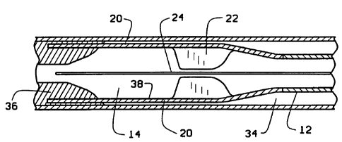
Figure 3 is a sectional view of catheter 10 in use within a body passage
34. In use, guidewire 24 is inserted into body passage 34 and advanced past an
obstruction 36. Catheter 10 is then inserted into body passage 34 over
guidewire
24 and advanced until distal end 18 is adjacent obstruction 36. Specifically,
catheter 10 is advanced until distal end 18 is positioned to contact
obstruction 36
or to be within about 2 mm of obstruction 36. The relative discance of distal
end
18 to obstruction 36 is determined using radiopaque markers and fluoroscopy,
or
other imaging techniques known in the art_ After flexible portion 20 is
correctly
positioned adjacent obstruction 36, balloon 22 is inflated by applying air
pressure
through tubing connecting balloon 22 with, for example, a syringe. Expansion
of balloon 22 causes the flexible portion 20 to expand in the radial dimension
as
the folds are unfolded creating a core retention portion 38 within flexible
portion
20, Balloon 22 is expanded until flexible portion 20 is adjacent to the inner
wall
of body passage 34, as determined in one embodiment by a fluoroscopy image.
The laser is energized so that laser energy travels through fibers 30 to
photoablate
regions of obstruction 36.
lteferring to Figures 4 and 5, flexible portion 20 is then advanced further
through obstruction 36 and the process is repea,I.ed. Specifically as shown in
Pigure 5, the end faces of optical fibers 30 in expanded flexible portion 20
are
CA 02340195 2007-03-06
74988-21
-8-
positioned to direct laser energy toward regions of obstruction 36 which
approach
ormeet the inner wall of passage 34.
Referring to Figure 6, catheter 10 photoablates regions of obstruction 36.
Catheter 10 is then advanced further along passage 34. A core 50 of material
from obstrniction 36 is formed as catheter 10 photoablates regions of
obstruction
36 and is advanced. If catheter 10 is advanced through entire obstruction 36,
or
if the entire length of flexible portion 20 is advanced through obstruction
36, air
pressure is removed from balloon 22_ As a result, flexible portion 20
contracts
and retains core 50 within core retention portion 38. More particularly, as
balloon 22 is deflated and flexible portion 20 contracts, core 50 is retained
within
the folds of flexible portion 20. Core 50 is then removed from body passage 34
by withdrawing catheter 10 from body passage 34. Figure 7 is a cross-sectional
view of eontracted flexible portion 20 within body passage 34, retaining core
50
after core 50 has becn formed.
In an alternative embodiment of the method, obstruction 36 is entirely or
substantially removed from body passage 34 by photoablation. Balloon 22 is
expanded and contracted to impart radial motion to fibers 30 during
photoabIation, thereby increasing the area of obstruction 36 which is exposed
to
laser energy on a single pass through body passage 34. In addition, shaftway
12
may be rotated to impart tangential motion to fibers 30 to fnrther facilitate
substantial photoablation of obstruction 36.
In another alternative embodiment of catheter 10, shaftway 12 may be
slidably inserted through an outer catheter body (not shown) to provide
rigidity
to flexible portion 20 as flexible portion 20 is advanced through body passage
34.
Catheter 10 is advanced through body passage 34 until flexible portion 20 is
within a defined distance of obstruction 36 as defined above. Flexible portion
20
is then extended out of the outer catheter body and is free to expand.
Alternatively, the outer catheter body may be partially retracted to free
flexible
portion 20. After expansion of flexible portion 20, photoablation and
formation
CA 02340195 2001-02-12
WO 00/09196 PCT/US99/17317
-9-
of core 50, flexible portion 20 retaining core 50 is retracted within the
outer
catheter body and catheter 10 is withdrawn from body passage 34.
In an alternate embodiment shown in Figure 8, catheter 60 utilizes
mechanical spring force to expand the distal end of the laser catheter.
Catheter
60 includes a stiff shaftway 62 having a proximal end (not shown) and a distal
end 64. A fin structure including fins 66A and 66B is attached to distal end
64.
Fins 66A and 66B extend from shaftway 62 and are configured to have a
retracted
position (not shown) and an extended position (shown in Figure 8). Stiff
shaftway 62 is capable of transmitting torque to the distal end and allows the
fin
structure to be rotated to facilitate advancement of the fin structure, and
complete
removal of the obstruction. Fins 66A and 66B have a substantially rolled shape
in both the retracted position and the extended position, allowing fins 66A
and
66B to substantially conform to the inner dimensions of the body passage, to
minimize damage to the inner wall of the body passage. Optical fibers 68,
similar
to optical fibers 30, extend from the proximal end to distal end 64 and are
attached and spread across fins 66A and 66B. Proximal ends of optical fibers
68
(not shown) may be configured to connect through an optical fiber port (not
shown) to a laser connector and a laser (not shown), such as an excimer laser,
Nd:YAG, holmium or COZ laser. Shaftway 62 is slidably positioned within an
outer catheter body 70 which is configured to retain fins 66A and 66B in the
retracted position. Shaftway 62 slidably extends over guidewire 72, which is
similar to guidewire 24.
In one embodiment, shaftway 62 is fabricated from conventional catheter
materials such as, for example, stainless steel hypodermic tubing. Fins 66A
and
66B are fabricated from a spring material such as a metal alloy foil. In one
embodiment, the foils generally have a thickness of about 0.001 inches and may
be fabricated from, for example, stainless steel, niatinol or precipitation
hardened
steel. Thus, fins 66A and 66B expand from the retracted rolled shape to the
extended rolled shape due to the mechanical spring force of the foil. Fins 66A
CA 02340195 2007-03-06
74988-21
- 10-
and 66B are attached to shaftway 62 by welding or brazing, are about 10 to
about
30 mm long, and extend about 1.5 mm to about 15 mm from their attachments
at shaftway 62. Outer catheter body 70 is fabricated from conventional
catheter
materials as known in the art.
In one embodiment, distal end 64 includes two fins 66A and 66B, but any
number of fins may be used. Distal ends of optical fibers 68 are attached to
the
spring material forming fins 66A and 66B so that the ends of optical fibers 68
are
at an angle, or parallel, to the central axis of the body passage. For
example,
the distal ends of optical fibers 68 may be directed in any desired direction
from
parallel to the central axis of the body passage, to perpendicular to the
inner wall
of the body passage. The desired direction is determined by the size and shape
of the obstruction to be excised. For example, it is particularly desirable to
aim
fiber ends at a substantial angle, incIuding substantially perpendicularly, to
the
body passage inner wall for photoablation of well-defined regions of
obstruction,
such as restenosis within a stent.
Figure 9 is a cross-sectional view of distal end 64 positioned within a body
passage 96 and adjacent to an obstruction 98, Distal end 64 is shown extending
beyond outer catheter body 70. Fins 66A and 66B are in the extended position.
Figure 10 is a sectional view of distal end 64 with f ns 66A and 66B in the
extended position as shown in Figure 9. In one embodiment, fins 66A and 66B
each have a leading edge 100, a trailing edge 102, and a peripheral edge . To
facilitate navigation of distal end 64 tltrough curves in body passage 96,
fins 66A
and 66B are tapered along leading edge 100 and trailing edge 102 so that fins
66A
and 66B are widest at their atrachments to shaftway 62 and shortest along
peripheral edge.
In use, guidewire 72 is inserted into body passage 96 and advanced past
obstruction 98_ Catheter 60 is then inserted into body passage 96 over
guidewire
72. Fins 66A and 66B are retained in the retracted position within outer
catheter
body 70. Catheter 60 is advanced within passage 96 until distal end 64 is
CA 02340195 2001-02-12
WO 00/09196 PCT/US99/17317
- 11-
positioned proximate obstruction 98, for example, within about 2 mm. Distal
end
64 is extended out of outer catheter body 70 by advancing shaftway 62, thereby
releasing the restraint on fins 66 and allowing fins 66 to extend and conform
to
the inner dimensions of body passage 96. The laser coupled to the proximate
ends of optical fibers 68 is energized so that laser energy travels through
fibers
68 to photoablate regions of obstruction 98. Distal end 64 may be rotated and
advanced further along passage 96 for photoablation of any remaining regions
of
obstruction 98. The spring material forming fins 66A and 66B protects against
damage to the passage inner wall by preventing direct contact by optical
fibers 68
with the inner wall. Once obstruction 98 has been photoablated, distal end 64
is
pulled back into outer catheter body 70 to retract fins 66A and 66B and
facilitate
removal from body passage 96.
Alternate embodiments of the laser catheters described herein may be used
to remove small or large regions of atherosclerotic plaque from blocked blood
vessels, or regions of tumor invading an esophagus, ureter, urethra, bile duct
or
other body passage. The catheters may also be used, for example, to aid in the
removal of excess or abnormal cartilage in body joints such as knees, or in
disc
spaces between vertebral bodies. The catheters may be used without guidewires
and instead with alternate guidance methods such as optical coherence
tomography (OCT), ultrasound, CT scanning or fluoroscopy. The catheters may
be surgically introduced to body passages or elsewhere in the body using known
instruments such as arthroscopes, endoscopes, colonoscopes, bronchoscopes,
laparoscopes, etc. The distal end faces of the optical fibers may be rounded,
or
square-cut, cut at an angle, or connected to an optical prism lens to more
precisely target certain regions of the obstruction.
To further minimize damage to the body passage inner wall, the flexible
portion substantially conforms to the inner dimensions of the body passage. In
an alternate method of use, the flexible portion is expanded and contracted
during
CA 02340195 2001-02-12
WO 00/09196 PCT/US99/17317
- 12-
photoablation to increase the area of obstruction which is removed by direct
photoablation in one pass through the body passage.
The above described laser catheter improves the efficacy and safety of
using laser energy to remove large areas of an obstruction from a body
passage.
The laser catheter of the present invention uses photoablation to create a
core of
an obstruction which is then removed as a single mass at one time. The laser
catheter therefore minimizes damage to body passage walls by obviating the
need
for multiple passes through the body passage. Alternate embodiments of the
method for using the laser catheter include expanding and contracting the
flexible
distal portion during photoablation of the obstruction, thereby photoablating
large
areas of obstructions in one pass and minimizing damage to body passage walls.
In addition, the laser catheter includes a distal end which substantially
conforms
to the inner dimensions of the body passageway and further minimizes damage
to body passage inner walls.
From the preceding description of various embodiments of the present
invention, it is evident that the objects of the invention are attained.
Although the
invention has been described and illustrated in detail, it is to be clearly
understood that the same is intended by way of illustration and example only
and
is not to be taken by way of limitation. Accordingly, the spirit and scope of
the
invention are to be limited only by the terms of the appended claims.