Note: Descriptions are shown in the official language in which they were submitted.
CA 02340433 2001-02-13
WO 00/14546 PCT/EP99/06592
TAU AS A MARKER FOR EARLY CNS DAMAGE
FIELD OF THE INVENTION
The present invention relates to the field of CNS damage. The present
invention relates to a new
method for the early diagnosis of CNS damage by detection and/or
quantification of tau.
BACKGROUND OF THE INVENTION
Damage of the central nervous system (CNS damage) is caused by various
inducing agents among
which different disease processes, physical or chemical agents, anoxia and
ischemia. Disease
processes include space occupying lesions, invasion or metastasis of the brain
caused by different
malignancies and/or infection by a number of organisms.
Tumors of the CNS, may originate locally (primary tumors) or may spread to the
CNS
(metastases). Primary tumors arise from glial cells (astrocytoma,
oligodendroglioma,
glioblastoma), ependymal cells (ependymoma) or supporting tissue (meningioma,
schwannoma,
papilloma of the choroid plexus). In childhood, tumors arise from more
primitive cells
(medulloblastoma, neuroblastoma, chordoma). Malignant astrocytoma or
glioblastoma is the most
common type of primary tumor in adults over age 20. Both benign and malignant
primary CNS
tumors are capable of producing neurologic impairment.
Leukemia is the most common type of cancer in children. During the last twenty
years, the
survival of children with leukemia has improved markedly based on the routine
use of intensive
chemotherapy alone or as combined treatment (radiotherapy and chemotherapy).
Currently, the
estimated overall 10-year survival rate is around 75%. Given the increasing
number of childhood
leukemia survivors, concern has arisen about long-term effects of anti-cancer
chemotherapy
and/or radiotherapy resulting in possible damage to the central nervous system
and the need for
an early quantitative determination of this CNS damage is increasing.
Bacterial meningitis may be defined as an inflammation in response to
bacterial infection of the
pia-arachnoid and the fluid residing in the space which it encloses and also
of the fluid in the
CA 02340433 2001-02-13
WO 00/14546 PCT/EP99/06592
2
ventricles of the brain. The incidence of bacterial meningitis is between 4.6
and 10 cases per
100000 persons per year. H. influenzae is the most frequent cause, followed by
N. meningitidis
and S. pneumoniae. Once developed, characteristic features of bacterial
meningitis include an
increase in intracranial pressure, disruption of the blood-brain barrier,
cerebral edema, and
alterations in cerebral blood flow. The longer the duration of meningitis and
the less effective the
treatment, the greater the chances that complications and neurologic residua
will develop.
Approximately 10 percent of infants and children who have bacterial meningitis
will be left with
persistent unilateral or bilateral sensory hearing loss. Approximately 30
percent of children who
have had bacterial meningitis later will turn out to have subtle learning
deficits (Wilson et al.,
1991).
Viruses can also affect the central nervous system in a variety of ways
resulting in a distinction
between viral meningitis, viral encephalitis, myelitis and CNS diseases due to
slow virus infection.
Other conditions that may cause CNS damage are chemical agents such as
pharmaceuticals,
chemotherapy or exposure to chemical compounds, and physical agents. Head
injuries are
frequent in industrialised countries, affecting many patients in the prime of
life. To appreciate the
medical and social magnitude of this problem it needs only to be recognised
that almost 10 million
Americans have head injuries yearly, about 20 percent serious enough to cause
brain damage.
Another cause for CNS damage may be anoxia or ischemia. Anoxic-ischemic
encephalopaty is a
common and often disastrous condition, caused by a lack of oxygen to the
brain, resulting from
hypotension or respiratory failure. Acute ischemic stroke causes neuronal
damage and is a major
cause of neurological handicap in western society. Perinatal asphyxia may be
associated with CNS
damage as well. To date, clinical, electroencephalographic and neuroradiologic
evaluation,
together with cerebral blood flow studies are the most readily available
methods. However, early
and accurate evaluation of the severity of brain damage after a hypoxic-
ischemic event, remains
one of the most difficult problems in neonatal care.
For the detection of CNS invasion in leukemic children current diagnostic
procedures include
lumbar puncture, eye fundoscopy and brain imaging (Raichle, 1998). However,
these diagnostic
methods only allow detection of the CNS damage in a more advanced stage while
already ongoing
CNS damage in the early stages may be missed by these methods. Therefore,
there is a need for
additional diagnostic methods that allow early detection of CNS damage.
CA 02340433 2001-02-13
WO 00/14546 PCT/EP99/06592
3
A number of neurological markers have recently become available which reflect
conditions of the
central nervous system, relating to cell death, axon growth/re-induction,
inflammation and/or
blood-brain barrier dysfunction. The microtubule-associated protein tau exists
in different
isoforms, of which 4 to 6 are found in adult brain but only 1 isoform is
detected in fetal brain. The
diversity of the isoforrns is generated from a single gene on human chromosome
17 by alternative
mRNA splicing (Himmler, 1989; Goedert et al., 1989; Andreadis et al., 1992).
The most striking
feature of tau protein, as deduced from molecular cloning, is a stretch of 31
or 32 amino acids,
occurring in the carboxy-terminal part of the molecule, which can be repeated
either 3 or 4 times.
Additional diversity is generated through 29 or 58 amino acid-long insertions
in the NH2-terminal
part of tau molecules (Goedert et al., 1989). In vivo tau promotes microtubule
assembly and
stability in the axonal compartment of neurons by interactions involving its
microtubule binding
domain which is localised in the repeat region of tau (255-381) (L,ewis et
al., 1988). In normal
circumstances adult brain contains 2 - 3 mol phosphate per mole of tau (Selden
and Pollard, 1983;
Ksiezak-Reding et al., 1992). Phosphorylation of different sites in normal tau
as studied in rat and
humans is dependent on the developmental state (Lee et al., 1991; Bramblett et
ai., 1993; Goedert
et al., 1993). Tau variants of 60, 64 and 68 kDa arising as a consequence of
phosphorylation have
been detected in brain areas showing neurofibrillary tangles (Delacourte et
al., 1990; Goedert et
al., 1992; Flament and Delacourte, 1990, Greenberg and Davies, 1990). These
brains contain 6-8
mol phosphate per mol tau (Ksiezak-Reding et al., 1992). In tau isolated from
paired helical
filaments, phosphorylation can occur at several positions (Iqbal et al., 1989;
Lee et al., 1991;
Hasegawa et al., 1992). Detection of normally and abnormally phosphorylated
tau in brain
extracts is done either via antibodies (Mab A1z50: Ghanbari et al., 1990; Mab
Ab423: Harrington
et al., 1991; Mab AT120 : Vandermeeren et al., 1993; Mab AT180; Mab AT270 :
International
application published under WO 95/17429 and Mab AT8 : International
application published
under WO 93/08302), or via the change in molecular weight {Flament and
Delacourte, 1990), or
else by functional assay (Bramblett et al., 1992). A combination of monoclonal
antibodies, each
recognising specific epitopes of tau, has been used to detect the presence of
normally and
abnormally phosphorylated tau in CSF (Van de Voorde et al., 1995). Tau has
been used as a
marker to discriminate dementia with altered cytoskeletal properties such as
Alzheimer's disease
from normal aged subjects or from patients with other types of dementia.
CA 02340433 2001-02-13
WO 00/14546 PCT/EP99/06592
4
AIMS OF THE INVENTION
It is an aim of the present invention to provide a method for the early
detection and/or
quantification of CNS damage in an individual, said CNS damage being caused by
space-
occupying lesions of the CNS, by invasion or metastasis of the CNS, by
organisms, by anoxia or
ischemia, by chemical agents, by physical agents, or by a combination of these
mechanisms.
It is a more specific aim of the present invention to provide a method for the
early detection
and/or quantification of CNS damage in an individual, said CNS damage being
caused a primary
brain tumour, benign or malignant, brain metastasis or a subdural haematoma.
It is another more specific aim of the present invention to provide a method
for the early detection
and/or quantification of CNS damage in an individual, said CNS damage being
caused by invasion
of the CNS by leukemia, lymphoma or breast cancer.
It is another more specific aim of the present invention to provide a method
for the early detection
and/or quantification of CNS damage in an individual, said CNS damage being
caused by bacteria
or viruses causing encephalitis or meningitis.
It is another more specific aim of the present invention to provide a method
for the early detection
and/or quantification of CNS damage in an individual, said CNS damage being
caused by stroke,
by cerebral infarction, by cerebral haemorrhage, by thrombosis, by perinatal
asphyxia, by
Binswanger disease or by vasculitis.
It is another more specific aim of the present invention to provide a method
for the early detection
and/or quantification of CNS damage in an individual, said CNS damage being
caused by
chemotherapy.
It is another more specific aim of the present invention to provide a method
for the early detection
and/or quantification of CNS damage in an individual, said CNS damage being
caused by trauma,
stroke, intracranial pressure or radiation.
It is another aim of the present invention to provide a method for the early
detection and/or
quantification of CNS damage in an individual, said CNS damage being caused by
space-
occupying lesions of the CNS, by invasion or metastasis of the CNS, by
organisms, by anoxia or
ischemia, by chemical agents, by physical agents, or by a combination of these
mechanisms in
order to evaluate the effect of treatment on said CNS damage.
CA 02340433 2001-02-13
WO 00/14546 PCT/EP99/06592
It is another aim of the present invention to provide a kit for the early
diagnosis of CNS damage
in an individual, said CNS damage being caused by space-occupying lesions of
the CNS, by
invasion of the CNS, by organisms, by anoxia or ischemia, by chemical agents,
by physical agents,
or by a combination of these mechanisms.
5 It is another more specific aim of the present invention to provide a kit
for the early diagnosis of
CNS damage in an individual, said CNS damage being caused a primary brain
tumour, benign or
malignant, brain metastasis or a subdural haematoma.
It is another more specific aim of the present invention to provide a kit for
the early diagnosis of
CNS damage in an individual, said CNS damage being caused by invasion of the
CNS by
leukemia, lymphoma or breast cancer.
It is another more specific aim of the present invention to provide a kit for
the early diagnosis of
CNS damage in an individual, said CNS damage being caused by bacteria or
viruses causing
encephalitis or meningitis.
It is another more specific aim of the present invention to provide a kit for
the early diagnosis of
CNS damage in an individual, said CNS damage being caused by stroke, by
cerebral infarction,
by cerebral haemorrhage, by thrombosis, by perinatal asphyxia, by Binswanger
disease or by
vascultitis.
It is another more specific aim of the present invention to provide a kit for
the early diagnosis of
CNS damage in an individual, said CNS damage being caused by chemotherapy.
It is another more specific aim of the present invention to provide a kit for
the early diagnosis of
CNS damage in an individual, said CNS damage being caused by trauma, stroke,
intracranial
pressure or radiation.
It is another aim of the present invention to provide a method to screen or
monitor the effect
of compounds which prevent or treat CNS damage.
All the aims of the present invention are considered to be met by the
embodiments as set out
below.
DETAILED DESCRIPTION OF THE INVENTION
The present invention relates to a method for the early detection and/or
quantification of CNS
CA 02340433 2001-02-13
WO 00/14546 PCT1EP99/06592
6
damage in an individual, said CNS damage being caused by space-occupying
lesions of the CNS,
by invasion of the CNS, by organisms, by anoxia or ischemia, by chemical
agents, by physical
agents or by a combination of these mechanisms. This method comprises the step
of determining
and/or quantifying the level of tau in an individual and comparing it to the
level of tau in control
S healthy individuals.
The present invention relates to the surprising finding that tau levels in CSF
samples from children
with leukemia are increased compared to upper limit values for healthy
individuals. These
increased tau levels are an indication of latent central nervous system
invasion and already on-
going CNS damage long before this CNS damage can be measured by the current
diagnostic
procedures. Also in individuals that were suffering space occupying lesions of
the CNS, invasion
or metastasis of the CNS, ischemia, stroke or meningitis, increased tau levels
were observed in
an early stage. Accordingly, tau can be used as an aspecific marker for the
early detection of CNS
damage caused by invasion of the CNS by leukemia and in general, as an
aspecific marker for the
early detection of CNS damage caused by CNS damaging agents such as space-
occupying lesion
of the CNS, invasion or metastasis of the CNS, organisms, anoxia or ischemia,
chemical agents,
physical agents, or a combination of these mechanisms.
The central nervous system (CNS) is that part of the nervous system which, in
vertebrates,
consists of the brain and spinal cord, to which sensory impulses are
transmitted and from which
motor impulses pass out, and which supervises and co-ordinates the activity of
the entire nervous
system.
The term "CNS damage" refers to any condition of the CNS which is associated
with a neuronal
malfimctioning and which is caused by a specific inducing agent or damaging
agent. More
specifically, CNS damage refers to disease processes that include but are not
limited to space-
occupying lesions of the CNS, invasion or metastasis of the CNS and/or
organisms. Space
occupying lesions may be, for example, primary brain tumours, benign or
malignant, brain
metastasis, parasite derived cysts such as Taenia solium or Echinococcus
granulosus,
hydrocephalus and/or subdural haematoma. Invasion or metastasis of the CNS can
be caused by
malignancies such as leukemia, lymphoma, breast cancer, lung cancer, melanoma
and/or gastro-
intestinal malignancies or other types of cancers. Organisms that may infect
the CNS and cause
CNS damage include but are not limited to prions, viruses, bacteria or
parasites. Bacteria and
viruses that infect the CNS may cause meningitis, encephalitis, neuro aids or
neuroborreliose.
CA 02340433 2001-02-13
WO 00!14546 PCT/EP99/06592
7
They include but are not limited to Neisseria meningitides, H. in. fluenzae,
S. pneumoniae, Herpes
simplex meningoencephalitis and Herpes simplex gluteolis. CNS damage can also
be caused by
anoxia or ischemia during anestesia, perinatal asphyxia, drowning, asthma,
stroke, cerebral
infarction, thrombosis, cerebral haemorrhage, CO poisoning, Binswanger disease
and/or vasculitis.
Chemical agents causing CNS damage include but are not limited to gene
therapy,
pharmaceuticals, chemotherapy and exposure to chemical compounds. CNS damage
can also be
caused by physical agents such as trauma, radiation, hypothermia,
hyperthermia, intracranial
pressure or stroke. It is also possible that more than one of the above
mentioned causing agents
are responsible for the CNS damage.
The present invention thus provides a method for the early detection and/or
quantification of said
CNS damage by determining the level of tau. "Early detection and/or
quantification of CNS
damage" means that the CNS damage is determined by a method that allows it to
be detected
before it is detectable by the current methods.
The term "tau" as referred to in the present application can be any form of
tau, including any state
of phosphorylation. The level of tau is determined qualitatively and/or
quantitatively as a measure
for the degree of CNS damage. Tau can be detected in vitro as well as in vivo.
The method for the early in vitro detection of CNS damage in an individual
comprises the steps
of obtaining a sample from said individual, determining and/or quantifying the
level of tau in said
sample and comparing it to the level of tau in a sample of control healthy
individuals. The term
"sample" refers to any source of biological material, for instance body
fluids, hair, epithelial cells,
peripheral blood or any other sample comprising tau protein. In a preferred
embodiment, tau can be
detected andlor quantified in vitro by analysis of the level of tau in a body
fluid sample of the
patient. The term "body fluid" refers to all fluids that are present in the
human body including but
not limited to blood, lymph, urine and cerebrospinal fluid (CSF). In a more
specific embodiment
of the present invention tau is detected and/or quantified in a cerebrospinal
fluid sample taken
from the patient. In another specific embodiment of the invention tau is
detected and/or quantified
in a sample of blood derivatives of the patient. The blood sample can include
the whole sample
as taken from the patient. More preferably the blood sample includes a plasma
sample or a serum
sample.
Tau can be detected and/or quantified by any method known, including but not
limited to the use
of antibodies, the change in molecular weight (Flament and Delacourte, 1990),
or else by
CA 02340433 2001-02-13
WO 00/14546 PCT/EP99/06592
8
functional assay (Bramblett et al., 1992). In a preferred embodiment tau can
be detected by an
immunoassay comprising at least the following steps:
- obtaining a sample from the patient; and
- bringing said sample into contact with a monoclonal antibody (primary
antibody or
S capturing antibody) recognising tau, under conditions being suitable for
producing an
antigen-antibody complex; and
- detecting the immunological binding of said antibody to said sample.
Advantageously, the monoclonal antibody used in the invention is in an
immobilised state on a
suitable support. Alternatively, the present process may be put into practice
by using any other
immunoassay format known to the person skilled in the art.
The process for the detection of the antigen can then be carried out by
bringing together said
antigen-antibody complex formed by the antigen and the primary antibody
recognising tau with:
a) a secondary antibody (or detector antibody)
*which can be a monoclonal antibody recognising an epitope of the tau-primary
antibody
1 S complex but not recognising the primary antibody alone, or
*which can be a polyclonal antibody recognising an epitope of the tau-primary
antibody
complex but not recognising the primary antibody alone, with said polyclonal
antibody
being preferably purified by immunoaffinity chromatography using immobilised
tau or the
tau-primary antibody complex.
b) a marker either for specific tagging or coupling with said secondary
antibody, with said
marker being any possible marker known to the person skilled in the art;
c) appropriate buffer solutions for carrying out the immunological reaction
between the
primary antibody and the sample, between the secondary antibody and the tau-
primary
antibody complex and/or between the bound secondary antibody and the marker,
and,
d) possibly also, for standardisation purposes, a purified protein or
synthetic peptide reactive
with the antibodies that recognise tau.
Advantageously, the secondary antibody itself carries a marker or a group for
direct or indirect
coupling with a marker.
The term "epitope" refers to that portion of the antigen-antibody complex that
is specifically
bound by an antibody-combining site. Epitopes may be determined by any of the
techniques
known in the art or may be predicted by a variety of computer prediction
models known in the
CA 02340433 2001-02-13
WO 00/14546 PCT/EP99/06592
9
art.
The expression "recognising", "reacting with", "immunological binding" or
"producing an
antigen-antibody complex" as used in the present invention is to be
interpreted that binding
between the antigen and antibody occurs under all conditions that respect the
immunological
properties of the antibody and the antigen.
Any monoclonal or polyclonal antibody that specifically recognises tau may be
used for the
detection of tau. Antibodies specifically recognising normally and/or
abnormally phosphorylated
tau include A1z50 (Ghanbari et al., 1990), Ab423 (Harrington et al., 1991),
AT8 (International
application published under WO 93/08302), AT120 (Vandermeeren et al., 1993);
AT180 and
AT270 (International application published under WO 95/17429) and AT100
(International
application published under WO 96/04309). But also other antibodies known in
the art that
specifically recognise tau can be used.
The method for the early in vitro detection and/or quantification of CNS
damage in an individual
can also be used to evaluate the effect of a certain treatment on the CNS
damage in said
1 S individual. Possible treatments that might influence the status of the CNS
include but are not
limited to drug treatments, chemotherapy, physical therapy, including
radiotherapy and gene
therapy.
The method for the early in vivo detection and/or quantification of CNS damage
in an individual
comprises the steps of determining and/or quantifying the level of tau in said
individual and
comparing it to the level of tau in control healthy individuals. In a
preferred embodiment, tau can
be detected in vivo by in vivo imaging. Tau can be visualised in situ by non-
invasive methods
including but not limited to brain imaging methods described by Arbit et al.
(1995), Tamada et
al. (1995), Wakabayashi et al. (1995), Huang et al. (1996), Sandrock et al.
(1996), Mariani et al.
(1997). These in vivo imaging methods may allow the localisation and
visualisation of tau, for
example, by use of labelled antibodies recognising tau.
Tau can also be used as an aspecific marker for in vivo imaging to evaluate
the effect of a certain
treatment on the CNS damage in an individual. Possible treatments that might
influence the status
of the CNS include but are not limited to drug treatments, chemotherapy,
physical therapy,
including radiotherapy and gene therapy.
The present invention fi~rther relates to the use of tau as an aspecific
marker for the manufacture
of a diagnostic kit for the early detection in an individual of CNS damage
caused by space-
CA 02340433 2001-02-13
WO 00/14546 PCT/EP99/06592
occupying lesions of the CNS, by invasion or metastasis of the CNS, by
organisms, by anoxia or
ischemia, by chemical agents, by physical agents, or by a combination of these
mechanisms.
The present invention fixrther relates to the use of tau as an aspecific
marker for the manufacture
of a diagnostic kit for the early detection in an individual of CNS damage
caused by a primary
5 brain tumour, benign of malignant, brain metastasis, or a subdural
haematoma.
The present invention further relates to the use of tau as an aspecific marker
for the manufacture
of a diagnostic kit for the early detection in an individual of CNS damage
caused by invasion or
metastasis of the CNS by leukemia, lymphoma or breast cancer.
The present invention fixrther relates to the use of tau as an aspecific
marker for the manufacture
10 of a diagnostic kit for the early detection in an individual of CNS damage
caused by bacteria or
viruses.
The present invention fizrther relates to the use of tau as an aspecific
marker for the manufacture
of a diagnostic kit for the early detection in an individual of CNS damage
caused by stroke, by
cerebral infarction, by thrombosis, by cerebral haemorrhage, by pe~natal
asphyxia, by Binswanger
disease or by vasculitis.
The present invention further relates to the use of tau as an aspecific marker
for the manufacture
of a diagnostic kit for the early detection in an individual of CNS damage
caused by
chemotherapy.
The present invention fi~rther relates to the use of tau as an aspecific
marker for the manufacture
of a diagnostic kit for the early detection in an individual of CNS damage
caused by trauma,
stroke, intracranial pressure or radiation.
The present invention fixrther relates to a kit for the in vitro or in vivo
diagnosis in an individual
of CNS damage caused by space-occupying lesions of the CNS, by invasion or
metastasis of the
CNS, by organisms, by anoxia or ischemia, by chemical agents, by physical
agents, or by a
combination of these mechanisms. Any kit that provides a tool for the
detection of tau can be used
for the diagnosis of the above-mentioned CNS damage.
A preferred kit for the in vitro diagnosis in an individual of CNS damage
caused by space-
occupying lesions of the CNS, by invasion or metastasis of the CNS, by
organisms, by anoxia or
ischemia, by chemical agents, by physical agents, or by a combination of these
mechanisms is
based on an immunoassay and comprises:
- at least a monoclonal antibody (primary antibody) which forms an
immunological
CA 02340433 2001-02-13
WO 00/14546 PCT/EP99/06592
11
complex with an epitope of the tau protein;
- a secondary antibody
* which can be a monoclonal antibody recognising an epitope of the tau-primary
antibody complex but not recognising the primary antibody alone, or
* which can be a polyclonal antibody recognising an epitope of the tau-primary
antibody complex but not recognising the primary antibody alone, with said
polyclonal antibody being preferably purified by immunoaffinity
chromatography using immobilised tau protein or immobilised tau-primary
antibody complex;
- a marker either for specific tagging or coupling with said secondary
antibody;
- appropriate buffer solutions for carrying out the immunological reaction
between the
primary antibody and a test sample, between the secondary antibody and the tau-
primary antibody complex, and/or between the bound secondary antibody and the
marker;
- possibly, for standardisation purposes, a purified protein or synthetic
peptide containing
one of more tau epitopes.
The present invention also relates to a method to screen or monitor the effect
of compounds
which prevent or treat CNS damage comprising the step of determining the level
of tau and
comparing it to the level of tau in a control sample.
The present invention will now be illustrated by reference to the following
examples that set forth
particularly advantageous embodiments. However, it should be noted that these
examples are
illustrative and can not be construed as to restrict the invention in any way.
CA 02340433 2001-02-13
WO 00/14546 PCT/EP99/06592
12
4
I
N M
0.
N pp
40
' N N N
L N N ~ ~t !p
~
_ _ _
M M M ~ ~ ~ ~O 'd'
~ ~ I I ~, ... I
I I Ov N v7 N ~O ~O o0 00
H .-~ .- 00 N M M 00 00 ~' M M M M
T T ~ ~ ~ ~ ~ ~ T T ~ T
cd l~ cd al ed cCeC CClt~f GC cd cV eC
'b 'b 'b 'b 'd 'd'b 'b 'd b "d 'b 'L7
O
0 0 0 0 > > ~ > o >
U
-p O -p ~p b N N O
,.~ ~ O ~ E E ~ ~ ~ O O ~ E O
~ O U O O O ~ '~ O O O O ~n U
~O c~ ~ N W ,--M ~D I~ e~
A
j
I
!
as i
o ~ ~
-o
c o o ~, '_'.~
I ~
H x O .O CY Q ty v k
on -o -d -G C~
a~ E_' . ~ . o ~ ~ E_'
~1 A., ~ ~ ~ o T ~
I ~ G1, c_ C~ U U
' W
a.
0
a
0
0 0
o
i o
c , ~ 'a
.C I 'C h G
I
y I ..,
~ V V
O O
C~ j _ _ I
~ ~ O
O ~ir I
Gr
CA 02340433 2001-02-13
WO 00/14546 PCT/EP99/06592
13
ao
N
II
C
M
cps
a
I w
N N ~ ~n O
O C
~
M b w Q~ N N p~
M N .. V V ~' cd
'n ~ N ~
N I I ~ ~ ~ I I b
N v0 ~D ~D o0o0
m ~ 00 CT ~~ N N 00 00 00 M M M M
c~ at ed ~ e~ e~ e~itd eatcd e~ catcded O
A '~ 'B 'fl 'L7 'Z7'O 'd 'b 'b 'b 'b '~'b
~r
3
~ ' (~
d0
c
N N N O
O
~ ~ N N N
h.0 E ~ ~ Cp
B ~
O p CD C,Db~ ~ ~ O O
~ ~ ~
~C v1 O U ~ O O O O V1v O
A N V~ Cd ~O M ~ .-.M r. ~ ~p I~cCi V1
I N N
N ~ b N
O O C v ~ C
~C >C Q.,.f o U
G1 C~ ~ wr ~ H H
~ E-'E-' ~ '~ ~ ~ o ~ '~ E
E ~ c :~ . ~, ~
.o ~ ~ ~1 ~ d W U ~o Q~ fl
C
0
0
~a
.~ a c '~ "c 0 ~
'o
s es ~ e!.~ ~n o
' ~
V 0
_ ~ C
C
C
.~ ~ CC V U r.~r
C~ ~ w j
L ~ air O
.~..~ O~ C..
r~ i
~
CA 02340433 2001-02-13
WO 00/14546 PC'T/EP99/06592
14
N
~o
'~ .~ a N
... '.
H ..... .-. r-. .-. .-. r- .-. N N N ~ N
T ~ ~ ~ ~ ~ T ~ T T
cC at ed cC e~ cd e~ cd ccS cd
b 'b 'b 'b 'b 'b 'b 'b 'b 'b 'fl 'L3
H ~ I ~ ? j
e~ ~ e~
-p N O O ~ N N O N ~O
N ~ by pp N ~ Op ~ pp
:p :d ~ ~ ~ ~ '~ S~ "d
N ~ O ~ O O O ~ ~ p E O ~ O
U U O O O U O U
O A ~O ~' M ectC~ ~O N V1 ~Dt~ M cd
Q1
U
U
x
b
' ~ N ~ ~. ~ o
o ~ o r ~ ~ o
V ~ 0 0 .~ o
O .N f3.~ U w .c~'ni3. ~ U
A '~ ~..O O 'C '~"p ~ O
w > U x ~. 7 U ~ x
w
~3
H
C Ar
U N
N
0 N
N H
O
C~
p .~
Q C O C 'i O
A
c ~. on U '.
y c
~" b a N
L
C IO N r~1 'b
E"'~ 'd
N ~ A C/:
L ;U C%~ ~
E-' i~F O
F. .~ U
CA 02340433 2001-02-13
WO 00/14546 PCT/EP99/06592
0., 4, 4.
V1 M
I
.n N "'iN I~ H .-~ .-.N N N .-~N
T T >, >, i~ ?, >,>, >, ~, >, T
A "~ "O b 'b 'Q 'G 'Ob b b '~ 'O
'B
.. O
E"" ~ Er ~--~' C ~ ~ r~ ~" ~ ~ ..O
rm --im -r ~--y , i-.r~--m--m..~~ r~
~.
c~
I
I
U
s
a~
own ~''~c'ow ~ -p ~ an N o0
O O O O O +.~
t. ~ ..r.r N w N ~ ~ ~
b~4 ~ b~AbOA~ ~ N ~ ~ C ~OO~ .C
b ~ ~ ~p ~ ~ '~ b ~ b
~O O O O O
O O V U O ~ O U O V
L.iCC! M c~ c~ .-~ ~O N V'1~D t~ M cC
(~
N
1..
U
C
.
N O 4.
C C G. .U cC
I
pp O U C N p O
' G ~ ~
x ~ I p" I
A ~ W '~ o p x I
E- x I
I c J U L~
~
~
0
'
i
p.
II
O
w
0
0
c
C
"p N
O 'n I
47 I
V
.
(,~ ~ , n
I U
N ~ ,.y ~ .~n w
y ~ .~.J,~ G
CC L ~ jF I .E~
E., j , ~ C --
E,.,V ~
U
CA 02340433 2001-02-13
WO 00/14546 PGT/EP99/06592
16
n
00
N o0 t~1 o0 ~ ~ I'~ V' ~?' 'd' d' ~ ~ M V'1
I I I ~ I i I I I I I I I
M tW0 .- .-. v7 ,~ ,-~ ,--~ .-. .... .-~ ...~ N ~ >, ~v~
>, ~, >, ?, ~, ~, >, J, >, ?, ~, ~, ~, >, >,
cd cd cd c~i cd cd cd cd cd tti cd e~i c~ c~ cd
A 'fl "O 'd 'b 'b 'i~ '~ "d 'z5 'C 'b 'b 'b 'b b ."' V' c~
4.
c~
II
U
7 > > E- > > > > > O O E- ~ > O U
a~
0
U
cd
-o
~ N N ~ N ~ ~ ~ ~ N N N .~ ~ ~ N II
N
E ~ ~ ~ ~° o ~ ~ ~ ~ ~ ~ ~° o ~ ~ r~
V ~ °o °o o ° ~ 0 0 0 °o o °o ~ ~ o c
o o
A N -- ~-~ cps M ~ N N ~ ~~ ~D ctt N -- V' ~1
c~
a
.UD
h
N
d U
U ~ U
C C ' V C ~ C vi
O O '~ cc '~ O
cd ."~ cd N
CD ~ GD
o ~ UU x.DUU x ~ U._D o ~UU v_c oU II
o ~ ~ o ~ ~ o
:., ~s ~ ~ ~e ~ o ~ s x ~ ~ -c ~ O
s. v. v-. Cm.. i.., c~ ~,. A.. ~-~ a) v.. w.. O. .r w.,
a a a ~ > a a ~ r~ a > ~ r~ a a > ~ a
0
>r
0
a >
0
p ~ .II
0 0 ~:. o
a~ ~ o ~ :r
. .,
a 6;,
w ~, L G H
s. H a~ C v
~., w C a, y C
~r RI
a ~ a .c ~C .C ~ c
o c .II
o ~. '~ L ~ ~~e E-
CA 02340433 2001-02-13
WO 00/14546 PCTJEP99/06592
17
Table 4. Level of tau in CSF samples of patients with possible CNS damage
caused by
different factors.
Cause of CNS damage' Centre' Patient Age Sex Tau level
(pg/ml)
SPACE OCCUPYING LESIONS,
INVASION OR METASTASIS
Subdural haematome O1 025 39 M 263
Anaplastic oligoastrocytomaO1 032 68 M 329
Cerebral metastasis 08 024 70 F 292
Oligodendroglioma WHO III 08 031 48 M 335
Metastasis of breast cancer08 020 52 F 1 SO
BLEEDING, INFARCTION OR
ISCHEMIA.
Stroke - CVA O1 021 62 F 451
Multiple lacunar stroke O1 026 56 F 250
Stroke - CVA 04 004 50 F 105
Acute but limited stroke OS 032 72 M 248
Subarachnoid hemorrhage 08 019 50 M 216
Chronic post-anoxia (vegetativeOS 051 37 F 446
state)
Binswanger disease 10 014 74 M 347
Vasculitis 10 009 57 F 1250
Cerebral ischemia 10 005 80 M 273
Ischemia 10 006 71 F 574
CnnPrinr caaittal cinuc Ol 023 41 M 773
thrnmhncis
CA 02340433 2001-02-13
WO 00/14546 PCT/EP99/06592
18
Table 4. Continued.
Cause of CNS damage' Centres PatientAge Sea" Tau level
(pg/ml)
ORGANISMS
Cryptococcal meningitis OS 033 29 M 1143
Neuroborreliose OS 045 16 M 141
Chronic meningitis, unknownOS 046 34 F 452
cause
Meningococcal meningitis OS 049 24 M 37
Bacterial meningitis OS 059 73 F 446
Neuro aids OS 060 45 F 237
Pneumococcal meningitis OS 063 70 M 111
Bacterial meningitis 06 026 25 M 1250
Bacterial meningitis 06 027 19 M 37
TBC meningitis 06 028 54 M 720
Bacterial meningitis 06 029 23 M 37
Bacterial meningitis 06 030 28 M 88
Viral meningitis 06 037 82 M 314
Bacterial meningitis 08 018 49 F 205
Viral meningitis 08 029 29 F 170
CONTROLS
Muscle weakness and hystericalO1 020 39 F 282
conversion
Facial palsy (peripheral OS 041 44 M 141
nerve
disease, normal CSF
Tension (psychogenic) headacheOS 031 49 F 37
Psychogenic headache and OS 036 56 F 131
neurosis
Psychogenic signs and symptomsOS 040 48 F 220
Major hysteria OS 042 47 F 471
t L 11 1 J _ a . _ _ lv f A I 1 A 1 l t
~1 A
f
CA 02340433 2001-02-13
WO 00/14546 PCT/EP99/06592
19
Table 4. Continued.
Cause of CNS damage' Centre' PatientAge Sex Tau level
(pg/ml)
CONTROLS
Control (associated SjogrenOS 065 52 F 107
disease)
Control (neurosis) OS 079 40 F 90
Depression 10 008 59 F 201
Depression 10 010 69 F 139
Myalgia, myositis 04 003 31 M 37
Myalgia, myositis 04 005 35 F 435
Malaise, fatigue 04 006 50 M 98
Neck pain, vertigo 08 O1 I 39 F 192
Headache 08 017 49 M 113
Depression 08 021 57 M 361
Headache, strabismus convergens08 033 32 F 239
Bell's palsy 06 031 38 M 90
Bell's palsy 06 032 22 F 131
Bell's palsy 06 033 73 F 231
Bell's palsy 06 034 74 F 144
Carpal tunnel syndrome 06 035 71 M 190
Carpal tunnel syndrome 06 036 90 M 665
' O 1: Prof. P. Cras, University Hospital, Neurology, Antwerp, Belgium and Dr.
A. Daniels, UIA,
Laboratory of Neurobiology, Antwerp, Belgium; 04: Dr. P.D. Mehta, Institute
Basic Research,
Staten Island, NY, USA; O5: Dr. A. Ivanoiu, UCL St-Luc, Laboratory of
Neurochemistry,
Brussels, Belgium; 06: Prof. P.P. De Deyn, UIA, Laboratory of Neurochemistry,
Antwerp,
Belgium; 08: Dr. C. Bancher, Lainz Hospital, Neurology, Vienna, Austria; 10:
Dr. J. Wiltfang,
Georg-August University, Gerontopsychiatry, Gottingen, Germany.
b F: female; M: male.
'In cases where multiple factors may contribute to the damage, only the factor
supposed to be
CA 02340433 2001-02-13
WO 00/14546 PCT/EP99/06592
most relevant is given.
5 FIGURE LEGENDS
Figure 1. Tau values at diagnosis, before any treatment was given: 1. Control
children; 2. AML;
3. AML-CNS+; 4. CML; 5. MDS; 6. B-NHL; 7. Non-B-ALL; 8. Non-B-ALL CNS+; 9. VHR
non-B-ALL.
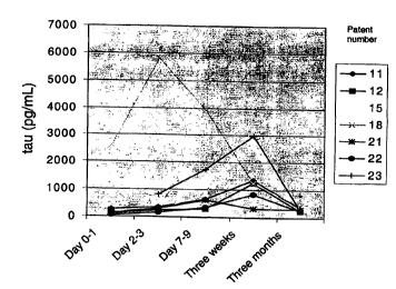
Figure 2. CSF-tau levels in seven patients after acute ischemic stroke. The
CSF samples were
collected at income (day 0-1), day 2-3, day 7-8, day 21-22 (3 weeks) and day
90-110 (3 months).
Figure 3. CSF-tau levels at the time of maximal release in relation to the
size of the infarction as
measured by CT scan.
EXAMPLE S
Example 1: Increased tau levels in children with leukemia
To evaluate the influence of chemotherapy on neuronal damage, a longitudinal
study was
conducted, involving 65 children with leukemia (aged 2 to 16 years) without
measurable central
nervous system involvement, and treated according to standard procedures. A
total of 377 CSF
samples were analysed. Before each injection, a small volume of fluid was
sampled for routine
laboratory analysis and the leftover was used in our study. These children
were being diagnosed
and then treated for their leukemia at the University Hospital of Leuven,
Belgium. Tau protein
in cerebrospinal fluid was assessed using the INNOTEST hTAU Antigen
(Innogenetics, Gent,
Belgium).
For all children suspected to have leukemia, a lumbar puncture was performed
before the start of
CA 02340433 2001-02-13
WO 00/14546 PCT/EP99/06592
21
the treatment to detect the possible presence of leukemic cells, indicative of
central nervous
system invasion. The tau levels measured at that time, and thus before
treatment, would serve as
the control level to compare chemotherapy-induced changes in the levels of
tau.
We observed that some children with leukemia already had very high tau levels
at diagnosis in
spite of the fact that no leukemic cells were detected in the central nervous
system. These children
constitute a new risk group having brain invasion or leukemia-induced CNS
damage, which
cannot always be found using current diagnostic procedures (imaging of the
brain, lumbar
puncture, eye fundoscopy). This was further supported by the increased tau
levels seen in one
patient having leukemia with proven cellular invasion into the brain
(malignant cells in the
cerebrospinal fluid).
Example 2: Increased tau levels in patients with leukemia before treatment
1. Subjects
Between August 1996 and June 1999, 510 samples of CSF were taken from 82
children being
treated for cancer at the Pediatric Hemato-oncology Department of the Catholic
University of
Leuven, Belgium. CSF samples were only taken in the course of scheduled lumbar
punctions
(LPs) for staging or treatment for malignancy.
Three groups of patients with hematological malignancies were studied. The
largest group
consisted of 48 patients with non-B-ALL, treated according to the EORTC
protocol 58881 (table
1 ). Of these children, 20 had CD 10(+) blasts (or common ALL), from whom two
patients had
also Down syndrome (DS) and 1 patient had the Brachmann-de Lange syndrome; 6
patients had
common B-cell blasts, 2 patients had common T-cell blasts, 2 patients had pro-
B-cell blasts, 9
patients had pre-B-cell blasts, and 9 patients had T-cell blasts. Fourty-two
children had leukemia,
6 patients had non-Hodgkin's lymphoma stage II (1 patient), Stage III (4
patients) or Stage IV
( 1 patient). One patient had overt CNS involvement (CNS+), defined according
to the study
protocol with malignant cells in the CSF. Five patients were considered as
very high risk (VHR)
patients according to the criteria defined in the protocol (2 patients with
t(9;22) and 3 patients
CA 02340433 2001-02-13
WO 00/14546 PCT/EP99/06592
22
with corticoid-resistance). As the first part of the induction treatment was
similar to the other
patients, they were included in the analyses according to the induction
chemotherapy. Twenty-
eight of the patients within non-B-ALL could be followed longitudinally during
their treatment
period.
S A second group of patients included 10 B-cell non-Hodgkin's lymphoma
patients, treated
according to the United Kingdom Children Cancers group (UKCCSG 9602) NHL
protocol (table
2}. Five patients had B-cell lymphomas, 3 patients had Burkitt's lymphoma, and
2 patients had
anaplastic large cell lymphoma (ALCL). All patients were treated with the same
protocol, except
one patient who had B-cell leukemia. Six of these patients were studied
longitudinally.
A third patient group consisted of 9 children with Acute Myeloid Leukemia -
myelodysplasia
(AML - MDS), of which 2 patients had CNS involvement and two patients had Down
syndrome.
There were 3 patients with M0, and 1 patient each with M1, M2, MSa or M7
phenotype. Two
patients had MDS, of which one had developed AML and was treated with
chemotherapy. All
these patients, except one MDS patient, were treated according to the EORTC
58921 protocol
(table 3) and 7 patients were followed longitudinally.
The other patients consisted of a heterogeneous group of children (n = 9) in
which for clinical
reasons an LP was performed. This group includes 3 children with
medulloblastoma (staging), 2
children with rhabdomyosarcoma (staging), 1 child with Langerhans cell
histiocytosis (LCH,
staging), ganglioglioma (staging), germinoma (staging), and retinoblastoma
with CNS metastasis
(staging and follow up). The control group consisted of 4 children from whom
CSF was taken
as part of the routine control for possible viral or bacterial infections, but
with negative results.
One patient with a localized retinoblastoma (staging) and one screened for
familial
hemophagocytic lymphohistiocytosis (HI,H) were also included in the control
group. The nearest
relatives of the patients gave oral informed consent for participation in the
study.
2. Methods
Liquor sampling. Lumbar punctures were performed just prior to the IT
administration of
therapeutics. Five ml of CSF was collected in different polypropylene tubes.
One sample was
centrifi~ged immediately at 1 S00 rpm for 2 minutes to eliminate cells and
other insoluble material.
The supernatant was stored at -70°C for subsequent analysis. The number
of freeze/thaw cycles
CA 02340433 2001-02-13
WO 00/14546 PCT/EP99106592
23
was restricted to a minimum.
Measurement of tau in the CSF. All biochemical analyses for the detection of
CSF-tau were
made without knowledge of the clinical diagnosis. Potentially confounding
factors, such as the
number of freeze/thaw cycles, recipient type, and volume of sample per assay
were standardized
for the whole study protocol. CSF-tau levels were determined using a sandwich
ELISA
(INNOTEST hTAU Antigen, Innogenetics, Gent, Belgium), that measured total tau
(both normal
and hyperphosphorylated tau). Samples were analyzed firstly alone, at the time
of each LP
Afterwards, all samples derived from one patient were analyzed again on one
immunoplate. The
correlation coefficient between the results from the first and the second
approach for a set of 104
samples was 0.901 (95%CI: 0.856 - 0.933). The increases of CSF-tau described
in the paper were
excluded to be general increases of proteins in the liquor.
3. Results
Normal upper limit levels for tau were firstly determined on CSF samples from
the 6 control
children. The mean CSF-tau value was 106.2 pg/ml (95%CI = 34.3-178.0).
Arbitrary cut-off
normal value was considered as 312 pg/ml (mean + 3 SD), which is in the range
of values
observed in adults (Hulstaert et al., 1999). Moreover, we did not find a
correlation between the
CSF-tau values at diagnosis and the age of the children (Pearson r = -0.161,
CI95% _ -0.3914
- 0.08836, n = 64), suggesting that age has no influence on CSF-tau levels.
Clearly pathological
CSF-tau values can be considered as above 500 pg/ml.
Tau levels at diagnosis were analyzed for each subgroup of patients (Fig. 1).
There was no
obvious difference for the tau levels in patients with and without Dow's
syndrome (not shown).
The two patients with overt CNS invasion (CNS+) had levels of CSF-tau at
diagnosis above 312
pg/ml. However, the 2 children with MDS, 7/28 children with non-B-ALL, 1/4 non-
B-ALL
patients with very high risk criteria, 1/5 patients with AML, and 2/8 patients
with B-cell NHL had
a level of tau above 312 pg/ml, while using classical diagnostic procedures,
CNS invasion was not
detected. Three patients with risen intracranial pressure (medulloblastoma),
and one patient with
germinoma, from which CSF was taken for staging, had high CSF-tau
concentrations
CA 02340433 2001-02-13
WO 00/14545 PCT/EP99/o5592
24
(823,1397,1500 and 442 pg/ml), in contrast to one patient with grade I
astrocytoma who had a
normal CSF-tau level (97 pg/ml). One patient with LCH had a normal CSF-tau
level of 112 pg/ml.
Two patients with rhabdomyosarcoma could be analyzed at diagnosis: one patient
with stage I
disease had a CSF-tau level of 279 pg/ml, the other patient with stage IV
disease had a level of
320 pg/ml. One patient with retinoblastoma and CNS involvement had a CSF-tau
level of 1800
pg/ml.
For 33 patients with non-B-ALL, CSF-tau levels at diagnosis did not correlate
with tumor burden,
as reflected by the white blood cell count (Pearson r = 0.04575, CD95%: -
0.3024 - 0.3831) or
serum LDH (Pearson r = -0.03002, CI95%: -0.3696 - 0.3166). In patients with B-
NHL, there was
no significant correlation between the LDH level and CSF-tau (Pearson r = -
0.3723, CD95%: -
0.8532 - 0.4507).
Egamule 3: Use CSF-tau as a marker for early detection of possible CNS damage
caused
by stroke
1. Subject
Seven patients, 3 men and 4 women, 63-81 years old (mean SD, 70.7~7.2 years)
with cerebral
infarctions admitted to the Unit of Neurology, Sahlgren's University Hospital,
Goteborg, Sweden,
were incorporated in the study. All patients were included within 72 h of
stroke onset.
2. Methods
CSF samples were collected using lumbar puncture. 12 ml was collected and
frozen in 0.5 ml
aliquots at -80°C until analyzed. CSF samples were collected at income
(day 0-1), day 2-3, day
7-8, day 21-22 (3 weeks) and day 90-110 (3 months). CSF-tau levels were
determined using a
sandwich ELISA (INNOTEST hTAU Antigen, Innogenetics, Gent, Belgium), that
measured total
tau (both normal and hyperphosphorylated tau).
The extent of brain damage was examined with computerized tomography (CT) of
the brain
during the first day of admittance. Clinical the patients were also examined
using the modified
CA 02340433 2001-02-13
WO 00/14546 PCT/EP99/06592
Scandinavian Stroke Scale Index (SSI; Scandinavian Stroke Study Group, 1985)
at onset of
stroke and 3 months later for the degree of disability with the Bartel Index
(BI; Mohoney and
Barthel, 1965).
5 3. Results
CSF-tau showed a marked increase after acute stroke, with a peak after 1-3
weeks and return to
normal after 3 months (Fig 2). There was also a correlation between CSF-tau
levels and the size
of the infarction as measured by CT scan (Fig 3). These results indicate that
CSF-tau reflects
10 neuronal damage and degeneration and the level in the CSF depends on the
amount of damaged
nerve cells. In this study no correlation was found between the clinical data
(SSI or BI) and CSF-
tau, probably due to small number of patients.
1 S Egamole 4: Use of tau as a marker for early detection of possible CNS
damage caused by
different dama ink agents
1. Subjects
20 A multicentre study was carried out at 8 European and 2 U.S. university
centres involved in CSF
research, based on residual CSF archived at the centres for research purposes.
CSF samples of
patients with a broad range of different neurological conditions were included
in order to get a
general idea about the specificity of the tau marker changes in a variety of
pathologies involving
the CNS. Neurological controls consisted of subjects without obvious CNS
damage (table 4).
The study was conducted in accordance with local clinical research
regulations. If required
additional local Ethics Committee or Institutional Review Board approval was
obtained by the
investigator prior to the start of the study.
2. Methods
CA 02340433 2001-02-13
WO 00/14546 PCT/EP99/06592
26
CSF samples were collected using lumbar puncture (LP). Only CSF samples
containing less than
S00 red blood cells per pl were included in the study. The CSF samples were
centrifuged at 2000
g for 10 minutes within 4 hours after LP and kept frozen without thawing. CSF
samples from
centre O1 had undergone an additional freeze-thaw cycle before analysis. The
concentration of
total tau comprising normal tau and paired helical filament-tau was measured
at the centres using
a sandwich ELISA technique (INNOTEST hTAU-Antigen, Innogenetics N. V.).
3. CSF-tau levels in patients with possible CNS damage compared to CSF-tau
levels in
neurological control subjects
Tau levels in the CSF samples of patients with possible CNS damage caused by
space occupying
lesions, by invasion or metastasis, by bleeding; infarction or ischemia or by
organisms and tau
levels in CSF samples of control subjects are shown in table 5. The group of
patients that had
possibly contracted CNS damage by space occupying lesions, by invasion or
metastasis, by
bleeding, by infarction or ischemia, or by organisms showed an overall higher
tau level than the
neurological control patients (p = 0.022, two sided Mann Whitney test).
REFERENCES
Andreadis A., Brown W., Kosik K. (1992) Structure and novel exons of the human
tau gene.
Biochem. 31: 10626-1063 3 .
Arbit E., Cheung N.K., Yeh S.D., Daghighian F, ,Shang J.J., Cordon-Cardo C.,
Pentlow K.,
Canete A., Finn R., Larson S.M. (1995) Quantitative studies of monoclonal
antibody targeting
to disialogangliosid GD2 in human brain tumours. Eur. J. Nucl. Med. 22: 419-
426.
Bramblett G., Trojanowski J., Lee V. (1992) Regions with abundant
neurofibrillary pathology in
human brain exhibit a selective reduction in levels of binding-competent tau
and accumulation of
abnormal tau isoforms (A68 proteins). Lab. Invest. 66: 212-222.
CA 02340433 2001-02-13
WO 00/14546 PCT/EP99/06592
27
Bramblett G., Goedert M., Jakes R., Merrick S., Trojanowski J., Lee V. (1993)
The abnormal
phosphorylation of tau at Ser396 in Alzheimer's disease recapitulates
phosphorylation during
development and contributes to reduced microtubule binding. Neuron. 10: 1089-
1099.
Delacourte A., Flament S., Dibe E., Hublau P., Sabionniere B., Hemon B.,
Sherrer V., Defossez
A. ( 1990) Pathological proteins Tau64 and 69 are specifically expressed in
the somatodendritic
domain of the degenerating cortical neurons during Alzheimer's disease. Acta
Neuropathol. 80:
I 11-117.
Flament S., Delacourte A. (1990) Tau Marker? Nature 346: 6279.
Ghanbari H., Kozuk T., Miller B., Riesing S. (1990) A sandwich enzyme
immunoassay for
detecting and measuring Alzheimer's disease-associated proteins in human brain
tissue. J. Clin.
Laboratory Anal. 4: 189-192.
Goedert M., Spillantini M., Jakes R., Rutherford D., Crowther R. (1989)
Multiple isoforms of
human microtubule-associated protein tau: sequences and localisation in
neurofibrillary tangles
of Alzheimer's disease. Neuron. 3: 519-526.
Goedert M., Cohen E., Jakes R., Cohen P. (1992) p42 Map kinase phosphorylation
sites in
microtubule-associated protein tau one dephosphorylated by protein phosphatase
2A1:
implications for Alzheimer's disease. FEBS Lett. 312: 95-99.
Goedert M., Jakes R., Crowther R., Six J., Lubke U., Vandermeeren M., Cras P.,
Trojanowski
J.Q., Lee V. (1993) The abnormal phosphorylation of tau protein at serine 202
in Alzheimer's
disease recapitulates phosphorylation during development. Proc. Natl. Acad.
Sci. (USA) 90:
5066-5070.
Greenberg S., Davies P. (1990) A preparation of Alzheimer paired helical
filaments that displays
distinct tau proteins by polyacrylamide gel electrophoresis. Proc. Natl. Acad.
Sci. USA 87: 5827-
5831.
CA 02340433 2001-02-13
WO 00/14546 PCT/EP99/06592
28
Harrington C., Mukaetova E., Hills R., Edwards P., Montejo de Garcini E.,
Novak M., Wischik
C. ( 1991 ) Measurement of distinct immunochemical presentations of tau
protein in Alzheimer's
disease. Proc. Natl. Acad. Sci. {USA) 88: 5842-584b.
Hasegawa M., Morishima-Kawashima M., Takio K., Suzuki M., Titani K., Ihara Y.
(1992)
Protein sequence and mass spectrometric analyses of tau in Alzheimer's disease
brain. J. Biol.
Chem 267: 17047-17054.
Himmler A. (1989) Structure of the bovine tau gene: alternatively spliced
transcripts. Mol. Cell
Biol. 9(4): 1389-96.
Huang Q., He G., Lan Q., Li X., Qian Z. Chen J. Lu Z., Du Z. (1996) Target
imaging diagnosis
of human brain glioma. Clinical analysis of 40 cases. Nucl. Med. Commun. 17:
311-316.
Hulstaert F., Blennow K., Ivanoiu A., Schoonderwaldt H.C., Riemenschneider M.,
De Deyn
P.P., Bancher C., Cras P., Wiltfang J., Mehta P.D., Iqbal K., Pottel H.,
Vanmechelen E.,
Vanderstichele H. (1999) Improved discrimination of AD patients using (3-
amyloid~l_a2~ and tau
levels in CSF. Neurology 52: 1555-1562.
IqbaI K., Grundke-Iqbal L, Smith A., George L., Tung Y., Zaidi T. ( 1989)
Identification and
localisation of a Tau peptide to paired helical filaments of Alzheimer's
disease. Proc. Natl. Acad.
Sci. (LJSA) 86: 5646-5650.
Ksiezak-Reding H., Liu W.K., Yen S.H. (1992) Brain Res. 597: 209-219.
Lee V., Balin B., Otvos L., Trojanowski J. (1991) A68: a major subunit of
paired helical filaments
and derivatized forms of normal tau. Science 251 (4994): 675-8.
Lewis S., Wang D., Cowan N. (1988) Microtubule-associated protein MAP2 shares
a microtubule
binding motif with Tau protein. Science 242: 936-939.
CA 02340433 2001-02-13
WO 00/14546 PCT/EP99/06592
29
Mariani G., Lasku A., Pau A., Villa G., Motta C., Calcagno G., Taddei G.Z.,
Castellani P.,
Syrigos K., Dorcaratto A., Epenetos A. A., Zardi L., Viale G.A. ( 1997) A
pilot pharmacokinetic
and immunoscintigraphic study with the technetium-99m labelled monoclonal
antibody BC-1
directed against oncofetal fibronectin in patients with brain tumours. Cancer
15: 2484-2489.
Mohoney F.L, Barthel D.W. (1965) Functional evaluation: The Barthel Index. Md.
State. Med.
J. 24: 61-69.
Raichle M.E. (1998) Behind the scenes of functional brain imaging: A
historical and physiological
perspective. Proc. Natl. Acad. Sci. USA 95: 765-772.
Sandrock D., Verheggen R., Helwig A.T., Munz D.L., Markakis E., Emrich D.
(1996)
Immunoscintigraphy for the detection of brain abscesses. Nucl. Med. Commun.
17: 311-316.
Scandinavian Stroke Study Group (1985) Multicenter trial of hemodilution in
ischemic stroke:
Background and study protocol. Stroke 16: 373-403.
Selden S., Pollard T. (1983) Phosphorylation of microtubule-associated
proteins regulates their
interaction with actin filaments. J. Biol. Chem. 258(11): 7064-71.
Tamada K., Fujinaga S., Watanabe R., Yamashita R., Takeuchi Y., Osano M.
(1995) Specific
deposition of passively transferred monoclonal antibodies against herpes
simplex virus type 1 in
rat brain infected with the virus. Microbiol-Immunol. 39: 861-871.
Vandermeeren M., Mercken M., Vanmechelen E., Six J., Van de Voorde A., Martin
J., Cras P.
(1993) Detection of tau proteins in normal and Alzheimer's disease fluid with
a sensitive sandwich
enzyme linked assay J. Neurochem. 61: 1828-1834.
Van de Voorde A., Vanmechelen E., Vandermeeren M., Dessaint F., Beeckman W.,
Cras P.
(1995) Detection of tau in cerebrospinal fluid. Research Advances in
Alzheimer's disease and
CA 02340433 2001-02-13
WO 00/14546 PCT/EP99106592
related disorders, Ed. Ibqal, Mortimer, Winblad & Wisniewski, John Wiley &
Sons Ltd.
Wakabayashi T., Yoshida J., Okada H., Sugita K., Itoh K., Tadokoro M., Ohshima
M. (1995)
Radioimaging of human glioma by indium-11 labelled G-22 anti-glioma monoclonal
antibody.
5 Noshuyo-Byori 12: 105-110.
Wilson J.D., Braunwald E., Isselbacher K.J., Petersdorf R.G., Martin J.B.,
Fauci A.S., Root
R.K. (1991) Harrison's Principles of Internal Medicine, 12th Edition, McGraw-
Hill Inc, NY,
USA.