Note: Descriptions are shown in the official language in which they were submitted.
CA 02341251 2001-02-20
WO 00/10475 PCTIUS99/19193
1
ELECTROCATHETER FOR IfNDUCING YESSEL STENOSYS HAYING TWO ARRAYS OF DIVERGING
ELECTRODES
This application is a continuation-in-part of co-penain~ appncation
serial no. 08/927,2~~1 filed on September 11, 1997 and application serial no.
08/958,766 filed on October 26, 1997.
BACKGROUND OF' THE INVENTION
The invention relates generally to a method and apparatus far applying
energy to shrink a hollow anatomical structure, such as a fallopian tube ar a
vein, including, but not limited to, superficial and perforator veins,
hemorrhoids, and esophageal varices, and more particularly, to a method and
apparatus using an electrode device having multiple leads for applying radio
frequency (RF) energy, microwave energy, or thermal energy.
The human venous system of the lower limbs consists essentially of the
superficial venous system and the deep venous system with perforating veins
connecting the two :>ystems. The superficial system includes the Long or great
saphenous vein and the short saphenous vein. The deep venous system
IS includes the anterior and posterior tibial veins which unite to form the
popliteal vein, which in turn becomes the femoral vein when joined by the
short saphenous vein.
The venous system contains numerous one-way valves for directing
blood flow back to tike heart. Venous valves are usually bicuspid valves, with
each cusp forming a sack or reservoir for blood which, under retrograde blood
pressure, forces the :free surfaces of the cusps together to prevent
retrograde
flow of the blood and allows only antegrade blood flow to the heart. When an
incompetent valve is in the flow path, the valve is unable to close because
the
cusps do not forma proper seal and retrograde flaw of the blood cannot be
stopped. When a venous valve fails, increased strain and pressure occur
within the lower venous sections and overlying tissues, sometimes leading to
additional valvular failure. Two venous conditions which often result from
CA 02341251 2001-02-20
WO 00/10475 PCT/US99/19193
2
valve failure are varicose veins and more symptomatic chronic venous
insufficiency.
The varicose vein condition includes dilation and tortuosity of the
superficial veins of the lower limbs, resulting in unsightly discoloration,
pain,
swelling, and possibly ulceration. Varicose veins often involve incompetence
of one or more venous valves, which allow reflex of blood within the
superficial system. This can also worsen deep venous reflex and perforator
refiux. Current treatments of vein insufficiency include surgical procedures
such as vein stripping, ligation, and occasionally, vein-segment transplant.
Chronic venous insufficiency involves an aggravated condition of
varicose veins which may be caused by degenerative weakness in the vein
valve segment, or by hydrodynamic forces acting on the tissues of the body,
such as the legs, ankles, and feet. As the valves in the veins fail, the
hydrostatic pressure increases on the next venous valves down, causing those
veins to dilate. As this continues, more venous valves will eventually fail.
As
they fail, the effective height of the column of blood above the feet and
ankles
grows, and the weight and hydrostatic pressure exerted on the tissues of the
ankle and foot increases. When the weight of that column reaches a critical
point as a result of the valve failures, ulcerations of the ankle begin to
form,
which start deep and eventually come to the surface. These ulcerations do not
heal easily because of poor venous circulation due to valvular incompetence in
the deep venous system and other vein systems.
Other related venous conditions include dilated hemorrhoids and
esophageal varices. Pressure and dilation of the hemorrhoid venous plexus
may cause internal hemorrhoids to dilate and/or prolapse and be forced
through the anal opening. If a hemorrhoid remains prolapsed, considerable
discomfort, including itching and bleeding, may result. The venous return
from these prolapsed hemorrhoids becomes obstructed by the anal sphincters,
which gives rise to a strangulated hemorrhoid. Thromboses result where the
blood within the prolapsed vein becomes clotted. This extremely painful
condition can cause°_ edema and inflammation.
CA 02341251 2001-02-20
WO 00/10475 PCT/US99/19193
3
Varicose veiins called esophageal varices can form in the venous system
with submucosa ot-' the lower esophagus, and bleeding can occur from the
dilated veins. Biee~ding or hemorrhaging may result from esophageal varices,
which can be difficult to stop and, if untreated, could develop into a life
threatening condition. Such varices erode easily, and Lead to a massive
gastrointestinal hemorrhage.
Ligation of ,~ fallopian tube (tubal ligation) for sterilization or other
purposes is typically performed by laparoscopy. A doctor severs the fallopian
tube or tubes and ties the ends. External cauterization or clamps may also be
used. General or regional anesthetic must be used. AIi of the above are
performed from outside the fallopian tube.
Hemorrhoids and esophageal varices may be alleviated by infra-luminal
ligation. As used herein, "Iigation" or "infra-luminal ligation" comprises the
occlusion, collapse, or closure of a lumen or hollow anatomical stz-ucture by
the application of energy from within the lumen or structure. As used herein,
"ligation" or "infra-~luminal ligation" includes electro-ligation. In the case
of
fallopian tube ligation, it would be desirable to perform the Iigation from
within the fallopian tube itself (infra-fallopian tube) to avoid the trauma
associated with external methods.
Ligation involves the cauterization or coagulation of a lumen using
energy such as that applied through an electrode device. An electrode device
is introduced into the lumen and positioned so that it contacts the lumen
wall.
Once properly positioned, RF energy is applied to the wall by the electrode
device thereby causing the Lumen to shrink in cross-sectional diameter. In the
case of a vein, a reduction in cross-sectional diameter of the vein, for
example
from 5 mm (0.2 in;l to 1 mm (0.04 in), significantly reduces the flow of blood
through a lumen and results in an effective occlusion. Although not required
for effective occlusion or ligation, the vein wall may completely collapse
thereby resulting in a full-lumen obstruction that blocks the flow of blood
through the vein. Likewise, a fallopian tube may collapse sufficient to effect
a
sterilizaraon of the v~patient.
CA 02341251 2001-02-20
WO 00110475 PCT/US99/19193
4
One apparatus for performing ligation includes a tubular shaft having
an electrode device attached at the distal tip. Running through the shaft,
from
the distal end to thc: proximal end, are electzical leads. At the proximal end
of
the shaft, the leads terminate at an electrical connector, while at the distal
end
of the shaft the leads are connected to the electrode device. The electrical
connector provides the interface between the leads and a power source,
typically an RF generator. The RF generator operates under the guidance of a
control device, usually a microprocessor.
The ligation apparatus may be operated in either a monopolar or
bipolar configuratso~n. In the monopolar configuration, the electrode device
consists of an electrode that is either positively or negatively charged. A
return
path for the current passing through the electrode is provided externally from
the body, as for example by placing the patient in physical contact with a
large
low-impedance pad. The current flows from the ligation device through the
I5 patient to the low impedance pad. In a bipolar configuration, the electrode
device consists of a pair of oppositely charged electrodes of approximately
equal size, separated from each other, such as by a dielectric material or by
a
spatial relationship. Accordingly, in the bipolar mode, the return path for
current is provided by an electrode or electrodes of the electrode device
itself.
The current flows from one electrode, through the tissue, and returns by way
of the oppositely charged electrode.
To protect against tissue damage; i.e., charring, due to cauterization
caused by overheating, a temperature sensing device is attached to the
electrode device. The temperature sensing device may be a thermocouple that
monitors the temperature of the venous ixssue. The thermocouple interfaces
with the RF generator and the controller through the shaft and provides
electrical signals to t:he controller which monitors the temperature and
adjusts
the energy applied t~o the tissue through the electrode device accordingly.
The overall effectiveness of an ligation apparatus is largely dependent
on the electrode device contained within the apparatus. Monopolar and
bipolar electrode devices that comprise solid devices having a fixed shape and
CA 02341251 2001-02-20
WO 00/10475 PCT/US99/19193
size can limit the effectiveness of the ligating apparatus for several
reasons.
Firstly, a fixed-size electrode device typically contacts the vein wall at
only one
point on the circumference or inner diameter of the vein wall. As a result,
the
application of RF energy is highly concentrated within the contacting venous
5 tissue, while the flow of RF current through the remainder of the venous
tissue
is disproportionately weak. Accordingly, the regions of the vein wall near the
point of contact collapse at a faster rate then other regions of the vein
wall,
resulting in non-uniform shrinkage of the vein lumen which can result in
inadequacy of the overall strength of the occlusion and the Iumen may
eventually reopen. To avoid an inadequate occlusion, RF energy must be
applied for an extended period of time so that the current flows through the
tissue generating thermal energy including through the tissue not in contact
with the electrode t:o cause that tissue to shrink sufficiently also. Extended
applications of energy have a greater possibility of increasing the
temperature
of the blood to an unacceptable level and may result in a significant amount
of
heat-induced coagu.lum forming on the electrode and in the vein which is not
desirable. This can be prevented by exsanguinatian of the vein prior to the
treatment, and through the use of temperature regulated power delivery.
Secondly, the effectiveness of an ligating apparatus having a fixed-size
electrode device is limited to certain sized veins. An attempt to ligate a
vein
having a diameter that is substantially greater than the electrode device can
result in not only non-uniform heating of the vein wall as just described, hut
also insufficient shrinkage of the vein diameter. The greater the diameter of
the vein relative to the diameter of the electrode device, the weaker the
energy
applied to the vein wall at points distant from the point of electrode
contact.
Accordingly the vein wall is likely to not completely collapse prior to the
venous tissue beconning over cauterized at the point of electrode contact.
While coagulation His such may initially occlude the vein, such occlusion may
only be temporary in that the coagulated blood may eventually dissolve
recanalizing the vein. One solution for this inadequacy is an apparatus having
interchangeable electrode devices with various diameters. Another solution
CA 02341251 2001-02-20
WO 00110475 PCT/US99/19193
6
would be to have a. set of catheters having different sizes so that one with
the
correct size for the diameter of the target vein will be at hand when needed.
Such solutions, however, are both economically inefficient and can be tedious
to use. It would be desirable to have a single catheter device that is usable
S with a large range of sizes of lumina.
Although described above in terms of a vein, the concepts are generally
applicable to other hollow anatomical structures in the body as well. For
consideration of avoiding unnecessary repetition, the above description has
been generally con~Ened to veins.
la Hence those skilled in the art have recognized a need for an expandable
electrode device and a method capable of more evenly distributing RF energy
along a circumferential band of a wall of the target anatomical structure
where
the wall is greater in diameter than the electrode device, and thereby provide
more predictable and effective occlusion of anatomical structures while
15 minimizing the formation of heat-induced coagulum. Such device and method
should be applicable to the ligation of all the veins in the body, including
but
not limited to perforator and superficial veins, as well as hemorrhoids,
esophageal varices, and also fallopian tubes. The invention fulfills these
needs
and others.
2~ SUMMARY OF THE INVENTION
Briefly, and in general terms, the present invention provides an
apparatus and method for applying energy along a generally circumferential
band of the wall of a hollow anatomical structure, such as a vein, fallopian
tube, hemorrhoid, or esophageal varix. The application of energy in
25 accordance with this apparatus and method results in a more uniform and
predictable shrinkal;e of the structure.
In a first aspect, an apparatus for applying energy to a hollow
anatomical structure comprises a catheter having a shaft with a working end at
which energy is applied to the structure, a first plurality of expandable
30 electrode leads mounted at the working end of the catheter, each lead
having
an electrode, a second plurality of expandable electrode leads mounted at the
CA 02341251 2001-02-20
WO 00/10475 PCTIUS99/19193
7
working end of the catheter separate from and longitudinally spaced apart
from the first plurality and longitudinally spaced apart from the first
plurality,
each lead having an electrode, wherein electrodes of the first and second
pluralities each have an expanded position at which the electrode is located
outward from the catheter shaft and a contracted position at which the
electrode is located nearer the shaft, and a deployment device mounted to the
catheter, the deplo~;rment device having a first position at which selected
electrodes are in the contracted position and a second position at which
electrodes are in the expanded position. In more detailed aspects, the
electrode leads of tlhe first plurality are formed such that they are urged
outwardly from the catheter shaft, wherein the deployment device comprises a
movable sheath having a First position at which the sheath surrounds the first
plurality of electrode leads over at least a portion thereof and confines the
surrounded leads to a contracted position, the movable sheath having a second
position at which tree first and second pluralities are permitted to expand
outwardly. Further, the electrode leads of the second plurality are formed
such that they are urged outwardly from the catheter shaft. The movable
sheath at its first position also surrounds the second plurality of electrode
leads
over at Least a portion thereof and confines the surrounded leads to a
contracted position.
In further aspects, each of the electrode Ieads of the first and second
pluralities is formedl with an outward bend that tends to expand the distal
portion of each lead outwardly with the second plurality of electrode leads
mounted to the catheter proximal to the first plurality, the movable sheath at
its first position in relation to the electrode leads is distal to the bends
of the
first and second pluralities of electrode Leads thereby retaining the first
and
second pluralities in contracted conf gurations. The movable sheath at its
second position is proximal to the bends of the first and second pluralities
thereby permitting t:he first and second pluralities to expand outyvardly.
In yet further detailed aspects, the electrode Leads axe mounted at the
working end in a cantilever arrangement. Each of the electrode leads of the
CA 02341251 2001-02-20
WO 00/10475
PCT/US99/1919,'
8
first and second pluralities are disposed in relation to the working end such
that when in the expanded position, the electrodes of the leads form a
substantially symmetric arrangement of substantially evenly-spaced electrodes.
Each electrode Ie~ad is formed of an electrically conductive material
insulated
along its length, .and each electrode lead includes an outwardly-facing
portion
at which no insulation is present thereby forming the electrode. The electrode
leads are formed of a material having a strength selected such that when the
sheath is in its second position, the Leads are strong enough to move into
apposition with the hollow anatomical structure, and the leads have a strength
IO such that they pe~:mit the hollow anatomical structure to shrink but remain
in
apposition with the shrinking structure.
The first plurality of electrode leads are mounted to a first electricalIy-
conductive mounting ring to which the electrodes of those Leads are
electrically
inter-connected. 'the second plurality of electrode leads are mounted to a
I5 second electrically-conductive mounting ring to which the electrodes of
those
leads are electrically inter-connected. A third electrically-conductive
mounting
ring is provided to which alternating electrode leads of a selected one of the
pluralities of elecn-ode leads are connected thereby resulting in adjacent
leads
of the selected plurality being connected to different mounting rings. A power
source is connected to the electrodes and a controller controls the power
source. A switch is connected to the controller, the switch having a first
position at which the controller applies different polarities to the mounting
rings, and a second position at which the controller applies the same polarity
to the mounting rings.
25 In yet another aspect, a power source is connected to the electrodes, a
controller controls the power source, and a temperature sensor is mounted to
an electrode lead, the temperature sensor providing temperature signals to the
controller wherein the controller controls the power source in response to the
temperature signals..
30 In further aspects, the controller is adapted to switch the electrical
polarity of the Iead:c as selected including controlling the output of the
power
CA 02341251 2001-02-20
WO 00/10475 PCT/US99/19193
9
source to the electrode leads such that adjacent electrodes of the first
plurality
of leads are of opposite polarities while maintaining the polarity of the
second
plurality of electrodes so that they are electrically neutral, switching the
polarity of the electrodes of the first plurality of leads so that they are
all of the
same polarity upon collapse of the hollow anatomical structzzre around the
first
plurality of leads, and controlling the power source so that the electrodes of
the second plurality of leads are of opposite polarity relative to the
electrodes
of the first plurality of leads upon performing the step of switching the
polarity
of the electrodes. The controller is further adapted to; in more detailed
aspects, control the power source sa that adjacent electrodes of the first
plurality are of opposite polarity, control the power source so that adjacent
electrodes of the second plurality are of opposite polarity, and control the
power source so that the polarities of the electrodes of the second plurality
are
selected so that opposite polarities are longitudinally aligned with the
electrodes of the fiyst plurality. In yet further aspects, the apparatus
further
comprises a backpl,ate located at a surface of the patient wherein the
controller
is further adapted to control the energy applied to one of the pluralities of
electrode leads so that the electrodes are of a first polarity and control the
energy applied to the backplate so that it is a second polarity.
In another aspect, the deployment device comprises a movable sheath
and an alignment device positioned inside the sheath, the alignment device
maintaining separation between the electrode leads wherein movement of the
sheath and alignment device in relation to each other controls whether the
electrode leads are expanded or contracted.
In accordance with other method aspects of the invention, there is
provided the steps of introducing into the hollow anatomical structure a
catheter having a shaft and a working end with a first plurality of electrode
leads disposed at the working end and a second plurality of electrode leads
disposed at the working end spaced longitudinally apart from the first
plurality, each lead having an electrode connected to a power source,
expanding leads of the first plurality outwardly from the working end of the
CA 02341251 2001-02-20
WO 00J1047S t'CT/US99/19193
catheter wherein the electrodes of the first plurality move away from each
other and into contact with the inner wall, and expanding the Leads of the
second plurality outwardly from the working end of the catheter, wherein the
electrodes of the second plurality move away from each other and into contact
5 with the inner wall at positions spaced apart longitudinally from the
contact
points on the inner wall of the first plurality. Further, in another aspect,
the
method comprises t:he step of applying energy to the inner wall from
electrodes of the electrode leads to collapse the hollow anatomical structure
to
effectively occlude the hollow anatomical structure.
10 A more detaiiled aspect includes the step of moving a sheath and the
first and second pluralities of electrodes in relation to each other to
selectively
expand the electrode leads outwardly or contract the electrode leads.
The method further comprises the step of moving the catheter in the
hollow anatomical structure while continuing to apply energy to the hollow
anatomical structure by the electrodes. Additionally, the step of compressing
the hollow anatomical sixucture to a desired size before and/or during the
step
of applying energy is provided. Further steps comprise compressing the hollow
anatomical structure with a tourniquet or elastic bandage before and/or
during the step of applying energy and monitoring the hollow anatomical
structure through an ultrasound window formed in the tourniquet or bandage.
More detailed aspects of the method comprise exsanguinating the hollow
anatomical structure before and/or during the step of applying energy, by
delivering fluid to diisplace blood from the anatomical structure, or by
compressing the hollow anatomical structure.
In addition, vthere are provided the steps of controlling the energy
applied to the electrodes of the first plurality of leads so that they have a
first
polarity and controlling the energy applied to the electrodes of the second
plurality of leads so~ that they have a second polarity different from the
first
polarity. In another aspect, provided are the steps of controlling the power
source so that adjacent electrodes of the first plurality of leads are of
opposite
polarity while maintaining the polarity of the second plurality of electrodes
so
CA 02341251 2001-02-20
WO 00/10475 PCT/US99/19193
11
that they are electrically neutral, switching the polarity of the electrodes
of the
first plurality of leads so that they are all of the same polarity upon
collapse of
the hollow anatomical structure around the first plurality of leads, and
controlling the power source so that the electrodes of the second plurality of
leads are of opposite polarity relative to the electrodes of the first
plurality of
leads upon perforniing the step of switching the polarity of the electrodes.
Further aspects include applying a backplate to a surface of the patient,
controlling the energy applied to one of the pluralities of electrode leads so
that the electrodes are of a first polarity, and controlling the energy
applied to
the backplate so that it is a second polarity. In another aspect, the method
comprises the steps. of controlling the power source so that adjacent
electrodes
of the first plurality are of opposite polarity, controlling the power source
so
that adjacent electrodes of the second plurality are of opposite polarity, and
controlling the power source so that the polarities of the electrodes of the
second plurality are selected so that opposite polarities are longitudinally
aligned with the electrodes of the first plurality.
In yet further detailed aspects, a method comprises the steps of sensing
the temperature at an electrode lead and controlling the application of power
to the electrode leads in response to the temperature sensed at the lead.
Additionally, there is provided the step of flushing the hollow anatomical
structure with fluid before the step of applying energy.
Further aspects include introducing a catheter having first and second
pluralities of longitudinally, spaced-apart expandable electrode leads into a
vein, introducing th.e catheter into a fallopian tube, introducing the
catheter
into a hemorrhoid, ~or introducing the catheter into an esophageal varix.
These and other aspects and advantages of the present invention will
became apparent from the following more detailed description, when taken in
conjunction with the accompanying drawings which illustrate, by way of
example, embodiments of the invention.
CA 02341251 2001-02-20
WO 00110475 PCT/US99119193
12
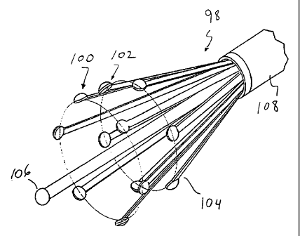
(BRIEF DESCRIPTION OF THE DRAWINGS
FIGURE 1 is a diagram of an energy application system with a partial
cutaway view of a catheter showing both the working end which includes a
plurality of outwardly expandable electrodes for applying energy to tissue and
S the connecting end', which is connected to a power source controlled by a
microprocessor controller for controlling the energy applied to the electrodes
of the working end;
FIG. 2 is a cross-sectional view of the working end of a first
embodiment of a catheter in accordance with aspects of the invention
depicting the electrodes in a fully expanded position;
FIG. 2a is an end view of the working end of the first embodiment of
the catheter taken ~aiong line 2a-2a of FiG. 2;
FIG. 3 is a cross-sectional view of the working end of the first
embodiment depicting the electrodes in a fully retracted position;
FIG. 4 is a cross-sectional view of the working end of a second catheter
in accordance with principles of the invention depicting the electrodes in a
fully expanded position;
FIG. 4a is an end view of the second embodiment of the invention taken
along line 4a-4a of FIG. 4;
FIG. 5 is a cross-sectional view of the working end of the second
embodiment of the catheter of FIG. 4 depicting the electrodes in a fully
retracted position;
FIG. 6 is a cross-sectional view of an anatomical structure containing
the catheter of FIG. 2 with the electrodes in apposition with the anatomical
structure;
FIG. 6a is an end view of the anatomical structure containing the
catheter taken along line 6a-6a of FIG. 6;
FIGS. 7a through 7c are cross-sectional views of the anatomical
structure containing a catheter in accordance with the first embodiment of the
invention and depicting the anatomical structure at various stages of
ligation;
CA 02341251 2001-02-20
WO 00/10475 PCT/US99/19193
I3
FIG. 8 is a cross-sectional view of an anatomical structure containing a
catheter in accordance with the second embodiment of the invention as
depicted in FIG 4;
FIG. 8a is an end view of the anatomical structure containing the
catheter taken along line 8a-8a of FIG. 8;
FIGS. 9a and 9b are cross-sectional views of the anatomical structure
containing the catheter in accordance with the second embodiment of the
invention and depicting the anatomical structure at various stages of
ligation;
FIG. I O is a cross-sectional view of the working end of a third
embodiment of a catheter in accordance with the invention depicting the
electrodes in a fully retracted position;
FIG. IOa is an end view of the working end of the third embodiment of
the catheter taken along line l0a-l0a of FIG. 10;
FIG. 11 is a cross-sectional view of the working end of the third
embodiment depicting the electrodes in a fully expanded position;
FIG. 12 is a cross-sectional view of an anatomical structure containing
the catheter of FIG. 10 with the electrodes in apposition with the anatomical
structure;
FIG. 13 is a cross-sectional view of the anatomical structure containing
the catheter of FIG. 10 where the anatomical structure is being iigated by the
application of energy from the electrodes;
FIG. 14 is a cross-sectional view of an anatomical structure containing
the catheter of FiG. IO with the electrodes in apposition with the anatomical
structure where external compression is being applied to reduce the diameter
of the hollow structure before the application of energy from the electrodes
to
ligate the structure;
FIG. 15 is a side view of another embodiment of an electrode catheter
having a balloon and a coaxial fluid channel;
FIG. 16 is a view of the balloon and catheter of FIG. 15 showing the
balloon inflation ports formed in an inflation sheath of the catheter, also
showing the inflation lumen that communicates with the inflation ports;
CA 02341251 2001-02-20
WO 00!10475 PCT/US99/19193
14
FIG. I7 is a cross-sectional view of an anatomical structure containing
another embodiment of the catheter having a balloon located proximal to
bowable arms with. electrodes, the portion of the catheter distal to the
balloon
having perfusion holes;
FIG. 18 is a side view of another embodiment of an electrode catheter
having a covering spanning the splayed leads of the electrodes extended out
the catheter;
FIG. 19 is a side view of another embodiment of an electrode catheter
having a balloon and a coaxial fluid channel located proximal to expandable
leads, the balloon having openings for receiving blood to maintain deployment
of the balloon;
FIG. 20 is a side view of another embodiment of an electrode catheter
having a balloon arid a coaxial fluid channel located proximal to expandable
leads, the balloon having openings for receiving blood to maintain deployment
of the balloon;
FIG. 21 is a partial cross-sectional side view of another embodiment of
an electrode catheter having an expandable section;
FIG. 22 is a partial cross-sectional side view of the embodiment of an
electrode catheter of FIG. 21 in an expanded condition;
FIG. 23 is a scide view of an embodiment of an electrode catheter having
two pluralities of longitudinally-separated expandable electrodes in a
retracted
condition;
FIG. 24 is a side view of the embodiment of the electrode catheter of
FIG. 23 with both pluralities of the electrodes in expanded configurations;
FIG. 25 is a partial cross-sectional view of the embodiment of an
electrode catheter of FIG 23;
FIG. 26 is a side view of another embodiment of an electrode catheter
having two pluralities of longitudinally-separated expandable electrodes in a
retracted condition;
CA 02341251 2001-02-20
WO 00/10475 PCT/US99/19193
FIG. 27 is a side view of another embodiment of an electrode catheter
having two pluralities of longitudinally-separated expandable electrodes in a
retracted condition;
FIG. 28 is a ;side view of another embodiment of an electrode catheter
5 having two pluralities of longitudinally-separated expandable electrodes in
a
retracted condition;
FIG. 29 is a view of a catheter used in a method in accordance with the
invention to treat a hemorrhoid;
FIG. 30 is a Yriew of a catheter used in a method in accordance with the
10 invention to treat an esophageal varix; and
FIG. 31 is a ~~iew of a catheter used in a method in accordance with the
invention for ligating a fallopian tube.
DETAILED DESCRIPTION OF THE EMBODIMENTS
Turning now to the drawings with more particularity wherein like
15 reference numerals indicate like or corresponding elements among the
figures,
shown in FIG. 1 is a catheter 10 for applying energy to an anatomical
structure
such as a vein. The catheter 10 includes an outer sheath 12 having a distal
orifice 14 at its working end 15. The connector end 17 of the outer sheath 12
is attached to a handle 16 that includes an electrical connector 18 for
interfacing with a power source 22, typically an RF generator, and a
microprocessor controller 23. The power source 22 and microprocessor 23 are
usually contained in one unit. The controller 23 controls the power source 22
in response to external commands and data from a sensor, such as a
thermocouple, located at an intraluminal venous treatment site. In another
embodiment, the user can select a constant power output so that automated
temperature control is not present and the user can manually adjust the power
output in view of th.e temperature on a display readout. The catheter 10
includes an expandable electrode device 24 (partially shown) that moves in
and out of the outer sheath 12 by way of the distal orifice 14. The electrode
device includes a plurality of electrodes which can be expanded by moving the
CA 02341251 2001-02-20
WO 00/10475 PCT/US99/19193
16
electrodes within the shaft, or by moving the outer shaft relative to the
electrodes. Although FIG. 1 illustrates a plurality of electrodes surrounding
a
single central electrode, different electrode configurations will be described
for
the catheter.
Contained within the outer sheath 12 is an inner sheath 28 or inner
member. A fluid port 21 communicates with the interior of the outer sheath
12. The catheter I O can be periodically flushed out with saline through the
port 21. The flushi~,ng fluid can travel between the outer sheath and the
inner
sheath. The port also allows for the delivery of drug therapies. Flushing out
the catheter prevents the buildup of biological fluid, such as blood, within
the
catheter 10. The treatment area of the hollow anatomical structure such as a
vein can be flushed with a fluid such as saline, or a dielectric fluid, in
order to
evacuate blood from the treatment area of the vein so as to prevent the
formation of coagulum or thrombosis. The use of a dielectric fluid can
minimize unintended heating effects away from the treatment area. The
dielectric fluid prevents the current of RF energy from flowing away from the
vein wall.
In one embodiment, the catheter 10 includes a lumen which begins at
the distal tip of the outer sheath 12 and runs substantially along the axis of
the
outer sheath 12 before terminating at the guide-wire port 20 of the handle 16.
A guide wire can be introduced through the lumen of the catheter i0 for use in
guiding the catheter to the desired treatment site. Where the catheter is
sized
to treat smaller veins, the outer diameter of the catheter may not allow for a
fluid flush between the outer sheath 12 and the inner sheath 28. However, a
fluid flush can be introduced through the lumen for the guide wire in such an
embodiment.
Referring now to FIGS. 2, 2a, 3, 4, 4a and S, the outer sheath 12
includes a shell 44 .and a tip portion 46. To provide an atraumatic tip for
the
catheter 10 as it is manipulated through the vein, the tip 46 is preferably
tapered inward at ins distal end or is "nosecone" shaped. The tip 46, however,
can have other shapes that facilitate tracking of the catheter I0 over a guide
CA 02341251 2001-02-20
WO 00/10475 PCT/US991i9193
17
wire and through the bends in the venous vascular system. The nosecone-
shaped tip 46 can, for example, be fabricated from a polymer having a soft
durometer, such as 70 Shore A. The shell 44 comprises a biocompatible
material having a low coefficient of friction. In one configuration, the outer
sheath I2 is sized to fit within a venous lumen and for example may be
between 5 and 9 French, which corresponds to a diameter of between 1.7 mm
(0.07 in) and 3.0 rnm (1.2 in), or other sizes as appropriate.
The electrode device 24 contains a number of leads, including insulated
primary leads 30 and, in some embodiments, a secondary lead 31. Preferably,
the leads are connected to the power source 22 (FIG. 1) such that the polarity
of the leads may bc: switched as desired. Alternately, a microprocessor
controller can be used to switch the polarity, as well as control other
characteristics of the power for the electrode device. Thus the electrode
device
can operate in either a bipolar or a monopolar configuration. When adjacent
primary leads 30 have opposite polarity the electrode device 24 operates as a
bipolar electrode device. When the primary leads 30 are commonly charged
the electrode device 24 can operate as a monopolar electrode device. When
the primary leads 3'~0 are commonly charged, and a secondary lead 31 has an
opposite polarity, the electrode device 24 operates as a bipolar electrode
device. The embodiment of the invention shown in FIGS. 2 and 3 depict an
electrode device 24 having four primary leads 30 and a secondary lead 31,
while the embodiment of the invention shown in FIGS. 4 and S depict an
electrode device 24 having only four primary leads. The invention is not
limited to four primary leads 30; more or fewer leads may be used in either
2S embodiment. The number of leads can be dependent on the size or diameter
of the hollow anatomical structure to be treated. The apposed electrodes
should be kept within a certain distance of one another. Larger vessels may
require more primary leads to ensure proper current density and proper heat
distribution.
The insulation on each of the leads 30, 31 may be removed at the distal
end 32, 33 to expose the conductive wire. In the first configuration as shown
CA 02341251 2001-02-20
WO OO/i0475 PCT/US99/19i93
18
in FIGS. 2, 2a, and 3, each electrode 34 has a hemispherical shape. In a
second configuration, the electrode can have either a generally spherical
shape
or a spoon shape. As shown in FIGS. 4, 4a and 5, the electrodes have a spoon
shape which can bfi combined to form a sphere or other shape so as to
minimize its prof le when the vein collapses. The electrodes 34 are either
integrally formed at the distal end 32, soldered, or otherwise formed to the
distal end of each primary lead 30. It is to be understood that when the
distal
end 32 is referred to as acting as an electrode, this is not limited to where
the
electrode 34 is integrally formed at the distal end 32. For example, the
distal
end can apply energy to the surrounding tissue where there is an electrode
integrally formed at the distal end, or where an electrode is separately
soldered to the distal end, or where there is another energy delivery device
located at the distal end. The electrode 34 typically has a diameter greater
than the diameter of the primary lead 30. For example; the primary lead 30
may have a diameter ranging from 0.18 mm (0.007 in.) to 0.28 mm (0.011
in.), while the electrode 34 has a diameter of 0:36 mm (0.014 in.) to 0.51 mm
(0.020 in.). The primary leads 30 and the electrodes 34 are preferably made
from a biologically-compatible material such as stainless steel. The
insulation
surrounding the primary leads 30 generally has a thickness of between 0.03
mm (0.001 in.) anc~: 0.06 mm (0.0025 in.}, resulting in a combined lead-
insulation diameter of between 0.23 mm (0.009 in.) and 0.41 mm (0.016 in.).
In an alternate configuration, as shown in FIGS. 2 and 3, each primary lead 30
is strip-shaped with a width from 0.76 mm (0.03 in.) to 1.0 mm (0.04 in) and
a thickness of approximately 0.13 mrn (0.005 in.), while the secondary lead 31
is typically tubular-shaped. It should be noted that these dimensions are
provided for illustrative purposes, and not by way of limitation. A
hemispherical electrode 34 is shaped at the distal end, as for example, by
sanding down a sixteenth-inch (1.6 mm) diameter sphere which is soldered to
the distal end 32 of the primary lead 30. The electrodes can also be
constructed by stamping the desired shape or configuration from the
conductive lead. The electrode is integral with the Lead, and the remainder of
CA 02341251 2001-02-20
WO 00/10495 PCTIUS99/19193
19
the lead is insulated. The distal end 33 of the secondary lead 31 preferably
includes a generally spherically-shaped electrode 35.
An alignment device 36 arranges the leads 30,37. such that they are
mounted to the catheter at only their proximal ends and so that separation is
maintained betlvee~n the leads within, and distal to the alignment device. The
leads can form cantilevers when mounted on the alignment device. A
preferred configurarion of the alignment device 36 includes a plurality of off-
center, axially-aligned lamina 38 which are substantially symmetrically
positioned relative to the axis of the alignment device 36. The alignment
device 36 is formecL, for example, by extruding the plurality of axially-
aligned
lamina 38 through a solid cylinder composed of a dielectric material, such as
polyamide. Each lead 30 passes through an individual off-center lumen 38
and exits out the rear of the alignment device 36. The alignment device 36
may further includE: a central lumen 48 that may be aligned with the axis. In
some embodiments the central lumen 48 is used for accepting a guide wire or
for the delivery or perfusion of medicant and cooling solution to the
treatment
area during application of RF energy. In other embodiments, the central
Iumen 48 may be used for the secondary Iead 31. The alignment device 36
may also further include an auxiliary lumen 47 for additional leads, such as
the leads of a thermocouple used as a temperature sensor. The alignment
device 36 comprises a dielectric material to prevent or minimize any coupling
effect the leads 30, 31 may have with each other and, if present, the guide
wire. The length of the alignment device is, for example, 12.5 mm (0.5 in.) to
19.0 mm (0.75 in.) in one embodiment. However, these dimensions are
provided For purposes of illustration and not by way of limitation.
In the embocLiment of the invention shown in FIGS. 2, 2a and 3, the
inner sheath 28 is attached to the alignment device 36 and extends beyond the
rear 37 of the alignment device. Preferably, the inner sheath 28 completely
surrounds the exterior wall of the alignment device 36 and is mounted to it by
adhesive or press fit or in other manner such that it remains in a fixed
position
relative to the inner sheath. The inner sheath and alignment device can act as
CA 02341251 2001-02-20
WO 00/10475 PCTIUS99/19193
an inner member relative to the outer sheath. The inner sheath 28 comprises a
biocompatible matE:rial with a low coefficient of friction. The inner sheath
28
provides a pathway for the interconnection between the leads 30, 31 and the
electrical connector 18 (FIG. 1). This interconnection may occur in any of
5 several ways. The leads 30, 3I themselves may be continuous and run the
entire length of the inner sheath 28. In the alternative (not shown), the
positively charged leads 30, 3I may couple with a common positively charged
conductor housed in the inner sheath 28. Likewise, the negatively charged
leads 30, 31 may couple with a common negative conductor. Preferably, the
10 leads 30, 31 are connected to a conductor that allows for the polarity of
the
leads to be switched. The conductor may comprise, for example, a 36 gauge
copper lead with a polyurethane coating. The coupling may occur at any point
within the inner shE;ath 28. To reduce the amount of wire contained in the
catheter, it is advantageous to couple the leads 30, 31 at the point where the
15 leads exit the rear 37 of the alignment device 36. To add further stability
to
the electrode device: 24, it is preferred that bonding material 40 surround
the
leads 30, 31 at the front end of the alignment device 36. In this embodiment,
the leads 30, 31 exit through the distal orifice 14 as the outer sheath I2 is
retracted backwards over the alignment device 36. The inwardly tapered tip
20 46 impedes the retracting movement of the outer sheath 12 to prevent the
exposure of the alignment device 36.
FIG. 3 shows the leads 30 and 31 in the retracted position where all
leads are within the nosecone-shaped tip portion 46 and the outer shell 44.
The alignment device 36 has been moved relative to the outer shell 44. The
soft nosecone provides an atraumatic tip for when the catheter is maneuvered
through the tortuous venous system. The electrode at the distal end of the
secondary lead 31 can be sized to approximately the same size as the opening
formed in the nosecone 46. The nosecone forms a closed atraumatic tip
together with the electrode of the secondary lead when the alignment device is
retracted into the outer sheath of the catheter. This can present an
atraumatic
CA 02341251 2001-02-20
WO 00/I0475 PCT/US99I19193
21
tip even where the nosecone is not constructed from a material having a soft
durometer.
Referring now to FIGS. 4 and 5, in another embodiment, the alignment
device 36 is attached to the outer sheath 12 and thereby remains immobile in
relation to it. The inner sheath 28 is movably positioned at the rear of the
alignment device 36 and again provides a pathway for the interconnection
between the primary leads 30 and the electrical connector 18 (FIG. I). In
some embodiments the inner sheath 28 contains a guide-wire tube 49 that
runs the entire length of the inner sheath. The guide-wire tube 49 is aligned
to
communicate with the central lumen 48 of the alignment device 36 at one end
and with the guide-wire port 20 (FIG. 1) at the other end. The primary leads
30 may be continuous and run the entire length of the inner sheath 28 or they
may be coupled to common leads as previously described. The primary leads
30 are secured to the front end 27 of the inner sheath 28, as fox example with
a potting material 5.0, so that the movement of the inner sheath 28 results in
a
corresponding movement of the primary leads 30 through the lumina 38 of the
alignment device 3fi. In this embodiment, the primary leads 30 are not
secured to the alignment device 36 and in essence are free-floating leads in
the
axial direction. The primary leads 30 travel through the alignment device 36
and exit through the distal orifice 14 as the front end of the inner sheath 28
is
moved toward the rear 37 of the alignment device 36.
In the above embodiments, the primary leads 30 are formed, e. g., arced
or bent, to move away from each other and thereby avoid contact. The "distal
portion" of the primary leads 30 is the portion of the Iead which extends from
the front end of the alignment device 36 when the leads are fully extended
through the distal orifice I4. It is preferred that the distal portions 42 are
formed to move radiaily outward from each other relative to the axis of the
alignment device 3fi and form a symmetrical arrangement. This is shown in
both the embodiments of FIG. 2a and FIG. 4a. The degree of arc or bend in
the primary leads 30 may be any that is sufficient to radially expand the
leads
as they exit the outf:r sheath 12 through the distal orifice 14. It is
essential
CA 02341251 2001-02-20
WO 00/10475 PCT/US99/I9193
22
that the degree of the arc or bend be sufficient to provide enough force so
that
the primary leads 30 expand through blood and the electrodes 34 come in
apposition with the vein wall. The electrodes are preferably partially
embedded in the vein wall to assure full contact. The rounded portion of the
electrode is embedded into the vein wall to achieve full surface apposition so
that the entire uninsulated surface area of the electrode is in contact with
venous tissue for effective current distribution. The surface area of the
electrodes in contact with the venous tissue preferably is sufficient to avoid
a
high current density which may lead to spot heating of the venous tissue. The
heating effect is preferably distributed along a circumferential band of the
vein. The apposed f:lectrodes should be spaced no more than 4 or 5
millimeters from an~e another along the circumference of the vein. Thus, the
electrode arrangemf:nt is related to the size or diameter of the vein being
treated. Other properties of the primary leads 30, such as lead shape and
insulation thickness;, affect the push force of the lead and the degree of arc
or
bend must be adjusted to compensate for these factors. For example, in one
configuration of the electrode device 24, a wire having a diameter of between
0.18 mm {0.007 in) and 0.28 mm (0.011 in) with a total insulation thickness
of between 0.05 rnm (0.002 in} to 0.13 mm (0.005 in) is arced or bent at an
acute angle to provide sufficient apposition with the anatomical structure. It
is
to be understood that these dimensions are provided for illustrative purposes,
and not by way of limitation.
Other techniques for expanding the leads outwardly once they have
been extended from the working end of the catheter may be possible. For
example, the leads nnay be straight but are mounted in the alignment device at
an angle such that they are normally directed outward.
For increased appositional force, it is preferred that the primary leads
be strip-shaped, that is rectangular in cross section, with dimensions, for
example, of a width from 0.76 mm (0.030 in.) to 1.0 mm (0.039 in} and a
30 thickness of approximately 0.13 mm {0.005 in.). The rectangular cross
sectian
provides increased resistance to bending in the width dimension but allows
CA 02341251 2001-02-20
WO 00/10475 PCTIUS99119193
23
bending more freely in the thickness dimension. This strip-shaped
configuration of thc~ primary leads 30 is shown in FIGS. 2, 2a, and 3 and
provides for increased stability in the lateral direction while allowing the
necessary bending in the radial direction. In FIGS. 2, 2a, and 3, each primary
S lead comprises a rectangular cross section mounted in relation to the
catheter
such that the thinner dimension of the rectangular cross section is aligned
with
the direction of expansion of the lead. The leads are less likely to bend
sideways when expanded outward, and a uniform spacing between leads is
more assured. Uniform spacing promotes uniform heating around the venous
tissue which is in apposition with the electrodes at the distal ends of the
leads.
The length of the distal portion of the leads 30 also affects the
configuration of the electrode device 24. The maximum distance between two
mutually opposed electrodes 34; i.e., the effective diameter of the electrode
device 24, is affected by the bend degree and length of the distal portion 42.
The longer the length of the distal portion 42 the greater the diameter of the
electrode device 24. Accordingly, by changing the distal portion 42 length and
arc or bend degree, the catheter 10 can be configured for use in differently
sized anatomical stzuctures.
Different numbers of leads 30, 31 can be employed with the catheter.
The number of leads 30, 31 is limited by the diameter of the alignment device
36 and the number of lumina 36, 38, 47 that can be extruded through the
alignment device. in a bipolar configuration, an even number of primary leads
are preferably available to form a number of oppositely charged electrode
pairs. The electrodes in apposition with the anatomical structure should be
25 maintained within a certain distance of each other. In a monopolar
configuration, any number of commonly charged leads 30 can be present. In
the monopolar mode, distribution of RF energy through the anatomical tissue
is obtained by creating a return path for current through the tissue by
providing a return device at a point external from the tissue, such as a large
30 metal pad.
CA 02341251 2001-02-20
WO 00/10475 FCT/US99/19193
24
Now referring again to FIG. 1, an actuator 25 controls the extension of
the electrode device: 24 through the distal orifice I4. The actuator 25 may
take the form of a switch, lever, threaded control knob, or other suitable
mechanism, and is preferably one that can provide fine control over the
movement of the outer sheath I2 or the inner sheath 28, as the case may be.
In one embodiment of the invention, the actuator 25 (FIG. 1) interfaces with
the outer sheath I2 (FIG. 2, 2a and 3) to move it back and forth relative to
the
inner sheath 28. In another embodiment the actuator 25 (FIG. 1) interfaces
with the inner sheath 28 (FIGS. 4, 4a and 5) to move it back and forth
relative
to the outer sheath 12. The relative position between the outer sheath and
inner sheath is thus controlled, hut other control approaches may be used.
Referring again to FIGS. 2, 2a, 3, 4, 4a and 5, the catheter 10 includes a
temperature sensor 26, such as a thermocouple. The temperature sensor 26 is
mounted in place on an electrode 34 so that the sensor 26 is nearly or is
substantially flush with the exposed surface of the electrode 34. The sensor
26
is shown in the drawings as protruding from the electrodes for clarity of
illustration only. The sensor 26 senses the temperature of the portion of the
anatomical tissue that is in apposition with the exposed electrode surface.
Monitoring the temperature of the anatomical tissue provides a good
indication of when shrinkage of the tissue is ready to begin. A temperature
sensor 26 placed an the electrode facing the anatomical tissue provides an
indication of when shrinkage occurs (70 ° C or higher) and when
significant
amounts of heat-induced coagulum may begin to form on the electrodes.
Therefore maintaining the temperature above 70 degrees Centigrade produces
a therapeutic shrinkage of the anatomical structure. Application of the RF
energy from the electrodes 34 is halted or reduced when the monitored
temperature reaches or exceeds the specific temperature that was selected by
the operator, typically the temperature at which anatomical tissue begins to
cauterize. The temperature sensor 26 interfaces with the controller 23 (FIG.
1) through a pair of sensor leads 45 which preferably run through the
auxiliary
lumen 47 and then through the inner sheath 28. The signals from the
CA 02341251 2001-02-20
WO 00/10475 PCT/US99/19193
temperature sensor 26 are provided to the controller 23 which controls the
magnitude of RF energy supplied to the electrodes 34 in accordance with the
selected temperature criteria and the monitored temperature. Other
techniques such as impedance monitoring, and ultrasonic pulse echoing can be
5 utilized in an automated system which shuts down or regulates the
application
of RF energy from t:he electrodes to the venous section when sufficient
shrinkage of the vein is detected and to avoid overheating the vein.
Impedance can be used to detect the onset of coagulum formation.
Referring now to FIGS. 6, 6a and 7a through 7c, in the operation of one
10 embodiment of the catheter 10, the catheter is inserted into a hollow
anatomical structure, such as a vein 52. The catheter is similar to the
embodiment discussed in connection with FIGS. 2 and 3. The catheter 10
further includes an external sheath 60 through which a fluid can be delivered
to the treatment site. In this embodiment, the fluid port {not shown)
15 communicates with the interior of the external sheath 60, and fluid is
delivered from between the external sheath 60 and the outer sheath 12. The
external sheath 60 surrounds the outer sheath 12 to form a coaxial channel
through which fluid may be flushed.
Fluoroscopy, ultrasound, an angioscope imaging technique, or other
20 technique may be used to direct the specific placement of the catheter and
confirm the position in the vein. The actuator {not shown) is then operated to
shift the outer sheath relative to the inner sheath by either retracting the
outer
sheath l2~backward or advancing the inner sheath 28 forward to expose the
leads 30, 31 through the distal orifice 14. As the leads 30, 31 exit the
distal
25 orifice 14, the primary leads 30 expand radially outward relative to the
axis of
the alignment device 36, while the secondary lead 31 remains substantially
linear. The primary Ieads 30 continue to move outward until apposition with
the vein wall 54 occurs and the outward movement of the primary leads 30 is
impeded. The prinnary leads 30 contact the vein along a generally
circumferential band of the vein wall 54. This outward movement of the
primary Ieads 30 occurs in a substantially symmetrical fashion. As a result,
the
CA 02341251 2001-02-20
WO 00/10475 PCT/IlS99/19193
26
primary-lead electrodes 34 are substantially evenly spaced along the
circumferential band of the vein wall 54. The central-lead electrode 35 is
suspended within the vein 52 without contacting the vein wall 54.
When the electrodes 34 are positioned at the treatment site of the vein,
the power supply 2 2 is activated to provide suitable RF energy. One suitable
frequency is 5I0 khtz. One criterion used in selecting the frequency of the
energy to be applied is the control desired over the spread, including the
depth, of the thermal effect in the venous tissue. Another criterion is
compatibility with filter circuits for eliminating RF noise from thermocouple
signals.
In bipolar operation, the primary leads 30 are initially charged such that
adjacent leads are oppositely charged while the secondary lead is electrically
neutral. These multiple pairs of oppositely charged leads 30 form active
electrode pairs to produce an RF field between them. Thus, discrete RF fields
are set up along the circumferential band of the vein wall 54. These discrete
fields form a symmcarical RF Field pattern along the entire circumferential
band of the vein wall 54; as adjacent electrodes 34 of opposite polarity
produce RF fields between each other. A uniform temperature distribution can
be achieved along tike vein wall being treated.
The RF ener~,ry is converted within the adjacent venous tissue into heat,
and this thermal effect causes the venous tissue to shrink, reducing the
diameter of the vein. A uniform temperature distribution along the vein wall
being treated avoids the formation of hot spots in the treatment area while
promoting controlled uniform reduction in vein diameter. The thermal effect
produces structural transfiguration of the collagen fibrils in the vein. The
collagen fibrils shorten and thicken in cross-section in response to the heat
from the thermal effect. As shown in FIG 7a, the energy causes the vein wall
54 to collapse around the primary-lead electrodes 34. The wall 54 continues
to collapse until further collapse is impeded by the electrodes 34. The
electrodes are pressed farther and farther together by the shrinking vein wall
54 until they touch and at that point, further collapse or ligation of the
wall 54
CA 02341251 2001-02-20
WO 00110475 PCTlUS99/19193
27
is impeded. Upon collapse of the vein wall 54 around the primary-Iead
electrodes 34, the polarity of the primary-lead electrodes is switched so that
all
primary-lead electrodes are commonly charged. The switching of polarity for
the leads need not be instantaneous. The application of RF energy can be
ceased, the polarir;~ switched, and then RF energy is applied again at the
switched polarity. The secondary-Iead electrode 35 is then charged so that its
polarity is opposite that of the primary-lead electrodes 34. The RF field is
set
up between the primary-lead electrodes 34 and the secondary-Iead electrode
35.
The catheter 10 is then pulled back while energy is applied to the
electrode device. As shown in FIG. 7b, while the catheter 10 is being pulled
back, the primary-lead electrodes 34 remain in apposition with the vein wall
54 while the secondary-lead electrode 35 comes in contact with the portion of
the vein wall previously collapsed by the primary-lead electrodes 34.
Accordingly, RF energy passes thraugh the vein wall 54 between the primary-
lead electrodes 34 and the secondary-lead electrode 35 and the vein wall
continues to collapse around the secondary-lead electrode 35 as the catheter
10 is being retracted. As shown in FIG. 7c, ligation in accordance with this
method results in an occlusion along a length of the vein 52. A lengthy
occlusion, as opposed to an acute occlusion, is stronger and less susceptible
to
recanalization.
A similar result is achieved when the catheter 10 having both primary
and secondary leads is operated in a monopolar manner. In a monopolar
operation, the secondary-lead electrode 35 remains neutral, while the primary
leads 30 are commonly charged and act in conjunction with an independent
electrical device, such as a large low-impedance return pad (not shown)
placed in external contact with the body, to farm a series of discrete RF
fields.
These RF fields are substantially evenly spaced around the circumference of
the vein and travel along the axial length of the vein wall causing the vein
wall
to collapse around the primary-lead electrodes. Upon collapse of the vein
wall,
the secondary-lead electrode is charged to have the same polarity as that of
the
CA 02341251 2001-02-20
WO 00/10475 PCT/i1S99/19193
28
primary-lead electrodes. The electrode device is retracted and the vein wall
collapses as described in the bipolar operation.
In either bipolar or monopolar operation the application of RF energy is
substantially symmetrically distributed through the vein wall, regardless of
the
diameter of the vein 52. This symmetrical distribution of RF energy increases
the predictability and uniformity of the shrinkage and the strength of the
occlusion. Furthermore, the uniform distribution of energy allows for the
application of RF energy for a short duration and thereby reduces or avoids
the
formation of heat-induced coagulum on the electrodes 34. The leads,
including the non-convex outer portion of the electrode, are insulated to
further prevent heating of the surrounding blood.
Fluid can be delivered before and during RF heating of the vein being
treated through a coaxial channel formed between the external sheath 60 and
the outer sheath 1~:. It is to be understood that another lumen can be formed
in the catheter to deliver fluid to the treatment site. The delivered fluid
displaces or exsanguinates blood from the vein so as to avoid heating and
coagulation of blood. Fluid can continue to be delivered during RF treatment
to prevent blood from circulating back to the treatment site. The delivery of
a
dielectric fluid increases the surrounding impedance so that RF energy is
directed into the tissue of the vein wall.
Referring now to FIGS. 8, 8a, 9a and 9b, in the operation of an
alternate embodiment of the catheter 10 that may be used with a guide wire
53. As in the previous embodiment, the catheter 10 is inserted into a hollow
anatomical structure, such as a vein 52. The guide wire 53 is advanced past
the point where energy application is desired. The catheter 10 is then
inserted
over the guide wire. 53 by way of the central lumen 48 and the guide wire tube
49 (FIG. 4) and is advanced over the guide wire through the vein to the
desired point. The guide wire 53 is typically pulled back or removed before RF
energy is applied to the electrode device 24.
The actuator 25 (FIG. 1) is then manipulated to either retract the outer
sheath 12 backward, or advance the inner sheath 28 forward to expose the
CA 02341251 2001-02-20
WO 00/10475 PCT/US99/19193
29
leads 30 through the distal orifice 14. The leads 30 exit the distal orifice
14
and expand radially outward relative to the axis of the alignment device 36.
The leads 30 continue to move outward until apposition with the vein wall 54
occurs. The leads 30 contact the vein along a generally circurnferential band
S of the vein wall 54. This outward movement of the leads occurs in a
substantially symmetrical fashion. As a result, the electrodes 34 are
substantially evenly spaced along the circumferentiai band of the vein wall
54.
Alternately, the elecarodes can be spaced apart in a staggered fashion such
that
the electrodes da not lie along the same plane. For example, adjacent
electrodes can extend different lengths from the catheter so that a smaller
cross-sectional profile is achieved when the electrodes are collapsed toward
one another.
When the electrodes 34 are positioned at the treatment site of the vein,
the power supply 2a: is activated to provide suitable RF energy to the
electrodes 34 so that the catheter 10 operates in either a bipolar or
monopolar
manner as previously described. As shown in FIGS. 9a and 9b, the energy
causes the vein wall 54 to collapse around the electrodes 34 causing the leads
to substantially straighten and the electrodes to cluster around each other.
The wall 54 continues to collapse until further collapse is impeded by the
electrodes 34 (FIG. !fib). At this point the application of energy may cease.
The electrodes can be configured to form a shape with a reduced profile when
collapsed together. The electrodes can also be configured and insulated to
continue applying RF energy after forming a reduced profile shape by the
collapse of the vein wall. The catheter 10 can be pulled back to ligate the
adjacent venous segment. If a temperature sensor 26 is included, the
application of energy may cease prior to complete collapse if the temperature
of the venous tissue rises above an acceptable Level as defined by the
controller
23.
Where the catheter includes a fluid delivery Lumen (not shown), fluid
can be delivered before and during RF heating of the vein being treated. The
fluid can displace blood from the treatment area in the vein to avoid the
CA 02341251 2001-02-20
WO 04/10475 PCT/US99/19i93
coagulation of blood. The fluid can be a dielectric medium. The fluid can
include an anticoagulant such as heparin which can chemically discourage the
coagulation of blood at the treatment site.
After completing the procedure for a selected venous section, the
5 actuator mechanisnn causes the primary Ieads to return to the interior of
the
outer sheath 12. Ei'.ther the outer sheath or the inner sheath is moved to
change the position. of the two elements relative to one another. Once the
leads 30 are within the outer sheath T2, the catheter 10 may be moved to
another venous section where the ligation process is repeated. Upon treatment
10 of all venous sites, i:he catheter 10 is removed from the vasculature. The
access point of the vein is then sutured closed, or local pressure is applied
until
bleeding is controlled.
Another emtrodiment of the catheter is illustrated in FIG. 10. The inner
member or sheath 28 is contained within the outer sheath 12. The inner
15 sheath is preferably constructed from a flexible polymer such as polyimide,
polyethylene, or nylon, and can travel the entire length of the catheter. The
majority of the catheter should be flexible so as to navigate the tortuous
paths
of the venous system. A hypotube having a flared distal end 39 and a circular
cross-sectional shape is attached over the distal end of the inner sheath 28.
20 The hypotube is preferably no more than about two to three centimeters in
length. The hypotube acts as part of the conductive secondary lead 31. An
uninsulated conductive electrode sphere 35 is slipped over the hypotube. The
flared distal end of the hypotube prevents the electrode sphere from moving
beyond the distal end of the hypotube. The sphere is permanently affixed to
25 the hypotube, such as by soldering the sphere both front and back on the
hypotube. The majority or the entire surface of the spherical electrode 35
remains uninsulate<i. The remainder of the hypotube is preferably insulated so
that the sphere-shaped distal end can act as the electrode. For example, the
hypotube can be covered with an insulating material such as a coating of
30 parylene. The interior lumen of the hypotube is lined by the inner sheath
28
CA 02341251 2001-02-20
WO 00/10475 PCT/US99/19193
31
which is attached to the flaired distal end of the hypotube by adhesive such
as
epoxy.
Surroundin~;.the secondary lead 31 and sphere-shaped electrode 35 are
a plurality of primary leads 30 which preferably have a flat rectangular strip
shape and can act as arms. As illustrated in FIG. lI, the plurality of primary
leads are preferably connected to common conductive rings 62. This
configuration maintains the position of the plurality of primary leads, while
reducing the number of internal electrical connections. The rings 62 are
attached to the inner sheath 28. The position of the rings and the primary
leads relative to the outer sheath follows that of the inner sheath. As
earlier
described, the hypo~tube of the secondary lead 31 is also attached to the
inner
sheath 28. Two separate conductive rings can be used so that the polarity of
different primary leads can be controlled separately. For example, adjacent
primary leads can be connected to one of the two separate conductive rings so
IS that the adjacent leads can be switched to have either opposite polarities
or the
same polarity. The rings are preferable spaced closely together, hut remain
electrically isolated from one another along the inner sheath. Both the rings
and the hypotube are coupled with the inner sheath, and the primary leads 30
that are connected to the rings move together with and secondary lead while
remaining electrically isolated from one another. Epoxy or another suitable
adhesive can be used to attach the rings to the inner sheath. The primary
leads
from the respective rings each alternate with each other along the
circumference of the inner sheath. The insulation along the underside of the
leads prevents an electrical short between the rings.
The ring and. primary Ieads are attached together to act as cantilevers
where the ring forms the base and the rectangular primary leads operate as the
cantilever arms. The leads 30 are connected to the ring and are formed to
have an arc or bent( such that the leads act as arms which tend to spring
outwardly away from the catheter and toward the surrounding venous tissue.
Insulation along the underside of the Ieads and the rings prevents unintended
electrical coupling between the leads and the opposing rings. Alternately, the
CA 02341251 2001-02-20
WO OO/10a7S PCT/US99/i9193
32
leads are formed straight and connected to the ring at an angle, such that the
leads tend to expand or spring radialIy outward from the ring. The angle at
which the leads are attached to the ring should be sufficient to force the
primary distal ends and electrodes 34 through blood and into apposition with
the vein wall. Other properties of the primary leads 30, such as lead shape
and insulation thickness, affect the push force of the lead and the degree of
arc
or bend must be adjusted to compensate for these factors. The rectangular
cross section of they leads 30 can provide increased stability in the lateral
direction while alic>wing the necessary bending in the radial direction. The
I0 leads 30 are less likely to bend sideways when expanded outward, and a
uniform spacing bEaween leads is more assured. Uniform spacing between the
leads 30 and the dl;stal ends promotes uniform heating around the vein by the
electrodes 34.
The distal ends of the primary leads 30 are uninsulated to act as
electrodes 34 having a spoon or hemispherical shape. The leads can be
stamped to produce an integral shaped electrode at the distal end of the lead.
The uninsulated outer portion of the distal end electrode 34 which is to come
into apposition with the wall of the anatomical structure is preferably
rounded
and convex. The flattened or non-convex inner portion of the distal end is
insulated to minimize any unintended thermal effect, such as on the
surrounding blood in a vein. The distal end electrodes 34 are configured such
that when the distil ends are forced toward the inner sheath 12, as shown in
FIG. 10a, the distal ends combine to form a substantially spherical shape with
a profile smaller than the profile for the spherical electrode 35 at the
secondary distal end.
The outer sheath I2 can slide over and surround the primary and
secondary leads 30, 31. The outer sheath 12 includes an orifice which is
dimensioned to hare approximately the same size as the spherical electrode 3S
at the secondary distal end which functions as an electrode. A close or snug
fit
between the electrode 35 at the secondary distal end and the orifice of the
outer sheath 12 is achieved. This configuration provides an atraumatic tip for
CA 02341251 2001-02-20
WO 00110475 PCT/US99/19193
33
the catheter. The electrode 35 secondary distal end is preferably slightly
larger
than the orifice. The inner diameter of the outer sheath 12 is approximately
the same as the reduced profile of the combined primary distal end electrodes
34. The diameter of the reduced profile of the combined primary distal end
electrodes 34 is preferably less than the inner diameter of the outer sheath.
A fluid part (not shown) can communicate with the interior of the outer
sheath 12 so that fluid can be flushed between the outer sheath 12 and the
inner sheath 28. Alternately, a fluid port can communicate with a central
lumen 48 in the hyPotube which can also accept a guide wire. As previously
stated, the catheter 10 can be periodically flushed with saline which can
prevent the buildup of biological fluid, such as blood, within the catheter
20.
A guide wire can be introduced through the lumen 48 for use in guiding the
catheter to the desired treatment site. As previously described, a fluid can
be
flushed or delivered though the lumen as well. If a central lumen is not
desired, the lumen of the hypotube can be filled with solder.
Preferably, t:he primary leads 30 and the connecting rings are connected
to a power source :22 such that the polarity of the leads may be switched as
desired. This allows for the electrode device 24 to operate in either a
bipolar
or a monopolar configuration. When adjacent primary leads 30 have opposite
polarity, a bipolar electrode operation is available. When the primary leads
30
are commonly charged a monopolar electrode operation is available in
combination with a large return electrode pad placed in contact with the
patient. When the primary leads 30 are commonly charged, and a secondary
lead 31 has an opposite polarity, a bipolar electrode operation is available.
More or fewer leads may be used. The number of leads can be dependent on
the size or diameter of the hollow anatomical structure to be treated.
Although not shown, it is to be understood that the catheter 10 can
include a temperature sensor, such as a thermocouple, mounted in place on
the distal end or electrode 34 so that the sensor is substantially flush with
the
exposed surface of the electrode 34. The sensor senses the temperature of the
portion of the anatomical tissue that is in apposition with the exposed
CA 02341251 2001-02-20
WO 00/10475 PCT/US99119193
34
electrode surface. Application of the RF energy from the electrodes 34 is
halted or reduced when the monitored temperature reaches or exceeds the
specific temperature that was selected by the operator, such as the
temperature at which anatomical tissue begins to cauterize. Other techniques
S such as impedance monitoring, and ultrasonic pulse echoing can be utilized
in
an automated system which shuts down or regulates the application of RF
energy from the electrodes to the venous section when sufficient shrinkage of
the vein is detected and to avoid overheating the vein.
Referring now to FIGS. 12 through 14, in the operation of one
embodiment of the catheter 10, the catheter is inserted into a hollow
anatomical structure, such as a vein. Fluoroscopy, ultrasound, an angioscope
imaging technique, or another technique may be used to direct and confirm
the specific placement of the catheter in the vein. The actuator is then
operated to retract the outer sheath I2 to expose the leads 30, 31. As the
outer sheath no longer restrains the leads, the primary leads 30 move outward
relative to the axis defined by the outer sheath, while the secondary lead 3I
remains substantially linear along the axis defined by the outer sheath. The
primary leads 30 continue to move autward until the distal end electrode 34 of
the primary leads are placed in apposition with the vein wall 54 occurs and
the
, outward movement of the primary leads 30 is impeded. The primary leads 30
contact the vein along a generally circumferential area of the vein wall 54.
This outward movement of the primary leads 30 occurs in a substantially
symmetrical fashion so that the primary distal end electrodes 34 are
substantially evenly spaced. The central-lead electrode 35 is suspended within
the vein without contacting the vein wall 54.
When the electrodes 34 are positioned at the treatment site ofthe vein,
the power supply 22 is activated to provide suitable RF energy. In a bipolar
operation, the primary leads 30 are initially charged such that adjacent leads
are oppositely charf;ed while the secondary Lead is electrically neutral.
These
multiple pairs of oppositely charged leads 30 form active electrode pairs to
produce an RF field. between them, and form a symmetrical RF field pattern
CA 02341251 2001-02-20
WO 00/10475 PCT/US99/19193
along a circumferential band of the vein wall to achieve a uniform temperature
distribution along the vein wall being treated.
The RF energy produces a thermal effect which causes the venous tissue
to shrink, reducing the diameter of the vein. As shown in FIG 13, the energy
5 causes the vein wall 54 to collapse until further collapse is impeded by the
electrodes 34. The electrodes are pressed closer together by the shrinking
vein
wall. The electrodes 34 are pressed together to assume a reduced profile
shape which is sufficiently small so that the vein is effectively Iigated.
Upon
collapse of the vein wall 54 around the primary-lead electrodes 34, the
polarity
10 of the primary-Iead electrodes is switched so that all of the primary-Iead
electrodes are commonly charged. The secondary-lead electrode 35 is then
charged so that its polarity is opposite that of the primary-lead electrodes
34.
Where the primary electrodes 34 and the secondary electrode 35 are spaced
sufficiently close together, when the vein wall collapses around the primary
15 lead electrodes, the electrode at the distal end of the secondary lead can
also
come into contact with the a portion of the vein wall so that an RF field is
created between the primary electrodes 34 and the secondary electrode 35.
The catheter 10 is pulled back to ensure apposition between the
electrodes at the distal ends of the leads and the vein wall. When the
catheter
20 10 is being pulled back, the primary-lead electrodes 34 remain in
apposition
with the vein wall 54 while the secondary-lead electrode 35 comes in contact
with the portion of the vein wall previously collapsed by the primary-lead
electrodes 34. RF e:nergy passes through the venous tissue between the
primary-lead electrodes 34 and the secondary-lead electrode 35. L,igation as
25 the catheter is bein;~ retracted produces a lengthy occlusion which is
stronger
and less susceptible to recanalization than an acute point occlusion.
In a monopolar operation, the secondary-lead electrode 35 remains
neutral, while the primary leads 30 are commonly charged and act in
conjunction with an independent electrical device, such as a large low-
30 impedance return pad (not shown) placed in external contact with the body,
to form RF fields substantially evenly spaced around the circumference of the
CA 02341251 2001-02-20
WO 00/10475 PCTIUS99119193
36
vein. The thermal effect produced by those RF fields along the axial length of
the vein wall causes the vein wall to collapse around the primary-lead
electrodes. Upon collapse of the vein wall, the secondary-lead electrode is
charged to have the same polarity as that of the primary-lead electrodes. The
S electrode device is retracted as described in the bipolar operation.
In either bipolar or monopolar operation the application of RF energy is
substantially symmetrically distributed through the vein wall. As previously
described, the elecarodes should be spaced no more than 4 or 5 millimeters
apart along the circumference of the vein, which defines the target vein
diameter for a designed electrode catheter. Where the electrodes are
substantially evenly spaced in a substantially symmetrical arrangement, and
the spacing between the electrodes is maintained, a symmetrical distribution
of
RF energy increases the predictability and uniformity of the shrinkage and the
strength of the occlusion.
As shown in FIG. 14, after the electrodes 34 come into apposition with
the vein wall (FIG,. 12), and before the energy is applied to ligate the vein
(FIG. 13), an external tourniquet, such as an elastic compressive wrap or an
inflatable bladder with a window transparent to ultrasound, is used to
compress the anatomy, such as a leg, surrounding the structure to reduce the
diameter of the vein. Although the compressive force being applied by the
tourniquet may effectively ligate the vein, or otherwise occlude the vein by
flattening the vein, for certain veins, this compressive force will not fully
occlude the vein. .A fixed diameter electrode catheter in this instance would
not he effective. The electrodes 34 which are expanded outward by the
formed Ieads 30 can accommodate this situation.
The reduction in vein diameter assists in pre-shaping the vein to
prepare the vein to be molded to a ligated state. The use of an external
tourniquet also exsanguinates the vein and blood is forced away from the
treatment site. Coagulation of blood during treatment can be avoided by this
procedure. Energy is applied from the electrodes to the exsanguinated vein,
CA 02341251 2001-02-20
WO 00110475 PCTIUS99119193
37
and the vein is molded to a sufficiently reduced diameter to achieve ligation.
The external tourni'.quet can remain in place to facilitate healing.
The catheter can be pulled back during the application of RF energy to
ligate an extensive section of a vein. In doing so, instead of a single point
S where the diameter of the vein has been reduced, an extensive section of the
vein has been painted by the RF energy from the catheter. Retracting the
catheter in this manner produces a lengthy occlusion which is less susceptible
to recanalization. T'he combined use of the primary and secondary electrodes
can effectively produce a reduced diameter along an extensive length of the
vein. The catheter can be moved while the tourniquet is compressing the vein,
of after the tourniquet is removed.
Where the catheter includes a fluid delivery lumen, fluid can be
delivered to the vein before RF energy is applied to the vein. The delivered
fluid displaces blood from the treatment site to ensure that blood is not
present
1S at the treatment site, even after the tourniquet compresses the vein.
Where the tourniquet is an inflatable bladder with a window
transparent to ultrasound; an ultrasound transducer is used to monitor the
flattening or reduction of the vein diameter from the compressive force being
applied by the inflating bladder. The window can be formed from
polyurethane, or a stand-off of gel contained betyveen a polyurethane pouch.
A gel can be applied to the window to facilitate ultrasound imaging of the
vein
by the transducer. Ultrasound visualization through the window allows the
operator to locate the desired venous treatment area, and to determine when
the vein has been effectively ligated or occluded. Ultrasound visualization
2S assists in monitoring any pre-shaping of the vein in preparation of being
molded into a ligatf:d state by the thermal effect produced by the RF energy
from the electrodes"
After completing the procedure for a selected venous section, the
actuator causes the leads 30 to return to the interior of the outer sheath 12.
Once the leads 30 are within the outer sheath 12, the catheter 10 may be
moved to another venous section where the ligation process is repeated.
CA 02341251 2001-02-20
WO 40/10475 PCT/US99/19193
38
In another embodiment, as illustrated in FIG. I5, a balloon 64 is located
on the catheter, and can be inflated through ports 66 to occlude the vein. The
inflated balloon obstructs blood flow and facilitates the infusion of a high-
impedance fluid to the vein in order to reduce the occurrence of coagulation
by directing the energy into the vein wall. The inflation of the balloon to
occlude the vein before the application of energy can obviate the use of the
tourniquet to occlude the vein. Furthermore, this also allows the vein to be
occluded even for the deep veins where a compressive tourniquet may not be
able to compress tye vein to occlusion. It is to be understood that other
mechanisms can be: used to expand the diameter of the catheter to create an
impermeable barriE:r that occludes the vein.
Fluid 61 can. be delivered after inflation of the balloon 64 and before
the RF heating of the vein being treated through a coaxial channel 62 formed
between the external sheath 60 and the outer sheath 12. It is to be understood
that another lumen can be formed in the catheter to deliver fluid to the
treatment site. For example, the lumen through which the guide wire is
passed may be used for the delivery of fluid. The delivered fluid displaces or
exsanguinates the remaining blood from the treatment area of the vein so as to
avoid heating and coagulation of blood. Fluid can continue to be delivered
during RF treatment to prevent blood from circulating back to the treatment
site. The delivery of a high-dielectric fluid increases the surrounding
impedance so that l~F energy is directed into the tissue of the vein wall.
Less
energy is used because the energy is directed to the target; i.e., the vein
wall,
rather than being dissipated in the blood. Therefore, the vein wall can reach
the desired temperature more rapidly than in the case where energy is
permitted to reach the blood, which has a cooling effect. Additionally, blood
clotting is avoided with this approach, because the blood has been replaced
with another fluid such as deionized water mixed with heparin to displace
blood and prevent t:he formation of blood clots.
A partial cross-sectional view of this embodiment is shown in FIG. 16,
where an inflation :.heath 70 surrounds the external sheath 60 to provide a
CA 02341251 2001-02-20
WO 00110475 PCTIUS99/19193
39
coaxial inflation lumen 72 for the balloon 64. The inflation lumen 72 is in
fluid communication with the ports 66. Saline or any other suitable fluid can
be used to inflate the balloon.
As shown in the FIG. 17, in one embodiment, the balloon 64 can be
used in combination with bowable members or arms 76 having electrodes,
where perfusion holes 78 are formed in the Gatherer between the balloon 64
and the bowable arms 76. The balloon 64 in this embodiment is inflated
through a balloon :inflation lumen 72 (as shown in FIG. 16). The use of
bowable arms fox treating veins is discussed in U.S. patent application serial
no. 08/610,911, which is hereby incorporated by reference. The arms can be
constructed so as to spring radially outward from the catheter, yet offer
little
resistance in moving back toward the catheter as the vein diameter is
diminished to occlusion. An anti-coagulant or saline or a high-impedance fluid
can be introduced or flushed through the perfusion holes 78 in the catheter.
As discussed earlier, the high-impedance fluid forces blood away from the
venous treatment area and prevents the energy from being dissipated in a
more conductive medium such as blood.
As shown in FIG. 18, in another embodiment, a flexible covering 80 is
wrapped around or inside the leads 30 of the electrodes 34 to prevent blood
flow in the vein. The covering 80 spans the area between the splayed leads
along the circumference of the catheter when the leads are extended out the
opening, such that the webbed covering blocks blood flow within the vein.
The covering may be thought of as webbing or an umbrella to keep blood on
one side away from the electrodes. When the electrodes come into apposition
with the vein wall, then the gap, if any, between the electrodes 34 and the
covering 80 should be eliminated or otherwise minimized. The covering 80
should be impermeable to fluid. Suitable materials include PET and nylon.
Elastomeric materials are also suitable as the leads will need to move close
together as they are retracted, and interference with the movement of the
leads as the vein diameter is reduced by the application of energy is
preferably
minimized. Although this embodiment is illustrated with only primary leads, it
CA 02341251 2001-02-20
WO 00110475 PCT/US99/19193
is to be understood. that this embodiment is not so limited and that a
secondary lead may be included with the catheter as well without affecting the
use of the covering.
As with the lballoon disclosed earlier, the covering occludes the vein
5 before the application of energy, such that the need for an external
compressive tourniquet is not required to stop blood flow. Furthermore, this
also allows the vein to be occluded even for the deep veins where a
compressive tourniquet may not be able to compress the vein to occlusion. A
high-impedance fluid such as deionized water, or an anti-coagulant such as
10 heparin or saline, or both, or heparin with deionized water may be infused
or
flushed through a central lumen (not shown) similar to that shown in FIG. 4 as
numeral 48 or to tl;uat shown in FIGS. 10 and 11 before the application of
energy as well. The electrodes extend through the shaft lumen which also acts
as a conduit for the fluid being flushed through a central lumen 48 (not
I5 shown). A sclerosing fluid may also be delivered to the venous treatment
site
to enhance the dlectro-Iigation effect from the application of RF energy. The
sclerosing fluid ma~~ be added in addition to, or in substitution of, the
previously discussed fluids.
In the embodiment shown in FIG. 19, a covering 80 having a parachute
20 shape can be oriented so that blood becomes trapped by the concave portion
of
the covering 80 and the volume of the blood maintains the deployment of the
covering. In this ex:ampIe, the covering is a balloon having openings 84 which
allow blood to gather in the balloon, and expand the balloon. The covering 80
can be permanently attached to the catheter shaft. The catheter can still be
25 moved along the vein, even with the balloon in an inflated state.
In the embodiment shown in FIG. 20, the covering 80 is coupled to an
outer cannula 82 surrounding the catheter shaft and connected to an actuation
mechanism or lever. The outer cannula 82 can be slid along the longitudinal
axis of the catheter to allow one end of the parachute covering 80 to be moved
30 axially along the catheter shaft. During insertion of the catheter, the
movable
end of the covering is pulled away from the connecting end of the catheter to
CA 02341251 2001-02-20
WO 00/10475 PCT/US99/19193
41
collapse the covering against the catheter. After the catheter is delivered to
the venous treatment site, the cannula 82 is slid toward the working end to
deploy the covering 80 which then fills with blood entering through the
openings 84, thereby occluding the vein. The covering expands as it fills with
blood, and when the covering comes into contact with the vein wall, the vein
is
occluded. Fluid, a.s before, can be infused either through perfusion holes 78
or
a coaxial channel ~48 (not shown).
In the embodiment shown in the cross-sectional view of FIG. 2I, the
catheter 10 includes an expandable section having a skeleton 90 disposed
along a portion of the working end of the catheter. The skeleton 90 is more
flexible than the surrounding shaft of the catheter, and can be constructed
from a metal or polymer braid. A flexible membrane 92 covers the skeleton
90, with the ends of the membrane attached to the shaft of the catheter
adjacent the skeleton. The membrane is preferably constructed from an
IS elastomeric material. As shown in FIG. 22, when the tip of the connecting
end
is moved toward the working end of the catheter, or vice versa, the skeleton
90
is deformed and farces the membrane 92 out into contact with the vein wall.
This embodiment does not require a separate lumen to provide an inflation
fluid to a balloon. The skeleton 90 is preferably resilient so that it returns
to
its original shape once the working end and connecting end are no longer
being forced toward one another. Mechanisms for moving the connecting end
toward the working end of the catheter for expanding the diameter of a
catheter are also discussed in. U.S. patent application serial no. 08/610,912,
which has been incorporated by reference. Although the expandable section
may be controlled separately from the extension of the electrode, the
expandable section can be controlled by the same mechanism which extends
the electrodes away from the catheter.
As illustrated in FIGS. 23 and 24, another embodiment of an
expandable electrode catheter 98 includes two sets of expandable electrode
leads 100 and I02, although additional sets of electrode leads may be
possible.
The electrodes 104 of this embodiment are simiiar to the electrodes of the
CA 02341251 2001-02-20
WO 00110475 PCT/US99/19193
42
embodiment illustrated in FIG. 2 having electrodes with a spoon-shaped
contact area. Other shapes for the electrode may be used, such as ellipses,
rounded, ovals, race tracks, and others. Although only one electrode is
indicated by numeral 104 in FIGS. 23 and 24, this is for purposes of clarity
in
the drawings only. All electrodes are meant to be indicated by numeral 104.
While each set of electrode leads may include as few as two electrode leads,
the illustrated embodiment includes six electrode Ieads per set, although more
than six electrode leads may be used as well.
In the embodiment shown in FIGS. 23 and 24, the sets of electrode
leads 100 and 102 are longitudinally separated from each other. Thus, the
electrodes within each set of electrode leads are separated from one another
radially and each of those electrodes is also separated from every electrode
in
the other set longitudinally, due to the longitudinal separation. There
therefore exists radlial separation and longitudinal separation of electrodes
at
the working end 105 of the catheter 98 in the arrangement shown in FIGS. 23
and 24.
With the configuration of electrode leads presented in FIGS. 23 and 24,
greater flexibility exists in establishing current flaws through the tissue of
a
patient. As in previous embodiments, the electrodes expand outwardly into
contact with patient tissue. Where all the electrodes of a first set of
electrode
leads have the same polarity, there may be an odd number of electrodes in the
set, or an even number. All electrodes in the set may be connected to a
common connection point, such as the conducting ring 62 shown in FIG. 11. A
single conductor from the connecting end of the catheter may power all
electrodes of the set by a single connection to that conducting ring. All
electrodes of a second set of electrode leads may also be commonly connected
at a respective conducting ring but to a different electrical potential than
the .
first set. Because nn~o different electrical potentials exist at the working
end of
the catheter, energy will flow through the patient tissue between those sets
of
electrode leads and a bipolar arrangement will exist. Thus, a length of
patient
CA 02341251 2001-02-20
WO 00/10475 PCT/US99/19193
43
tissue, at least as long as the distance between the first and second sets of
electrode leads, w ll receive the energy.
A monopolar arrangement may also be established if desired by setting
all electrodes of all electrode leads to the same electrical potential and
establishing a different electrical potential outside the patient, such as at
a
"backplate" in contact with the skin of the patient at a selected location.
Energy from the working end 105 of the catheter will then flow through the
patient to the retu;~n provided by the backplate.
In another arrangement in polarizing or controlling the electrical
potential at the electrodes, the electrodes in the first set of electrode
leads may
be individually controlled so that there are electrode pairs of differing
potentials in the set of leads. This would establish a bipolar approach within
the first set of leads itself. If the electrodes of the second set of leads
are
likewise connected. for different potentials among themselves, they too would
provide a bipolar approach in their own set and currents would flow through
patient tissue between the electrodes in each set of leads. If the electrodes
having a first polarity in the first set are aligned with the electrodes
having a
different polarity in the second set of leads, energy would not only flow
between the bipolar electrodes within the set but would also flow to the
electrodes in the other set resulting in two bipolar arrangements at the
single
working end of the catheter. Patient tissue of a length at least as great as
the
distance between the first and second sets of electrode leads will receive
energy as well as patient tissue between electrodes within each set of leads
itself.
A further arrangement coupled with the bipolar approach just described
would be to also use a backplate at a different electrical potential to
provide
further control over the energy flow through the patient's tissue. In this
case,
energy would flow between the electrodes within each set of leads, between
electrodes in different sets of leads, and between electrodes and the
backplate.
In yet a further arrangement, each of the electrodes may be individually
connected to a power source (22, FIG. 1) and the electrical potential at each
CA 02341251 2001-02-20
WO 00/10475 PCT/US99119193
44
electrode can be individually controlled (23, FIG. I). This arrangement may
yield even more precise control over the current densities through patient
tissue. As an example, where less current flow is desired between certain
electrodes of a set of leads but more current flow is desired between those
electrodes and electrodes of a second set of leads, the potential between the
electrodes of the same set may be reduced but the potential between those
electrodes and the electrodes of the second set of leads may be increased
resulting in the desired current flow densities. In the case where a backplate
is
also used, the electrodes may be controlled so that energy flows between such
electrodes and the backplate. Because each electrode is individually
controlled, the level of energy it imparts to the tissue at its location is
controllable.
One factor treat could affect the number of electrodes per set of
electrode leads is the diameter of the vein being treated. The design of the
contact pad for the electrode leads could also affect the desired number of
electrodes for a givE:n procedure.
In this embodiment, the electrode leads 100, 102 are formed to expand
outwardly into apposition with the target tissue, yet as the target tissue
shrinks, the electrodes maintain contact with that tissue and are moved
inwardly by that tissue. Because of this arrangement, the leads compensate for
variations in the diameter of the vein. They are therefore capable of
maintaining apposition with the tissue whether or not compression of the vein
or anatomical structure exists, such as by use of a pressure cuff or
tourniquet.
The tip 106 of the electrode catheter 98 should have a hemispherical or
another atraumatic shape. The tip I06 may be electrically neutral, and may be
fabricated from a polymer or it may be fabricated of stainless steel. Because
the tip 106 has a rounded shape and is located at the distal extreme of the
catheter, it may perform a guiding function when introducing the catheter to
the patient.
The double <.~et of expandable electrodes can be used to ligate veins or
other hollow anatomical structures in a manner similar to that previously
CA 02341251 2001-02-20
WO 00/10475 PCT/US99119193
described. The outer sheath 108 can be pulled back to allow the electrode to
expand out~n; ardly from the catheter and into apposition with the wall of the
lumen being treated. The two sets of electrodes 100 and 1.02 apply energy to
the lumen to cause it to shrink to a reduced diameter. The catheter can be
5 moved or pulled back while the energy is being applied to treat an extended
area of the lumen. When the desired area of the lumen or vein is treated
(e.g.,
ligated) energy is no longer provided to the electrodes, and the outer sheath
108 is pushed forward to force the expanded electrodes back to an
unexpanded condition. The catheter can then be removed from the patient, or
10 another section of the vein can be treated.
The description of the component parts discussed above are for a
catheter to be used in a vein ranging in size from 3 mm (0.12 in) to 10 mm
(0.39 in) in diameter. It is to be understood that these dimensions do not
limit
the scope of the invention and are merely exemplary in nature. The
15 dimensions of the component parts may be changed to configure a catheter 10
that may used in various-sized veins or other anatomical structures.
Referring now to FIG. 25, there is shown a partial cross-section view of
the catheter of FIG;. 23 and 24. Two pluralities of electrodes 100 and I02 are
shown with the electrodes of the first plurality 100 being indicated by
numeral
20 104 and the electrodes of the second plurality 102 being indicated by
numeral
150. Each electrode is formed from an electrically-conductive electrode lead
152 and 154 respectively that is electrically insulated along its length
except at
its distal end at which point no insulation exists thus forming the electrode.
Each lead has an outward bend (not shown). An inner tube 156 includes a
25 lumen I58 through which fluid may flow for flush or other purposes, or
through which a guide wire may be positioned. A hypotube 160 is positioned
over the inner tube: and layers of insulation 162 are mounted over the
hypotube. The first plurality 100 of electrode leads 152 extend proximally to
a
first mounting ring; 164 to which all are connected. The second plurality 102
30 of electrode leads '154 extend proximally to a second mounting ring 166 to
which all are connected. The rings I64 and 166 are mounted over the
CA 02341251 2001-02-20
WO 00!10475 PCT/US99/19193
46
hypotube insulation so that no electrical conduction path exists between the
two. Wire conductors 168 and 170 extend from the proximal end of the
catheter to each ring so that all electrode leads connected to a particular
ring
are interconnected Electrically.
Alternate arrangements are possible and in one, alternating electrodes
of a particular plurality are connected to two different rings. Each ring is
separately connected to the power source and the polarities of the rings may
therefore be made dlifferent to establish a bipolar approach within the
plurality. One electrode may be a "+" polarity while the two adjacent
electrodes may be a "-" polarity. In this case then, there would be a total of
three rings for all electrodes. In another arrangement, both pluralities would
have two rings for i1a respective electrodes with alternating electrodes
connected to different rings so that bipolar approaches within each plurality
may be established. In this case, there would exist a total of four rings for
the
two pluralities of electrodes.
An outer movable sheath 172 when slid in the distal direction to the
point shown in FIG. 25 will cause the electrode leads to contract to the
position shown. When slid in the proximal direction a sufficient distance, the
sheath 172 acts as a deployment device in that it will move past the bend (not
shown) in each of the electrode Leads of the second plurality T 02 permitting
all
electrode leads to expand outwardly as shown in FIG. 24.
The electrode leads are formed of stainless steel in this embodiment and
with the thin insulation layer and the outward bend, have enough strength to
automatically move outwardly through blood flow (in a venous application)
and into apposition with the inner wall of the target tissue. As the inner
wall
shrinks due to the application of heat by the electrodes, the inner wall will
farce the electrode :leads toward their contracted position but the electrodes
will automatically stay in apposition with the inner wall during the entire
ligation process duE: to their outward bends and the material of which they
are
formed.
CA 02341251 2001-02-20
WO 00110475 PCTIUS99/19193
47
Other embodiments of electrode catheters having two pluralities
of longitudinally-separated expandable electrodes are illustrated in FIGS. 26,
27, and 28. These embodiments are similar to the embodiment of the
electrode catheter described in connection with FIGS. 23- 25. The
configuration of the tip 106 for the embodiments of the electrode catheters
illustrated in FIGS. ~:6-28 are different. The electrode catheter illustrated
in
FIG. 26 includes a ti;p 106 having a radius which matches, or is slightly less
than, the inner radius of the collapsed electrodes 100. The electrodes 100
collapse one the molded tip 106. The electrode catheter illustrated in FIG. 27
includes a tip 106 having a rounded and cut shape. The radius of the rounded
and cut tip matches the inner radius of the collapsed electrodes 100, and the
electrodes 100 collapse behind the cut of the tip 106. The electrode catheter
illustrated in FIG. 28 includes a tip 106 having a more hemispherical shape
than the tip illustrated in FIG. 27.
Referring now to FIG. 29, when treating the veins of the lower
hemorrhoidal region, the access site is prepared. A guide wire 120 is passed
into the vein, and advanced through to the venous treatment site.
Alternatively, the catheter may be inserted into the vein directly and
manipulated without a guide wire. The guide wire can be advanced
retrograde to the venous treatment site. Several intravenous paths may be
taken to the hemorrhoidal treatment site, and it is to be understood that
other
access sites can be used to treat either internal or external hemorrhoids.
A partial cross-sectional view of the venous system leading to the
hemorrhoidal region is shown in FIG. 29. Hemorrhoids are generally defined
as internal or external depending on whether they are formed above or below
the dentate line DL, respectively. Internal hemorrhoids IH are commonly
formed when the smaller veins draining to the superior hemorrhoidal vein SHV
or the middle hemo:rrhoidal vein MHV become dilated. External hemorrhoids
are commonly formed when the smaller veins draining to the inferior
hemorrhoidal vein IHV become dilated.
CA 02341251 2001-02-20
WO 00/10475 PCT1US99/I9193
48
One method of delivering the catheter 122 and guide wire 120 is to
introduce the guides wire 120 into the external iliac vein EI on the side
opposite
to the dilated veins of the hemorrhoid. The guide wire is steered across the
bifurcated branch of the inferior vena cava 1VC to the inferior iliac vein II.
The
S guide wire is then maneuvered into either the middle hemorrhoidal vein MI-N
to treat internal hemorrhoids, or the pudendal vein PV and then the inferior
hemorrhoidal vein IHV to treat external hemorrhoids. The guide wire 120 is
deployed and manE:uvered into the middle hemorrhoidal vein MHV to treat an
internal hemorrhoid. The guide wire I20 is maneuvered through the venous
system until it reaches the dilated veins of the hemorrhoid. The catheter 122
is then delivered to the venous treatment site over the guide wire 120, as
shown in FiG. 29. 'The working end 124 of the catheter I22 includes a
plurality of leads and electrodes for applying RF energy once properly
positioned at the venous treatment site to ligate or occlude the vein. The
catheter should be flexible to allow tracking of the catheter aver the guide
wire
and through bends in the venous vascular system. Fluoroscopy, x-ray,
ultrasound, or a similar imaging technique could be used to direct the
specific
placement of the catheter and to confirm position within the vein.
Another method of delivering the catheter and guide wire is to
introduce the guide wire into the superior hemorrhoidal vein and maneuver
the guide wire through the superior hemorrhoidal vein SHV to the
hemorrhoidal region. The guide wire is maneuvered into position, and the
catheter is then delivered over the guide wire to the venous treatment site
for
the internal hemorrhoid. The venous treatment site is within the lumen of a
dilated vein, and the electrode leads expand away from the body of the
catheter to come ir.~to apposition with the wall of the dilated vein.
When the electrode leads of the catheter I22 are positioned at the
venous treatment site, an RF generator is activated to provide suitable RF
energy to cause heating of the surrounding venous tissue. The energy emitted
from the electrodes is converted within the venous tissue into heat. As
CA 02341251 2001-02-20
WO 00/10475 PCT/US99/19193
49
previously discussed, the application of energy causes the vein to collapse
and
become effectively occluded or ligated.
In another anatomical region, varicose veins called esophageal varices
can form in the venous system along the submucosa of the lower esophagus,
and bleeding can occur from the swollen veins. When treating the veins of the
Iower esophageal region, the access site is prepared, and a guide wire 120 is
passed into the vein and advanced through to the venous treatment site. The
guide wire can be deployed and manipulated so as to reach the treatment site
for treating the esophageal varices. The venous treatment site is preferably
IO within the lumen of a dilated vein. The wire is advanced to the venous
treatment site which is to be repaired. Preferably, the guide wire and
catheter
are advanced antegrade to the esophageal treatment site. Alternatively, the
catheter may be inserted into the vein directly and manipulated without a
guide wire. Fluoroscopy, x-ray, ultrasound, ar a similar imaging technique
could be used to direct the specific placement of the catheter and to confirm
position within the vein. A properly sized catheter 122 delivers the electrode
leads to the site of venous dysfunction along the esophageal varix. The
electrodes apply RF energy or other forms of energy at a suitable power or
frequency to cause the vein to collapse and become effectively occluded or
ligated.
As shown in FIG. 30, in a partial view of the venous system leading to
the esophageal region, the catheter 122 is advanced over the guide wire 120 to
a dilated section of the vein. One method of delivering the catheter and guide
wire is to introduce the guide wire through the superior mesenteric vein SMV
to the portal vein PV and coronary vein CV which branches and leads to the
lower esophagus E to form the esophageal veins EV. As an alternate route, the
guide wire could bc~ introduced into the inferior mesenteric vein, and routed
.
through the splenic: vein SV, the portal vein PV, and the coronary vein CV to
arrive at the esophageal varix to be treated.
Referring now to FIG. 32, ligation of a fallopian tube is shown. A guide
wire 120 has been located in the fallopian tube F and the catheter 122 is also
CA 02341251 2001-02-20
WO 00/10475 PCT/US99/19193
positioned in the fallopian tube F through the fallopian tube ostium 126. The
shaft of the catheter has been introduced through the uterus U with an
endoscope 128. The working end of the catheter may now be energized as
described above to perform an intra-fallopian tube ligation. The second
5 fallopian tube may then also be ligated in the same manner.
Prior to or during the application of energy to the hollow anatomical
structure, the structure may be compressed or pre-sized. Additionally, the
Iumen may be exsanguinated by compression or by a fluid flush, One method
for compressing thf: lumen of the fallopian tube by external means is with
10 pnuemoperitoneurn. In a further feature, the lumen, particularly the lumen
of
a fallopian tube, may be compressed by the application of negative pressure
through the cathetE:r. For example, in the open end catheter such as that
shown in FIG. 25, a negative pressure may be applied to the lumen at that
open end. If desired, an inflatable balloon may be mounted to the shaft of the
15 catheter proximal to the orifice through which the negative pressure is
applied
to assist in its application to the lumen. Other arrangements are possible,
such
as the use of ports in the wall of the catheter through which negative
pressure
may be applied.
Although described above as positively charged, negatively charged, or
20 as a first polarity; opposite polarity, or as a positive conductor or
negative
conductor, these terms are used for purposes of illustration only. These terms
are generally meant to refer to different electrode potentials and are not
meant
to indicate that any particular voltage is positive or negative. Furthermore;
other types of energy such as light energy from fiber optics or microwaves can
25 be used to create a thermal effect in the hollow anatomical structure
undergoing treatment. While the particular hollow anatomical structure may
be a vein (e.g., var:icose veins, hemorrhoids, esophageal varices, etc.) or a
fallopian tube, it is to be understood that other anatomical structures can be
ligated using the system disclosed herein.
30 While several particular forms of the invention have been illustrated
and described, it will be apparent that various modifications can be made
CA 02341251 2001-02-20
WO 00/10475 PCT/US99/19193
S1
without departing f=rom the spirit and scope of the invention. Accordingly, it
is
not intended that tlhe invention be limited, except as by the appended claims.