Note: Descriptions are shown in the official language in which they were submitted.
CA 02342047 2008-02-14
723.026W01
DEVICE WITH FIELD-MODIFYING STRUCTURE
This invention relates to devices and particularly medical devices for the
reception of electromagnetic radiation. These devices are used (generally at
radio
frequencies) to obtain a variety of field responses in field-of-view magnetic
resonance images of a localized regions within a natural organism (such as
within a
human). Medical devices may be used in conjunction with such magnetic
resonance
imaging devices, such as catheters and other devices for delivery of drugs and
other
therapeutic agents to areas of interest within the body or in an artificial
environment.
Throughout this specification, the term MR is used to mean "Magnetic
Resonance" and "MR microcoil" is used to denote a magnetic resonance device
used for imaging from within a patient. MR coils are conventionally used
externally
to the body in order to generate MR images, while the MR microcoil may be
mounted at the tip of a catheter or other insertion device used commonly to
probe
the interior of a body so as to provide quick and direct access to the region
where
imaging is required. It is essential during the course of medical procedures
such as
image-guided and minimal access surgery, performed within small regions of a
patient's anatomy, to be able to visualize the procedure being performed by
the
surgeon and the neighborhood of the anatomical region being treated
surgically.
While several methods, including x-ray imaging and fiber optic viewing offer
possible alternative means of performing the visualization, magnetic resonance
imaging methods are a particularly convenient means of doing this, especially
given
the highly localized nature of the procedures being performed. Extended x-ray
exposures are harmful to the patient, and fiber optic viewing is not well
suited either
to viewing within small confines or to volume visualization. Both of these
limitations may be circumvented by magnetic resonance imaging.
U.S. Patent Nos. 6,026,316 and 6,061,587 describe the use of improved
Magnetic Resonance Imaging (MRI) techniques and devices enables a real-time
visualization of compositional changes in the molecular composition of small
regions within patients. The compositional changes may be caused by delivery
of
1
CA 02342047 2008-02-14
drugs or active chemicals, or by the stimulation of local chemical production
by
tissues or organs in the patient. MRI can actually enable visualization of
minute
concentration changes within the body, particularly intracranial regions of
the
patient.
U.S. Patent Number 5,271,400 describes a tracking system for the position
and orientation of an invasive device within a patient. The device includes a
receiver coil and an MR active sample.
In MR imaging of blood vessels with an intravascular coil', J. Mag. Res.
Imag., 1992, Vol. 2, pages 421-429, A.J. Martin, D.B. Plewes and R.M.
Henkelman
describe an opposed solenoid design for an intravascular MR microcoil. This
paper
describes microcoils made of a pair of helical windings arranged in opposed
fashion
at the tip of a catheter, shown to be suitable for magnetic resonance imaging
purposes. The term "opposed coil" means a coil in which the relative winding
of
two coil segments is opposite in sense, and the current flow in each opposed
coil
winds in opposite directions about the coil axis (relative to moving towards
or away
from the core or axis of the coil). That is, viewing the coils looking down an
axis of
the core around which the coils are disposed, one will be wrapped clockwise
and the
other will be wrapped counterclockwise, with a common lead between the two
segments. The field-of-view of this coil is roughly cylindrical about the
opposed
solenoidal windings. The coil is essentially radio frequency insensitive
beyond the
longitudinal extent of the windings since the magnetic field in this design is
squeezed out of the gap between the windings and is only significantly large
in a
cylindrical region that does not extend too far beyond this gap.
E. Atalar et al. describe a catheter receiver coil in 'High resolution MRI and
MRS by using a catheter receiver coil', Mag. Res. Med., 1996, Vol. 36, pages
596-
605. The gain of this coil falls off rapidly with distance from the coil so
that noise
levels in an image adjusted for the signal may still vary widely across the
image
2
CA 02342047 2008-02-14
U.S. Patent No. 5,964,705 describes an opposed solenoid design for an MR
microcoil with helical windings whose pitch varies along the length of the
winding
with the aim of achieving homogeneity. However, the optimization method given
there for finding a suitable pitch variation assumes a 'sheet current'
distribution of
the current along the catheter tip, which may not be realized in practice.
Accordingly, the homogeneity of the field produced by the microcoil can be
improved by other means, such as the invention described and claimed herein.
In summary, while microcoils for such internal imaging have been described
before, the said device here has been designed specifically for very
significant
homogeneity of the radial magnetic field around the MR microcoil and a
concommitant optimal sensitivity and signal strength of the received signal.
This
tends to provide a uniformly large field-of-view within which the said device
provides a strong signal-to-noise ratio that is useful for imaging purposes.
BRIEF DESCRIPTION OF THE INVENTION
A microcoil configuration, preferably on a medical device to be inserted into
a patient, has an opposed pair of microcoils. At least one or each microcoil
of the
opposed pair of microcoils has at least a region where a diameter
circumscribed by a
first winding is greater than the diameter circumscribed by at least one
complete
second winding, especially an adjacent winding displaced from the first
winding
along an axis or core of the medical device or an axis of the microcoil. The
second
winding is nearer to or farther from an intermediate region between the
microcoils
that define the pair of microcoils. For example, it is common to have a
connecting
(usually straight or non-wound) lead between the two microcoils, and this lead
may
be used to define an intermediate region. The microcoil configuration with
varying
circumference between windings (especially adjacent windings) is generally
referred to herein as a dumb-bell or horn configuration because of its general
appearance and the individual microcoils are referred to as a horn microcoil,
again
because of the visual appearance of the microcoil.
3
CA 02342047 2008-02-14
78944-6
In one broad aspect, there is provided a device
adapted for insertion within an organism during a magnetic
resonance procedure comprising: an RF coil for magnetic
resonance imaging for use with the device, the RF coil
comprising at least one pair of radially opposed microcoils
being wound in a helical and in an opposite sense, said at
least one pair of radially opposed microcoils being arranged
along a common longitudinal axis, each microcoil of said at
least one pair of microcoils being spaced apart from each
other along said common longitudinal axis, wherein a winding
diameter of each microcoil increases with increasing
distance from the other microcoil of said at least one pair
of microcoils.
3a
CA 02342047 2008-02-14
Figure 1 shows a side view of one embodiment of a microcoil according to
the present invention described in detail a below.
Figure 2 shows a three-dimensional plot of the microcoil geometry where
the varying cross section of the microcoil is easily discernible.
Figure 3 shows plots of the transverse magnetic field profile at different
orientations around the coil section, at a certain fixed distance (e.g., of
three times
the coil's outer end radius) from the coil axis.
Figure 4 shows plots of the transverse magnetic field at different
orientations
around the coil section, at a fixed distance (of five times the coil's outer
end radius)
from the coil axis.
Figure 1A shows a three dimensional plot of the microcoil geometry with a
single pair of radially-opposed spirals.
Figure 2A shows a plot of the transverse magnetic field in a 2 cm x 2 cm
planar region placed symmetrically about the axis of the coil at a distance of
1 cm
forward from the distal end of the coil.
Figure 3A indicates the location with respect to the coil of the planar region
chosen to make the plot in Figure 2. For clarity, the variation in transverse
magnetic
field intensity across the chosen planar region is shown here as a variation
in color
within the slice.
Figure 4A shows a plot of the transverse magnetic field in a 2 cm x 2 cm
planar region placed symmetrically about the axis of the coil at a distance of
2 cm
forward from the distal end of the coil.
Figure 5A is a three dimensional plot of the microcoil geometry employing
two pairs of radially-opposed spirals.
Figure 6A shows a plot of the transverse magnetic field in a 2 cm x 2 cm
planar region when two spiral pairs are used, with the planar region placed
symmetrically about the axis of the coil at a distance of 1 cm forward from
the distal
end of the coil.
Figure 1B shows a three dimensional plot of the microcoil geometry.
4
CA 02342047 2008-02-14
Figure 2B shows a semi-logarithmic plot of the transverse magnetic field
profile at a given cross section (at two-tenths of the coil length) through
the
microcoil.
Figure 3B shows a semi-logarithmic plot of the transverse magnetic field at a
transverse cross section passing through the middle of the microcoil.
Figure 4B shows a depiction of the field of view of the microcoil in
comparison to its size.
Figure 5B shows some alternative winding constructions within the practice
of the present invention.
Figure 6B also shows an alternative winding constructions within the
practice of the present invention.
The practice of certain aspects of the present invention are applicable to all
medical devices which might be used with magnetic resonance imaging-based
viewing procedures occurring concurrently with the primary medical procedure.
Features of the present invention which may individually have this general
applicability within the medical device field include the types of RF-
responsive
coils and associated circuitry provided to medical devices to assure their MR-
compatibility, and means for directing the said microcoil within or with a
catheter
device. The preferred construction uses a pair of opposed, non-uniformly-
cylindrical coaxial coils with varying cross section and separated by a space,
with
the current flowing in opposite (rotationally angularly opposite, not linearly
opposite with respect to the axis of the center of the microcoil or catheter)
senses in
the two coils. The two opposed coils may be connected by a bridging conductor
(also referred to herein as an intermediate zone) so that the entire
configuration is
built from a single conductor. Thus, when that configuration is viewed from
one
end of the common coil axis, the currents in the two coils are such that the
current
direction in one of the coils is clockwise around this axis and that in the
other is
anticlockwise (counterclockwise) around the axis. This is due to the opposite
sense
of winding of the conductor used in the coils. Although this opposite sense of
CA 02342047 2008-02-14
windings is generally used in the constructions of the present invention, it
is clearly
possible to further alter the structural configurations (and hence the
resultant field)
by other configurations still using a horn shaped microcoil(s). For example,
the
connections have been heretofore specifically described as between a pair of
microcoils from interior to interior sections or leads of the individual
microcoils (as
shown in Figure 2, between the smallest windings of both microcoils). In
alternative embodiments still characterized by the presence of at least one
horn-
shaped microcoil, the electrical connection between adjacent microcoils or
separate
electrical leads to adjacent microcoils, at least one of which is horn-shaped,
may
comprise connectors from exterior-to-exterior positions of the microcoils,
interior-
to-exterior positions, or exterior-to-interior position on adjacent
microcoils. For
example, the connections may be between a largest diameter position winding on
a
microcoil to the smallest diameter winding on an adjacent microcoil, or
between the
largest windings of both microcoils or between the smallest windings on the
adjacent microcoils. The visible appearance along an axis (e.g., A-A')
would'see'
the coils as a) clockwise and counterclockwise, b) clockwise and clockwise, c)
counterclockwise and counterclockwise or d) counterclockwise and clockwise
when
constructed from amongst these various mechanisms of electrical
interconnection
between adjacent microcoils or with separate electrical connections among the
microcoils.
As noted earlier, there may be a single horn shaped microcoil, a pair of
adjacent microcoils where at least one is horn-shaped, a pair of microcoils
where
both are horn-shaped, multiple pairs of microcoils in which at least one
microcoil or
microcoil pair is horn-shaped, and the like. Each use of a horn-shape
microcoil will
alter the response field in the novel manner described herein.
A unique feature of the present invention is the non-cylindrical nature of the
horn-shape microcoil and the winding due to the changing winding radius along
the
coil axis. This permits designing the field for maximal homogeneity within a
cylindrical spatial region surrounding the coil and allows for more variations
among
vertical and horizontal spacing of the windings and degrees of control over
the
6
CA 02342047 2008-02-14
shape of the responsive field. While many alternative variations of the
winding
radius are possible, a radius variation in which the radius is greatest at the
outer end
of each coil of the opposed coil pair and is least at the inner end of each
coil is
found to be most optimal. There may be some 'collapse' (or reduced winding
diameter) at the large end of the horn or a bulge at the smallest end of the
horn,
without any significant variation from the practice of the present invention.
In fact,
such geometries may be necessary because of design issues in certain medical
instrument constructions. The envelope of the winding profile is horn shaped
in this
configuration. The shape of this horn may be chosen to follow different
profiles,
although that of a quarter sine wave is most preferred. The lack of a perfect,
uniform, traditional, tubelike cylindrical nature of the microcoil winding in
the
present invention, which novel configuration produces magnetic field lines
whose
radial components outside the microcoil are distributed over and around a
significant proportion of the length of the microcoil, is a fundamental
principle
behind the present invention, and may be efficiently used to yield a
significantly
uniform field profile, constituting a substantial advance in the design of
such
devices. This unique design may be used in combination with other design and
structure variables within the coil, microcoil and winding configurations.
Among
the non-exclusive design variations that may be used are a) variable winding
thickness, b) variable diameters, c) variable winding translation and
separation
along the microcoil axis, d) varying rate of diameter increase along the
microcoil
axis, e) the number of microcoil pairs, f) and the combination of different
microcoil
geometries in combination with at least one horn-shape microcoil.
There are certain terms used in the description of the present invention that
should be defined to assist in a better understanding of the invention. A coil
or
microcoil is a set of wires or electrical elements forming a continuous
conducting
path around a device. The coils or microcoils comprise at least windings or
wires
that form the structural content of the coils or microcoils. The windings or
wires
have a thickness (which can be referred to as a diameter or cross-section, but
will be
referred to as a thickness) and the windings as they form the coils or
microcoils
7
CA 02342047 2008-02-14
form shapes that have a diameter (loci of distances) about a core or axis of
the
device around which the windings pass. The space between adjacent windings can
be measured in three different ways. The space between windings can be
measured
linearly along the axis of the core of the windings (equivalent to measuring
the Z
value in a two dimension graph), by vertical separation (equivalent to
measuring the
Y value in a two dimensional graph), and by direct linear separation
(equivalent to
measuring the literal distance between points on a two dimension graph [e.g.,
{the
distance may be physically measured of by taking the square root of (ZI -Z2)2
+
(Y1-Y2) 2}]). The three different distances shall be referred to as the Z
distance, the
Y distance and the literal distance, respectively. As the windings move along
the Z
direction, their diameters at any point along the winding (with respect to the
axis of
the microcoil or device) vary from adjacent windings at the same rotational
position
(an angle of rotation, clockwise or counterclockwise, with respect to a
reference 00
position looking down the axis of the coil or microcoil). When the term
diameter of
a winding or diameter of a single wrap of the microcoil is used, that diameter
can be
measured (especially in comparison to the adjacent winding diameter) at a
specific
angle or rotation (with the same angle of rotation used in comparing adjacent
winding diameters) or by averaging (by integration) the diameter of a complete
encircling of the axis by a winding. These terms will be referred to herein as
adjacent diameters and average diameters, respectively. The Z distance between
adjacent windings within a microcoil will be referred to herein as a
frequency,
expressed as either distance (that is distance between windings) or as
windings per
linear Z distance.
One general description of the present invention may be as a device for use
within an organism, the device comprising an element having at least one pair
of
opposed RF receiver microcoils having a space (usually measured along the Z
axis,
parallel to the axis of the microcoil or core of the device) between each
microcoil of
the pair of microcoils. The coils of the microcoils comprise at least orie
adjacent
pair of windings on at least one microcoil in which a diameter of one winding
on the
at least one microcoil increases in winding diameter with respect to a
diameter of an
8
CA 02342047 2008-02-14
adjacent winding. This increase in adjacent diameter is seen as adjacent
windings
are compared along the Z axis. Increased benefits are obtained when at least
some
progressive windings have progressively increasing diameters in this
construction.
It is not essential that every winding in the series of windings that form the
microcoil increase in average diameter as compared to the average diameter of
the
adjacent winding. The increases may be in step form, with one or two adjacent
pairs of windings having the same diameter, or even one pair decreasing in
average
diameter as the general trend of the windings is to increase in average
diameter.
The device may, for example, comprise a catheter having at least one lumen,
and the
at least one horn-shape microcoil or the at least one pair of microcoils is
radially
located about the at least one lumen and the windings have thicknesses of
greater
than 0.1 mm. At least some of the windings may have a diameter of greater than
0.1 mm and less than 2.4 mm. A particularly useful device has at least one
drug
delivery port present within said device. A particularly good location for the
at least
one drug delivery port is a position where at least some drug which is
delivered
through the port is delivered away from the device within a space between said
pair
of opposed microcoils. One additional design benefit is to have at least some
microcatheters present within said device which extend outside of said device
to
deliver at least some liquid material within a volume bordered by planes
extending
radially from the catheter (at ends of the at least one pair of microcoils).
The volume
where the liquid material could be defined as between these planes, the
surface of
the catheter and a distance of anywhere from 0.1 to 6 mm from the surface of
the
catheter (thereby defining an annular volume). This volume where the liquid
material is to be delivered defines a delivery space between each microcoil
within
said at least one pair of microcoils. A slope or gradient may also be measured
on
the horn microcoil by passing a line (with the most appropriate geometric
averaging) through a point on each coil at the same rotational angle. The
slope of
the line drawn through points on the windings may, for example only, be
between 5
and 85 degrees, between 10 and 80 degrees or between 15 and 80 degrees with
respect to the axis of the coil. Other ranges such as between 5 and 70
degrees,
9
CA 02342047 2008-02-14
between 10 and 60 degrees, and the like may also be used as the slope of the
horn
shape.
The device, in response to radiofrequency transmission, generates a
reception field which has a more uniform transverse field strength within said
volume bordered by planes than in a comparable volume surrounding a catheter
having equally spaced windings of similar winding diameters (that is, the
average
winding diameters of all windings are the same). At least one horn-shape
microcoil
or at least one pair of microcoils may be embedded within a binder material
that
surrounds said lumen. At least one pair of microcoils may be electrically
connected
to a preamplifier within a portion of said device that may be inserted into an
organism. That is, the location of the preamplifier is such that when the
device is
inserted into the body of a patient, the preamplifier is also inserted along
with the
device. As a result of some desirable methods of construction of the circuitry
(e.g.,
photolithographic etching and deposition, mask sputtering, electrodeposition,
and
other like positive or negative imagewise deposition or removal of materials),
electrical connections may be present and at least some of the electrical
connections
may have been formed in situ within the device.
Another way of describing a device of the present invention is as a device
for use within an organism, the device comprising an element having at least
one
pair of opposed RF receiver microcoils having a space between the pair of
microcoils. The RF receiver microcoils each comprise at least three individual
windings. The at least three individual windings of said microcoils having
diameters so that adjacent windings have winding diameters that increase by at
least
1% with respect to an adjacent winding. The diameters may increase by at least
2%,
at least 4%, at least 8%, at least 10% or more between adjacent windings, with
the
total increase in diameters from the smallest average diameter to the largest
average
diameter being from 5 to 200%, with any intermediate value (e.g., 8%, 15%,
25%,
50%, 75%, 100%, 150%, etc.) being useful.
Figure 1 shows a side view of the preferred coil geometry; other choices are
CA 02342047 2008-02-14
possible for the variability of the radius. A medical device 2 is shown
comprising a
catheter 4 and a microcoil element 6. The microcoil element 6 is shown to
comprise
a pair of opposed microcoils 8 and 10 (although a single horn could be used,
alone
or in combination with a microcoil of a different configuration, such as
concentric
windings in an approximately planar configuration or a set of windings of
coequal
diameters wrapped in sequence about the core, or a series of windings being
spaced
as one of the windings in the pair of opposed microcoils in U.S. Patent No.
5,964,705). These shown two horn-shaped microcoils 8 and 10 are generally
disposed about an axis comprising the catheter 4 and the axis A-A' of the
catheter 4.
Looking directly down the axis A-A', one coil (e.g., coil 8) would appear to
move in
a counterclockwise direction about the axis A-A', while looking the same
direction
down the axis A-A', the other microcoil 10 would appear to move in a clockwise
direction about the axis A-A'. Microcoil 10 is shown with five (5) windings
(12, 14,
16, 18 and 20) or complete rotations of the wires 21 that comprise the
microcoi110.
The diameter d of the last, outermost winding 20 is shown as the outermost
distance
(away from the axis A-A') attained in two opposite apogees in the traversal of
the
wires 21 in forming one complete (360 degrees) angular rotation of the
windings
(12, 14, 16, 18 and 20) of the microcoils around the core. At least two of the
diameters of adjacent windings (within 12, 14, 16, 18 and 20) are different,
with at
least one pair of adjacent windings (within 12, 14, 16, 18 and 20) being of
different
diameters. As determined from their respective distance from an intermediate
zone
Zi, at least some adjacent microcoil windings should have increased diameters
as
they are farther from the intermediate zone Zi. The intermediate zone Zi in
this
Figure is shown as an electrical bridge between the two microcoils 8 and 10.
It is
preferred that each microcoil (e.g., 8 and 10) have at least three windings
that
increase in diameter as the windings move farther from the intermediate zone
Zi,
but there are no absolute upper limits to the number of windings, although
certain
uses might find 10, 20, 30 or fifty windings to be reasonable limits in
combination
with the minimum of three windings.
11
CA 02342047 2008-02-14
In Figure 1, it is also shown as an option in the practice of the present
invention, as shown in U.S. Patent No. 5,964,705, the frequency or spacing
(22, 24
and 26) between adjacent windings (e.g., within 12, 14, 16, 18 and 20) may be
different, and in this Figure 1 are shown with the distance (22, 24 and 26)
between
adjacent windings (e.g., within 12, 14, 16, 18 and 20) decreasing as the
windings are
positioned farther from the intermediate zone Zi. The frequency may also
decrease
(the separation increasing) and the diameter of the windings may decrease
between
adjacent windings (e.g., within 12, 14, 16, 18 and 20) as the windings are
positioned
farther from the intermediate zone Zi as an option for special configurations
and
uses, or the windings may both increase and decrease in frequency and
diameters for
other configurations.
As is noted in Figure 1, a space S between microcoils is most conveniently
measured along a straight line of the winding, at points on the winding with
approximately the same angle of rotation and where the windings are no longer
oriented in a path circumscribing the axis A-A'. The points from which this
measurement is taken is an approximation of an end of a microcoil, with no
further
wrapping of the wire about the axis A-A'.
In Figure 1, the first coil starts to wind from a larger radius ri to a
smaller
radius r2 with the difference between the two radii preferably ranging
approximately
from 0.1rl to 0.7r1, or being represented by an at least 4% change, at least
5%
change, at least 10% change, or a change of from 10% to 70% between either
adjacent windings or amongst a gradiently changing group of windings in a
series.
The radius rl may be chosen to be as small as 0.5 mm or as large as 4 mm.
While
this is a preferable range for many applications, some applications may use
values
outside this range. As seen from the end of the coil corresponding to the
larger
radius, the sense of winding of the first coil is clockwise around the common
coil
axis. There must be at least two complete windings, usually at least three or
at least
four windings, and any number of windings may be used (e.g., up to 6, 8, 10,
20,
and the like) with diminishing returns being eventually met. Figure 2
illustrates the
12
CA 02342047 2008-11-18
78944-6
sense of winding of the first coil with an end view of the same. The bridging
conductor between the coils runs for a length that preferably lies in the
approximate
range ri to 8r1. The second coil starts winding in an anticlockwise sense
beginning
from a radius of r2 with the winding radius then increasing to the value rl.
The
radius of the helical winding in each coil may increase or decrease steadily
or in
stepwise fashion. Furthermore, the pitch of the winding, or the distance along
the
axis between two successive turns of coil, may vary along the length of each
of the
coils. The pitch of the very first turn of coil may range from t to r2 , where
t is the
width of the conductor used in the coil. In the design shown, the pitch of
each
successive turn of coil increases in geometric fashion from the outer end to
the inner
end. This geometric ratio of successive pitch lengths may vary preferably in
the
approximate range from 1 to 3.5, with increasing pitch from outer to inner end
being
preferred. The variation in radius, as stated earlier, could preferably take
various
generally horn shaped forms.
Although the above description discusses a single pair of opposed coils, it is
possible to use multiply wound configurations while maintaining the horn-
shaped
profile of the windings.
In Figure 2, looking down the coil axis A-A', let the coil winding start at an
orientation defined to be the 6 o'clock orientation, and this orientation will
be
labeled C-C'. Then 9 o'clock (B-B'), 12 o'clock (C'-C) and 3 o'clock (B'-B)
orientations are defined in clockwise fashion with orientation labels 29 (B-
B'), 28
(C'-C) and 31 (B'-B), respectively. These orientations are marked in Figure 2,
where A-A' is the coil axis, and B-B' and C-C' are mutually perpendicular axes
which are also orthogonal to A-A'. The shown winding angle 27 is then measured
away from C-C', clockwise as seen looking down A-A', and 28, 29, 30 and 31
define the 6 o'clock, 3 o'clock, 12 o'clock and 9 o'clock orientations
respectively,
discussed just above.
13
CA 02342047 2008-02-14
Figure 3 shows the typical radial magnetic field profiles generated by the
coil along four lines parallel to the coil axis and at a distance of 3r, from
it, and the
coil extending from 0 to approximately 14ri in the axial direction. Due to the
nature
of the coil and the winding used, the transverse magnetic field is mostly
radial.
Figure 4 shows the radial field profile along lines at different orientations
around the coil axis and parallel to it, at a distance of 5r1 from the axis.
It can be
seen that the field profile does not change very much around the coil, and is
uniform
over a substantial fraction of the length of the coil.
Fewer than 50 number percent of windings in the practice of the present
invention may, for example, intersect a single geometric plane perpendicular
to a
common axis between adjacent microcoils. In fact, fewer than 40%, fewer than
30%, fewer than 20%, fewer than 10% and 0% of windings within a single
microcoil should intersect such a geometric plane perpendicular to said common
axis.
The fabrication of the coil may be accomplished using many different
methods familiar to those skilled in the art, including winding conducting
wires or
filaments of wire over a catheter tip whose cross sectional variation follows
the horn
profile, deposition and etching processes, masked deposition,
microlithography, and
such other techniques known to practitioners of the art, on different
substrates,
including flexible films, and possibly followed by rolling to achieve the
final
configuration of the coil geometry. The conductor may be of copper, silver,
gold or
other electrically highly conducting material possibly including alloys,
composites
or platings. The coil itself may be enclosed by or encased within tightly
fitting
protective or insulating material such as a polymer.
The number of turns in each of the opposed coils may range between 2 and
25, although preferably between 5 and 14. The width of the conductor used in
the
winding may range from 10 micrometers to 2 millimeters. The leads connecting
to
14
CA 02342047 2008-02-14
the ends of the coil may be arranged geometrically in such suitably convenient
fashion as the specific application for which the invention is used demands.
CA 02342047 2008-02-14
This invention relates to medical devices for the reception of radio frequency
electromagnetic radiation. These devices are used to obtain a local but very
wide
field-of-view magnetic resonance image of a region within a natural organism
(such
as within a human) or elsewhere. Medical devices such as catheters and other
devices for delivery of therapeutic agents and monitoring of metabolic
activity may
be used together with such magnetic resonance imaging devices. The use of
novel
microcoil configurations in the devices alter the response characteristics of
the
microcoils.
In summary, while microcoils for such internal imaging have been described
before, the said device here advances the art by virtue of being designed
specifically
to maximize the field of view as well as the signal gain within the field of
view.
This requires a tradeoff with homogeneity of the radial receptive field, which
may
however be corrected for if the receptive field (magnetic field B transverse
to the
main MR magnetic field produced by unit current in the coil) is known. This
correction may be performed by dividing the reconstructed image intensity at a
given pixel location by the gain corresponding to that pixel (which is
proportional to
the magnetic field B at that location), and repeating this for all pixels.
Microcoil designs are provided that enable unique RF response Field profiles
that are particularly useful in MRI imaging procedures, particularly where
fields of
view outside of the medical device are desirable. These devices are
particulatly for
use within an organism, the device comprising an element having at least one
RF
receiver, the coils of said microcoils defining a cross-section that lies in a
plane
oriented at 0 to 90 (or 0 to 80) degrees to the longest axis of the device.
Another
way of describing the device is as a device for use in an organism, the device
comprising an element having at least one wound microcoil with at least three
windings on the microcoil. Each winding has an aspect ratio of greater than
one.
The aspect ratio of each winding is measured as the ratio of longest to
shortest
dimension in a cross section situated approximately transverse to the winding
axis
of the coil windings, the winding axis also being transverse to the longest
axis of
said device
16
CA 02342047 2008-02-14
Another way of describing the device for use within an organism is as a
device comprising an element having at least one RF receiver microcoil, the
coils of
the microcoils defining a cross-sectional contour having an alignment value of
at
least 0.75 with the longest axis of the device. The device may have the cross-
section comprise a geometric shape, such as a curvilinear shape, a polygon
(regular
or irregular), or a polygon where corners on the polygon are softened (e.g.,
slighly
rounded). The device may comprise a catheter having at least one lumen. At
least
one microcoil should be located with its longest dimension defining a
longitudinal
spatial extent (direction) parallel to the at least one lumen and the coils
having a
conductor thickness of greater than 0.01 mm and less than 2.4 mm.
A preferred construction uses an MR Responsive coil having a significant
distribution of surface area and orientation along an axis generally parallel
to the
major axis (longest dimension) of an underlying medical device, such as the
tube
length of a catheter or stent. In particular, the coil is wound such that the
cross
section of the winding transverse to the winding axis possesses a high aspect
ratio.
The winding axis is generally transverse to the long axis of the underlying
medical
device. By high aspect ratio, it is meant that when viewed along the axis
about
which the coil is wound, the coil presents a cross section in the form of a
contour
whose ratio of largest width to smallest width is at least 4. The microcoil
may be
wound in any configuration that presents a substantial distribution parallel
to the
body of a major axis of the medical device. A simple example of such a
construction would be to have the microcoils rectangularly wound (especially
with a
high aspect ratio) about the device, for example, with the coil embedded
within or
attached to the outside of the catheter or other device, with the long side of
the
rectangle being parallel to the catheter axis. Thus the coil's winding axis is
transverse (e.g., between 0 and 85 degrees, or between 0 and 60 degrees or
between
0 and 30 degrees) to the catheter axis (e.g., from parallel to the catheter
axis to the
larger degree angle) (shown in Figure 1 B and further described below). The
catheter axis is essentially the axis of the catheter along its longest
dimension
(usually the axis of the generally cylindrical body forming the catheter,
shown as B-
17
CA 02342047 2008-02-14
B' in Figure 1). This winding geometry creates a large field of view
transverse to
the catheter axis.
One general description of the practice of the present invention is included
in
the following text. A device according to the invention includes its use
within an
organism, the device comprising an element having at least one wound microcoil
with an aspect ratio larger than four the winding coils of said microcoils
defining a
polygonal cross-section of three to twelve sides, and the aspect ratio being
defined
as the ratio of largest diameter to smallest diameter of said polygonal
section of said
microcoil. By polygonal, it is not meant that the transition from one
generally linear
face to another generally linear face is necessarily a sharp angle, as there
may be a
generally polygonal shape with rounded transitions from one face to another.
The
term polygonal, unless restricted by the term "rigid polygonal" allows for
generally
polygonal shapes, e.g., a multiplicity of non-continuously arcing surfaces,
and
generally straight linear elements or faces, with rounded transitions between
the
generally straight linear elements or faces. For example, a square with its
corners
rounds up to forty percent of the length of each side would still be polygonal
according to this definition. The term also includes cross-sections where the
rounding of the edges extends 35% or less, 30% or less, 20% or less, 10% or
less,
and 5% or less of each side, and rigid polygonal shapes. The device may
comprise a
catheter having at least one lumen, and the at least one microcoil is located
with its
longest dimension defining a longitudinal spatial extent parallel to said at
least one
lumen while the conductor making up the coils has a thickness of greater than
0.01
mm and less than 2.4mm. The device may include at least one drug delivery port
present within it. The device may have the at least one drug delivery port
located so
that at least some drug which is delivered through said port is delivered away
from
the device within a volume bordered by planes extending transverse to the
device at
the ends of said longitudinal spatial extent. The device may have
microcatheters
present within the device. These may extend outside of the device to deliver
at least
some liquid material within a volume bordered by planes extending radially
from
the catheter at the ends of said longitudinal spatial extent. In response to
radiofrequency transmission, the device may generate a reception field which
has an
18
CA 02342047 2008-02-14
average strength that diminishes by a factor of at least 10 from an area
defined by a
cylinder of 1.5 cm about a core axis of the cylindrical device to an area
defined by a
cylinder of 4.0 cm about the core axis of the cylindrical device. The device
may
have at least one high aspect ratio wound microcoil embedded within a binder
material and may have the at least one high aspect ratio microcoil
electrically
connected to a preamplifier within a portion of the device which may be
inserted
into an organism. The device may also comprise an element having at least one
high aspect ratio microcoil with its longest dimension transverse to its
winding axis
and defining a longitudinal spatial extent, the winding coils of said
microcoils
defining a polygonal cross-section of four to six sides.
A unique feature of the present invention is the high aspect ratio of the
winding in a cross section transverse to the winding axis, which allows for a
large
receptive field in a roughly cylindrical region surrounding the catheter axis.
The
gain in this cylindrical region falls off with increasing distance from the
catheter
axis and can be very high within a cylindrical slab of diameter about 2 cm
surrounding the catheter axis. Within this slab, the gain can be as much as
100
times larger than that correspondingly obtained from a typical head coil. This
choice of winding geometry also yields a very homogeneous field profile along
a
direction parallel to the catheter axis. While a high-aspect-ratio rectangular
shape
for the winding pattern is most preferred, other high-aspect-ratio geometries
may
also be used. These alternative designs are shown in Figures 5B and 6B. In
Figure
5B, the aspect ratio would be the ratio of the length (Lo) to the width or
diameter
(w) of the particular winding. By high aspect ratio is meant that the ratio of
length
to diameter in a winding is at least 4, at least 5 or higher. Configuration a)
shows
the highest cosine profile and therefore the highest percent of the coil
winding that
would be aligned with the catheter axis (B-B'). The determination of the
percentage
of alignment of the winding with the catheter axis is determined by a formula
later
described herein. This configuration 5Ba) would provide a percentage alignment
of
about 90% or more.
Figure 5Bb) and 6B show other configurations that also provide for higher
winding orientation with the catheter axis. These configurations 5Bb) and 6B
show
19
CA 02342047 2008-02-14
that regular polygon shapes are not essential and that curvilinear shapes and
irregular shapes may be used. The integration of the function of the cosine
over the
surface of the wire is done with regard to the angle ~, measured at each point
along
the winding (B and C, for example) with respect to the angle ~ of the tangent
T at
that point (e.g., Tc and TB). Greater orientation of the wire of the microcoil
and the
winding along with the axis of the catheter A-A' increases the performance due
to
the field configuration according to the present invention.
One way of defining the distribution of the microcoils according to the
present invention is as follows. When looking at a cross-section of each
winding, if
~ is the angle made by the tangent to the winding at each point on it with the
long
axis of the device (e.g., the axis of the catheter), the integration of the
absolute value
of the cosine of the winding(s) with respect to the long axis of the device
must equal
at least 0.75. That is,
Alignment Value (AV)=E(jcos ~J)#/E(d~)
with E denoting an integral taken over the length of the entire winding. It is
desirable that the alignment value equals, at least 0.70, at least 0.75, at
least 0.80, at
least 0.85, and at least 0.90. It is not possible to have an alignment value
of exactly
100% because of the need for the return of the coil, but the closer the value
to
100%, the more specific is the field generated from the design. This alignment
value is with respect to the integration from 0 to 27E radians about each
winding or
all windings within the microcoil. This can be seen also in Figures 5 and 6,
as
herein explained. A circular winding would have an alignment value of 2/n
(about
0.64).
Besides the monitoring of small quantities of locally delivered therapeutic
agents, including those used in gene and cell based therapies, an important
application area of the microcoil described herein is the monitoring of cell
and
tissue function by means of magnetic resonance spectroscopy methods. These
methods directly measure metabolic activity in tissue.
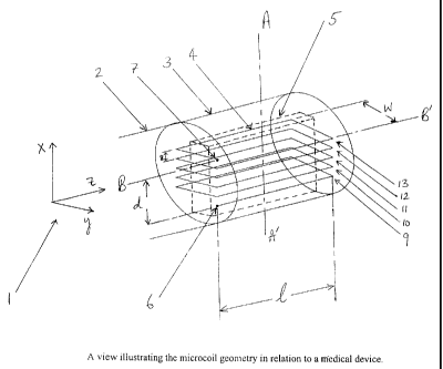
Figure 1B shows a three dimensional view of the preferred microcoil
geometry where the rectangular geometry of the winding is apparent. In Figure
113,
CA 02342047 2008-02-14
a coordinate system (also shown as 1) is chosen such that the z-axis is
parallel to the
long side of the coil (or to the catheter axis B-B').
The winding axis A-A' of the microcoil is parallel to the x-axis. Figure 1 B
shows the microcoil 4 as consisting of five (5) complete turns of winding (9,
10, 11,
12 and 13) between the coil terminals 6 and 7, about the coil axis A-A', with
uniform spacing (shown as 8) between adjacent turns. We will call the
dimension
of the entire wound coil in the y-direction with the width w of the coil and
the
dimension of the entire coil in the x-direction the depth d of the coil. The
width of
the coil is the smallest diameter presented by the coil in a section
transverse to the
winding axis, including the thickness of the conductor. The depth of the coil
is the
sum of (a) all the spacing distances between successive turns of winding and
(b) the
product of the number of winding turns and the conductor thickness. The aspect
ratio of the microcoil 4 is the ratio (1/w). The length Z of the coil (longest
dimension
in the z-direction) may vary in the range 0.5 cm to 6 cm and may more
preferably lie
between 1 cm and 4 cm. The width w of the coil, chosen so as to fit within a
catheter or affixed to the outside of a catheter, may range from 0.5 mm to 6
mm, and
more preferably may lie between 1 mm and 4 mm. The depth d of the entire coil
(including all the winding turns and the spaces between them) may range from
0.3
to 6 mm, and more preferably may lie between 1 mm and 4.5 mm. The conductor
used in the winding may be electrically highly conducting material such as
copper,
silver or gold, or it may be suitable alloys, composites or plated
combinations of
conducting materials. The thickness of the conductor used in the windings may
range from 0.01 mm to 2 mm and the gap between adjacent turns of conductor may
be between 0.01 mm and 1.5 mm. The number of turns in the winding may, for
non-limiting example only, vary between one and fifteen, between one and ten,
between two and ten, between two and eight, between three and ten and between
three and eight.
The sensitivity or gain of the coil is proportional to the component of the
magnetic field that is transverse to the main field of a magnetic resonance
imaging
system, produced by unit current flowing through the coil. We compute below
the
magnetic field transverse to the main field of a magnetic resonance system and
21
CA 02342047 2008-02-14
produced by unit current in the microcoil for the case when its long axis is
parallel
to the main magnetic field. For other orientations of the microcoil with
respect to
the main field the magnetic field may be obtained by an appropriate rotation.
The
magnetic field may be determined in a specified region surrounding the coil by
using the Biot-Savart law:
B = ( /4n) E[(dl x r)/(IrI^3)J
with the integral E taken over the entire length L of the conductor in the
coil, where
B is the magnetic field at a specified point in space, dl is a current-
carrying length
element and r is the distance vector from the current element to the specified
point.
A study of the distribution of the magnetic field around the coil is useful
for
assessing the field profile. In particular, with the choice of coil
orientation with
respect to the main magnetic field of the imaging system as stated above, it
is the
component of the magnetic field transverse to the main magnetic field that is
relevant for signal construction. For this purpose, we have therefore mapped
the
transverse component of the magnetic field along sections transverse to the
catheter
axis, at various locations along the coil length.
Figure 2B is a plot of the logarithm of the transverse magnetic field in a 4
cm x 4 cm transverse section located at a distance of 20 % along the coil
length
measured from the end of the catheter with the coil terminals at that end. The
five-
turn coil shown in Figure 1B with approximate dimensions 1=2 cm, x-2 mm, d=1.5
mm was used in this computation. The transverse field is very sharply peaked
at the
center and consequently we have illustrated the field profile in a logarithmic
plot.
The significance of the plot is that the transverse field profile, and thence
the
sensitivity, is still substantial at the outer edges of this section.
Figure 3B is similar to Figure 2B except that it is the transverse field
profile
at a section located at the middle of the coil. It is evident from these two
figures
that the field profile changes very little along the length of the coil.
Figure 4B shows the size of the sensitive region in relation to that of the
coil
itself. The ends of the cylinder bounding this cylindrical region are shown.
The
22
CA 02342047 2008-02-14
signal-to-noise ratio at the edge of this region is comparable to that
obtained from a
typical head coil used in magnetic resonance imaging.
Figures 5B and 6B show possible variations in the winding pattern which
may be followed.
23
CA 02342047 2008-02-14
This invention relates to primary medical devices for the reception of radio
frequency
electromagnetic radiation, particularly medical devices used to obtain a
magnetic
resonance image of a region in front of the device and within a natural
organism or
patient (such as within a human) or elsewhere, and secondary medical devices
such as
catheters and secondary devices for delivery of therapeutic agents and
monitoring of
metabolic activity. The use of magnetic resonance primary medical devices to
provide
enhanced imaging within the region of interest in conjunction with the
deployment of
secondary medical devices offers a particularly effective means of delivering
targeted
therapy.
While microcoils for internal imaging have been described before, the device
of
the present invention advances the state of the art by design features that
maximize the
field of view in a direction forward to and beyond the spatial extent of the
coil itself (e.g.,
parallel to the axis or the core of the device), as well as improving the
signal gain within
this field of view. The signal power falls off with distance in the forward
direction
(forward being defined as a direction outwardly directed from the device along
the core
axis of the microcoils). For volume imaging purposes, this fall-off can be
adjusted for by
dividing the reconstructed image intensity at a given voxel location by the
gain
corresponding to that voxel (which is proportional to the magnetic field B at
that
location), and repeating this for all voxels.
An aspect of the invention comprises a device to be inserted into a patient
comprising a solid body having at least one microcoil, or at least one pair of
radially
opposed microcoils physically associated with the solid body, each microcoil
having an
outside microcoil diameter of 6mm or less, all individual windings of said
each microcoil
together defining a geometric plane of each microcoil, and the plane of each
microcoil
being parallel to the plane of another microcoil in each pair of radially
opposed
microcoils.
One form of construction in the practice of the present invention uses a
single
electrically conducting path, configured so as to yield one microcoil spiral
winding or a
multiplicity of pairs of spiral windings with a common coil axis, with the
spirals in each
pair being radially opposed. The term "radially opposed" as used in the
practice of the
present invention is defined as meaning that the pairs have their windings
positioned in
24
CA 02342047 2008-02-14
the sense that one member of each spiral pair winds radially inward and the
other
member of the pair winds radially outward about the coil axis (when moving
along the
length of a continuous electrical path). There cannot be an intermediate coil
between the
two coils of the pair of radially opposed coils that has an opposite winding
sense if those
two microcoils are to be considered `radially opposed' in the practice of the
invention, or
those adjacent coils would not be radially opposed with a uniform sense of
winding. The
term "radially-opposed" will be used to describe this arrangement of each
spiral pair (that
is a pair on a common coil axis) with the spirals in each pair being radially
opposed in the
sense that one member of each spiral pair winds radially inward and the other
member of
the pair winds radially outward about the coil axis when considered along a
common,
continuous electrical path. The electrical path need not be continuous (e.g.,
the same
current passing through one microcoil and then the other in the path) for a
radially
opposed pair to be formed. For example, there may be two separate conductive
paths
from the same direction. One electrically conductive path may connect to the
exterior
winding of a microcoil, and the other electrically conductive path may connect
to the
interior winding of the adjacent microcoil. These adjacent microcoils could
then be
considered radially opposed. It is important to note, however, that the sense
of the
winding of each of the spirals in the entire configuration or at least at one
end of a
configuration or at one end of a device is the same (all clockwise or all anti-
clockwise
about the coil axis as seen from the proximal end of the coil, looking down
the"axis).
Another unique feature of the present invention is the potential for use of
multiple
radially-opposed spiral pairs in a single electrically conducting path and all
wound with
the same sense of winding. Thus, even widely separated pairs of spirals in
this
arrangement would have the same winding sense, although sufficiently separated
pairs of
coils at distant positions along a single device could have pairs with
different winding
senses, especially where the distance is great enough that there would not be
a significant
(greater than l0%) overlap in magnetic fields of MRI significance. This
winding
configuration allows for a significantly large receptive field in a roughly
cylindrical
region forward from the distal end of the microcoil or forward from the
catheter when the
microcoil is placed at the distal tip of the catheter. The gain in these
regions falls off with
CA 02342047 2008-02-14
increasing distance from the distal end of the microcoil device. The gain is
significantly
large in a cylindrical region of length of more than 2 cm beyond the distal
end of the coil,
and of diameter more than 2 cm around the coil axis. Within a distance of 1 cm
forward
from the distal end of the coil, the average improvement in signal-to-noise
ratio with a
single radially-opposed spiral pair can be larger by a factor of about 40
compared with
the typical construction of a standard head coil. At a distance of 2 cm
forward from the
distal end of the coil, the signal-to-noise ratio with the use of a single
spiral pair is
comparable to that obtained with a standard head coil. The use of radially-
opposed spiral
pairs allows for optimally squeezing forward the magnetic field produced by a
current in
the conductor, and thence the increased sensitivity in the forward direction.
An aspect of the invention comprises a device to be inserted into a patient
comprising a
solid body having at least one pair of radially opposed microcoils physically
associated
with the solid body, each microcoil having an outside microcoil diameter of
6mm or less,
all individual windings of said each microcoil together defining a geometric
plane of each
microcoil, and the plane of each microcoil being parallel to the plane of
another microcoil
in the pair of radially opposed microcoils. Parallelism and planarity of
microcoils are
approximate and do not have to be mathematically precise. For example, it
would be
difficult to have every single winding (a single complete encirclement around
an axis or
core) exactly and uniformly concentric about that core or preceding windings
and within
a single plane with every other winding within a microcoil. The fact that the
elongated
element (e.g., wire) that constitutes the winding is three-dimensional means
that a
literally two-dimensional plane cannot exist that encompasses all dimensions
of the
winding. There may also be some wobble or axial shifting of windings.
Similarly,
parallelism between the planes of adjacent microcoils allows for some
significant (e.g.,
10-20 degrees) angularity between the planes defined by the windings in the
microcoil,
and absolute parallelism, although probably desirable, is neither likely nor
essential.
In the practice of the present invention, the direction pointing along the
catheter (or
microcoil) axis from the proximal end of the microcoil to the distal end of
the microcoil
will be referred to as the 'forward' direction. The forward direction will
often be
26
CA 02342047 2008-02-14
associated with the forward tip of the catheter or medical element alsm. The
large
forward extension of the field of view and the high sensitivity of the coil in
a significantly
larger region within the field of view constitute a substantial advance in the
design of
magnetic resonance microcoils.
Figure 1A shows a three dimensional view of the preferred microcoil geometry
where the
radially-opposed geometry of the windings 4 and 6 in the spirals is apparent.
In the
figure, a coordinate system is chosen such that the z-axis is along the axis
of the coil (or
the catheter axis). The distance between the individual windings 8 in the
spirals of a
radially-opposed pair 2 may range from about 0.6 mm to 4 mm. The spiral
winding of the
first coil 10 may be represented by the equation
x = -r(0) sin 0
y = r(O) cos 0
with
r(A) = rO + (k/2n) 0
where k is a constant, x and y are the Cartesian coordinates, rO is the
beginning radius and
0 is the winding angle. The winding angle for the spiral goes from 0 to 2nn
where the
winding number n may be as small as 1 or as large as 25; the representation
may be
similar for the other spiral 12. The beginning radius rO can lie in the range
between 0.07
mm and 1.7 mm and more preferably between 0.09 mm and 1.4 mm. The constant k
can
range from 0.02 mm and 1.7 mm or more preferably between 0.03 mm and 1.3 mm or
still more preferably between 0.05 mm and 1 mm. Similar values apply for the
second
spiral of the pair except that the radius of the second coil decreases as
r(0) = ro - (k/2n) 0.
The entire diameter of the coil may range from 0.3 mm to 6 mm, and more
preferably
may lie between 1 mm and 4.5 mm. The conductor used in the winding may be an
electrically highly conducting material that is capable of being shaped or
wound into a
microcoil or spiral, such as copper, silver or gold, or it may be suitable
alloys or plated
composites, polymers, or combinations of highly conducting materials. The
thickness of
the conductor used in the windings generally may range from 0.05 mm to 2 mm,
but this
size is dependent upon the selection of the material and the particular needs
of the device.
27
CA 02342047 2008-02-14
Figure 2A is a plot of the transverse magnetic field in a 2 cm x 2 cm
transverse section
located at a distance of 1 cm measured from the distal end of the coil. Coil
dimensions as
in Figure 1 were used in this computation. The transverse field is minimal at
the center of
the chosen section and reaches a maximal value in a rotationally symmetric
fashion about
the center of the section. The significance of the plot is that the
transverse, field profile,
and thence the sensitivity, is still substantial at the outer edges of this
section.
Figure 3 illustrates the location of the slice chosen in Figure 2A (over which
the magnetic
field is plotted there) with respect to the coil. The variation of the
magnetic field over the
slice is also depicted here by means of a color density, with the spectrum of
colors from
violet to red indicating a variation in intensity from low to high
respectively. For
illustration of scale the coil is also shown in this Figure. Figure 4A is
similar to Figure 2A
except that it is the transverse field profile at a section located at a
distance of 2 cm distal
to the distal end of the coil. At this distance the average signal-to-noise
ratio attained is
comparable to that of a typical commercial head coil.
Multiple pairs of radially-opposed spirals may be employed for further
enhancement of
signal-to-noise, and to extend the field-of-view in the forward direction.
Thus the number
of spiral pairs actually used may range from 1 to 20, and more preferably from
1 to 10.
As an example, Figure 5A shows, in a three dimensional perspective, a
microcoil
configuration 20 which employs two pairs (22 and 24) of radially-opposed
spirals (e.g.,
26 and 28 for opposed spiral pair 22), which effectively increases the signal-
to-noise ratio
within the sensitive region or the receptive field corresponding to that
provided by the
coil by more than 50 percent as compared to a single radially-opposed spiral
pair. This
effect is further highlighted in Figure 6A, which shows the transverse field
in a section
located 1 cm forward from the distal end of the coil. The enhancement of the
magnetic
field is apparent.
28