Note: Descriptions are shown in the official language in which they were submitted.
CA 02342344 2001-02-28
WO 00/13133 PCT/US99/18825
Method and System for the Computerized
Analysis of Bone Mass and Structure
The present invention was made in part with U.S. Goven~ment support under
grant
numbers. This study was supported in parts by USPHS Grants RO1 AR42739 and T32
CA09649. The U.S. Government has certain rights in this invention.
Field of the Invention:
The invention relates generally to a method and system for the computerized
analysis
of bone mass and structure. Specific applications are given for the analysis
of the trabecular
mass and bone pattern for the assessment of bone strength and/or osteoporosis
and as a
predictor of risk of fracture. Novel techniques involve the merging of various
features
including those related to bone mass, bone geometry, bone structural
information, and
subject's age. Additional techniques include the application of Minkowski
Dimension and an
artificial neural network to aid in the computerized fractal analysis of the
bone structure. In
addition, an estimate of the volumetric BMD is presented incorporating bone
mass and bone
geometry.
The present invention generally relates to computerized techniques for
automated
analysis of digital images, for example, as disclosed in one or more of U.S.
Patents
4,839,807; 4,841,555; 4,851,984; 4,875,165; 4,907,156; 4,918,534; 5,072,384;
5,133,020;
5,150,292; 5,224,177; 5,289,374; 5,319,549; 5,343,390; 5,359,513; 5,452,367;
5,463,548;
5,491,627; 5,537,485; 5,598,481; 5,622,171; 5,638,458; 5,657,362; 5,666,434;
5,673,332;
5,668,888; and 5,740,268; as well as U.S. patent applications 08/158,388;
08/173,935;
08/220,917; 08/398,307; 08/428,867; 08/523,210; 08/536,149; 08/536,450;
08/515,798;
08/562,087; 08/757,611; 08/758,438; 08/900,191; 08/900,361; 08/900,362;
08/900,188; and
08/900,189, 08/900,192; 08/979,623; 08/979,639; 08/982,282; 09/027,468;
09/027,685;
09/028,518; 09/053,798; 09/092,004; 09/098,504; 09/121,719; and 09/131,162 all
of which
are incorporated herein by reference.
The present invention includes use of various technologies referenced and
described
in the above-noted U.S. Patents and Applications, as well as described in the
references
identified in the appended APPENDIX and cross-referenced throughout the
specification by
CA 02342344 2001-02-28
WO 00/13133 PCT/US99/18825
reference to the number, in brackets and bold print, of the respective
reference listed in the
APPENDIX, the entire contents of which, including the related patents and
applications listed
above and references listed in the APPENDIX, are incorporated herein by
reference.
Discussion of the Backg o ~nd~
Although there are many factors that affect bone quality, two primary
determinants of
bone mechanical properties are bone mineral density (BMD) and bone structure.
Among the
density and structural features extracted from bone using various techniques,
researchers
agree that BMD is the single most important predictor of bone strength as well
as disease-
conditions such as osteoporosis. Studies have shown correlation between BMD
and bone
strength (Carter and Haye, 1977 [4]; Beck et al., 1989 [2]; Keaveny and Hayes,
1993 [9]). To
this purpose, a range of techniques have been developed to measure BMD to
evaluate fracture
risk, diagnose osteoporosis, monitor therapy of osteoporosis, and predict bone
strength (Beck
et al., 1989 (2]; Ross et al., 1990 [14J; Adams, 1997 [1]; Grampp et al., 1997
[7]).
The standard technique for noninvasive evaluation of bone mineral status is
bone
densitometry. Among various techniques for bone densitometric measurement,
dual energy
X-ray absorptiometry (DXA) is relatively inexpensive, low in radiation dose (<
$ pSv
effective dose equivalent), and of high accuracy (= 1 %) and precision {= 1 %)
(Sartoris and
Resnick, 1990 [15]; Adams, 1997 [1]; Lang, 1998 (10]). DXA has gained
widespread clinical
acceptance for the routine diagnosis and monitoring of osteoporosis (Adams,
1997 [1]). In
addition, DXA can be directly used to measure whole bone geometric features
(Faulkner et
al., 1994 [6]; Sieranen et al., 1994 [17]; Karlsson et al., 1996 [8]; Lang,
1998 [10]). The
BMD measurement from DXA, however, is only moderately correlated to bone
mechanical
properties and has limited power in separating the patients with and without
osteoporosis-
associated fractures (Cann et al., 198$ [3]). DXA provides an integral measure
of cortical and
trabecular bone mineral content along the X-ray path for a given projected
area, but DXA
only measures bone mass, not bone structure. As a consequence, DXA
measurements are
bone-size dependent and yield only bone mineral density per unit area (g/cmz)
instead of true
density, i.e., volumetric bone mineral density (g/cm'). Therefore, if the BMD
measurements
of patients with different bone sizes are compared. the results can be
misleading.
Although the effect of bone size on area BMD using DXA is apparent (Carter et
aL,
-2-
CA 02342344 2001-02-28
WO 00/13133 PCT/US99/18825
1992 [5]; Seeman, 1998 [16]), only a few studies (Nielesn et al., 1980 [13];
Martin and Buff,
1984 [11]; Carter et al., 1992 [5]) have been performed to account for such a
bias. To
compensate for the effect of bone size for vertebral bodies, Carter et al. (
1992) (5] developed
an analysis method and suggested a new parameter, bone mineral apparent
density (BMAD),
as a measure of volumetric bone mineral density.
Also, one of the functions of bone is to resist mechanical failure such as
fracture and
permanent deformation. Therefore, biomechanical properties are fundamental
measures of
bone quality. The biomechanical properties of trabecular bone are primarily
determined by
its intrinsic material properties and the macroscopic structural properties
(Cowin et al., 1987
[24]; Chakkalakl et al., 1990 [23]; Brandenburger, 1990 [21]; Keaveny and
Hayes, 1993 [9)).
Extensive efforts have been made toward the evaluation of bone mechanical
properties by
studying bone mineral density (BMD) and mineral distribution.
Since bone structural rigidity is derived primarily from its mineral content
(Elliott et
al., 1989 [27]), most evaluation methods have been developed to measure bone
mass (mineral
content or density) and to relate these measures to bone mechanical properties
(Carter and
Haye, 1977 [4]; Bentzen et al., 1987 (20]; Hvid et al., 1989 [32]; Keaveny and
Hayes, 1993
[9]; Keaveny et al., 1994 [36]). Results from in vivo and in vitro studies
suggest that BMD
measurements are only moderately correlated to bone strength (Carter et al.,
1992 [5]).
However, studies have shown changes in bone mechanical properties and
structure
independent of BMD (Goldstein, 1987 (30]; Faulkner et al., 1991 [28]).
Moreover, because
density is an average measurement of bone mineral content within bone
specimens, density
does not include information about bone architecture or structure.
Various methods have been developed for in vitro study of two- or three-
dimensional
architecture of trabecular bones using histological and stereological analyses
(Whitehouse,
1974 [43]; Feldkamp et al., 1989 [29]; Goulet et al., 1994 [31]; Croucher et
al., 1996 [25]).
These studies have shown that, by combining structural features with bone
density, about 72
to 94 percent of the variability in mechanically measured Young's moduli could
be explained.
However, these measurements are invasive.
For the noninvasive examination of trabecular bone structure, investigators
have
developed high-resolution computed tomography (CT) and magnetic resonance
imaging
(MRI) (Feldkamp et al., 1989 [29]; Durand and Ruegsegger, 1992 [26J; Majumder
et al.,
-3-
CA 02342344 2001-02-28
WO 00/13133 PCT/US99/18825
1998 [38]). However, due to cost and/or other technical difficulties, these
techniques are
currently not in routine clinical use. The potential of using X-ray
radiographs to characterize
trabecular bone structure has also been studied. Although the appearance of
trabecular
structure on a radiograph is very complex, studies have suggested that fractal
analysis may
yield a sensitive descriptor to characterize trabecular structure from x-ray
radiographs both in
in vitro studies (Majumdar et al, 1993 [37]; Benhamou et al., 1994 (19];
Acharya et al., 1995
[18]; Jiang et al., 1998a [33]) and in an in vivo study (Caligiuri et al.,
1993 (22]).
Different methods, however, exist with which to compute fractal dimension.
Minkowski dimension, a class of fractal dimension that is identical to
Hausdroff dimension
(Mandelbrot, 1982 [39]), is particularly suitable for analyzing the complex
texture of digital
images because it can be formally defined through mathematical morphology and
easily
computed using morphological operations (Serra, 1982 [42]; Maragos, 1994
(40]). The
Minkowski dimension computed from an image, regardless of texture orientation,
gives a
global dimension that characterizes the overall roughness of image texture.
Similarly, the
Minkowski dimensions computed from different orientations yield directional
dimensions
that can be used to characterize the textural anisotropy of an image {Jiang et
ai., 1998a [33]).
SUMMARY OF THF TNVENTI[ON
Accordingly, an object of this invention is to provide a method and system for
the
computerized analysis of bone mass and/or structure.
Another object of this invention is to provide a method and system for
estimating
bone strength.
Another object of this invention is to provide a method and system for
estimating a
volumetric bone mass measure using bone geometry.
Another object of this invention is to provide a method and system for
incorporating
Minkowski Dimension into the analysis of the bone structure pattern.
Another object of this invention is to provide a method and system for
extracting
information from fractal-based texture analyses.
Another object of this invention is to provide a method and system for merging
information on bone mass, bone geometry, bone structure and/or subject age in
order to
obtain measures of bone strength.
-4-
CA 02342344 2001-02-28
WO 00/13133 PCT/US99/18825
These and other objects are achieved according to the invention by providing a
novel
automated method, storage medium storing a program for performing the steps of
the method,
and system in which digital image data corresponding to an image of the bone
are obtained.
Next there is determined, based on the digital images, a measure of bone
mineral density
{BMD) and at least one of a measure of bone geometry, a Minkowski dimension, a
trabecular
orientation, and subject data. The strength of the bone is estimated based
upon the measure
of BMD and at least one of the measure of bone geometery, the Minkowski
dimension, the
trabecular orientation, and the subject data. Preferably, a normalized BMD
corresponding to
a volumetric bone mineral density of the bone as the measure of BMD is
determined, and the
strength of the bone is estimated based at least in part on the normalized
BMD.
To improve bone texture analysis, the present invention also provides a novel
automated method, storage medium storing a program for performing the steps of
the method,
and system in which digital image data corresponding to an image of the bone
is obtained,
and a region of interest (ROI) is selected within the bone. A fractal
characteristic of the
image data within the ROI using an artificial neural network is extracted. The
strength of the
bone is estimated based at least in part on the extracted fractal
characteristic.
To perform bone analysis with an improved measure of bone mineral density, the
present invention also provides a novel automated method, storage medium
storing a program
for performing the steps of the method, and system in which digital image data
corresponding
to an image of the bone is obtained. A measure of normalized bone mineral
density (BMD)
corresponding to a volumetric bone mineral density of the bone is determined,
and the
strength of the bone based is estimated based at least in part on the
normalized BMD.
BEEF DE~CRI_PTION OF THE DRAWINGS
A more complete appreciation of the invention and many of the attendant
advantages
thereof will be readily obtained as the same becomes better understood by
reference to the
following detailed description when considered in connection with the
accompanying
drawings, wherein:
Figure 1 (a) is a flowchart of the inventive method for analyzing bone mass
and
structure;
Figure 1 (b) is a schematic showing how the present invention combines various
types
-S-
CA 02342344 2001-02-28
WO 00/13133 PCT/US99/18825
~of data to analyze bone mass, bone geometry, and/or structure;
Figure 2(a) is a histogram showing the distribution, in an exemplary database,
of
diseases leading to total hip arthroplasty;
Figure 2(b) is a histogram showing the distribution of cases in the exemplary
database
in terms of bone strength;
Figures 3(a) and 3(b) are schematic diagrams that show the setups used to
radiograph
the femoral neck specimens, wherein the setup in Figure 3(a) simulates the
femoral neck as it
would appear in a clinical hip radiograph, and the setup in Figure 3(b) was
used to produce a
high-resolution radiograph of the specimens;
Figure 4(a), Figure 4(b), and Figure 4(c) are respective images of (a) a pre-
operative
film, (b) a specimen film using the "simulated clinical" setup, and (c) a
specimen film using
the "verification" setup, wherein the regions-of interest shown in Figure 4(b)
and Figure 4(c)
are the regions from which the texture measures are calculated;
Figure S(a) and Figure S(b) are respective illustrations of (a) a side view of
a specimen
showing how, for strength testing, the bone cube is initially cut into bone
disks having a
height of 6.5 mm with the most inferior cut aligned with the bottom of the
lead bead placed
on the medial surface of the specimen, and (b) a top view of a bone disk
showing how the
disk is cut into 6.5 centimeter thick columns which were subsequently cut into
6.5 centimeter
cubes (the arrows on the left indicate the projection of the ROI that was
selected on the
radiograph);
Figure 6 is a graph showing the how load-to-failure is determined from
mechanical
testing;
Figure 7 is an image showing an ROI and several geometric measures from the
proximal femur of a subject;
Figure 8 is a graph showing the linear relationship between femoral neck width
(BB)
and femoral shaft width (CC);
Figure 9(a), Figure 9(b), and Figure 9(c) are respective plots showing (a) the
dependency of BMD on bone size, (b) the dependency of BMD on femoral neck
width, and
(c) the dependency of BMD on femoral shaft width;
Figure 10(a), Figure 10(b), and Figure 10(c) are respective plots showing (a)
the linear
relationship between bone strength and the area-based BMD, (b) the power law
relationship
-b-
CA 02342344 2001-02-28
WO 00/i3133 PC'T/US99/18825
between bone strength and the BMD normalized with the femoral neck width
(nBMDN), and
(c) the power law relationship between bone strength and the BMD normalized
with the
femoral shaft width (nBMDs);
Figure 11 (a) and Figure 11 (b) are respective images of (a) a radiograph of
the femoral
neck specimen from the femur, and (b) a selected ROI from the neck radiograph;
Figure 12 is a graph showing the relationship between the normalized volume
and the
scale and showing the slope used to determine the Minkowski dimension;
Figure 13(a) and Figure 13(b) are respective illustrations of (a) a squared
structuring
element of 3x3 pixels used to compute the global Minkowski dimensions, and (b)
a horizontal
structuring element of 3x1 pixels used to compute the directional Minkowski
dimensions;
Figure 14 is a graph showing the directional Minkowski dimension as a function
of
the angle of a structuring element for a single ROI;
Figure 15 is a graph showing the parameters of an ellipse used in
characterizing the
plot shown in Figure 14;
Figure 16 is an image of a pelvis radiograph showing the orientation from the
Minkowski dimension analysis relative to the direction of the ROI submitted
for mechanical
testing;
Figure 17(a) is an image of a representative ROI where BMD = 0.2054, DM[fJ =
2.59,
and 9~ = 34°;
Figure 17(b) is an image of a representative ROI where BMD = 0.2052, DM[fJ =
2.73,
and 8~ = 149°;
Figure 17(c) and Figure 17(d) are plots of the ellipse fitting data for Figure
17(a) and
17(b), respectively;
Figure 18 is a plot showing the relationship between bone strength and global
Minkowski dimension where RZ = 0.17 and p = 0.016;
Figure 19 is a graph showing the relationship between nBMD2 and DM[fJ where RZ
=
0.04 and p = 0.10;
Figure 20(a) is a graph showing the relationship between log area and log
relative
length from the surface area fractal analysis of an ROI;
Figure 20(b) is an illustration showing how the data from the graph in Figure
20(a) are
used as inputs for an artificial neural network (ANN);
_7_
CA 02342344 2001-02-28
WO 00/13133 PCT/US99/18825
Figure 21 is a graph showing ROC curves that illustrate the relative
performances of
the conventional fractal analysis method, the ANN method, and bone mass alone,
for
distinguishing between strong and weak bone;
Figure 22 is a block diagram of a system for implementing the inventive
method; and
Figure 23 is a schematic illustration of a general purpose computer 300
programmed
according to the teachings of the present invention.
DETAILED DESCRIPTION OF THE PREFERRED EMBODI~N~j~TS
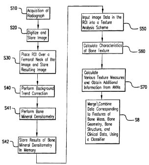
Referring now to the drawings, and more particularly to Figure 1 (a) thereof,
a
flowchart describing an inventive method for the analysis of bone is shown.
Figure 1 (b) is a
schematic showing how the present invention incorporates various types of data
to analyze
bone mass, bone geometry, and/or structure.
With the inventive method described in Figure 1 (a), the characteristics of
the bone,
geometry, and trabecular pattern are extracted using computer analysis of
image data from
digital images of bony parts of the body such as the hip. The overall scheme
includes an
initial acquisition of a radiographic image of the hip in step S I 0. The
image is digitized and
stored in memory in step S20. Alternatively, steps S I 0 and S20 may be
combined into a
single step by directly acquiring a digital radiographic image of the hip. A
region of interest
(ROI) is then placed over a femoral neck on the image and the corresponding
image data are
stored in memory in step S30. Background trend correction is performed in step
S40 to yield
the underlying fluctuations, i.e., the trabecular pattern, in the bone. In
step S41 bone mineral
densitometry, including BMD, is also performed on the bone. Then, in step S42
the results of
bone mineral densitometry are stored in memory. Next, in step S50 the image
data in the
ROI are then input to a texture analysis scheme, and then, in step S60
characteristics of the
bone texture are calculated. In step S70 various texture measures are
calculated using texture
schemes such as Minkowski Dimension, and additional information is obtained
from the use
of artificial neural networks (ANNs).
The image data in memory (from step S20) is also used to extract bone geometry
yielding such features as femoral neck thickness and femoral shaft thickness.
These features
can also be used to normalize BMD and to yield an estimate of volumetric BMD.
In step S80
data corresponding to the features of bone mass, bone geometry, bone
structure, and clinical
_g_
CA 02342344 2001-02-28
WO 00/13133 PCT/US99/18825
data (e.g., the subject's age) are merged/combined using one or more
classifiers such as a
linear discriminant function and/or an artificial neural network (ANN) to
yield an estimate of
bone strength and thus the likelihood of risk of future fracture.
Figure 22 is a block diagram illustrating a system 1000 for implementing the
inventive method for analysis of bone mass and bone trabecular structure. The
method and
the hardware used to implement the method and system 1000 are discussed in
greater detail
below under the various section headings that follow the description of Figure
22.
Refernng to Figure 22, an image acquisition device 2000 inputs a radiographic
image
of an object into a digitization circuit 2000a. An image memory 2001 stores
the digitized
image. If the radiographic image is obtained with a direct digital device,
then there is no need
for the digitization circuit 2000a. The image memory 2001 sends stored images
to an ROI
selection circuit 2002 for placing ROIs on images. The ROI selection circuit
sends images
with ROIs to a nonlinear detection system correction circuit 2003 for
performing background
trend correction. The nonlinear detection system correction circuit 2003 sends
image data,
for which background trend correction has been performed, to a bone structure
circuit 2006
for determining structural features of bone (including the trabecular
orientation) represented
by the image data. The bone structure circuit sends the extracted structural
features to a
texture circuit 2020 which generates texture information including the
Minkowski dimension.
An ANN fractal measure circuit 2040 determines, among other things, the
fractal nature of
the bone texture information generated in the texture circuit 2020.
The image memory 2001 also sends stored image data to a bone mass circuit 2004
for
calculating BMD. Additionally, the image memory 2001 sends stored image data
to a bone
geometry circuit 2005 for calculating various measures of bone geometry
including femoral
neck width and femoral shaft width. A normalization circuit 2007 calculates
the normalized
BMD based on the BMD and the bone geometry information generated in the bone
mass
circuit 2004 and bone geometry circuit 2005, respectively. The normalized BMD
provides an
estimate of the volumetric bone mineral density.
A data memory 2009 stores data regarding BMD, normalized BMD, bone geometry,
and the fractal nature of the bone texture. This data may be weighted in a
weighted sum
circuit (not shown) before being stored in the data memory 2009. Patient
clinical data is also
input and stored in the data memory 2009.
-9-
CA 02342344 2001-02-28
WO 00/13133 PCTNS99/18825
A classifier circuit 2050 estimates bone strength (and thus the likelihood for
risk of
future fracture) based on the measures of bone mass, bone geometry, bone
structure, and/or
patient data. An image memory (now shown) stores any image data generated by
the various
components of the system. A display system (for example, the monitor 302 in
Figure 23,
discussed later) converts the digital image data generated by the system's
components into
analog data and displays the resulting images. A superimposing circuit (not
shown)
superimposes the results of the system's calculations onto the displayed
images, stores the
results in file format, or provides the results in a text-only format.
Figure 2(a) is a graph showing the distribution of diseases in a database on
which the
present invention was tested. The database included femoral neck specimens.
The specimens
were excised from patients undergoing total hip arthroplasties. The ages
ranged from twenty
to ninety-four years with a mean age of fifty-eight years. Each patient case
also contained a
standard pre-operative pelvis radiograph. The clinical findings necessitating
hip replacement
for the individuals included osteoarthritis (n=30), avascular necrosis (n=12),
and rheumatoid
arthritis (n=2). Since many of the specimens were obtained from individuals
with joint
disease, rather than bone disease, the strengths of the bone ranged from very
strong to very
weak. The range of ages of the individuals from which the specimens were
obtained was 20-
94 years with a median age of 63 years and an average age of 59 years. The
wide range in
age yielded a large variation in bone mechanical properties.
Figure 2(b) is a histogram showing the distribution of cases in the exemplary
database
in terms of bone strength.
Bo~~e_mineral densl~a~d bone radioQranhv
The overall method for calculation of volumetric BMD includes conventional
area-
based BMD from DXA and the extraction of geometric measures from pelvic
radiographs.
Area-based BMD was performed on each femoral neck specimen. Each femoral neck
specimen was positioned in a Styrofoam cup by an orthopedic surgeon to match
the
angulation and anteversion presented on the standard pelvis radiograph of the
patient.
LUCITE with a thickness of five centimeters was added below each specimen to
simulate the
-10-
CA 02342344 2001-02-28
WO 00/13133 PCT/US99/18825
soft tissue in clinical BMD measurements. A Lunar DPX-IQ (Lunar Corp., Madison
WI)
densitometer was used to scan each specimen. After a specimen was scanned, a
region of
interest (ROI) was identified, and the area BMD (g/cm~) within the ROI in the
anteriorposterior direction was obtained using the analysis software available
on the Lunar
DPX system. Each of the ROIs was selected to match the site from where the
trabecular bone
cubes would be machined from the femoral neck specimen for mechanical testing
(discussed
below).
The excised femoral neck specimens were radiographically exposed under two
conditions: a "simulated clinical" setup and a "verification" setup. A
schematic diagram of
the "simulated clinical setup used to radiograph the specimens is shown in
Figure 3(a).
LUCITE was used as a scattering material to simulate soft tissue. The geometry
of the setup
and choice of screen-film system and grid are those that are currently used in
the Department
of Radiology at the University of Chicago Hospitals. A Lanex medium/TMG
(Eastman
Kodak; Rochester, NY) screen-film system was used with an 8: I focused grid.
The distance
from the focal spot of the X-ray tube to the film cassette was 100 cm, and the
distance
between the film cassette and the bottom of the first sheet of LUCITE was 7.6
cm. Placement
of the specimens (angulation and anteversion) was performed by an orthopedic
surgeon such
that the orientation of the femoral neck was similar to its position in a
standard pelvis
radiograph. The specimens were held in this orientation by securing them in a
polystyrene
foam cup. The specimens were also radiographed using a high-resolution film (X-
Omat TL,
Eastman Kodak; Rochester, NY) with the specimen in direct contact with the
film. Direct
exposure (i.e., no screen or grid) was used to produce this high-quality
radiograph, referred to
here as the "verification" setup. The "verification" setup is shown
schematically in Figure
3(b). The "verification" setup yields a high spatial resolution image with
minimal x-ray
scatter due to the absence of LUCITE and no light diffusion due to the absence
of a screen.
The pre-operative pelvis films of some patients were available. However,
because the
objective of these pre-operative films was to show the geometry of the hip
joint, the films
frequently displayed poor image quality in terms of density and contrast. An
example of a
pre-operative film is shown in Figure 4(a). Figure 4(b) shows a "clinical"
specimen
radiograph corresponding to the pre-operative film of Figure 4{a). Figure 4(c)
shows a
"verification" radiograph corresponding to the pre-operative film shown in
Figure 4(a). From
-11-
CA 02342344 2001-02-28
WO 00/13133 PCT/US99/18825
Figures 4(a), 4(b), and 4(c), one can visualize the location of the bone
specimen relative to the
rest of the pelvic anatomy. The regions of interest shown in Figure 4(b) and
Figure 4(c) are
the regions from which the texture measures are calculated.
Biomechanical testin~for the establi m nt of bone strengtt~(i a "truth"1
The cancellous (trabecular) bone were precisely cut into 6.5 mm cubes with an
Isomet-2000 saw cutting system (Beuler Corp. Lake Bluff, IL). The specimens
were first cut
into disks in the plane perpendicular to the axis of the femoral neck
specimen. The inferior
cut of the first disk was aligned with the bottom of the lead bead as shown in
Figure 5(a).
As depicted in Figure 5(b), each disk was then cut into 6.5 mm columns from
anterior
to posterior (Columns A, B, and C in Figure 5(b)). Each column was then cut
into cubes.
Medial femoral cortical bone was excluded from all specimens. For each femoral
neck,
multiple cubic specimens were machined along the anterior-posterior (AP)
direction within a
region corresponding to the ROI where the BMD was initially measured. Specimen
cubes
that corresponded to the ROI extracted on the digitized radiograph (discussed
in greater detail
in conjunction with computerized analysis below) were used to determine the
strength of the
specimen. The method for compressive strength testing is based on the method
described by
Linde et al. (1988) [45]. The compressive strength testing was performed with
an Instron
8500 plus (Instron Corp., Park Ridge, IL) materials testing system. The cubes
were placed
between the platens so that compressive testing was performed in the superior-
inferior
direction. The specimens were first pre-loaded to a load of five Newtons. For
pre-
conditioning, the specimens underwent twenty cycles of compression to 0.5%
strain and then
relaxation at a rate of 0.2 cycles per second. After preconditioning, the load
was returned to
five Newtons, and then destructive testing was performed by increasing the
strain at a rate of
0.1% strain per second until the specimen failed. All specimens machined from
alI femoral
necks were tested destructively under compressive load using the same testing
conditions,
and the mechanical properties (the Young's modulus and the strength) were
obtained for each
bone cube. For each femoral neck, the overall Young's modulus (E) and strength
(S) were
computed by averaging the values obtained from all bone cubes (two to four
cubes) within the
corresponding ROIs.
Using the load-strain information shown in the graph of Figure 6, the
destructive
-12-
CA 02342344 2001-02-28
WO 00/13133 PCTNS99/18825
modulus was calculated as the maximal slope of the load-strain curve divided
by the cross-
sectional area of the specimen. The stress to failure of the specimen was
obtained from the
peak of the stress-strain curve. The strength value used for assessing the
performance of the
texture features was taken to be the average value of the strength measures of
the cubes that
had at least fifty percent of their area within the ROI from the radiographs.
Bone Qeometrv and volumetr,r hnnP minPr~l .io"~;t«
Femur geometry was measured from the standard pelvic radiograph for each
patient.
The radiographs were digitized with a laser film digitizer (LD4500, Konica
Corp., Tokyo
Japan) to a spatial resolution of i 21 x 121 p,m and 10-bit quantization
levels. An interactive
display program was developed using IDL (Research Systems, Inc., Boulder CO}
software in
order to measure femur geometry as suggested by Karlsson et al. (1996) [8].
All the
measures were performed by a musculoskeletal radiologist. The geometric
measures shown
in Figure 7 were used to normalize the area-based BMD. These geometric
measures included
the femoral neck width (BB) and the femoral shaft width (CC) measured right
below the
lesser trochanter.
The femoral neck and the femoral shaft from which the widths were measured are
nearly circular, and thus, the values of BB and CC can be treated as diameters
of the
corresponding regions. The normalized BMD (nBMD, g/cm') was computed from the
measured area BMD (g/cm2) normalized by the diameter, i.e.
nBMD __ BMD
BB , or (1}
ftBMDs = BMD .
CC (2)
Since the BMD was measured from the femoral neck, it is desirable to use
femoral neck
diameter to obtain nBMDN. However, in some cases osteophytes were observed on
the
medial and lateral sides of the necks. In these cases, the measurement of neck
width could be
-13-
CA 02342344 2001-02-28
WO 00/13133 PCTNS99/18825
biased. Specifically, the measured neck width in the medial-lateral (ML)
direction could be
greater than the actual neck width in the AP direction. Therefore, the femur
shaft width was
also investigated as a measurement with which to normalize BMD.
Analysis of variance was performed to show the mean difference in the measured
femoral neck width and shaft width. Regression analyses were performed between
either the
BMD or the normalized BMD values, and the mechanical properties of the bone.
Both linear
and squared power law models were used in the regression analyses. The
coe~cient of
determination (RZ) was used to measure the explanatory or predictive power of
bone
mechanical properties by the area BMD and volumetric BMD.
The descriptive statistics of measured femoral neck width (BB) and shaft width
(CC)
are shown in Table 1. Although, analysis of variance showed that the measured
widths of BB
and CC were significantly different (p-value less than 0.02), the absolute
mean difference in
the measured widths were quite small. The average neck width was only 8%
larger than the
average shaft width. Table I also demonstrates large patient-to-patient
variations in the
measured bone size, e.g., the maximum shaft width was 60% larger than the
minimum shaft
width and that was nearly twice as large for the measurement of neck width.
Figure 8 shows
strong correlation between the neck and shaft widths. The coefficient of
determination (RZ)
was 0.65. Figures 9 (a) and (b) show the relationship between the area-based
BMD and bone
size. Table 1 shows a descriptive statistics of the geometrical measurements
and BMD's from
the proximal femora.
Variables Means Standard
Minimum Maximum
deviation
BB (mm) 43.77 6.67 31.57 62.79
CC (mm) 40.50 4.23 32
38
. 51.81
BMD 0.98 0.22 0.52 1
2 53
(g/cm .
)
nBMDN 0.23 0.05 0
09
. 0.3 5
(~cm')
nBMDs 0.24 0.06 0.10 0
' 42
(~cm .
)
-14-
CA 02342344 2001-02-28
PCT/US99/18825
WO 00/13133
nBMDN - BMD normalized using femoral neck width (BB);
nBMDs - BMD normalized using femoral shaft width (CC).
Figure 10 (a) shows the relationship between strength and the area-based BMD.
The
coefficients of determination (R2) of the generalized linear regressions for
the area-based
BMD and strength are shown in Table 2, and for the area-based BMD and Young's
modulus
are shown in Table 3. The RZ's for both linear and power law relationships are
presented in
the tables. It is clear that the power law models explain more variability in
bone mechanical
properties. Compared with the linear models, the power law models improved the
RZ's by
22% and 13% for predicting Young's modulus and strength, respectively. Table 2
shows
coefficients of determination (Rz) between strength (S) and bone density (D)
in linear and
power law relationships. Table 3 shows coefficients of determination (R'-)
between Young'
modulus (E) and bone density (D) in linear and power law relationships.
Squared Power
Predictor Linear Model Law Model
BMD (g/cm2) 0.238 0.268
nBMDN {g/cm') 0.300 0.363
nBMDs (g/cm') 0.319 0.372
Note: nBMDN - BMD normalized using femoral neck width (BI3)
nBMDN - BMD normalized using femoral shaft width (CC).
(p-value < 0.001 for all models)
Squared Power
Predictor Linear Model . Law Model
BMD (g/cm2) 0.251 0.306
nBMDN (g/cm') 0.291 0.381
nBMDs (g/cm') 0.338 0.431
Note: nBMDN - BMD normalized using femoral neck width (BB)
-15-
CA 02342344 2001-02-28
WO 00/13133 PCTNS99/18825
nBMDN - BMD normalized using femoral shaft width (CC).
(p-value _< 0.001 for all models)
The effects of normalized BMD on the prediction of bone strength are
graphically
shown in Figure 10(b) and Figure 10(c). It is apparent that the normalization
reduced data
variability and revealed a more linear trend between the strength and either
nBMDN or
nBMDs. The percent variation in strength explained by the normalized BMD using
both
linear and power law models, as quantified by the RZ's, are also presented in
Table 2. For the
linear model, normalization increased the Rz's by 26% and 34% for the area-
based BMD
normalized by the neck width (nBMDN) and by the shaft width (nBMDs),
respectively. For
the power law model, the increases in RZ's were 35% and 39% using nBMDN and
nBMDs,
respectively. As with bone strength, the normalization caused a similar
improvement in the
correlation between bone density and Young's modulus as shown in Table 3.
Since the BMD measure produced by DXA is an area-based density, it is valid to
compare the BMDs of patients with similar bone size. However, test results
showed that the
variation in bone size could be very high, e.g. the largest neck width was
nearly twice as large
as the the smallest one. In addition, as suggested by Figure 9(a) and Figure
9(b), there is a
clear trend that BMD is a function of bone size. As a consequence, the BMD
measurements
of patients with different bone sizes could be misleading. Therefore, a
normalization
procedure is useful for relative comparison. Test results showing increased RZ
between the
mechanical properties and the normalized BMD further verify this argument.
Osteophytes were observed on femoral necks for some of the cases. The
osteophytes
were mainly in the medial and lateral surfaces of femoral necks. Therefore,
the measured
neck width could be larger than the actual width for these cases. The large
variation in the
neck width measures (see, for example, the standard deviations in Table 1 ) as
compared to
that of the shaft width measures may be due to this phenomenon. As a
consequence, the
nBMDN (using femoral neck width) was expected to be less accurate than the
nBMDs. Since
a normalization method was sought for relative comparison rather than
measuring true
volumetric density, femoral shaft width appeared to be a better measure for
the normalization.
The justification for this choice is based on the following reasons: (1)
femoral neck width and
shaft width are virtually identical (8% difference in the means) so that shaft
width represents
-16-
CA 02342344 2001-02-28
WO 00/13133 PCT/US99/18825
bone thickness in the neck region; (2) femoral neck width and shaft width are
linearly
correlated (Figure 8, RZ=0.65) even with the inclusion of osteophytes in the
measurement of
neck width; (3) no osteophytes were observed in the lesser trochanter region
from where the
shaft width is extracted; and (4) femoral shaft width can be measured either
from pelvis
radiographs or directly from DXA scans (Faulkner et al., 1994 [6); Karlsson et
al., 1996 [8])
so that a noninvasive evaluation is possible.
The results obtained from analyzing the database suggest two ways in which the
clinical evaluation of bone quality can be improved. First, BMD can be
normalized using a
squared power law relationship. Substantial improvement was achieved by simply
normalizing the measured BMD with bone size. In the prediction of bone
strength, the RZ
was 0.372 when nonmalized BMD with the power law model was used. Using Rz as a
basis
for comparison, the use of normalized BMD with the power law resulted in a 56%
improvement over the simple model that did not use normalization (RZ was only
0.238).
Although in the setup, the BMD measured in the femoral neck region was
normalized, the
results strongly support the analytic approach developed by Carter et al. (
1992) [5] for
predicting BMD of whole vertebral bodies.
Although various power law relationships with different exponents have been
reported
in the literature, our data are best described by a squared power law
relationship. Many
reports (e.g., Carter and Haye, 1977 [4]; McBroom et al., 1985 [12]) have
shown that, using
BMD as a single predictor, the squared power law relationship best describes
both modulus
and strength. With the present invention, the power law models improved the
RZ's from 13%
to 30% in comparison to the simple linear models,.
With the present invention, RZ values between bone mineral density and
mechanical
properties ranged from 0.24 to 0.31 for both linear and squared models. In
comparison with
the typical RZ values reported in literature (which range from 0.4 to 0.8 as
summarized by
Keaveny and Hayes (1993) [9]), the Rz's obtained with the present invention
were quite low.
This is not surprising because, in most of the reports, both the BMD and
mechanical testing
were conducted on the cubic specimens as opposed to the simulated femoral neck
setup. The
present invention incorporates the femoral neck setup to measure the BMD. As a
result, the
BMD obtained by the present invention is an integral measurement of area
density that
includes both cortical and trabecular bone in the entire thickness of the
femoral neck.
-17-
CA 02342344 2001-02-28
WO 00/13133 PCT/US99/18825
Further, mechanical testing was performed only on the trabecular bone cubes
machined from
the bone region that corresponded to the ROI where the BMD was measured.
Consequently,
both the bone size variation and the misalignment between the ROI and the
cubes may have
contributed to lower R--''s.
The purpose of the present invention is not to develop a method for measuring
true
volumetric bone mineral density. Instead the inventors of the present
invention have tried to
(1) emphasize the problem of using area-based BMD, and (2) establish the
feasibility of using
DXA and radiography to assess bone quality in clinical applications. Standard
clinical pelvis
radiographs were used for the measurement of the bone geometry. However,
because of the
high spatial resolution obtained from DXA (Lang, 1998 [10]), DXA can be
directly used to
measure both BMD and the bone geometry so that the need for an additional
imaging
modality can be avoided.
Using BMD and geometric bone data, the results obtained with the inventive
method
suggests that the use of DXA-based bone densitometry to predict bone mineral
status can be
improved with the inventive method. The area-based BMD obtained using DXA was
normalized by a geometric measure obtained from standard pelvic radiographs.
Results show
notable improvement in predicting bone mechanical properties using the
normalized bone
mineral density (i.e., volumetric BMD). The inventors have concluded that the
inventive
method, which is essentially a simulated in vivo method, is a simple and cost-
effective
modif cation of bone densitometry, and holds potential for enhancing the
performance of
DXA for clinical applications.
Radiographs were digitized with a Konica LD4500 laser film digitizer (Konica
Corp.;
Tokyo, Japan) with 0. 12 1-mm pixel size and 10-bit quantization. Regions-of
interest
(ROIs) of dimension 64 x 64 pixels were selected in the medial portion of the
femoral neck
by an orthopedic surgeon. An example of ROI placement is shown in Figures
4(b). The
ROIs were positioned to avoid overlapping structures (e.g. osteophytes).
Correction was
performed for the possible nonlinear nature of the detector's characteristic
response {the H&D
curve for radiographic films as detector) and for the background trend within
the ROI image
data. Background trend correction is necessary since the variation in optical
density within
_I8_
CA 02342344 2001-02-28
WO 00/13133 PCT/US99/18825
the ROI in hip images includes variations due to the gross anatomy of the
human body
(background trends) and variations due to the fine underlying texture which is
related to the
trabecular pattern of the bone. The nonuniform background trend can be
determined using a
2-dimensional surface fitting technique (such as one with a second degree
polynomial
function) (Katsuragawa et al., 1988 [35]). The fitted trend is subtracted from
each ROI in
order to yield the underlying fluctuations, i.e., the trabecular pattern.
Prior to any
computerized texture analysis, this background correction was performed on the
ROIs.
The ROI was selected in the medial portion of the neck where the cubic bone
specimens were machined for mechanical testing (Figure I 1 (a)). Figure 11 (b)
shows a
selected ROI from the neck radiograph in Figure 11 {a).
Fractal analysis was performed on the ROIs using either Minkowski dimension or
surface area based methods.
For a ROI image f of 64x64 pixels in size, the global Minkowski dimension,
DM[fJ, is
computed by {Maragos, 1994 [40]),
log[Vg(e)/E3] ( )
Dn,,[~ -lE o log(1/e) ~ 3
where for a structuring element g at scale E, VB(E) is the "volume" between
two processed
versions of f obtained using morphological operators. The volume V6(e) is
computed by
64 64
Vg(c) - ~ ~ {{f~eg)-(f~eg)) ~ (4)
m=0 n=0
where (f~Eg) and (f~eg) are the dilated version and the eroded version,
respectively, of the
image obtained using a structuring element g at scale E. Note that Vs(e) is
the volume arising
from the difference between the dilated and eroded surfaces. Finding the slope
of the least-
square fitted line between log[Vs(e)/E3] and log( 1 /e) gives the estimated
fractal dimension as
shown in Figure 12.
To compute the directional Minkowski dimension, the ROI image is rotated from
8 = 0 ° to 360 ° with a I 0 ° increment (Jiang et al.
1998b [34]). For each rotation 8, the
volume, Vg(E)e, is calculated by
-19-
CA 02342344 2001-02-28
WO 00/13133 PCT/US99/18825
64 64
ug{~)e = ~ ~ ({fe~~g)-(f~~~g)) ~ (5)
m=0 n=0
where fe is the original ROI image rotated by 0. The directional Minkowski
dimension as a
function of 8, DM[fJe, is then computed from Equation {3) using the calculated
volume from
Equation (5) for each rotation.
A squared structuring element of 3x3 pixels (Figure 13a) and a horizontal
structuring
element of 3x1 pixels (Figure 13b) were used to compute the global (Equation
(2)) and
directional (Equation (3)) Minkowski dimension, respectively (Jiang et al.,
1998a [33]). The
resulting plot of 6 vs. the directional Minkowski dimension is shown in Figure
14. The
directional fractal dimension as a function of 8 was fit to an ellipse using a
least-square fitting
method to describe the textural anisotropy of the X-ray images. The ellipse
parameters, the
major and minor diameters (a and b), eccentricity (e = sqrt (az-bz)/a), and
ellipse orientation
(~~), were used to describe the image texture which, in turn, characterizes
trabecular structure
(Figure 15).
Since the machined bone cube and the selected ROI from the neck radiograph
were at
different orientations as shown in Figure 11 (a), the actual ellipse
orientation (B J was
computed relative to the direction of mechanical testing. Thus, 6a varies from
0 to 90 degrees
based on the original ellipse orientation (8~) and the angle (T) of the
femoral neck axis. T
was determined by a radiologist for each case using the pelvic radiographs
(Figure 16).
Overall, the various computer-extracted, fractal-based features obtained from
each
ROI image included a global description of image roughness, DM(fJ, and the
measures, a, b, e,
and 8" to characterize the anisotropy of the image texture.
The ROI's from two different cases that have identical BMD's are shown in
Figure 17
(the nBMD's are 0.2054 and 0.2052 for the cases in Figures 17 (a) and 17 (b),
respectively).
However, the global Minkowski dimension (DM[fJ) and the orientation (q~) are
quite different
for the ROI's. The DM[fJ and 6~ are 2.59 and 34 °, respectively, for
the ROI in Figure 17(a),
and the DM[fJ and 6~ are 2.73 and 149 °, respectively, for the ROI in
Figure 17(b). The
mechanical strengths are also different, the bone cubes corresponding to the
ROI's in Figures
17{a) and 17(b) having strengths of 0.93 and 7.47 MPa, respectively. The
results of ellipse
-20-
CA 02342344 2001-02-28
WO 00/13133 PCf/US99/18825
fitting show that the directional Minkowski dimensions fit to the ellipses
very well. The
coefficient of determination, Rz, used to measure the goodness of fit of the
ellipse fitting,
yielded a mean of 0.966 with a minimum, maximum, and standard deviation of
0.917, 0.990
and 0.016, respectively. Figure 17(c) and 17(d) show the fitted ellipse data
for the ROI's in
Figures 17(a) and 17(b), respectively.
Pearson correlations (r) among the mechanical properties, BMD, and image
texture
features are shown in Table 4. The following relationships were observed.
Among density
and structural features, the nBMD2 had the highest correlation with both
strength and
modulus; followed by Minkowski dimension, orientation (6J, and age in a
decreasing order.
The relationship between the strength and DM[fJ is shown in Figure 18.
Trabecular bone gets
stiffer and stronger with an increase in both BMD and DM[fJ (positive
correlation
coefficients), and with a decrease in both age and trabecular orientation
(negative correlation
coefficients). Although DM[f) had some correlation with BMD, it became quite
independent
when the BMD was normalized and squared (r = 0.30) as suggested by Figure 19.
BMD was
found to be nearly uncorrelated with both age and trabecular orientation (r = -
0.2). Table 4
shows correlation (Pearson) coefficients among the mechanical properties and
the density and
computer-extracted structural image features.
Table 4
Strength Modulus BMD nBMD nBMDZ Age DM[fJ 0, a b
Modulus 0.92'
BMD 0.51' 0.52
nBMD' 0.58' 0.60' 0.92'
Age 0.63' 0.67' 0.89' 0.95'
DM[fj -0.26' -0.36' -0.0T -0.l0' -0.12'
e, 0.41' 0.38' 0.42' 0.31' 0.30' 0.11°
a (ellipse -0.28' -0.28° -0.14' -0.19° -0.20' 0.22° 0.23'
major axis)
a (ellipse -0.19' -0.21' -0.19° -0.24' -0.26' -0.31' 0.10° 0.24'
major axis)
a(ellipse 0.02' -0.11° -0.13' -0.08' -0.0T 0.19' -0.01°
0.07° 0.43
major axis)
a (exentricity) -0.23' -0.31' -0.12° -0.22' -0.25' 0.24' 0.09' 0 1 T 0
76' -0 242
Note: 'p-value < 0.001; Zp-value < 0.01;'p-value < 0 ~l ; 4p-value z 0.1.
-21-
CA 02342344 2001-02-28
WO 00/13133 PCT/US99/18825
estirr~ates of bone s rengthg
Statistical analyses including general linear regression, stepwise regression,
best subset
selection, and correlation, were performed between the various descriptors of
bone quality
including BMD, age, computer-extracted radiographic features, and
biomechanical properties
(S and E). Stepwise regression and best subset selection were used to select
and merge the
various descriptors of bone mineral density and structural features into a
single index, which
was then evaluated as a predictor of the biomechanical properties. Although
linear
combinations of features have been described above, artificial neural networks
can also be
used to merge the information corresponding to each of the various features,
as illustrated in
Figure 1 (a) and Figure I (b).
For unbiased comparisons, the coefficients of determination were adjusted by
the
number of predictors and the sample size (Meter et al., 1990 [41]) and the
adjusted RZ's were
used for all subsequent comparisons. Stepwise regression and best subset were
used to select
the best predictors for the models (Meter et al., 1990 [41]). From the
computer-extracted
structural features, the global Minkowski dimension and trabecular orientation
were selected
as the best structural features in predicting both modulus and strength. In
addition to these
two structural features and density, patient age was also selected as a good
predictor.
Table 5 shows the best regression models and RZ's for predicting the Young's
modulus.
The squared relationship using normalized BMD (nBMDz) showed substantial
improvement
over the model using area BMD directly. By adding more predictors to the model
using
nBMD2 alone (RZ = 0.431 ), one at a time using stepwise regression, the RZ's
were improved by
16%, 25%, and 29% using two, three, and four predictors, respectively. By
including nBMDz,
age, Minkowski dimension, and trabecular orientation into the model, an R2 of
0.554 was
achieved. Compared with the model using just area BMD, the four-predictor
model (nBMD2,
age, DM[fJ, a J improved the RZ by more than 120%. Table 5 shows regression
equations and
the coefficients of determination (RZ) between Young's modules (E) and bone
density &
structural features.
-22-
CA 02342344 2001-02-28
WO 00/13133 PC'TNS99/18825
Predictors RZ RZ (adjusted) p-value
BMD 0.274 0.251 <0.002
nBMD 0.358 0.338 <O.ppl
nBMDz 0.448 0.431 <0.001
nBMD2, DM[fJ 0.481 0.447 <0.001
nBMD2,Age 0.531 0.501 <0.001
nBMDz,DM[fJ,6a 0.525 0.477 <0.001
nBMDZ,Age,DM[fJ 0.5 83 0.541 <0.001
nBMDZ,Age,DM[fJ,6a0.608 0.554 <0.001
Similar results were also obtained in the regression for the prediction of
bone strength
as shown in Table 6. Squared relationship using normalized BMD also showed
substantial
improvement over the model using area BMD directly. Adding more predictors
into the
model using nBMD2 alone (Rz = 0.372) improved the RZ's by 5%, 20%, and 29%
using two,
three, and four predictors, respectively. The highest R2, which was 0.48, was
achieved by
incorporating nBMD2, age, Minkowski dimension and trabecular orientation into
the model.
The improvement in Rz using the four-predictor model over the single predictor
model of just
area BMD was approximately 100%. Table 6 is a regression equations and the
coefficients of
determination (RZ) between strength (S) and bone density & structural
features.
Predictors RZ f'.RZ (adjusted)p-value
BMD 0.261 0.238 <0.002
nBMD 0.340 0.319 <0.001
nBMDZ 0.391 0.372 <0.001
nBMD2, DM[fJ 0.445 0.409 <0.001
nBMD2,Age 0.426 0.389 <0.001
nBMD2,DM[fJ,6a 0.501 0.451 <0.001
-23-
CA 02342344 2001-02-28
WO 00/13133 PCT/US99/18825
nBMD2,Age,DM[f] 0.496 0.446 <0.001
nBMD2,Age,DM[fJ,6a 0.538 0.480 <0.001
In Tables 5 and 6, the best two- and three-predictor models without using
patient age
are also presented. For predicting Young's modules, both two- and three-
predictor models
with age performed better than models that did not use age. However, for
predicting strength,
the models without age performed slightly better than the models with age. For
both modules
and strength, adding more predictors into the four-predictor models made a
negligible
improvement in the models' predictive power. Positive regression coefficients
for density and
Minkowski dimension were found for all models, and negative regression
coefficients for age
and orientation were found for all models. Residual analyses showed that the
data used in all
models were nearly normally distributed and had a random nature.
An attempt was made to integrate a normalized BMD (representing volumetric
BMD)
with computer-extracted structural features to yield a potentially relevant
method for bone
quality evaluation. The results of the attempt suggest the potential of using
these bone
features for clinical application since good correlation with bone strength
was obtained.
Among all features investigated, bone density was the strongest single
predictor in the
prediction of bone mechanical properties {Table 1 ). Normalization of area BMD
with bone
size has been shown to be very important, and the power law relationship
(i.e., nBMD2)
further improved the correlation between bone strength and density.
Among the fractal-based structural features evaluated, the global Minkowski
dimension, DM[fJ, yielded the highest predictor for bone mechanical
properties. The global
Minkowski dimension, in principle, characterizes the textural roughness of an
image. The
textural roughness is a function of the trabecular elements projected onto the
X-ray image
plane. Therefore, trabecular bone with a higher global Minkowski dimension or
rougher
image texture is healthier and stronger.
Trabecular bone possesses strong anisotropy and bone mechanical properties are
related to trabecular orientation. Thus, trabecular bone is expected to be
stiffer and stronger in
the direction where most trabecular elements are aligned, but more susceptible
to crushing in
other directions. Although three-dimensional trabecular orientation of in
vitro bone (Jiang et
al. (1998b) [34]), is more closely related to bone strength, such methods are
invasive or
-24-
CA 02342344 2001-02-28
WO 00/13133 PCTNS99/18825
destructive. With the present invention, texture orientation, as calculated
from a projection
radiograph (i.e. from a two-dimensional image), was used to characterize the
three-
dimensional orientation of the trabecular network. The results suggest that
the texture
orientation extracted from a radiograph is related to bone strength, and the
global Minkowsici
dimension and texture orientation together, better describe trabecular
structure.
Using multiple-predictor models, analysis of the database in accordance with
the
present invention showed that both density and structural features contribute
to bone
mechanical properties. Although bone density is the most important feature,
only a portion of
the variability in bone modulus and strength can be explained by the
normalized BMD (i.e.,
volumetric BMD). The structural features extracted from bone radiographs and
age explain
the additional variation in bone quality that can not be explained by bone
density alone. Age
may contain additional information on mechanical properties that cannot be
explained by
either the noninvasively measured density and/or structural predictors. The
independence of
the structural features from bone density as seen in Figure 19 and the
progressively improved
RZ's in the mufti-predictor models validate the importance of the inventive
models.
The resultant RZ's in this example were lower than those reported in
literature as
summarized by Keaveny and Hayes (1993) [9J. Several factors may be responsible
for this
difference. First, the whole bone thickness was used to measure bone mineral
density. Even
though area BMD is normalized, the volumetric density is a gross measure
because it
integrates bone minerals from the entire thickness of the femoral neck which
includes cortical
bone. Note, however, that bone mechanical properties were only obtained from
the trabecular
bone cubes. Therefore, the measured BMD of the femoral neck is not exactly the
BMD of the
bone cubes. Second, although careful attention is given to matching the
locations for
measuring BMD, selecting the ROI on the radiographs, and machining bone cubes,
it is
impossible to match these locations exactly. Because the amount of trabecular
bone and
trabecular arrangement may vary dramatically in the neck region, slight
mismatching could
change the actual BMD,DM[fJ and/or trabecular orientation. Third, to estimate
trabecular
orientation, it was assumed that the femoral neck axis as measured from the
pelvic radiograph
coincided with the loading direction in the mechanical testing. However, due
to anteversion
and rotation shown on the radiograph and the presence of osteophytes around
the neck in
some of the cases, the femoral neck axis measured from the pelvic radiograph
potentially may
-25-
CA 02342344 2001-02-28
WO 00/13133 PCT/US99/18825
not agree with the direction for mechanical testing. Such misalignment can
introduce error in
the estimation of trabecular orientation, and therefore decrease the
predictive power of
trabecular orientation.
~,~rsis of fractal-based systems using artificial neural networks
The fractal dimension of the bone ROIs can be estimated by the Minkowski
Dimension, as discussed above, or by using a surface area technique, as
described elsewhere
(Caliguiri et al., 1994) [44]. In the surface area based technique, the gray
level of each pixel is
regarded as a "height" with pixel size as "length" and "width" to calculate a
"surface area" for
each ROI. Adjacent pixels are then combined to yield an effectively larger
pixel size with a
new gray level averaged from these combined pixels. A new "surface area" is
then calculated
for each ROI, and the process is successively repeated, combining adjacent
pixels from earlier
steps, and calculating the resultant surface area for each new effective pixel
size (Fig. 20).
The fractal dimension (D) for each ROI is calculated using D = 2 - H, where H
is the slope of
a least-squares line fitted to the relationship of log surface area versus log
pixel size for each
ROI. The number 2 is the topological dimension of the gray level surface.
With both of these fractal based technique, one is required to determine a
slope (Figure
12) or multiple slopes (Figure 206) if the texture is muitifractal in nature.
This may be
difficult due to the number of limited data points used in determining the
slope (see Figures 12
and 20(a). However, we present here a technique for the incorporation of an
ANN to
determine the fractal nature of the texture and relate it to bone strength and
risk of fracture. A
feed-forward back-propagation neural network is demonstrated for the surface-
area technique.
(Similar use can be performed with the Minkowski dimension volume technique.)
The data
points from the surface area vs. effective pixel size plot of Figure 20(a) are
used as the input
nodes to an ANN as shown in Figure 20(b) (six input nodes are used in this
example). There
exists one hidden layer with three nodes and a single output node trained on
the truth data, i.e.,
the bone mechanical strength data. Continuous load-to-failure data are used as
the desired
output for the ANN. Using round-robin testing, specimens were classified as
strong or weak
based on the load-to-failure values. Table 7 shows the correlation of the
conventional
calculation of slope method and the ANN method with load-to-failure, which
yields
correlation coefficients of -0.53 and 0.77, respectively. The correlation of
bone mass (BMD)
-26-
CA 02342344 2001-02-28
WO 00/13133 PCT/US99/18825
with strength is also given (0.51 ) for comparison. Table 8 and Figure 21 show
the
performances of the conventional fractal method and the new ANN method in
terms of ROC
analysis. A cutoff of 300 Newtons was used to divide the specimens into 7
strong and 27
weak bones. Again, the ANN method of extracting the fractal dimension from the
surface (or
volume) plots outperformed the conventional method as well as the use of BMD
alone. These
results indicate that computerized texture analysis of trabecular bone pattern
on digitized
radiographs can provide information on bone strength. A statistically
significant improvement
over BMD was found using a fractal-based neural network system in the task of
distinguishing
between strong and weak bone.
-27-
CA 02342344 2001-02-28
WO 00/13133 PCT/US99/18825
Correlation with load to failure
Method Correlation with strength p-value
Slope Method -0.53 0.0010
ANN 0.77 <0.0001
BMD 0.51 0.0018
In distinguishing between strong and weak bone
Method AZ p-value'
Slope Method 0.85~0.06 0.126
ANN 0.88+0.07 0.007
BMD 0.72~0. i 1 -
'p-value in comparison with BMD
This invention may be conveniently implemented using a conventional general
purpose digital computer or micro-processor programmed according to the
teachings of the
present specification, as will be apparent to those skilled in the computer
art. Appropriate
software coding can readily be prepared by skilled programmers based on the
teachings of the
present disclosure, as will be apparent to those skilled in the software art.
The present invention includes a computer program product which is a storage
medium including instructions which can be used to program a computer to
perform processes
of the invention. The storage medium can include, but is not limited to, any
type of disk
including floppy disks, optical discs, CD-ROMs, and magneto-optical disks,
ROMs, RAMs,
EPROMs, EEPROMs, magnetic or optical cards, or any type of media, including
hard drives,
suitable for storing electronic instructions.
-28-
CA 02342344 2001-02-28
WO 00/13133 PCT/US99/18825
Fig. 23 is schematic diagram of a general purpose computer 300 which can be
used to
implement the present invention. In Fig. 23, the computer 300, for example,
includes a
display device 302 (such as a touch screen monitor with a touch-screen
interface), a keyboard
304, a pointing device 306, a mouse pad or digitizing pad 308, a hard disk 310
(or other fixed,
high density media drives, connected using an appropriate device bus, such as
a SCSI bus, an
Enhanced IDE bus, a PCI bus, etc.), a floppy drive 312, a tape or CD ROM drive
314 with
tape or CD media 316 (or other removable media devices, such as magneto-
optical media,
etc.), and a mother board 318. The motherboard 318 includes, for example, a
processor 320, a
RAM 322, and a ROM 324. The computer 300 also includes I/O ports 326 and
optional
specialized hardware 328 for performing specialized hardware/software
functions (such as
sound processing, image processing, signal processing, neural network
processing, etc.), a
microphone 330, and a speaker or speakers 340.
Stored on any one of the above described storage media (computer readable
media),
the present invention includes programming for controlling both the hardware
of the computer
300 and for enabling the computer 300 to interact with a human user. Such
programming may
include, but is not limited to, software for implementation of device drivers,
operating
systems, and user applications. Such computer readable media further includes
programming
or software instructions to direct the general purpose computer 300 to perform
tasks in
accordance with the present invention.
The programming of general purpose computer 300 may include a software module
for
digitizing and storing images obtained from an image acquisition device.
Alternatively, it
should be understood that the present invention can also be implemented to
process digital
image data obtained by other means, for example from a PACS.
The invention may also be implemented by the preparation of application
specific
integrated circuits or by interconnecting an appropriate network of
conventional component
circuits, as will be readily apparent to those skilled in the art.
In clinical application, because of bone size variation, it is impossible to
measure true
volumetric BMD with DXA. Nevertheless, for the purpose of comparing
individuals with
different bone sizes, it is possible to normalize the area-based BMD with a
geometric
dimension that is proportional to bone thickness in a noninvasive manner. In
the present
invention, area-based BMD and volumetric BMD are used as predictors of bone
mechanical
-29-
CA 02342344 2001-02-28
WO 00/13133 PCT/US99/18825
properties. Further a method for noninvasively normalizing the BMD values for
use in
clinical applications is provided.
The present invention provides a new and improved method and system for the
analysis of bone. Specific applications are given for the analysis of regions
within the femoral
hip. The techniques employed include novel features that characterize the
volumetric bone
mineral density (BMD) of bone and allow extraction of bone geometry features.
The
techniques also include incorporation of Minkowski Dimension in the analysis
of the bone
structure pattern and extraction of information from fractal-based texture
analyses. These
features of bone mass, bone geometry, bone structure, and/or subject age are
then merged
using artificial neural networks in order to yield an estimate of bone
strength. Incorporation
of these features make the system desirable for in vivo screening (for
osteoporosis, bone
strength, and risk of future fracture).
The results obtained from implementing the present invention demonstrate the
important contributions of normalized BMD, structural features, and age to
bone mechanical
properties, e.g., bone strength. In addition, the limitation of fractal-based
analyses is
overcome with the use of an ANN to extract fractal information.
Obviously, numerous modifications and variations of the present invention are
possible in light of the above technique. It is therefore to be understood
that within the scope
of the appended claims, the invention may be practiced otherwise than as
specifically
described herein. Although the current application is focused on radiographic
medical images,
the concept can be expanded to analysis in other images of the human body.
-30-
CA 02342344 2001-02-28
WO 00/13133 PCT/US99/18825
[1] Adams, J.E. Single and dual energy X-ray absorptiometry. Eur. Radiol.
7(suppl.
2):S20-531; 1997.
[2] Beck, T.J., Ruff, C.B., Warden, K.E., Scott, W.W. and Rao, G.U. Predicting
femoral
neck strength from bone mineral data, a structural approach. Investigative
Radiology
25:6-18; 1989.
[3] Cann, C.E., Genant, H.K., Kolb, F.O. and Ettinger, B. Quantitative
computed
tomography for the prediction of vertebral body fracture risk. Bone 6:1-7;
1985.
[4] Carter, D. and Haye, W. The compressive behavior of bone as a two-phase
porous
structure. J. Bone Joint Surg. 59A:954-962; 1977.
[5] Carter, D.R., Bouxsein, M.L. and Marcus, R. New approaches for
interpreting
projected bone densitometry data. J. Bone Miner. Res. 7:137-145; 1992.
[6] Faulkner, K.T., McClung P. and Cummings S.E. Automated evaluation of hip
axis
length of predicting hip fracture. J. Bone Miner. res. 9:1065-1070; 1994.
[7] Grampp, S., Genant, H.K., Mathur, A., Lang, P., Jergas, M., Takada, M.,
Gluer C.C.,
Lu, Y. and Chavez, M. Comparison of noninvasive bone mineral measurements in
assessing age-related loss, fracture discrimination, and diagnostic
classification. J.
Bone Miner. Res. 12:697-71 1; 1997.
-31-
CA 02342344 2001-02-28
WO 00/13133 PCTNS99/18825
[8] Karlsson, K.M., Sernbo, L, Obrant, K.J., Redlund-Johnell, I. and Johnell,
O. Femoral
neck geometry and radiographic signs of osteoporosis as predictors of hip
fracture.
Bone 18:327-330; 1996.
(9] Keaveny, T.M. and Hayes, W.C. A 20-year perspective on the mechanical
properties
of trabecular bone, Trans. of ASME 11 S: 534 542; 1993.
(10] Lang, T.F. Summary of research issues in imaging and noninvasive bone
measurement. Bone 22:159S-160S; 1998.
(11] Martin, R. and Burr, D. Non-invasive measurement of long bone cross-
sectional
moment of inertia by photon absorptiometry. J. Biomech. 17:195-201; 1984.
[12] McBroom, R., Hayes, W., Edwards, W., Goldberg, R. and White, A.
Prediction of
vertebral body compressive fracture using quantitative computed tomography. J.
Bone
Joint Surg. 67A: 1206-1214; 1985.
[13] Nielesn, H., Mosekilde, L., Melsen, B., Christensen, P. and Melsen, F.
Relations of
bone mineral content, ash weight and bone mass: implications for correction of
bone
mineral content for bone size. Clin. Orthop. 153: 241-247; 1980.
[14] Ross, P.D., Davis, J.W., Vogel J.M. and Wasnich R.D. A critical review of
bone mass
and the risk of fracture in osteoporosis. Calcif. Tissue Int. 46:149-161;
1990.
[15] Sartoris, D.J. and Resnick, D. Current and innovation methods for
noninvasive bone
densitometry. Radiologic Clinics of North America 28:257-278; 1990.
(16] Seeman, E. Editorial: Growth in bone mass and size- are racial and gender
differences
in bone mineral density more apparent than real? J. Clin. Endocrinol. Metab.
83:1414-
1419; 1998.
-32-
CA 02342344 2001-02-28
WO 00/13133 PCT/US99/18825
[17] Sieranen, H., Kannus, P., Oja, P. and Vuori, I. Dual-energy X-ray
absorptiometry is
also an accurate and precise method to measure the dimensions of human long
bones.
Calcif. Tissue Int. 54: 101-105; 1994.
[18] R.S.A. Acharya, A. LeBlanc, L. Shackelford, V. Swarnarkar, R.
Krishnamurthy, E.
Hausman and C. Lin, "Fractal analysis of bone structure with application to
osteoporosis and microgravity effects," SPIE 2433, 388-403 (1995).
[19] C. L. Benhamou, E. Lespessailles, G. Jacquet, R. Harba, R. Jennane, T.
Loussot, D.
Tourliere and W. Ohley, "Fractal organization of trabecuiar bone images on
calcaneus
radiographs," J. Bone and mineral research 9, 1909-1918 (1994).
[20J S. M. Bentzen, I. Hvid and J. Jorgensen, "Mechanical strength of tibial
trabecular bone
evaluation by x-ray computed tomography," J. Biomech. 20, 743-752 (1987).
[21] G. H. Brandenburger, "Clinical determination of bone quality: is
ultrasound an
answer," Calcif. Tissue Int. 53, S 151 -S 156 ( i 990).
[22] P. Caligiuri, M. L. Giger, M. J. Favus, H. Jia, K. Doi, and L. B. Dixon,
"Computerized
radiographic analysis of osteoporosis: preliminary evaluation," Radiology 186,
471-
474 (1993).
[23] D.A. Chakkalakl, L. Lippiello, R.F. Wilson, R. Shindell and J.F.
Connolly, "Mineral
and matrix contributions to rigidity in fracture healing," J. Biomech. 23, 425-
434
( 1990).
[24] S.C. Cowin, W.C. Van Buskirk and R.B. Ashman, "Properties of bone," In
Handbook
ofBioengineering: edited by R. Skalak and S. Chien, 2.1-2.28, (McGraw-Hill,
NY,
1987).
[25] P.I. Croucher, N. J. Garrahan and J. E. Compston, "Assessment of
cancellous bone
-33-
CA 02342344 2001-02-28
WO 00/13133 PC'T/US99/18825
structure: comparison of strut analysis, trabecular bone pattern factor, and
marrow
space star volume," J. Bone Miner. Res. 11, 955-961 (1996).
[2b] E.P. Durand and P. Ruegsegger, "High-contrast resolution of CT images for
bone
structure analysis," Med. Phys. 19, 569-573 (1992).
[27] J.C. Elliott, P. Anderson, R. Boakes and S.D. Dover, "Scanning X-ray
microradiography and microtomography of calcified tissue," In Calcified
Tissue:
edited by D. W. L. Hukins, (CRC Press, inc. Boca Raton, Florida, 1989).
[28] K.G. Faulkner, C. Gluer, S. Majumdar, P. Lang, K. Engelke and H.K.
Genant,
"Noninvasive measurements of bone mass, structure, and strength: current
methods
and experimental techniques," AJR 157, 1229-1237 (1991).
[29] A. Feldkamp, S.A. Goldstein, A.M. Parfitt, G. Jesion, and M. Kleerekoper,
"The direct
examination of three-dimensional bone architecture in vitro by computed
tomography," J. Bone Miner. Res. 4, 3-11 {1989).
[30] S.A. Goldstein, "The mechanical properties of trabecular bone: dependence
on
anatomical location and function," J. Biomech. 20, 1055-1061 (1987).
(31] R.W. Goulet, S. A. Goldstein, M.J. Ciarelli, J.L. Kuhn, M.B. Brown and
L.A.
Feldkamp, "The relationship between the structural and orthogonal compressive
properties of trabecular bone," J. Biomech. 27, 375-389 (1994).
[32] I. Hvid, S.M. Bentzen, F. Linde, L. Mosekilde and B. Pongsoipetch, "X-ray
quantitative computed tomography: the relations to physical properties of
proximal
tibial trabecular bone specimens," J. Biomech. 22, 837-944 {1989).
(33] C. Jiang, R.E. Pitt, J.E.A. Bertram, and D.J. Aneshansley, "Fractal-based
image texture
analysis of trabecular bone architecture," Medical & Biological Engineering &
-34-
CA 02342344 2001-02-28
WO 00/13133 PCTNS99/18825
Computing, Submitted ( 1998a).
[34] C. Jiang, R.E. Pitt, J.E.A. Bertram, and D.J. Aneshansley, "Fractal
characterization of
trabecular bone structure and its relation to mechanical properties," J.
Biomech.,
Submitted ( 1998b).
[35] S. Katsuragawa, K. Doi. and H. MacMahon, "Image feature analysis and
computer-
aided diagnosis in digital radiograph: detection and characterization of
interstitial lung
disease in digital chest radiographs," Medical Physics 15:311-319 (1988).
[36] T.M. Keaveny, E.F. Wachtel, C.M. Ford and W.C. Hayes, "Differences
between the
tensile and compressive strengths of bovine tibial trabecular bone depend on
modulus," J. Biomech. 27, 1137-1146 (1994).
[37] S. Majumder, R.S. Weinstein and R.R. Prasad, "Application of fractal
geometry
techniques to the study of trabecular bone," Med. Phys. 20, 1611-1619 (1993).
[38] S. Majumder, M. Kotharl, P. Augat, D.C. Newitt, T.M. Link, J.C. Lin, T.
Lang, Y. Lu
and H.K. Genant, "High-resolution magnetic resonance imaging: three-
dimensional
trabecular bone architecture and biomechanical properties," Bone 55, 445-454
(1998).
[39] B.B. Mandelbrot, The fractal geometry of nature, (Freeman, San Francisco,
CA,
1982).
[40] P. Maragos, "Fractal signal analysis using mathematical morphology,"
Advances in
Electronics and Electron Physics 88, 199-246 (1994).
[41] J. Neter, W. Wasserman and M.H. Kuter, Applied linear statistical models
(3rd
edition), (Richard D. Irwin, Inc., 1990).
[42] J. Sema, Image Analysis and Mathematical Morphology. (Academic, London,
1982).
-35-
CA 02342344 2001-02-28
WO 00/13133 PCT/US99/18825
[43] W.J. Whitehouse, "The quantitative morphology of anisotropic trabecular
bone," J.
Microsc. 101, 153-168 (1974).
[44] Caligiuri P., Giger M.L., Favus M., "Multifractal Radiographic Analysis
of
Osteoporosis," Medical Physics 21: 503-508, 1994.
[45] F. Linde, C. B. Gothgen, I. Hvid, B. Pongsoipetch, and S. Bentzen,
"Mechanical
properties of trabecular bone by a non-destructive compressive testing
approach,"
Eng.Med. 17, 23-29 ( 1988).
-36-