Note: Descriptions are shown in the official language in which they were submitted.
CA 02342868 2004-04-29
Method and Apparatus for Producing Diffracted-Light Contrast
Enhancement in Microscopes
BACKGROUND OF THE INVENTION
1. FIELD OF THE INVENTION
This invention pertains most generally to optics systems and elements, and
more
particularly to illumination systems within bright-field microscopes. Through
the
teachings of the present invention, light control within the optical path of
the microscope
is achieved by rotating the opaque, convex element through the light path to
produce a
highly beneficial contrast enhancement.
2. DESCRIPTION OF THE RELATED ART
Microscopes are well-known to provide magnification of small portions or
samples of living or inanimate material. A sample prepared for viewing through
the
microscope is most generally referred to as the specimen, and may be a living
biological
organism, or may
1
CA 02342868 2001-03-01
WO 00/13055 PCT/US99/20163
alternatively be other matter, whether organic or inorganic in origin. Optical
bright-field
microscopes, which are the subject of the present invention, magnify images
formed from light
passing through and about a sample for viewing of features that are ordinarily
too small to be
seen clearly with the naked eye. The sample may be translucent, transparent,
or have some
combination of varying opacity that may include opaque material as well,
though with bright-
field microscopes as referred to herein, the samples must have some
translucence through
which light may pass for viewing. The sample may also vary greatly in size,
though in most
instances the specimen is a relatively small sample of matter such as may be
readily placed
upon a carrier referred to as the slide. For those less familiar with
microscopy, the slide acts
as a holder substrate upon which the relatively small specimen may be
supported, for transport
to the microscope for viewing and, depending upon the specimen, potentially
for subsequent
storage or archiving. In bright-field microscopy, the slide will most
preferably be of an
optically transparent or translucent material, and is frequently fabricated
from transparent glass.
Within the bright-field microscope, light generated by a light source is
typically
gathered by a collector lens and concentrated by a condenser upon the stage of
the microscope.
The specimen is mounted upon the stage, and the light passes through and about
the specimen.
The image is then magnified through a combination of objective lens and
eyepiece or ocular
lens, for subsequent viewing or photographing.
Bright-field microscopy is quite old, and is not limited to the inclusion of
condensers
or collector lenses. Prior art microscopes have been used with light-gathering
mirrors and other
structures that use alternative light sources such as sunlight and other
natural light, as well as
artificial lights that have been generated from lanterns and candles as well
as electric light
bulbs. As is known to those working in the field, electric light bulbs offer a
particularly
2
CA 02342868 2001-03-01
WO 00/13055 PCT/US99/20163
convenient and predictable source of light, and so today most laboratory grade
microscopes
include some combination of bulb, collector lens and condenser.
Various adaptations and techniques have been developed through time to enhance
bright-field microscopes. A frequent goal is to improve detection and
differentiation of features
within a specimen. Among the more well documented methods are staining of
biological
specimens, illumination at oblique angles, and various contrast enhancing
techniques such as
phase-contrast, differential interference contrast, and single-sideband
microscopy. By staining
a specimen, differences in permeability and/or absorption of the stain lead to
visual distinctions
between various components of the specimen, and can assist greatly in the
identification of the
specimen. Unfortunately, once stained, the specimen is not readily returned to
the state it was
in prior to staining. As a result, a single specimen may not be readily
analyzed by multiple
methods including staining unless the staining is preserved for a last action.
Unfortunately
then, all other data desired to be gathered must be completely collected prior
to staining, other
than that derived from the staining, and no second party verification or
confirmation is possible
once the staining is complete. If the staining should reveal a need for
further testing, absent the
stain, such testing will not be possible on that sample. Particularly where
samples are only
available for testing in limited supply, or where independent review at
different times is
preferred, this drawback of staining can be quite undesirable.
Unlike staining, other methods are non-destructive and do not alter the
specimen.
Illumination at oblique angles produces visible reflection and refraction at
the interfaces
between materials having even relatively small differences in indices of
refraction. Several
techniques have been proposed for oblique illumination, including the use of
an eccentric
mount in association with the condenser aperture, variously referred to as the
iris diaphragm
3
CA 02342868 2001-03-01
WO 00/13055 PCT/US99/20163
or condenser diaphragm, and herein referred to as the aperture diaphragm. By
using an
eccentric mount, the aperture diaphragm may be shifted from a central
position, which passes
an equal amount of light from all directions about the central optical axis,
to an off-axis position
which only passes illumination from one side of the central optical axis
through the condenser
to the stage. This technique, which is discussed for example by H.N. Ott in
U.S. patent
863,805, does result in a shadowcast image with improved contrast. However,
resolution of
smaller features within the specimen is sacrificed, and depth of field is
undesirably increased
due to the reduced numerical aperture of the condenser. For those less
familiar with bright-field
microscopes, depth of field represents the distance which is in focus along
the axis of light
transmission through the sample. For an infinitely thin sample, depth of field
is not particularly
significant. However, as one might imagine, when the sample gets thicker along
the axis of
light transmission, which it will in all living samples, there will be more
and more features
within the optical path. If many of these features remain in focus, which is
what happens as the
depth of field increases, then the image will become progressively more
cluttered. Since a more
cluttered viewing field makes identification of features more difficult, an
increased depth of
field is usually quite undesirable.
A similar technique is also illustrated by Ott in U.S. patent 1,503,800, as
well as by
Diggins in U.S. patent 2,195,166, where they each illustrate a concave-shaped
oblique light
diaphragm which is mounted adjacent the iris diaphragm. The oblique diaphragm
includes a
leaf which partially and progressively blocks light from one side of the
diaphragm as the leaf
rotates into the light path from one side thereof. Unfortunately, while the
oblique light
diaphragm is an improvement which less reduces the numerical aperture of the
condenser than
the earlier Ott patent, the depth of field is still increased by these Ott and
Diggins inventions,
4
CA 02342868 2001-03-01
WO 00/13055 PCT/US99/20163
and the resultant image is less than desirable. Furthermore, and as will be
described in more
detail hereinbelow with reference to the present invention, the concave
surface illustrated by
Ott and Diggins offers undesired interference in the resultant light path,
which results in less
contrast and a more two-dimensional image.
Rehm, in U.S. patent 3,490,828 illustrates another oblique illumination
method, this
time varying the light source from an on-axis mirror to a second off-axis
mirror, the off-axis
mirror which may be positioned for diverse angles of light incident upon the
stage and
specimen. While this invention offers the advantage of not significantly
altering the depth of
field which is in focus, thereby allowing a viewer to focus on relatively
narrow vertical sections
within a specimen without visual clutter, the Rehm invention requires a
specially designed
microscope, and may not be readily retrofit onto existing microscopes.
Further, the Rehm
invention does not offer advantages which are inherent in the use of
diffracted light, this feature
which will be discussed more fully hereinbelow with regard to the present
invention. Instead,
the Rehm invention is limited to oblique, full wave incident light. A similar
off-axis mirror
system is illustrated by Greenberg in U.S. patents 5,345,333 and 5,592,328,
which also suffers
from the same disadvantages and drawbacks.
Other various contrast enhancing techniques modify the illuminating beam,
generally
by altering the condenser by the inclusion of special apertures, polarizers
and prisms, or half-
masks. The resulting image is then filtered or modulated at the image plane of
the objective
lens. These techniques require several additional components and, frequently,
fairly
sophisticated image analyzers or electronic contrast enhancement. Examples of
these are found,
for example, in U.S. patents 4,407,569 to Piller et al; 5,394,263 to Galt et
al; 5,673,144 and
5,715,081 to Chastang et al; 5,684,626 and 5,706,128 to Greenberg; 5,703,714
to Kojima; and
CA 02342868 2001-03-01
WO 00/13055 PCT/US99/20163
5,729,385 to Nishida et al. While many of these techniques offer improved
image properties,
the complexity and cost associated with these methods limit their application
to only a few
special purpose research grade microscopes. The techniques are not readily
adapted to existing
microscopes or lower cost student or general laboratory applications.
SUMMARY OF THE INVENTION
In a first manifestation, the invention is a combined device for enhancing
contrast of a
refractive specimen. The device includes a microscope having a stage for
locating a specimen
within an optical path, a source of light, and a means for forming an enlarged
virtual image of
the specimen. The microscope is combined with a convex edge plate within the
optical path.
The convex edge plate alters light travelling through the optical path to
produce diffracted light,
which illuminates the specimen. According to further features of the first
manifestation, the
convex edge plate is sufficiently wide that diffracted light is passed from
only one edge onto
the specimen, while the plate is also sufficiently narrow so as to only block
a minority of light
passing through the optical path.
In a second manifestation, the invention is a method for enhancing contrast of
a
refractive specimen comprising the steps of diffracting light within an
optical pathway and
defocussing the condenser lens by relative motion between the diffracting
means and the
condenser lens to illuminate a portion of the refractive specimen with
diffracted light.
In a third manifestation, the invention is a diverging chromatic light source
formed
adjacent a juncture between a dark shadow and a bright field which interacts
with a refractive
specimen to form distinctive optical illumination maximums and minimums, in
combination
with an optical display for displaying the distinctive illumination as a major
part of the field of
6
CA 02342868 2001-03-01
WO 00/13055 PCT/US99/20163
view within the display.
OBJECTS OF THE INVENTION
A first object of the present invention is to provide a contrast-enhancing
illumination
method. A second object is to enhance contrast without altering a specimen,
such that the
specimen may readily be preserved unaltered for future or alternative
analysis. A third object
of the invention is to provide apparatus which may be placed within both new
and existing
microscopes at various locations within the optical path, and which is not
limited to only one
or a few types or brands of microscopes. A further object of the invention is
to provide a low-
cost apparatus which is readily purchased by owners of existing microscopes
and which offers
image enhancement comparable to much more costly systems of the prior art.
These and other
objects of the invention are achieved by the preferred embodiment. which is
described
hereinbelow and which will be best understood in conjunction with the appended
drawing
figures.
BRIEF DESCRIPTION OF THE DRAWINGS
Figure 1 illustrates a photographic microscope in combination with a preferred
embodiment apparatus of the invention.
Figure 2 illustrates a preferred embodiment apparatus of the invention of
figure 1 from
a top projected plan view.
Figure 3 illustrates the preferred combination of figure 1 from a close-up
view
illustrating the relative proportion of the preferred embodiment apparatus of
the invention
relative to the field diaphragm of the photographic microscope.
7
CA 02342868 2001-03-01
WO 00/13055 PCT/US99/20163
Figures 4a - 4c illustrate a method step of the invention in association with
the preferred
combination.
Figure 5 illustrates the diffraction of light adjacent the edge of an
alternative
embodiment apparatus from a projected view.
Figure 6 illustrates the diffraction of light into the bright field region
from the
alternative embodiment of figure 5, from a top plan view.
Figure 7 illustrates the diffraction of light into the bright field region
from a second
alternative embodiment apparatus from a top plan view.
Figure 8 illustrates the diffraction of light into the bright field region
from the preferred
embodiment apparatus from a top plan view.
DESCRIPTION OF THE PREFERRED EMBODIMENT
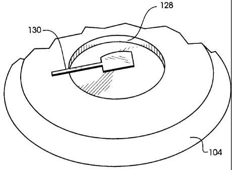
As shown in figure 1, microscope 100 includes a body 102 extending vertically
at a first
end from base 104. Body 102 supports at a second end a body tube 110, which,
along with base
104, is sufficiently rigidly attached to body 102 to provide support for the
remaining
components of microscope 100. A stage 106 is supported between body tube 110
and base 104
which locates a specimen 150 within the optical pathway along optical axis
190. Stage 160 is
typically supported from within appropriate structure within body 102 so as to
be vertically
adjustable closer to and further from objective 160 by rotation of objective
focus adjustment
knob 112. Carried with stage 106 is condenser 142 and aperture diaphragm 140
which are
moved relative to stage 106 by rotation of condenser focus adjustment knob 114
to move
condenser 142 and aperture diaphragm 140 either closer to or further from
stage 106.
The optical components of microscope 100 may be thought of as originating at
light
8
CA 02342868 2001-03-01
WO 00/13055 PCT/US99/20163
source 120, which will typically have some type of electric filament 122 and
may typically be
a lamp such as a halogen or tungsten bulb. The particular nature of light
source 120 is not
critical to the invention, and other sources of light are known to work for
particular
applications, even, in some instances, providing preferable results. Many of
the available
sources are mentioned hereinabove in the background of the invention, though
nearly any
source of illumination could conceivably be used. As is the normal practice, a
collector lens
124 is preferably provided adjacent light source 120 to gather as much light
as possible from
light source 120, thereby maximizing the efficiency of light source 120 and
reducing the
amount of power and cooling required for operation of microscope 100. Mirror
126 serves to
direct horizontally oriented optical energy from light source 120 along a
vertical axis and
through field diaphragm 128. Field diaphragm 128 serves primarily to control
the total amount
of light which is ultimately delivered to specimen 150, and, in the preferred
embodiment
combination of figure 1, field diaphragm 128 will most preferably be left in a
wide-open
position to allow maximum illumination. Reducing the field diaphragm 128
aperture will
diminish the three-dimensional shadowcast effect which predominates in the
present invention.
In the most preferred combination, edge plate 130 is located adjacent field
diaphragm
128. Edge plate 130 is located within the general optical pathway indicated by
optical axis 190,
and as a result does block some light which would otherwise have passed
through field
diaphragm 128 and into condenser 142. Nevertheless, edge plate 130 will most
preferably only
block a minor percentage of the light passing through field diaphragm 128.
After passing
through field diaphragm 128 and interacting with edge plate 130, light will
next pass through
aperture diaphragm 140 which is adjacent condenser 142. Aperture diaphragm 140
will, in the
preferred combination, also be left as open as possible so as to admit the
maximum amount of
9
CA 02342868 2001-03-01
WO 00/13055 PCT/US99/20163
light through condenser 142. Restricting aperture diaphragm 140 has the strong
effect of
"stopping down" condenser 142. At low magnifications, this will diminish the
three-
dimensional shadowcast effect. At high magnifications this will also
undesirably increase the
depth of field. Condenser 142 serves to focus light through specimen 150 into
objective lens
160, which in turn forms a first virtual image of specimen 150. This image is
further magnified
by eyepiece 170, which might, for exemplary purposes only, include eyepiece
field lens 172
and eyepiece eye lens 174. For standard viewing in accord with the preferred
embodiment, no
additional structure is necessary. However, if the microscope is so equipped,
photographs may
be taken of the magnified specimen through the use of a camera or film holder
180 having a
shutter 182 and film plane 184.
As can be seen in figure 2, edge plate 130 includes a handle 132 which allows
the
manipulation of edge plate 130 by human hand, without adverse interaction with
or
contamination of adjacent optical components. Handle 132 will most preferably
be made much
thinner than body 134, to reduce the asymmetric disruption of the illuminating
beam and any
resulting astigmatism that would otherwise disrupt the image. Body 134 is
bordered by inactive
edges 135, 136 and 138. These inactive edges must be sufficiently spaced from
active edge 137
to prevent any optical interaction within the active region of light cast by
edge 137, as will be
described hereinbelow. Otherwise, it is most preferable to maintain these
edges as closely
spaced as possible to minimize asymmetric blockage of light and resultant
astigmatism. Active
edge 137 is most preferably convex in geometry, as shown in figure 2. The
thickness of edge
plate 130 is not critical to the invention, though edge plate 130 is
preferably formed from a
relatively thin and lightweight sheet material such as black anodized
aluminum, which is
selected for the characteristics of low cost, ease of manufacture, durability,
and inherent optical
CA 02342868 2001-03-01
WO 00/13055 PCT/US99/20163
absorption. Handle 132 may also be stamped simultaneous with the balance of
edge plate 130,
or may alternatively be comprised by a small rod or other handle.
Nevertheless, other materials
having different properties and relative thickness may be used satisfactorily
in the perfonnance
of the invention. While the most preferred embodiment edge plate 130 uses only
a single active
edge, it is noted herein that more than one edge may be designed to act as an
active edge. For
example, edge 138 may also be designed to be active though it will be
understood that in order
to prevent optical interaction between various edges, edge 138 will be active
in a different
region of specimen 150 than edge 137.
Figure 3 illustrates the placement of edge plate 130 from a projected view, to
better
illustrate the arrangement and relative sizes of components. As can be seen
therein, edge plate
130 forms a minor portion of the cross-section taken along optical axis 190,
thereby admitting
a majority of light through to condenser 142. As illustrated, handle 132 is
not fixedly attached
to microscope 100. Nevertheless, it will be understood that, when desired, one
of ordinary skill
will be able to modify handle 132 and microscope 100 to include various
attachment points and
mechanism, or other devices such as but not limited to bearing structures,
that may be used to
position edge plate 130 fixedly to microscope 100. One benefit of the smaller
surface area of
edge plate 130 which blocks light and the central location of edge plate 130,
as shown in figure
3, is that astigmatism within the image is reduced. This only further benefits
the clarity of the
image formed by microscope 100.
Figures 4a - 4c illustrate the method of the invention as seen through
eyepiece eye lens
174. In the preferred embodiment of figure 1, edge plate 130 will be at the
level of field
diaphragm 128. When microscope 100 has condenser 142 adjusted to its usual
Koehler
position, shown in figure 4a, a dark region 210 is evident, which is the
shadow cast by edge 137
11
CA 02342868 2001-03-01
WO 00/13055 PCT/US99/20163
of edge plate 130. In the Koehler position, field diaphragm 128 will usually
be in focus, and
since edge plate 130 is at the same level, edge plate 130 will also be in
focus and will cast a
sharp shadow onto specimen 150 as represented by dark region 210. As can be
seen in figure
4a, there is very little evidence of specimen 150 visible, though a light
outline of cell 240 can
be detected near the border between bright-field region 220 and dark region
210. However,
when condenser focus adjustment knob 114 is adjusted to move condenser 142
either slightly
up or down from the Koehler position, dark region 210 and bright-field region
220 become
separated by a chromatic region 230. The initial adjustment produces only a
small chromatic
region 230 as shown in figure 4b, but nevertheless, an additional cell 241
becomes visible, and
greater detail of cell 240 becomes visible, including not only membrane but
also nucleus.
Further defocussing results in a broadened chromatic region 230, as shown in
figure 4c. This
chromatic region 230 may be adjusted to completely cover the field of view
through eyepiece
170. A much larger number of cells within specimen 150 are now visible, and
once again the
features within the first visible cells 240 and 241 are now much clearer. The
chromatic region
230 will typically take on a relatively monochromatic blue color if the
condenser is positioned
just below the Koehler position, and a red color if the condenser is
positioned just above the
Koehler position, with edge plate 130 at the level of field diaphragm 128.
While the invention
is not solely limited to any particular theory, the chromatic light is
believed to result from
diffraction along active edge 137 of edge plate 130. Since the overall
intensity of light within
chromatic region 230 is reduced relative to the bright field region 220, it is
plausible to increase
the intensity of the light output by light source 120, though this may not be
necessary in many
cases.
Edge plate 130 may be positioned at any point essentially throughout the sub-
stage
12
CA 02342868 2001-03-01
WO 00/13055 PCT/US99/20163
illumination path. However, the most preferred location is as shown in figure
1, away from
condenser 142. The image of edge plate 130 will be just slightly out of focus
through
condenser 142 with specimen 150. A second preferred location for edge plate
130 is between
light source 120 and collector lens 124. Most preferably, and in either of the
foregoing more
preferred locations, edge plate 130 is supported by body 102 at base 104, and
does not move
when objective focus adjustment knob 112 is rotated, nor when condenser focus
adjustment
knob 114 is rotated. Several significant benefits are enured by this
arrangement. First and
foremost, there is no need for special spacings or clearance for edge stop
130. Were edge stop
130 to move together with condenser 142, there would have to be sufficient
clearance to allow
the motion of edge plate 130. Otherwise edge plate 130 must be positioned much
more closely
to condenser 142. And yet when edge plate 130 is located closer to condenser
142, edge plate
130 begins to adversely affect the optical characteristics of microscope 100,
including in
particular the depth of field and resolution. In addition, movement of the
condenser could
undesirably upset the positioning of edge plate 130, or could require more
complex attachment
between edge plate 130 and the surrounding support of microscope 100.
Furthermore, not all
microscopes have ready access at any one or more of the preferred locations.
The placement
of the preferred edge plate 130 of the present invention is very
unrestrictive, allowing the
present invention to be benefited from with a very wide variety of microscopes
while not
interfering with pre-existing components. Other benefits may additionally be
gained by the
relative motion between condenser 142 and edge plate 130. So, while edge plate
130 could be
placed anywhere in the substage illumination path between light source 120 and
stage 106, the
most preferred region is at the level of field diaphragm 128. The otherwise
more preferred
placement includes anywhere between field diaphragm 128 and light source 120.
13
CA 02342868 2001-03-01
WO 00/13055 PCT/US99/20163
While not wishing to be bound by any particular theory in those aspects of the
invention
which are otherwise understood and demonstrated to be operational using the
techniques
described and illustrated herein, figure 5 does illustrate broader basic
principles of the
invention. These broader features will be understood by those skilled in the
art, upon a review
of the present disclosure, to not be limited to any single physical structure
or apparatus.
Instead, these features of the present invention enable those skilled in the
art to design a
potential myriad of embodiments, which are, nevertheless, within the scope of
the present
invention and claims, and are enabled herein. As shown in figure 5, edge plate
130' has a
straight active edge 137' that extends beyond field diaphragm 128. Light rays
10 are blocked
by field diaphragm 128 and edge plate 130', while rays 12 pass unobstructed by
either edge
plate 130' or field diaphragm 128. A portion of the optical rays 14 will also
be diffracted by
edge plate 130' along active edge 137', forming a diffraction wave represented
by rays 14a, 14b
and 14c. The diffraction waves that result from interaction with an edge,
represented by rays
14a - 14c, are known to have regions of chromaticity. These chromatic regions
are relatively
monochromatic as a result of the diffraction occurring at edge plate 130'. As
shown in figure
5, ray 14c has a vertical component 18 and a horizontal component 16. The
diffracted rays 14
which form chromatic region 230 are then observed to interact with specimen
150 at regions
of varying refractive index, such as at cell membranes and within the nucleus
of the cell. The
diffracted light is demonstrated herein to interact with these regions of
varying refractive index
to form new constructive (bright) or destructive (dark) interference patterns,
or more simple
additive and subtractive illumination regions. In either case, the net effect
is substantially
enhanced contrast which includes both brightening and darkening of various
entities and
regions within specimen 150.
14
CA 02342868 2001-03-01
WO 00/13055 PCT/US99/20163
Several additional features of the present invention serve to further refine
and enhance
the imaging of a specimen, and these features are illustrated in figures 6- 8.
As shown in figure
6, alternative embodiment edge plate 130' having a straight active edge 137 is
shown to have
a diffraction pattern in the lateral direction transverse to optic axis 190
towards bright field
region 220, as illustrated by arrows 16'. Arrows 16' neither diverge nor
converge. As shown
in figure 7, edge plate 130" has a concave active edge 137" which has a
diffraction pattern in
the lateral direction shown by rays 16" towards bright field region 220. As is
evident, these
rays tend to converge. As shown in figure 8, edge plate 130 has active edge
137 which is
convex in shape. Rays 16 which are diffracted from active edge 137 towards
bright field region
220 tend to diverge.
The use of convex active edge 137 has been demonstrated to provide
substantially better
contrast and more three-dimensional images within an ordinary biological
specimen 150 than
obtained with straight active edge 137, while straight active edge 137'
provides clearer
resolution than achieved with concave active edge 137". The use of convex edge
137 therefore
provides substantial additional advantage. While not wishing to be bound by
any particular
theory of operation, this advantage is believed to be due to the nature of
diverging rays 16. In
a theoretically perfect optical system, the relationship between rays 16 will
hold throughout the
optical system. However, since all lenses are imperfect due to aberrations, a
certain amount of
angular deflection will occur between rays emanating from adjacent areas of
the edge.
Furthermore, all real edges are also imperfect, including edge 137, and will
have optically
significant defects therein which can misdirect adjacent rays 16. Finally,
specimen 150 may
also have imperfections that would tend towards generating undesired
interference. Rays 16
which diverge are less prone to optical interference with adjacent rays, due
to the slight
CA 02342868 2001-03-01
WO 00/13055 PCT/US99/20163
divergence. The slight divergence tends to negate the effects of optically
detectable
imperfections present in edge 137 and optical defects present in the remaining
optical
components of microscope 100. This benefit is further enhanced by the relative
monochromaticity of chromatic region 230, which limits interfering refraction
from other
wavelengths that might otherwise tend to blur or even completely mask the
specimen image of
the present invention. Other edge geometries which offer benefit of diverging
rays 16 similar
to convex edge 137 are also contemplated, and will be understood by those
skilled in the art to
be included herein.
While, in most cases, it will be desirable to utilize a convex edge, the
present invention
contemplates and enables application of various edge plates each having
differently configured
active edges. For example, and as illustrated in figures 6 - 8, there may be
applications
requiring edge plate 130' with straight edge 137' or edge plate 130" with
concave edge 137".
Owing to the mechanical simplicity of the invention, various edge plates may
be inserted during
one viewing session, which will allow multiple perspectives to be taken of a
single specimen.
Because edge plate 130 is most preferably fabricated from a durable material
such as anodized
aluminum, other surface treated metals, or even plastics, ceramics, composites
or any other
suitable material, edge plate 130 will be resistant to damage or breakage.
This can be
particularly important in the applications such as school laboratories, where
the tools should
be both durable and of low cost.
As demonstrated by the preferred embodiment, the relatively monochromatic
diffracted
light which is interacted in an additive and subtractive or constructive and
destructive way
provides far better contrast enhancement and resolution of specimens than
heretofore available
with other techniques. As a result of the interaction between diffracted
light, bright-field light
16
CA 02342868 2001-03-01
WO 00/13055 PCT/US99/20163
and specimen, and the further combination of benefits from convex active edge
137, the present
invention exceeds contrast enhancement achieved by oblique illumination, and
equals or
exceeds that achieved by the much more complex and expensive research
techniques such as
differential interference contrast. Since field of depth is not adversely
impacted by the
relatively small edge plate 130, the image remains clear and uncluttered, as
demonstrated by
figures 4a - 4c herein.
INDUSTRIAL APPLICABILITY
Within the region jointly accompanied by diffracted rays 14c and rays 12 from
figure
5, the specimen has greatest contrast. The simultaneous additive and
subtractive nature of
reflection and/or interference patterns that are created within chromatic
region 230 due to the
interaction between diffracted light 14, bright-field illumination 12 and
specimen 150 yields
astounding contrast. Owing to the simple nature of the apparatus required to
generate this
enhanced contrast, there are many applications for which the present invention
is suited, only
one of which is in the area of biological analysis and observation. Other
known applications
of microscopy which have heretofore been difficult due to insufficient
contrast, but which
provide specimens having varying optical properties, will also be served by
the present
invention. Since the objects of the invention are, as described in the
description of the preferred
embodiment, achieved by the present invention, the present invention is
applicable industrially
not only to new microscopes, but also to low-cost retrofitting of existing
microscopes. This
retrofit enables enhancement of contrast sufficient to bring heretofore
invisible features into full
view through an ordinary eyepiece in an ordinary microscope.
While the foregoing details what is felt to be the preferred embodiment of the
invention,
17
CA 02342868 2001-03-01
WO 00/13055 PCT/US99/20163
no material limitations to the scope of the claimed invention are intended.
Further, features and
design alternatives that would be obvious to one of ordinary skill in the art
are considered to
be incorporated herein. For example, while the preferred embodiment
illustrates the use of a
compound microscope having an internal light source 120, the edge plate of the
present
invention may be implemented in other light paths that originate from other
types of sources,
and in other optical arrangements besides the preferred compound microscope,
as will be
ascertainable by those skilled in the art upon a review of the present
disclosure. Structures and
configurations that provide the equivalent effects as the present edge plate
are contemplated
herein. Rather than be limited by the disclosure of a single preferred
embodiment, the full
scope of the invention is instead set forth and described in the claims
hereinbelow.
18