Note: Descriptions are shown in the official language in which they were submitted.
CA 02342880 2001-03-02
WO 00/15122 PCT/US99/17839
CELLULAR SUBLIMATION PROBE AND METHODS
BACKGROUND OF THE INVENTION
The invention relates generally to the field of electrosurgery,
s and in particular to electrosurgical procedures which are performed within a
body
cavity which is filled with a liquid. In one particular aspect, the invention
relates to
the vaporization and cauterization of tissue in a body cavity which is filled
with a
conductive medium.
Electrosurgical devices are currently being used to treat a variety of
ailments. For example, electrosurgical devices are successfully being used to
cut
and ablate tissue, as well as to provide coagulation. one exemplary
electrosurgical device that is useful in treating the endometrial lining of
the uterus
(among other applications) is described in U.S. Patent No. 5,456,689 and in
co-pending U.S. application serial no. 08/322,680, filed October 13, 1994, the
i5 disclosures of which are herein incorporated by reference. One embodiment
described in U.S. Patent No. 5,456,689 includes a wire loop electrode that may
be used to cut tissue when current is supplied to the electrode and the
electrode
is moved through tissue.
In some circumstances it may be desirable to perform
ao electrosurgical procedures in locations that are filled with an
electrically
conductive medium. For example, co-pending U.S. Patent Application Serial Nos.
08/678,412, filed July 2, 1996; 08/822,901, filed 3/20/97; and 09/046,298,
filed
March 23, 1998 describe exemplary electrosurgical devices and methods for
treating tissue in an having an electrically conductive medium. The complete
25 disclosures of all these references are herein incorporated by reference.
While the above referenced electrosurgical devices have proven to
be extremely successful, it is desirable to provide other electrosurgical
devices for
other applications and treatments. In one aspect, it would be desirable'to
provide
an electrosurgical device which can be used at high power settings to vaporize
3o tissue and provide coagulation effects. Such a device should have an
electrode
which is durable and robust so that it will not materially degrade when used
at
high power settings. Further, it would be desirable if such a device were
useful in
either a conductive or a nonconductive medium. In another aspect, it would be
SUBSTITUTE SHEET (RULE 26)
CA 02342880 2001-03-02
WO 00/15122 PCT/US99/17839
desirable to provide an electrosurgical device that is useful on .an
outpatient basis
so that tissue may be treated without requiring a prolonged stay in a
healthcare
facility. In still another aspect, it would be desirable to provide a device
with a
relatively large electrode so that larger areas of tissue may be treated in a
more
efficient manner.
SUMMARY OF THE INVENTION
The invention provides exemplary electrosurgical probes and
methods for their use. The probes of the invention are particularly useful in
io vaporizing or cauterizing tissue, particularly within a physiologic
(conductive)
distention medium, although the probes are also useful in non-conductive
media.
In one exemplary embodiment, an electrosurgical probe is provided which
comprises a probe body having a proximal end, a distal end and at least one
lumen. An electrode assembly is operably coupled to the distal end, with the
~s electrode assembly comprising an electrode and a jacket disposed to cover
at
least a portion of the electrode. The jacket and the electrode have a combined
mass that is sufficient to dissipate heat produced during operation of the
electrode
so that the electrode does not experience material degradation. Further, the
jacket provides insulation between the electrode and a conductive medium such
ao that the electrode is operable within the conductive medium.
The electrode is preferably constructed of a metal or metal alloy,
while the jacket is preferably constructed of a ceramic material. In one
particularly
preferable aspect, the jacket and the electrode have a combined mass that is
greater than about 0.04 gram, and more preferably, in the range from about 0.1
zs gram to about 0.2 gram. Such a combined mass allows for the electrode
assembly to be operated at relatively high power settings without materially
degrading the electrode. For instance, when the jacket and the electrode have
such a combined mass, the heat that is produced during an electrosurgical
procedure may be effectively dissipated by the electrode assembly without
3o causing degradation to the electrode.
In another aspect, the electrode preferably has an exposed surface
SUBSTITUTE SKEET (RULE 26)
CA 02342880 2001-03-02
WO 00/15122 PCTNS99/17839
3
area that is in the range from about 0.07 in2 to about 0.125 in2. Such a
surface
area is particularly useful in allowing the electrode assembly to be
effectively used
in vaporizing or cauterizing tissue.
In still another aspect, the probe body has a working lumen, a fluid
s inflow lumen, and a fluid outflow lumen. In this way, the inflow and outflow
lumens may be used to introduce distention or other media as well as to
enhance
visual clarity of the hollow viscus. In one particular aspect, the probe body
has an
diameter that is in the range from about 0.07 inch to about 0.3 inch. Such an
outer diameter is particularly useful in that the probe may be inserted into a
variety of commercially available sheaths. Further, the probe diameter is
smaller
than or equal to the natural inner cervical diameter, thereby facilitating
atraumatic
introduction and implementation.
In another particular aspect, the electrode assembly is coupled to
the distal end with a fixed mount such that the position of the electrode
assembly
i5 relative to the probe body is fixed. Alternatively, the electrode assembly
may be
coupled to the distal end with a movable mount such that the electrode
assembly
is movable relative to the probe body. Use of such a movable mount allows
substantially all of the area of the electrode to be consistently maintained
in
contact with the wall of the body cavity, thereby minimizing procedure time.
zo The electrosurgical probe as described above may be included as
part of an electrosurgical system which includes an imaging scope that is
receivable in the working lumen of the probe body. In this manner, the tissue
to
be treated may be visualized during the treatment process. The working lumen
preferably has a diameter in the range from about 0.03 inch to about 0.16
inch.
z5 Such a diameter allows for the introduction of a variety of imaging scopes,
such
as flexible fiberscopes, rigid telescopes, and the like.
The electrosurgical system includes an electrosurgical unit to supply
current to the active electrode. A source of conductive fluid is also provided
which is connectable to the fluid inflow lumen to introduce an electrically
3o conductive distention medium into the body cavity. A vacuum source is also
provided and is connectable to the fluid outflow lumen to withdraw fluids from
the
body cavity to improve the visualization during a procedure.
The invention further provides a method for electrosurgically treating
SUBSTITUTE SHEET (RULE 26)
CA 02342880 2001-03-02
WO 00/15122 PCT/US99/17839
4
tissue. According to the method, an electrosurgical probe is provided which
comprises a probe body having a proximal end, a distal end, at least one
lumen,
and an electrode assembly operably coupled to the distal end. The electrode
assembly in turn comprises an electrode and a jacket disposed to cover at
feast a
s portion of the electrode. The probe is introduced into a body cavity, and a
conductive medium is introduced into the cavity through the lumen. Current is
then supplied to the electrode, and the electrode is positioned near or
against
tissue to treat the tissue. As the electrode is activated, the jacket provides
insulation between the electrode and the conductive medium. Further, heat
io produced during treatment of the tissue is dissipated by the jacket and the
electrode to prevent the electrode from materially degrading.
To vaporize or cauterize tissue, the electrode is preferably operated
at a power setting that is in the range from about 86 watts to about 300
watts. fn
one aspect, the probe body further includes a working lumen, and an imaging
scope is introduced into the working lumen to visualize the position of the
electrode within the body cavity. The probe body preferably also includes an
aspiration lumen to allow the conductive medium to be withdrawn from the body
cavity to improve the field of vision. In still another aspect, the electrode
assembly
is pivotally coupled to the distal end so that as the electrode is moved along
ao tissue, the electrode assembly will pivot to accommodate the shape of the
tissue.
BRIEF DESCRIPTION OF THE DRAWINGS
Fig. 1 is a side view of an exemplary electrosurgical device
according to the invention.
25 Fig. 2 is an end view of the device of Fig. 1.
Fig. 3 is a side view of an imaging scope that may be used with the
device of Fig. 1 according to the invention.
Fig. 4 illustrates the scope of Fig. 3 inserted into the device of Fig. 1.
Fig. 5 is a top view of a distal portion of the device of Fig. 1.
3o Figs. 5A-5E are cross sectional views showing various
embodiments of electrode assembly configurations according to the invention.
Figs. 6A-6F are cross sectional views showing various electrode
configurations according to the invention.
SUBSTITUTE SHEET (RULE 26)
CA 02342880 2001-03-02
WO 00/15122 PCT/US99/17839
Fig. 7 illustrates an electrode assembly coupled to a probe body
with a single axis swivel mount according to the invention.
Fig. 8 illustrates an electrode assembly coupled to a probe body
with a two axis swivel mount that is joint supported according to the
invention.
s Fig. 9 illustrates an electrode assembly coupled to a probe body
with a two axis swivel mount that is strut supported according to the
invention.
Fig. 10 is an end view of the probe body and the electrode assembly
of Fig. 9.
Fig. 11 illustrates the electrosurgical device of Fig. 1 being used to
~o vaporize tissue according to the invention.
Fig. 12 is a front view of the device of Fig. 11 when vaporizing
tissue.
Fig. 13 illustrates the electrosurgical device of Fig. 1 with a larger
sized electrode that is aligned with an imaging lumen while vaporizing tissue
i5 according~to the invention.
Fig. 14 is a front view of the device of Fig. 13.
Fig. 15 illustrates the electrosurgical device of Fig. 13 showing the
swivel motion of the electrode to allow the electrode to vaporize tissue which
is
situated at various angles relative to the device according to the invention.
zo Fig. 16 illustrates the electrode assembly and probe body of Fig. 10
when vaporizing tissue according to the invention.
Fig. 17 is a side view of another embodiment of an electrosurgical
device according to the invention.
Fig. 18 is a front view of the device of Fig. 17.
zs Fig. 19 is a side view of an alternative electrode assembly that may
be used with the device of Fig. 17.
DETAILED DESCRIPTION OF THE SPECIFIC EMBODIMENTS
The invention provides exemplary electrosurgical probes,
3o electrosurgical systems, and methods for their use. The probes of the
invention
are particularly useful in sublimating or vaporizing tissue. The probes of the
invention include a relatively large electrode that may be operated at
relatively
high power settings to create an arc which, when contacting tissue, causes the
SUBSTITUTE SHEET (RULE 26)
CA 02342880 2001-03-02
WO 00/15122 ! PCT/US99/17839
cells to rupture, thus vaporizing the tissue. Although primarily useful in
vaporizing
tissue, the probes of the invention may also be used to cauterize tissue.
The probes of the invention preferably comprise a semi-rigid or
flexible body having an active electrode at a distal end and a single or
multiple
s port connection on the proximal end. The probe body preferably has a length
in
the range from about 4 inches to about 24 inches, and more preferably at about
12 inches, for applications within the uterine cavity.
The active electrode at the distal end preferably has a disc or
spherical shape and is coupled to the probe body such that the active surface
of
to the electrode may be positioned in the direction of, and in apposition to,
the
ablatable tissue. The opposing side of the electrode is preferably covered
with a
dielectric refractory material, such as a ceramic material, a high temperature
polymer, such as Teflon, glass, glass former, and the like. The dielectric
refractory
material is particularly advantageous in that it dissipates heat produced
during
ablation. Moreover, the material acts as an insulation layer between the
physiologic distention media and the active electrode.
As just mentioned, the probe is preferably used within a hollow
viscus that is filled or distended with a physiologic, i.e., conductive,
distention
media. Exemplary conductive media include normal saline solutions, lactated
ao Ringer's solutions, and the like. Use of such solutions are advantageous in
that
the normal cellular sodium concentration may be maintained during the
procedure
so as not to disrupt hemostasis. Although particularly useful in physiologic
distention media so as not to disturb the sodium balance (hyponatremia), it
will be
appreciated that the probes of the invention may also be used with
z5 non-conductive distention media as well, including sorbitol, mannitol,
glycine, and
the like.
Although the probes of the invention will find their greatest use in
treating tissue within the uterine cavity, the probes may be used within any
hollow
viscus, including the prostate, and the like. In one particular embodiment,
the
so outer profile of the probe is minimized so that it may be introduced into
the uterine
cavity with minimal cervical dilation and trauma.
The probe body may include one or more lumens, of which one
preferably allows the passage of an imaging scope. Other lumens may be used to
SUBSTITUTE SHEET (RULE 26)
CA 02342880 2001-03-02
WO 00/15122 PCT/US99/17839
7
communicate hydraulically between the proximal connection and the distal end,
thereby allowing the introduction of distention and visualization media. Other
lumens may be used to move other fluids into or out of the body cavity and to
allow for the passage of various other devices. The connection at the proximal
end is preferably used to couple the imaging scope to the sublimating probe
body.
Construction of the probes of the invention provide a number of
design advantages. For example, the electrode preferably has a relatively
large
size which allows for operation at high electrosurgical power settings without
material degradation. For example, the electrode may be operated at power
o settings in the range from about 40 watts to about 400 watts to vaporize
tissue.
The electrode and dielectric jacket preferably have a combined mass than is
greater than about 0.04 gram, and more preferably in the range from about 0.1
gram to about 0.2 gram. Such a mass is sufficient so that the electrode will
be
able to withstand the relatively high power settings without material
degradation.
15 The ceramic jacket is further advantageous in that it allows for the
electrode to be operated in physiologic distention media. The ceramic jacket
serves as an insulator so that the current may concentrate at the non-
insulated
surface areas of the electrode which are to be placed in contact with or in
apposition to the ablatable tissue. If such a jacket were not employed, the
current
zo would disperse equally throughout the conductive media, thereby reducing
the
current concentration in the regions which are used to vaporize the tissue.
A further advantage of the probes of the invention is that the
working lumen allows for the passage of various devices into the body cavity,
including imaging scopes, biopsy tools, fluids, and the like. The inflow and
outflow
zs lumens also allow for the delivery of fluids, such as radiopaque fluids,
drugs,
imaging media, distention media, and the like, into the body cavity.
Still another advantage is that the probe body may be constructed to
have a relatively small outer diameter which is compatible with most
commercially
available sheaths. Preferably, the probe body has an outer diameter in the
range
s o from about 0.07 inch to about 0.30 inch. If the probe body includes only a
central
working lumen, the outer diameter may be made especially small to facilitate
its
introduction into small body cavities. Moreover, by employing a flexible
fiberscope
as the imaging device, the outer diameter of the probe body may be kept at a
SUBSTITUTE SHEET (RULE 26)
CA 02342880 2001-03-02
WO 00/15122 PCT/US99/17839
minimum. As an example, the scope lumen may have a diameter in the range
from about 0.03 inch (for fiberscopes) to about 0.16 inch (for rigid
telescopes). In
this way, the scope lumen may be adapted to receive a variety of commercially
sized imaging devices, such as 1-4 mm fiberscopes, 2.7 mm telescopes, 4.0 mm
telescopes, and the like. Further, the connection at the proximal end may be
configured to have a mount type which is compatible with the most domestic and
international scope brands, such as Circon, Storz, Wolf, and the like.
Another particular design advantage of the probes of the invention
is that the electrode assembly may either be fixedly or movably attached to
the
~o probe body. Such configurations allow for electrode maneuverability with
respect
to the probe body so that the probe may be employed to effectively treat
tissue
having a variety of shapes and configurations.
The design advantages described above provide the probes of the
invention with a variety of clinical advantages. For example, for cases within
the
i5 uterus, the probe diameter is smaller than or equal to the natural inner
cervical
diameter. In this way, the probe may be introduced into the uterus and then
operated in an atraumatic manner. The small probe dimensions also enable
physicians to perform myoma treatment in office settings in an outpatient
manner.
In this way, procedures which are now typically performed within a hospital
may
so be performed in an outpatient manner, thereby significantly reducing the
costs to
the patient.
Another clinical advantage is that the probe may be introduced and
positioned at a desired location under direct vision using flexible or rigid
scopes.
In this manner, orientation and navigation of the probe may be optimized.
z5 As described above, the apposition of the electrode with tissue may
be maintained due to the maneuverability of the electrode. In this way, a
variety of
tissue surfaces may be effectively vaporized without the need for reorienting
the
probe body, thus minimizing procedure time. To provide maneuverability to the
electrode, the electrode may be pivotally mounted to the probe body to provide
3o either single or double axis electrode swivel action. Such configurations
allow
substantially the entire active area of the electrode to be consistently
maintained
in contact with the surface of the body cavity, thereby minimizing procedure
time.
SUBSTITUTE SHEET (RULE 26)
CA 02342880 2001-03-02
WO 00/15122 PC'T/US99/17839
9
Procedure time is also minimized due to the large area "high current
density" electrode which delivers energy to a large surface area. By being
able to
vaporize large areas of tissue, operation time is minimized. In one particular
aspect, the exposed surface area of the electrode is preferably in the range
from
s about 0.007 in2. to 0.125 in2.
The use of inflow and outflow lumens provides distention
capabilities and also enhances the visual clarity of the hollow viscus. In a
further
clinical advantage, a variety of electrode configurations may be provided as
discussed below to allow for cellular vaporization as well as combined
coagulative
i o effects.
Referring now to Figs. 1 and 2, an exemplary embodiment of an
electrosurgical device 10 will be described. Device 10 comprises an elongate
probe body 12 having a proximal end 14 and a distal end 16. A working lumen 18
extends between the proximal end 14 and distal end 16. Probe body 12 may be
constructed to be either semi-rigid or flexible, with preferable materials for
constructing probe body 12 comprising polymers, such as polyolefins,
polyesters,
nylons and the like. Coupled to proximal end 14 is a scope mount 20 to which
an
imaging scope may be coupled as described in greater detail hereinafter.
Coupled
to distal end 16 is an electrode assembly 22. As best shown in Fig. 2,
electrode
ao assembly 22 comprises an electrode 24 and a jacket 26. A pair of amls 28
and 30
couple electrode assembly 22 to distal end 16. Electrode 24 is preferably
constructed of a metal or metal alloy and is employed to produce an electrical
spark to vaporize or cauterize tissue. Jacket 26 is preferably constructed of
a
dielectric material, such as ceramic, which serves to dissipate heat created
during
as vaporization and to insulate a portion of electrode 24 from an electrically
conductive medium.
Probe body 12 further includes a fluid inflow lumen 32 and a fluid
outflow lumen 34. Conveniently, a connector tube 36 is coupled to inflow lumen
32 to allow fluids to be introduced into the body cavity. Although not shown,
a
3o similar tube is connected to outflow lumen 34 so that fluids may be
withdrawn
from the body cavity. In this way, various distention or imaging media may be
introduced into the body cavity to distend the body cavity or to clear fluids
to
improve visualization.
SUBSTITUTE SHEET (RULE 26)
CA 02342880 2001-03-02
WO 00/15122 PCT/US99/17839
to
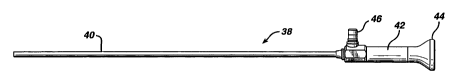
Referring now to Fig. 3, an exemplary imaging scope 38 which may
be inserted through working lumen 18 of probe body 12 (see Figs. 1 and 2) will
be
described. Imaging scope 38 comprises an elongate shaft 40 which may include
fiberscopic bundles or other optics associated with commercially available
s telescopes. In this way, shaft 40 may be constructed to be either flexible
or rigid.
Coupled to shaft 40 is a housing 42 which includes an eyepiece 44 and a light
coupling 46 to allow a light source to be coupled to housing 42. As is known
in the
art, imaging scope 38 may comprise any one of a variety of commercially
available scopes, including 2.7-4.0 mm rigid telescopes, 1.6 mm flexible
~o fiberscopes and the like.
As illustrated in Fig. 4, shaft 40 is insertable through working lumen
18 until housing 42 is coupled with scope mount 20. In this way, a hydrostatic
seal
is provided between scope 38 and device 10 to allow the body cavity to be
distended while preventing liquid from leaking from working lumen 18.
Moreover,
when scope 38 is coupled to device 10, electrode assembly 22 may be
visualized,
both during introduction of device 10 into a body cavity as well as during an
electrosurgical procedure where electrode 24 is employed to vaporize or
cauterize
tissue.
In an exemplary procedure, the inner lining of the uterine cavity may
ao be treated by first introducing a sheath through the cervical canal to
provide
access to the uterine cavity as is known in the art. Scope 38 is then inserted
through working lumen 18 and the combined scope 38 and device 10 are inserted
through the sheath to gain access into the uterine cavity. A distention medium
is
then introduced through tube 36 and into fluid inflow lumen 32 to distend the
z5 uterus, preferably with a physiologic distention media. If needed, fluids
may be
withdrawn through fluid outflow lumen 34 to improve the visibility within the
uterine
cavity. At any time, visualization of electrode assembly 22 may be gained by
looking through eyepiece 44. When situated to vaporize tissue,
electrost~rgical
current is provided to electrode 24 which is placed in apposition to the
desired
3 o tissue.
To prevent any of the pressurized fluid from leaking between the
sheath and device 10, proximal end 14 preferably includes a housing having a
SUBSTITUTE SHEET (RULE 26)
CA 02342880 2001-03-02
WO 00/15122 PCT/US99/17839
11
tapered portion which mates with and provides a seal with the hub of the
sheath.
In this way, device 10 may be used with essentially any type of standard or
commercially available sheath. If device 10 needs to be translated within the
sheath, a distensible O-ring may be fitted to the outer diameter of device 10
to
s provide hydrostasis. Device 10 is preferably constructed to have a length
that
exceeds the length of standard sheaths so that distal end 16 will extend
beyond
the tip of the sheath.
In some cases, the fluid may be introduced into the body cavity
through a fluid inflow lumen in the sheath. In this way, fluid inflow lumen 32
may
io be eliminated. In this manner, the overall size of device 10 may be reduced
for
profile optimization and device performance.
Fig. 5 illustrates a top view of an electrode assembly 47 in
schematic form. A section line 49 is provided to illustrate the orientation of
Figs.
5A-5E (which illustrate various embodiments of electrode assembly designs).
The
is embodiments illustrated in Figs. 5A-5E are shown to illustrate the various
ways in
which the jacket may be coupled to the electrode to provide exemplary heat
transfer characteristics so that heat produced during the vaporization
procedure
may be effectively dissipated so as not to materially degrade the electrode.
As
shown in Fig. 5A, a generally rectangular electrode 48 is surrounded on three
of
so its sides by a ceramic jacket 50. In Fig. 5B, an electrode 52 includes a
pair of
steps 54 that may be locked with a ceramic jacket 56 having an elliptical
outer
surface. In Fig. 5C, an electrode 58 is provided with a plurality of fingers
60 which
serve as heat sinks to facilitate the transfer of heat from electrode 58 to a
jacket
62. Fig. 5D shows an electrode 64 having a wedge shape to key with a jacket
66.
25 Finally, Fig. 5E shows an elliptical electrode 68 which is keyed with a
ceramic
jacket 70 to lock electrode 68 relative to jacket 70.
Hence, the electrode configuration shown in Figs. 5A-5E are
provided to enhance the heat transfer from the electrodes to the jackets so
that
the electrode will not materially degrade during a vaporizing procedure. The
so various electrode assemblies may be manufactured by any one of a variety of
processes. For example the electrodes may be mill machined, EDM machined,
coined, forged, cast, and the like. The jackets may be formed by mill
machining,
SUBSTITUTE SHEET (RULE 26)
CA 02342880 2001-03-02
WO 00/15122 PCT/US99/17839
12
metal oxide spraying, dip coating, electrostatic deposition, chemical
deposition,
vapor deposition, and the like.
Referring now to Figs. 6A-6F, further electrode assembly designs
will be described. The embodiments of Figs. 6A-6F are provided to illustrate
s different active surface area configurations to enhance the treatment of
tissue.
Although not shown, it will be appreciated that any of the elements of the
embodiments described in Figs. 5A-5E may be incorporated into the
embodiments illustrated in Figs. 6A-GF and vice versa. In Fig. 6A, a generally
rectangular electrode 72 is recessed within a jacket 74. In this manner, a
to fulguration recess is provided so that tissue may be fulgurated without
directly
contacting electrode 72 with tissue.
In Fig. 6B, an electrode 76 has a generally planar surface 78 which
projects from a jacket 80. In this way, planar surface 78 serves as a
vaporizing
surface, with the corners adjacent planar surface 78 serving to concentrate
the
is current to provide high-energy edges may be employed to both vaporize and
cut
tissue.
In Fig. 6C, an electrode 82 has a protruding vaporizing surface 84
with high energy planes 86 where current tends to concentrate. In this way,
high
energy planes 86 may be used to more effectively vaporize or ablate tissue. In
2o Fig. 6D, an electrode 88 and a jacket 90 are shown which are similar to the
embodiment of Fig. 6C except that electrode 88 and jacket 90 have a greater
mass to enhance heat transfer to reduce the chances of materially degrading
electrode 88 during a vaporization procedure.
In Fig. 6E, an electrode 92 has a curved active surface which is
25 useful in performing procedures involving both vaporization and
cauterization.
Electrode 92 is surrounded by a jacket 94 and include no high energy edges. As
such, electrode 92 may be used to perform functions similar to a standard
roller-ball or roller-barrel type electrode. In Fig. 6F, an electrode 96 has a
plurality
of lobes 98 where current tends to concentrate to more effectively vaporize
the
3o tissue. Fulguration surfaces 100 are provided between lobes 98 to fulgurate
tissue while lobes 98 are vaporizing tissue. Electrode 96 is disposed within a
jacket 102. In Fig. 6G, an electrode 104 and jacket 106 are essentially
identical to
those in Fig. 6F except for the addition of a dielectric material 108 which is
SUBSTITUTE SHEET (RULE 26)
CA 02342880 2001-03-02
WO 00/15122 PCT/US99/17839
13
disposed between lobes 110. In this way, a multi-lobe electrode surface is
provided. In Fig. 6F, an electrode 112 has a vaporizing/coagulation surface
114
and a lobe 116. A dielectric material 118 is disposed between surfaces 114 and
116. The jacket 120 is disposed about electrode 112.
s It will be appreciated that the various embodiments illustrated in
Figs. 5A-5E and 6A-6F are not exhaustive. Indeed, a wide variety of electrode
surfaces and heat transfer designs may be provided to enhance the
functionality
of the electrode assembly. For example, the electrode surfaces may be
recessed,
planar, lobed, spherical, conical, cylindrical, triangular, multi-surfaced,
combined
to planar/lobed, and the like. By providing various electrode configurations,
a
treatment system having a wide assortment of electrode configurations may be
provided at a relatively small cost and used during the same procedure. For
example, one device may have an electrode with edges and may be used to
provide high-energy vaporization and "cutting/ablating". This device may
quickly
i5 be swapped with another device having an electrode which provides for pure
"high efficiency" coagulation.
As previously described, the electrode assemblies of the invention
may be fixedly mounted relative to the probe body or may be pivotally or
swivel-mounted relative to the probe body to provide multiple degrees of
ao rotational freedom. For example, Fig. 7 illustrates a probe body 122 having
a pair
of arms 124 and 126 to which an electrode assembly 128 is hingedly mounted. In
this way, electrode assembly 128 may swivel about an axis extending between
arms 124 and 126. In Fig. 8, a probe body 130 includes a pair of arms 132 and
134 which are joined together at a point 136. An electrode assembly 138 is
as swivel-mounted to point 136, such as with a ball and socket arrangement. In
this
way, electrode assembly 138 may pivot to provide multiple degrees of freedom
of
movement. In Figs. 9 and 10, a probe body 140 includes a strut arrangement 142
to which an electrode assembly 144 is swivel-mounted in a manner similar to
the
embodiment of Fig. 8. In this manner, electrode assembly 144 may swivel about
3o multiple degrees of freedom. As shown in Fig. 10, use of strut arrangement
142 is
particularly advantageous in that it increases the field of view from a scope
146.
In addition to the fixed mountings or rotational mountings as just
described, the electrode assemblies may be mounted relative to the probe
bodies
SUBSTITUTE SHEET (RULE 26)
CA 02342880 2001-03-02
WO 00/15122 PCT/US99/17839
14
so that they are either aligned with or eccentric to the field of vision
provided by
the scope. For example, Figs. 11 and 12 illustrate the electrosurgical device
of
Figs. 1 and 2 when vaporizing tissue. Electrode assembly 22 is positioned at a
bottom portion of the field of view of scope 38 as illustrated in Fig. 12 so
that the
s electrode assembly is eccentric to the image. In this way, the physician may
view
above and beyond the tissue being vaporized. Electrode assembly 22 may be
either fixedly or pivotally mounted to arms 28 and 30 to provide various
degrees
of rotational freedom when the electrode assembly is placed in apposition to
tissue.
to Shown in Fig. 13 is electrosurgical device 10 having modified arms
28' and 30' so that electrode assembly 22 is aligned with the field of vision
of
probe 38. Such a configuration is particularly advantageous for tissue which
is at
an angle relative to probe body 12 as illustrated in Fig. 13. Another
advantage of
positioning electrode assembly 22 along a central axis of device 10 is that
the
is active electrode may be constructed to have a larger surface area.
Fig. 15 illustrates device 10 having modified arms 28' and 30' and
with electrode assembly 22 being pivotally attached to arms 28' and 30'. In
this
way, electrode assembly 22 may be swivelled perpendicular to the axis of probe
body 12 as shown. Such a configuration is particularly advantageous in
treating
ao tissue distal and perpendicular to the central axis of the device.
Fig. 16 illustrates probe body 140 and electrode assembly 144 of
Figs. 9 and 10 when used to treat tissue which is generally parallel to the
axis of
probe body 140. Due to the swivel mount, electrode assembly 144 is able to
maintain apposition to the tissue during vaporization. Such a configuration is
25 particularly advantageous in allowing for lateral, anterior/posterior
access.
Referring now to Figs. 17 and 18, an alternative embodiment of an
electrosurgical device 150 will be described. For convenience of discussion,
only
a distal end 152 of a probe body 154 is shown. Device 150 includes an imaging
scope 156 and an electrode assembly 158. Electrode assembly 158 comprises an
3o elongate shaft 160 which includes a conductor which may be coupled to an
electrosurgical unit. Coupled to shaft 160 are a pair of arms 162 and 164 to
which
an electrode 166 having a jacket 168 is coupled. As best shown in Fig. 18,
electrode 166 is disposed below scope 156 to provide a clear field of vision
for
SUBSTITUTE SHEET (RULE 26)
CA 02342880 2001-03-02
WO 00/15122 PCT/US99/17839
scope 156. As with other embodiments, electrode 166 may be fixedly mounted or
pivotally mounted to arms 162 and 164.
Shown in Fig. 19 is an alternative embodiment of an electrode
assembly 170 having a shaft 172 and a pair of arms 174 which have a S-shaped
5 configuration. Arms 174 are coupled to a jacket 176 and an electrode 178.
The
S-shaped configuration of arms 174 provides an alternative way to dispose
electrode 178 below the field of vision of the scope.
The invention has now been described in detail for purposes of
clarity of understanding. However, it will be appreciated that certain changes
and
to modifications may be made within the scope of the invention. Therefore, the
scope and content of the invention are to be determined in light of the
appended
claims and as well as the full equivalence to which those claims are entitled.
SUBSTITUTE SHEET (RULE 26)