Note: Descriptions are shown in the official language in which they were submitted.
CA 02343192 2001-03-06
WO 00/13577 PCT/U599I20236
PULSE OXIMETER SENSOR COMBINIED WITH
OROPHARYNGEAL AIRWAY AND BITE BLOCK
1. FIELD OF THE INVENTION
The invention relates to a combination oropharyngeal airway and bite block
s with pulse oximetry capabilities. More particularly, the invention relates
to a
device that allows for intraoral application of pulse oximeter sensors to a
patient
while establishing a ventilatable airway for the patient andlor maintaining
separation between the patient's upper and lower teeth.
II. BACKGROUND ART
to With a few exceptions, tradition and technology have favored
transillumination pulse oximetry in the operatinc,~ theater. The principle of
operation
of the pulse oximeter is fairly simple but is arguably the most important
develapment in anesthesia monitoring in the twentieth century. Two wavelengths
of light (usually fi60 nm and 940 nm} arse used to spectrophotometrically
~s determine the ratio of oxidized to reduced hemoglobin noninvasively as well
as to
determine the pulsatility of blood plethysmographically. Presently, the most
common application of this in the operating theater is via transillumination
through
the capillary bed of a peripheral digit. HowevE:r, it is not unusual far
multitrauma
and thermally injured patients to either have severe peripheral
vasoconstriction or
2o to have severely damaged (or missing due i:o amputation) peripheral
vascular
beds. Reflectance oximetry rather than transiilumination oximetry was the
earliest
investigative form of the technique. Transillumination pulse aximetry, without
question, is the most effective form when oximetry is obtained through skin.
However, when skin is not interposed as a barrier to capillary bed access,
2s reflectance pulse oximetry easily can be achieved with very accurate
results. The
effect is achieved by the backscattering of incident bispectral light that
traverses
and, on reflection from nonabsorptive collagenous tissues, retraverses formed
elements in the blood back to the oximetric detector.' Rather than superseding
transillumination pulse oximetry, this technique broadens the scope of
possible
3o monitoring sites, adding to the clinician's armarnentarium.
~~ ms~nm rr~ sN~~,- ~m n ~ ~~~
CA 02343192 2001-03-06
WO 00113577 PCTIUS99/20235
Previously, three devices were needed to accomplish the functions
provided by this invention. An orapharyngeal aurway and a bite block are sold
as
two separate pieces that are used at different times and in different
situations. A
pulse oximeter sensor is used to take readings for the determination and
s measurement of oxygen saturation in the blood without taking a blood sample.
Prior art devices have combined the oropharyngeal airway with the capability
to
perform transiiluminance pulse oximetry through the posterior tongue or have
placed oximeter sensors farther down the trachea then is proposed by this
invention.
to The oropharyngeal airway is used during surgical anesthesia. If the
oropharyngeal airway is inserted prior to induction of anesthesia or left
inserted
upon emergence from anesthesia, then there is the possibility that the patient
could be stimulated to vomit and aspirate storrEach contents resulting in an
often
fatal event for the patient. Also, the oropharyngeal airway will cause
is uncomfortable stimulations deep in the throat and thus cause gagging and
the
impingement of the teeth upon the endotracheal tube prior to extubation if the
patient awakens from anesthesia. Thus the oropharyngeal airway may not be
inserted until the patient is profoundly sedated and must be removed once the
patient begins to awaken. The oropharyngeal airway establishes a ventilatabte
2o airway in a patient who is unconscious.
The bite block maintains an oral aperture for suction and the passage of air
or vomit. The bite block is also used to prevent biting of an endotracheal
tube.
The bite block does not stimulate the posterior tongue or pharynx.
Prior pulse oximeter sensors inserted through the mouth are usable only
2s when the patient is under general anesthesia: 'These pulse oximeter sensors
are
inserted to reach the larynx area, for example, tJ.S. Patent No. 5,282,464 to
Brain
et al. Another known method uses transillumination pulse oximetry of the
posterior tongue, but this method may not be used with a patient, who is
awake,
for example, U.S. Patent No. 5,205,287 to Buchanan. Also, the posterior tongue
3o is not the most accessible body part to take oximetric measurements.
Conventional pulse oximetry in the severely burned patient can be a
significant challenge, yet this monitoring data is vital in operating room and
2
SUBSTrrUTE SHEET lIRULE 2~1
CA 02343192 2001-03-06
WO 00/13577 PCT/US99/2fl235
intensive care settings. Most current oximetric approaches depend upon
available peripheral sites permitting transiiluminatian oximetry and indeed,
this
method is sufficient for most surgical conditions and procedures.
Unfortunately,
patients with severe burns often have few sites for the effective placement of
the
s transilluminating pulse oximeter sensor. In addition, these patients often
have
severe circulatory compromise rendering they peripheral pulse oximeter less
efficient.
Recent studies indicate that oral pulse oximetry is a superior modality when
compared to peripheral transiilumination pulse oximetry. A variety of studies
have
to shown that oral pulse oximeters are more reliiably and rapidly responsive
than
peripheral pulse oximeters. However, these studies use oral transillumination
pulse oximetry, held in place via complex devices or pieces of improvised
malleable metal. Oral secretions, equipment failure, and placement difficulty
often
render these techniques ineffective.
~s Reflectance oximetry can be a useful tool where a capillary bed is easily
accessible. Indeed, it is used commonly and effectively among intrapartum and
neonatal patients whose capillary beds are easily accessed through their skin.
The technique has also been applied to adult and pediatric burn patients by
placing the reflectance sensor in wounds or ovE~r hyperemic sites such as
healed
2o partial thickness burns.
There are other often overlooked capillary beds readily accessible in most
adult burn patients that are as amenable to reflectance oximetry as the
forehead
of the premature infant. The buccal surface, po;;terior soft palate, hard
palate and
proximal posterior pharynx of a burned patient are seldom compromised no
2s matter how severe the burn, and the capillary beds are very close to the
surface in
those areas. Transiliumination pulse oximetry of the tongue and cheek has been
documented as a viable method of monitoring, but not everyone has the
equipment available to place a transilluminating~ pulse oximeter on the tongue
or
cheek. A reflectance pulse oximeter has the bislpectral emitter and the sensor
in a
3o side-by-side configuration rather than in opposition. The device may be
placed
flat upon a suitable capillary bed and it thus becomes a reflectance pulse
oximeter. In this manner, a standard disposable finger pulse oximeter probe
may
3
suesn~uTE sHESr tF:u~.~ z~~
CA 02343192 2001-03-06
WO 00/13577 PCT/US9912fl235
simply be placed flat against the buccal surface, thus rendering it a
reflectance
rather than a transiiluminating device.
Notwithstanding the usefulness of the above-described devices, and the
above-identified recognized viability of transilluminating buccal pulse
oximetry, a
s need exists for a more convenient device that combines a bite block with an
oropharyngeal airway. Additional convenience is obtained by including a pulse
oximeter sensor with a device that includes the bite block and the
oropharyngeal
airway.
III. DISCLOSURE OF THE INVENT10N
io This invention solves the ongoing prok>lems of using multiple devices to
perform intraoral oximetry measurements b~y providing a single device for
performing such measurements. The invention while addressing the problems of
the prior art obtains advantages that were not achievable with the prior art
devices.
is The invention encompasses a combined bite block and oropharyngeal
airway in one device. In accordance with a second embodiment, the invention
includes a pulse oximeter sensor with the combined bite block and
oropharyngeal
airway; thus achieving greater simplicity and convenience not possible when
three
separate devices were required to be on hand <~nd used.
2o An object of this invention is to simplifiy the amount and type of medical
devices that are required to be stocked by a medical facility or emergency
crew.
Another object of the invention is to obtain a decrease in costs resulting
from having one combination device instead of multiple devices.
Another object is the use of reflectance ,pulse oximetry in the oral cavity
for
2s a variety of freld, emergency, surgical, anesthetic, or critical care
procedures or
situations to include patients that are awake', sedated or undergoing general
anesthesia.
Still another object of the invention is to~ monitor oxygen levels in severely
burned ICU patients who are difficult to monitor.
4
SU6ST1TUTE gH~S'f I!Rtl>_E ?fl
CA 02343192 2001-03-06
WO 00113577 PCT/US99/20235
An advantage of the invention is an improvement in the quality of care
resulting from elimination of the need to switch devices during the course of
taking
oximetry measurements.
Another advantage of the invention is that EMS crews and personnel will
s be able to use this invention easily in the field during emergency
situations.
Another advantage of the invention is improved pulse oximetry readings.
Another advantage of the invention is reflectance pulse oximetry requires
less power to function and thus less heat is produced, which decreases the
risk
the patient will be burned. If the patient is burned, then the blood flow and
saliva
~o production will facilitate regeneration of the capillary bed quicker than
in other
tissue areas.
The invention accomplishes the above objectives and achieves the
advantages. The invention is easily adapted to a wide variety of situations.
Furthermore, intraoral (i.e., lingual, buccal or proximal posterior
is pharyngeallpalatal) placement of a disposable pulse oximeter probe in a
configuration relying upon reflectance will provide pulse oximetry
measurements
comparable to those obtained by peripheral pulse oximetry. The invention and
test
data suggest that buccal and proximal posterior pharyngeaUpalatal reflectance
pulse oximetry provides a simple, accurate means of monitoring arterial oxygen
2o saturation in the severely burned patient where oximetric monitoring
presents a
challenge.
Given the following enabling description of the drawings, the apparatus
should become evident to a person of ordinary skill in the art.
IV. BRIEF DESCRIPTION OF THE DRAWIINGS
2s Figure 7 illustrates a side view of a preferred embodiment.
Figure 2 illustrates a front view of the embodiment shown in Figure 1.
Figure 3 illustrates a rear view of the embodiment shown in Figure 1.
Figure 4 illustrates a top view of the embodiment shown in Figure 1.
SUBSTTrUTE SHEET iRtILE 26)
CA 02343192 2001-03-06
WO 00/13577 PCT/US99/2d?t35
Figure 5 illustrates a side view of another embodiment.
Figure 6 illustrates a front view of the embodiment shown in Figure 5.
Figure 7 illustrates a rear view of the emlbodiment shown in Figure 5.
Figure 8 illustrates a top view of the embodiment shown in Figure 5.
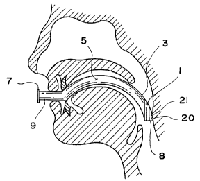
s Figure 9 illustrates the embodiment shown in Figure 5 in use as an oral
airway.
Figures 10(a) and (b) illustrate the embodiment shown in Figure 5 in use as
a bite btock.
Figure 11 illustrates a side view of another embodiment.
io Figure 12 illustrates a partial front view of the distal end of the
embodiment
shown in Figure 11.
V. BEST MODES FOR CARRYING OUT THE DESCRIBED EMBODIMENTS
Figures 1-4 illustrate an embodiment of the invention directed to a
combination bite block and oropharyngeal airway. As depicted, the device
is includes a base 7, a straight portion 9, and a palatal and proximal
pharyngeal
contour portion 5 preferably arched to be physiologically compatible with the
palate and pharynx. The contour portion 5 includes arched section 2 having an
outer distal curve 3 and a distal end 8. The contour portion 5 is preferably
integrally formed with straight portion 9, which includes a proximal end
abutting
2o the base 7. The base 7 preferably is large enough to allow the device to be
manipulated by the user.
A centrat passageway or channel 6 maiy be formed within the device to
extend from the distal end 8 to the base 7. As is apparent to one of ordinary
skill
in the art in view of the present disclosure, the: cross-section and
dimensions of
2s the passageway 6 may be selected to maximize the airtlow through the
passageway without reducing the integrity of the: device.
To facilitate operation of the device as a bite block, bilateral grooves (or
recesses) 1 into which the teeth may fit are preferably disposed in opposing
6
SUBSTITUTE SHEET fl3ULE 26~
CA 02343192 2001-03-06
WO 00/13577 PCT/US99/202_35 ,
relationship to each other and formed in the contour portion 5. More
preferably,
the bilateral grooves extend from the distal end' 8 to a point along the outer
distal
curve 3. The bilateral grooves 1 may be filled with a sponge-like or soft
material,
e.g., foam or rubber to protect the teeth. A significant advantage associated
with
s this embodiment is that it may be employed as both a bite block and an
oropharyngeal airway.
In accordance with a particularly advant<~geous feature of the invention, to
accomplish a change between the two modEa, the device only needs to be
repositioned within the patient thus avoiding the need to exchange devices as
to required with present devices. For example, during anesthesia, it may be
desirable to use the device as an oropharyngeal airway to establish a
ventilatible
airway for the patient. When used as such, thE: device is preferably inserted
into
the patient's mouth such that it impinges upon i:he posterior soft palate
andlor the
posterior pharynx along the outer distal curve 3.
is In addition, before, on induction of, during, on emergence from and after
anesthesia, it may be desirable to employ the device as a bite block. When
used
as such, the device is preferably inserted into the patient's mouth such that
the
bilateral grooves 1 on the sides of the contour portion 5 may be inserted
between
the molars andlor bicuspids on one side of the mouth. The outer distal curve 3
in
2o this mode abuts the buccai mucosa, as shown in Figure 7 0(a). The
alternative
preferred insertion method is to place the device such that the bilateral
grooves 1
are inserted between the molars and/or bicuspids on the side of the mouth that
the base 7 is located. The outer distal curve ;3 in this mode wilt abut the
lingual
surface of the tongue, as shown in Figure 10(b). Neither mode will stimulate
the
2s posterior tonguelpharynx.
Figures 5-9 illustrate a second embodiment of the combination bite block
and oropharyngeal airway that includes pulse oximeter sensor elements. This
embodiment has the same basic structure as the previously described
embodiment. in accordance with an aspect of the invention, pulse oximeter
3o elements 20 and 21 reside in the posterior di stal curvature of the device.
The
pulse oximeter elements 20 and 21 include a light source 20, which preferably
emits light with wavelengths of fi60 nm (red) and 940 nm (near infrared), and
a
7
SUBS~'CTtJTE SHEET (MULE 26)
CA 02343192 2001-03-06
WO 00/13577 PCT/US99/24235
light detector 21. The placement of the light source 20 and the light detector
21
may be switched with each other with respect to the placement shown in Figures
5, 7, and 9. Preferably, these pulse oximeter elements are embedded in the
body
of the device along the outer distal curve side 3 facing radially outward with
a
s cover protecting them. Preferably the cover is a clear, fluid impermeable
plastic.
Alternatively, the pulse oximeter elements 20 and 21 may be disposed within
passageway 6 adjacent the outer distal curve 3.
The light source 20 may include more than one emitter. The light source
20 is preferably one or more of the following: two light emitters such as
light
~o emitting diodes {LED), a bispectral emitter, a dual spectral emitter, a
photoemitter,
or a semiconductor die. However, any light source that facilitates reflectance
pulse oximetry may be employed. When the light source 20 is one light emitter
then the light emitter, for example, preferably uvould emit two frequencies of
light
at about 660 nm and about 940 nm. Typically, the two emitter arrangement will
~s include a red LED near 660 nm and a near-infrared LED emitting in the range
of
890 nm to 950 nm. The light source 20 may emit light having a bandwidth in the
range of 20 nm to 50 nm.
A light detector 21 detects light emitted by light source 20. Electrical
signals
representing the detected light are transmitted by light detector 21 to a
2o spectrophotometer or pulse oximeter that discriminates between the relative
intensity of these emissions and provides an iindex as to the degree of oxygen
saturation of hemoglobin in blood. Preferably, the tight detector 21 may be
one of .
the following: photoelectric receiver, photodetector, or a semiconductor die.
The pulse oximeter elements 20 and 211 may be disposed in a variety of
2s locations along the passageway in accordance with the desired application.
Preferably, the pulse oximeter elements 20 andl 21 are placed closer to the
distal
end 8 of the device so that the readings may be taken from the post pharynx
area,
the buccal surface, or the lingual surface of the patient. As the pulse
oximeter
elements 20 and 21 are moved towards the apex of the arched section A, the
3o readings more likely will be taken from the soft ~pafate of the patient.
The dividing
tine between these regions is highly dependent on the internal dimensions of
the
patient. However, the readings obtained from each area work equally well in
8
SUBSTITUTE SHEET (E~ULE 26)
CA 02343192 2001-03-06
WO OOII3577 PCTlUS99/2~?.35 ,
terms of accuracy. Also, the closer to the apex of the arched section A the
pulse
oximeter elements 20 and 21 are, the more difl~icult it is for the device to
contact
the buccal surtace or the lingual surface when the device is~ used as a bite
block.
When the pulse oximeter elements 20 and 21 acre positioned away from the apex
s of the arched section A towards the proximal end abutting the base 7, the
readings will be taken from the hard palate, which also will provide accurate
pulse
oximetry readings.
Figures 5-9 depict wiring 24 connecting i:he pulse oximeter elements to an
external cord 22. Such wiring 24 is preferably also embedded in the body of
the
io contour portion 5. The wiring 24 may include conductive lines and contact
electrodes. The external cord 22 preferably is insulated and connects to the
wiring 24 at the proximal end. The external cord 22 may include a standard
plug
designed to engage a pulse oximetry spectrophotometer or other external
device.
The spectrophotometer provides the electrical signals for controlling the
pulse
is oxirneter elements 20 and 21. Alternatively, pulse oximeter elements 20 and
21
may be in wireless communication with the pulse oximetry spectrophotometer or
other external device.
As previously mentioned, the pulse oximeter elements may be disposed in
the passageway . A disposable pulse oximeter sensor like the Nellcor~
2o Oxisensor~ Il N-25 may be stripped of its surroundings to leave only the
pulse
oximeter elements. The pulse oximeter elements are then feed along the topside
of the passageway 6. Although the pulse oximeter elements and wiring may be
present in the passageway 6, there will be sufficient airtlow capacity in the
passageway to supply oxygen to the patient. The N-25 pulse oximeter sensor
2s when installed in this manner does not overdrive as a result of the emitted
brightness from the light source, because of the optical effects provided by
the
oropharyngeal airway.
To facilitate operation of the device as a pulse oximetry sensor, a plastic
bag, protective cover or similar item may be placed around the distal end 8.
This
3o embodiment is particularly useful when there; is excess moisture that might
interfere with the operation of the pulse oximeter sensor elements.
9
SU8ST1TUTE SHEET (RULE 26)
CA 02343192 2001-03-06
WO 00113577 PCT1US99120~35
In accordance with an aspect of the invention, the passageway may have
an I-beam construction as shown in Figures 11 and 12. The I-beam structure
includes a first wall 30, a second wall 40, and a third wall 50. The first
wall 30
runs parallel to the second wall 40 with the third wall 50 running
perpendicular to
s and between the first two walls. Each wall preferably includes a straight
portion
32, 42, and 52 and a distal curve portion 34, 44, and 54 configured to fit the
contour of the palatal and proximal pharyngeal. At the end opposite the distal
end
8 is a base 7. A passageway 6', as shown in Figure 11, is formed on either
side
of the third wall 50 and is framed by the first and second walls 30 and 40. A
to groove 1' may be provided in the first wall 30 to provide a recess for the
teeth to
pass through to facilitate operation as a bite block.
Preferably, the pulse oximeter elements 20 and 27 are located within the
first wall 30 in the distal curve portion 34. Preferably, the first wall 30 is
thickened
in the area around the pulse oximeter elements 20 and 21 slightly relative to
the
is second wall 40 to better house the poise oximeaer elements 20 and 21. This
area
may include translucent material to allow for light to travel through the
first wall 30.
As one of ordinary skill in the art will appreciate, the pulse oximeter
elements 20
and 21 may be placed within the third wall 50i in the distal curve portion 54
(not
shown). The pulse oximeter elements 20 and 21 are positioned to perform
2o reflectance pulse oximetry. The pulse oximeter elements 20 and 21 may be
placed anywhere along the length of the first and third walls 30 and 50 in a
manner similar to the previous embodiment. The wiring 24 connected to the
pulse
oximeter elements 20 and 21 preferably is within the same watt as the pulse
oximeter elements 20 and 21. The wiring 2~4 may extend from the base 7 to
2s connect to an external device.
When the device is used as the oropharyngeal airway, the pulse oximeter
elements act as a reflectance pulse oximeter on the palate or proximal
posterior
pharynx. While in the case of the bite block, the pulse oximeter elements act
as a
reflectance pulse oximeter sensor on the buccal or lingual surfaces of the
mouth
so depending on the orientation and placement of the device. Consequently, the
pulse oximeter elements are able to act upon respective capillary beds to
provide
pulse oximetric data whether the patient is awab;e or under anesthesia.
SUBSTITUTE gHE~' (1RULE 26)
CA 02343192 2001-03-06
WO 00113577 PCT/US99120235
The base oropharyngeal airwaylbite block structure is preferably
manufactured using polypropylene material that is either molded or extruded.
Molding will produce a more rigid structure 'than extrusion. The sponge-like
material, e.g., foam or rubber in the recesses may be added after forming the
s base oropharyngeal airwaylbite block. Both molding and extrusion will allow
the
pulse oximeter sensor elements to be embedded in the oropharyngeal airwaylbite
block structure.
The invention may be used in a variety of surgical, anesthetic, combat or
critical care procedures or situations that include patients that are awake,
sedated
~o or undergoing general anesthesia. in particular, the invention may be used
throughout the pre-induction, induction, during, emergence from, and after
anesthesia without switching devices. This .advantage is accomplished while
avoiding uncomfortable stimulation deep in thae throat, which prevents
gagging,
vomiting, aspiration, and impingement of the teeth upon the endotracheal tube
is prior to extubation.
A method of taking pulse oximeter readings from different surfaces within a
patient has been submitted to actual testing iin the below-described
population
and according to the following protocols.
The first protocol involved taking readings from the buccal surface. Nine
2o patients were monitored via buccal reflectance noise oximetrv r,~Ar ~n
consecutive surgical procedures, which procedures consisted of burn excision
and grafting. Patients ranged in age from 23 to 56 years (Mean = 34.8,
Standard
Deviation (SD) = 11.2) and ranged from 17 to 'l5 percent total body surface
area
(%T8SA) burned (Mean = 44.3%, SD = 28.9). Each patient received from one to
2s eight operations (Mean = 4.01 ). Five of these nine patients arrived at the
operating room intubated for all of the operations in this study.. Four
patients were
induced and intubated in a standard fashion for all surgical procedures.
A NeUcorC~ Oxisensorfl tl D-25 (Nellcor Puritan BennettC~, Inc., Pleasanton,
California) was placed intraoraly between the lower teeth and the left or
right
3o buccal surface of the cheek and lip, with the bispectrai emitter and sensor
facing
the buccal surface. This poise oximeter orientation was used for the duration
of
each case. !n addition, a similar disposable oximetric probe was placed on a
11
SU6ST1TUTE SHEET ('RULE 26)
CA 02343192 2001-03-06
WO OOII3577 PCTIUS99/202_35 ,
peripheral digit in the commonly accepted transillumination configuration. At
five
minute intervals throughout the case, values for both oximetric probes were
coded
on the anesthesia record.
The differences between the peripheral and buccal Sp02 (oxygen
s saturation of hemoglobin) values were insignificant by t-tests for
correlated
means. Concordance rates as percent agreements were calculated for all cases.
Average percent agreement was 84% ranging from 25% to 100%. Three of the
20 samples had percent agreements less than 91 %. In each of these cases, the
peripheral pulse oximeter sensor appears to have failed, in two cases
secondary
io to sepsis, and in another secondary to peripheral vasoconstriction in the
face of a
norepinepherine infusion. Buccai Sp02 readings in all three cases continued to
be 97% or greater.
This data suggests that buccai reflectance oximetry is a simple, accurate
means of monitoring arterial oxygen saturation in the severely burned patient
is where oximetric monitoring presents a challenge. Given that central
oximetry has
been shown in numerous studies to be more rapidly responsive to oxygen
saturation variability than peripheral oximetry, as well as more directly
reflective of
central oxygen saturation, there are few drawbacks and considerabie benefit
from
this method. Indeed, in the three examples in this study where percent
2o agreements were tow, the peripheral oximetric; probes were returning
apparently
erratic andlor generally low values while buce:al oximetric readings remained
at
97% or higher. All three of these patients had peripheral vascular compromise
.
secondary to sepsis andlor a vasoconstricting <~gent (norepinepherine
infusion).
It may appear from the study results, at first blush, that a full range of
Sp02
2s values was not tested and that the continuouslly high Sp02 readings are
spurious
to the technique. On the contrary, in order to obtain a Sp02 value greater or
less
than 85% a very specific set of relationships must be present relative to the
bispectral emitter and light sensing oxirnetric elements. Thus, spuriously
high
values in particular do not consistently occur. High SpOz values require the
3o presence of saturated hemoglobin. Unlike linc,~ual oximetry, this technique
is not
necessarily limited, to intubated patients as a flat disposable oximetric
probe could
be placed between the cheek and teeth of an awake patient. In addition to
12
SUBSTITUTE SHEET f RULE 26)
CA 02343192 2001-03-06
WO 00/13577 PCT/US99/~OZ35
operating room considerations, ventilated patients in intensive care settings
could
benefit from this technique, especially given the more rapid response of a
centrally placed pulse oximeter over a peripheral one.
The second protocol involved comparing posterior pharyngeal reflectance
s pulse oximetry to conventional peripheral transillumination pulse oximetry
in
difficult to monitor burn patients. Eight patients' records were reviewed over
fourteen consecutive surgical procedures, all consisting of excision and
grafting.
Patients ranged in age from 9 to 43 years and ranged from 14.5% to 77.5% TBSA
burned (Mean = 30.4, SD = 22.1 ). The number of operations per patient ranged
~o from one to four.
A NelIcorO Oxisensor~ It pulse oximeter probe was placed in the distal
lumen of an appropriately sized oropharynge;al airway with sensor and emitter
facing the posterior pharynx. A similar probe was placed on a peripheral digit
as a
transiiluminating pulse oximeter. Sp02 values were noted at five-minute
intervals.
is Concordance statistics as well as a t test for correlated means were
calculated
between the simultaneously obtained Sp02 values.
The mean differences between pharyngeal reflectance and peripheral
digital transiilumination SpOz values were insignificant for all cases.
Concordance
statistics were as follows: 0.75 (n = 1 ) and 1.0 (n = 12).
2o Given the near perfect concordance ;statistics in this study, this data
suggests that posterior pharyngeal reflectance oximetry is a simple, highly
accurate means of monitoring arterial oxygen saturation in the severely burned
patient where oximetric monitoring presents a clhallenge.
The third protocol involved taking readings from the lingual surface. Data
2s was reviewed for eight difficult to monitor patients who were monitored via
lingual
reflectance pulse oximetry over twenty-five consecutive surgical procedures,
al!
consisting of burn excision and grafting. PatiE;nts ranged in age from 26 to
57
years (Mean = 36.0, SD = 10.3). Patients rangE:d from 20% to 92% TBSA burned
(Mean = 66.75%, SD = 26.42). Number of operations per patient ranged from one
3o to five (Mean = 3..13, SD = 1.55). Six of these eight patients arrived to
the
13
SUBSTITUTE SHEET (i~ULE 26)
CA 02343192 2001-03-06
WO 00/13577 PCT/US9912-0235
operating room intubated for all of the operations in this study. Two patients
were
induced and intubated in a standard fashion.
In each case, a NelIcorO Oxisensor~ II D-25 was centered flat on the
superior lingual surface with sensor and bispectral emitter facing the lingual
s surface. This pulse oximeter configuration vvas used for the duration of
each
case. When clinically indicated, an arterial blood gas (ABG) sample was drawn
and the Sp02 noted for clinical monitoring and prior to transfusion in every
case.
All had multiple ABG's drawn and all patients were transfused. The ABG SaOz
(oxygen saturation of arterial blood) was noted in each case.
io Descriptive statistics and a concordance rate as well as a t-test for
correlated means were calculated between the simultaneously obtained SpOz and
Sa02 values. The difference between tine SpOz and Sa02 values was
insignificant by t-test for correlated means (t = 1.25, df = 24, NS). Upon
inspection, the means were very close and the standard deviations were very
Is small as were the SEM's, all suggesting very little difference or
variability between
these two measures of oxygen saturation. A concordance rate of 92% was
calculated (+ 1.5%) showing a high degree of relationship between lingual and
ABG Sa02.
This data suggests that lingual reflectance oximetry is a simple, accurate
2o means of monitoring arterial oxygen saturation in the severely burned
patient
where oximetric monitoring presents a challenge. An existing disposable pulse
oxirneter was utilized in this study saving the cast of specially designed
equipment. Given that central oximetry has. been shown to be more rapidly
responsive to oxygen saturation variability than peripheral oximetry, there
are few
2s drawbacks and considerable benefit from this method. One drawback is that
the
technique is probably limited to intubated patiE:nts, as awake, extubated
patients
could find the presence of a lingual pulse oximeter irritating. However, this
limitation would hold with lingual transillumination pulse oximetry as well.
!n
addition to operating room considerations, ventilated patients in intensive
care
~o settings could benefit from this technique, especially given the more rapid
response of a centrally placed pulse oximeter over a peripheral one.
14
SUBSTITUTE SHEET (RULE 26)
CA 02343192 2001-03-06
WO 00/13577 PCT/US99/20~35 ,
VI. INDUSTRIAL APPLICABILITY
The invention is particularly useful for monitoring the blood oxygen content
of a subject. The invention may be used by hospital personnel, emergency
medical crews, in-home medical personnel, laboratory and veterinary personnel
s and battle field medical personnel.
Those skilled in the art will appreciate that various adaptations and
modifications of the above-described preferred embodiments can be configured
without departing from the scope and spirit of the invention. Therefore, it is
to be
understood that, within the scope of the appended claims, the invention may be
to practiced and constructed other than as specifically described herein.
SUBSTITUTE SNEE'r (RULE 26)