Note: Descriptions are shown in the official language in which they were submitted.
CA 02343266 2001-04-05
EARLY-FETAL-HEARTBEAT-DETECTION DEVICE
AND METHOD
BACKGROUND OF THE INVENTION
1. Field of the Invention
The present invention relates to a portable Doppler fetal heartbeat
measurement device and
a method of use for early detection of fetal heartbeat. More specifically,
this invention relates to the
adaptation of an intravaginal probe for use with a standard Doppler fetal
heartbeat measurement
device base unit and a method of use for detection of fetal heartbeat from
seven to twelve weeks
gestation.
2. Description of the Related Art
Christian Doppler first described what is now known as the Doppler effect in
the 19th century.
In the 20th century Doppler's principle has been harnessed in the optical,
radio and ultrasound
sciences to produce many useful devices. Doppler ultrasound techniques for
medical diagnostic
purposes are well known. For example, see Atkinson and Woodcock, DOPPLER
ULTRASOUND
AND ITS USE IN CLINICAL MEASUREMENT, Academic Press, New York City (1982).
Also,
Durley III (U.S. Patent No. 4,413,629; issued 1983) disclosed a portable,
ultrasonic Doppler device
for detecting fetal heartbeat and measuring its rate. The hand-held Doppler
fetal heartbeat
measurement device (hereafter referred to as a conventional doppler) has since
become standard
equipment at nearly every obstetrics practice in the United States, because it
is a useful, simple and
relatively inexpensive device. The conventional doppler consists of
essentially two components: (I)
a hand-held probe containing one or more transducers or transducer arrays for
generating and
detecting ultrasonic waves; and (ii) an electronic base unit (hereafter
referred to as a base unit)
capable of converting the electrical signal from the probe to an audible
response meaningful to the
human ear. In use, the hand-held probe is held against the mother's abdomen,
its transducer end
facing in the direction of the fetus. An ultrasonic wave stream (with a
typical frequency of 3 MHZ)
is directed towards the fetus through the abdominal wall. A portion of this
wave stream is reflected
1
CA 02343266 2001-04-05
back to the probe; the portion of this reflected stream that comes from the
fetal heart region. To the
extent that the heart is pumping, the movement of the heart (and blood through
the heart chambers)
results in a frequency shift ("Doppler shift") in the waves reflected from
that region. This Doppler
frequency shift varies proportionally with the instantaneous velocity of the
heart chambers. An
audible signal is generated by the base unit from the modulating Doppler
frequency shift. The base
unit may also display a visual readout, e.g. a digital display, of fetal heart
rate.
In a typical pregnancy, the conventional doppler is incapable of reliably
detecting the fetal
heartbeat until about 12 weeks gestation. This is owing primarily to the low
level of ultrasonic energy
reflected from the first trimester fetal heart and to the high degree of
dampening of that energy
(ultrasonic impedance) by the abdominal wall of the mother. In the event of
complications, or the
observation of a uterus size that does not correlate to the date of the last
menstrual period, a
physician's only option to determine fetal viability is to order a sonogram
with an intravaginal probe.
This technique allows early visualization and measurement of fetal cardiac
activity. It has significant
disadvantages, however, in that it is expensive and often requires the patient
to be sent to another
facility which is inconvenient and delays verification of fetal viability.
The primary approach to solving this problem has been directed at increasing
the signal to
noise ratio of the conventional doppler. Lee et al. (U.S. Patent 5,630,418;
issued 1997) ["Lee I"]
discloses a controller for muting break noise in a conventional doppler. Break
noise is generated
when the probe is moved across the skin surface causing the probe/skin
interface to be momentarily
broken. Eliminating break noise increases the signal to noise ratio and makes
it easier to identify the
fetal heartbeat while the physician is seeking the fetal heart (which involves
moving the probe across
the abdomen). Lee et al. (U.S. Patent 5,827,969; issued 1998) ["Lee II"]
teaches a device that uses
an abdominal probe with selective power settings, enabling the user to
increase the power of the
transmitted ultrasonic energy, which increases the reflected signal from the
fetal heart. Another
approach has been aimed at improving the signal processing techniques used to
distinguish the low
level fetal heartbeat component from the background noise. While these
approaches have improved
the sensitivity of the conventional doppler they are inherently limited by the
impedance of the
2
CA 02343266 2001-04-05
mother's abdominal wall and the distance between the probe and fetus,
especially in obese patients.
The fetal heart begins beating at approximately six to seven weeks gestation.
Medically, it
is desirable to detect and measure fetal heartbeat in the patient's first
office visit (typically
approximately eight weeks gestation). It is particularly desirable in cases of
spotting, cramping, pain
or other complications to determine whether fetal heartbeat is present and its
nature, as a
demonstration of fetal viability and as a means of reducing subsequent risk of
miscarriage. Further,
it offers early peace of mind for the patient, especially those with a history
of miscarriage. Further
still, it may lead to earlier detection of ectopic and other abnormal
pregnancies, possibly allowing
non-invasive treatment options and better preservation of patient fertility.
As indicated above,
however, present technology, though available in the form of full sonography
systems, is expensive
and inconvenient. The capital investment in sonography equipment is 100 to 200
times the cost of
the conventional doppler that is present in every ob-gyn office, even those in
rural areas ofthe country
far from medical centers. As a consequence, the charges to the patient are
comparably much more
expensive. Furthermore, patients in rural areas usually have to travel to
another town or city to have
this procedure carried out in those instances where the early detection of
fetal heartbeat is imperative.
One consequence is that on many occasions where it is deemed to be helpful
(and in retrospect would
have been very useful) it is simply not done.
Therefore, what is needed is an inexpensive, portable device that is capable
of reliably
detecting fetal heartbeat and measuring its rate as early as seven weeks
gestation.
SUMMARY OF THE INVENTION
It is an object of the present invention to provide an inexpensive and
convenient method and
device for detecting fetal heartbeat and measuring its rate in the first
trimester of pregnancy. More
particularly, it is the object of the present invention to provide such a
method and device capable of
3
CA 02343266 2001-04-05
detecting and quantifying fetal heartbeat as early as eight weeks gestation
for the majority of
pregnancies.
The present invention meets its objectives by adapting the conventional
doppler. The present
inventor has found, through experimentation, that with some modifications to
the conventional probe,
this new device is highly capable of detecting fetal heartbeat and measuring
its rate at seven to eight
weeks gestation. Consequently, one embodiment of the method of the present
invention is to adapt
the probe of the conventional doppler so that it can be used intravaginally.
Although there are a
number of probe designs for the conventional doppler, all terminate in a
blunt, flat region intended
to be placed against the outside of the abdomen, and then moved along the
outside abdominal wall.
Nevertheless, it is possible in many instances, after taking standard steps to
ensure an antiseptic
surface, to introduce the end of one of the conventional-doppler probes into
the vagina and thereby,
in many cases, obtain an early detection of the presence and rate of the fetal
heartbeat. This is a
procedure that has not been previously disclosed or taught.
Although it is possible to use the conventional doppler in the abovedescribed
fashion, it tends
to be awkward and not applicable to every woman. The device of the present
invention is designed
to overcome these problems, by adapting the conventional doppler to
incorporate a true intravaginal
probe. The intravaginal probe of the present invention contains the following
elements: (I) a rigid or
semi-rigid, elongated body portion, (ii) one or more transducers or transducer
arrays for emitting and
detecting ultrasonic energy and (iii) a means for transmitting intravaginal
probe signal information to
the base unit.
By using the conventional doppler base in connection with a modified probe,
the device of the
present invention achieves its goal of enabling every obstetrician's office to
afford a means of early
fetal-heart detection. The device of the present invention will cost an amount
comparable to the
traditional doppler device for detecting fetal heartbeat externally through
the abdominal wall: about
$600 in the present market. There are a number of enhancements that can
enhance the device and
method of the present invention. Though these embodiments will increase the
cost somewhat of the
4
CA 02343266 2001-04-05
device, the total price will still be about two orders of magnitude below that
of the sonography
equipment to which physicians and patients must now turn to for early
detection of fetal activity. The
enhancements include such things as providing wireless communication between
the transducer-
containing probe and the electronic base unit. This would give the ultimate in
hand-held convenience
for the person manipulating the probe. The wireless communication would be
carried out by any one
of the many known methods presently in use in computer technology and
elsewhere, such as radio-
based, infrared-light-based and the myriad of other telemetry methods.
The method of the present invention for detecting and quantifying fetal
heartbeat as early as
seven to eight weeks gestation has the following steps: (I) gently insert the
intravaginal probe into the
vagina, (ii) activate the electronic base unit so as to produce ultrasonic
waves from the probe, (iii) by
manipulating the probe, direct an ultrasonic wave stream towards the fetus,
(iv) detect the ultrasonic
waves reflected from the fetal heart at the intravaginal probe and (v) convert
the signal from the
intravaginal probe to an audible and/or visual display of fetal heart rate at
the base unit. One
advantage of the present inventive method is that the ultrasonic impedance
between the intravaginal
probe and the fetus is low, owing to their close proximity. Therefore, the
reflected signal from the
fetal heart is more easily separated from background noise. This enables the
fetal heartbeat to be
reliably detected and its rate quantified several weeks earlier than that of
the conventional abdominal
method.
The present invention has been found to be useful in detecting and quantifying
the fetal
heartbeat in early pregnancy. This means, for example, that if complications
arise early in pregnancy,
e.g. 8 weeks gestation, the present invention may be used to confirm the
presence of fetal heartbeat
in any obstetrician's office, without resorting to transvaginal sonography.
This has the significant
advantages of reducing patient expense, quickly confirming fetal viability
(providing immediate peace
of mind for the patient) and reducing patient inconvenience.
5
CA 02343266 2006-10-10
In accordance with one aspect of the present invention, there is provided an
ultrasonic
Doppler-shift-based fetal heartbeat detection and measurement device, said
device comprising:
a probe and activatable probe contents, wherein said probe has an external
shape adapted to
facilitate vaginal insertion of said probe and wherein said activatable probe
contents are
essentially limited to an activatable Doppler circuit; a base unit; and a
cable for conveying
signals between said activatable Doppler circuit and said base unit; wherein
said activatable
Doppler circuit consists essentially only of those components necessary to
generate outgoing
ultrasound of an emitted frequency, to detect incoming ultrasound of a
reflected frequency, to
continuously determine a frequency difference between said emitted frequency
and said
reflected frequency, to generate a data signal varying in time with said
frequency difference,
and to deliver said data signal to said means for conveying signals, said
means for conveying
signals when activated causing said data signal to be delivered to said base
unit.
In accordance with another aspect of the present invention, there is provided
A method
for inexpensive early detection and measurement of fetal heartbeat in a
patient thought to be
pregnant, said method comprising the steps of: a) selecting a Doppler probe
containing an
activatable Doppler circuit, where said Doppler probe is selected from a set
consisting of i) a
conventional Doppler probe from a conventional Doppler system and ii) a
modified
conventional Doppler probe differing from said conventional Doppler probe in
that said
modified conventional Doppler probe has an outer surface shaped to facilitate
intravaginal use
thereof; b) connecting said Doppler probe to a base unit of a conventional
Doppler system by
means of a signal-transmitting cable substantially identical to cables used
with conventional
Doppler systems; c) vaginally inserting said Doppler probe into said patient,
d) activating said
Doppler circuit using said base unit, e) positioning said Doppler probe so
that said Doppler
circuit is as close to a location where a fetus is suspected to reside as is
permitted by
intervening tissue, f) listening to and interpreting signals emanating from
said base unit, so as
to derive information regarding presence and nature of said fetal heartbeat.
5a
CA 02343266 2001-04-05
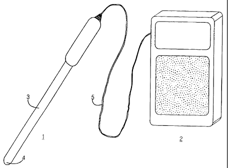
BRIEF DESCRIPTION OF THE DRAWING
Figure 1 is an illustration of the device corresponding to the Preferred
Embodiment of the
present invention.
DETAILED DESCRIPTION OF THE PREFERRED EMBODIMENT
The Preferred Embodiment of the device of the present invention is shown in
Fig. 1. The
device includes an intravaginal probe 1 and a conventional base unit 2.
Located at the distal end of
the probe 1 and internal to the probe 1 is a piezoelectric transducer capable
of converting an electrical
signal the signal from the intravaginal probe to an audible response
meaningful to the human ear.
The device of the Preferred Embodiment of the present invention includes an
intravaginal
probe 1 that is hand-held and portable and contains the following elements:
(I) A rigid or semi-rigid
elongated body portion 3, (ii) a transducer 4 for converting electrical
signals into ultrasonic energy,
and then emitting and detecting ultrasonic energy, and (iii) a means for
communicating signal
information pertaining to the fetal heartbeat to the base unit. The
intravaginal probe 1 of the
Preferred Embodiment of the present invention is longer and more slender than
the conventional
abdominal probe so as to minimize patient discomfort., and to enable greater
range to the physician
or other caregiver who is manipulating the probe in search of fetal heart
action. In particular, in the
Preferred Embodiment the elongated body portion 3 has a diameter less than 2.0
cm and a length
greater than 12.5 cm. The transducer 4 can be made in accord with any of the
transducers and
transducer arrays known in the art and presently used in conventional
abdominal Doppler fetal
heartbeat measurement systems and Doppler imaging systems. In the Preferred
Embodiment, they
will reside in a transducer housing at the end of the probe 1. The transducer
housing is shaped so as
to provide good contact with the apex of the vagina when directed towards the
uterus. The means
for communicating with the base unit in the Preferred Embodiment is a cord 5
containing electrical
wires running between the intravaginal probe 1 and the base unit 2. (In other
embodiments of the
invention, the intravaginal probe 1 is cordless, providing greater flexibility
and freedom to the
obstetrician, with the means for communicating with the base unit 2 being an
infrared, radio or other
6
CA 02343266 2001-04-05
telemetric signal. ) In the Preferred Embodiment, the intravaginal probel
contains a handle 6 to allow
for easy rotation of the intravaginal probel in the vagina.
In the Preferred Embodiment, a standard conventional doppler fetal heartbeat
base used for
the base unit 2. This conventional unit is capable of converting the signal
from the intravaginal probe
to an audible response meaningful to the human ear.
The present invention further relates to a method for early detecting and
quantifying of fetal
heartbeat. In the Preferred Embodiment of the present invention, this method
consists of the
following steps: (I) gently inserting an intravaginal probe 1 into the
patient's vagina, (ii) directing an
ultrasonic wave stream towards the fetus, (iii) detecting ultrasonic waves
reflected from the fetal heart
at the intravaginal probe 1 and (iv) converting the signal from the
intravaginal probe to an audible
signal at the base unit 2.
The details that have been provided here regarding the Preferred Embodiment of
the present
invention are in no way intended to limit the claimed invention. Anyone
skilled in the art can foresee
from a reading of the SUMMARY and other portions of this document many
equivalent ways of
implementing and practicing the present invention.
7