Note: Descriptions are shown in the official language in which they were submitted.
CA 02343279 2001-03-07
WO 00/31577 PCT/GB99/02284
1
TITLE: SCANNING CONFOCAL OPTICAL MICROSCOPE SYSTEM
Field of the Invention
This invention relates to a scanning confocal optical microscope system.
Background to the Invention
The confocal scanning optical microscope is now widely used. In its essentials
it consists
of a means for focussing a beam of light to a small spot on a specimen and
means for
collecting the emitted or reflected emission from that spot in order to build
up an image
by systematic scanning of the spot over a specimen. A defining feature of the
confocal
instrument is the presence of a beam-limiting aperture in front of the
detector, which
serves to limit detection of emitted light to that emerging from the immediate
vicinity of
the focus of illumination. White, in U.S. Patent 5 032 720, taught the use of
a variable
iris as such an aperture, allowing a compromise to be sought between best
optical depth
discrimination, which is obtained with the aperture maximally closed, and high
signal
strength, which is obtained with it open. An analysis of this advantage has
been
published by Sandison et al, pp 39-51 in Handbook of Biological Confocal
Microscopy,
IInd Edn., Plenum Press, New York and London, 1995. White also taught the
division
of the emitted light in a confocal microscope into more than one beam
according to
wavelength, by means of chromatic reflectors. This principle has proved to
have many
applications, chiefly in the imaging of a plurality of fluorescent stains
simultaneously
present in the same specimen.
In a widely-used commercial form of White's instrument, there are two or three
such
beams, each passing light to a separate variable iris, i.e. there are two or
three confocai
beam-limiting apertures. The value of having more than one aperture is that
the diameter
may be set differently in each. This is of value because the above-mentioned
compromise
may be sought according to the brightness of each individual stain. Also, the
theoretical
CA 02343279 2001-03-07
WO 00/31577 PCT/GB99/02284
2
optimum width of the aperture scales with wavelength, so a single diameter can
never be
precisely optimum for all wavelengths (van der Voort, H.T.M. & Brakenhoff,
G.J.
(1990), J. Microscopy 158, pp 43-54).
In White's microscope system, the separation of beams is achieved by the use
of chromatic
reflectors and further colour separation is achieved by means of barrier
filters. Since it
is accepted to be desirable to be able to distinguish colours, the use of a
spectrometer in
the emission path of a confocal microscope is an obvious development.
Brakenhoff, in
a diagram published on page 189 of Confocal Microscopy, edited by T. Wilson,
Academic
Press 1990, showed how a spectrometer could be used. Figure 1 of the
accompanying
drawings is redrawn from Brakenhoff's figure and serves to clarify the
placement of
components_in a confocal microscope.
In Figure 1, Iight from a laser 11 is passed through a lens 12 and a first
aperture,
consisting of a pinhole 13, from which the light emerges as an expanding beam
which is
rendered parallel by lens 14. From lens 14 the light is reflected by chromatic
reflector
15 so that it passes into a microscope objective lens 16 and is brought to a
focus, normally
as a diffraction-limited spot on a specimen at 17. Some of the light emitted
from the
specimen passes into lens 16 and the chromatic performance of the reflector
.15 is chosen
such that the emitted light is transmitted by this reflector 15 rather than
reflected. The
transmitted beam passes through a barrier filter 18, which absorbs unwanted
laser light,
and is focussed by the lens 19 on a confocal aperture at 20. It is essential
for the proper
functioning of a confocal microscope that this aperture lies in an optically
conjugate
position to the focus on the specimen; in other words the specimen is focussed
on this
aperture. In conventional optical terminology, the confocal aperture is an
image plane
stop.
Figure 1 shows how the light which has passed through the confocal aperture at
20 is
passed, in Brakenhoff's scheme, through a spectral dispersing means, such as a
monochromator 21, and the outgoing light is finally passed to a unitary
detector such as
a photomultiplier tube 22. The photocurrent in the photomultiplier tube 22 is
used as a
measure of the intensity of the light in the range of wavelengths selected by
the
CA 02343279 2001-03-07
WO 00/31577 PCT/GB99/02284
3
monochromator 21 and allows the construction of an image in computer memory if
the
spot of light is scanned systematically over the specimen.
In order to be able to record images from the spectrometer (photomultiplier
tube 22) at
more than one wavelength simultaneously, it is a possible development of the
system
proposed by Brakenhoff that the spectrometer should be of the multichannel
type. This
was proposed explicitly by Engelhardt in PCT Application WO 9S/07447. This
combination of the known art of spectrometry with known apparatus for confocal
microscopy works well and has the advantage of being more flexible than the
fixed-
reflector design taught by White. It is, however, inferior to White's design
in that a
plurality of confocal apertures, each for a different wavelength range, cannot
be used.
Since all the detected light is passed through a single confocal aperture the
previously
mentioned advantages of multiple apertures are lost.
The present invention aims to overcome this difficulty, allowing the use of
multiple
confocal apertures in conjunction with multichannel spectral detection. It
effectively
consists of a form of imaging spectrometer, which is simple in construction
and easily
integrated into a scanning confocal optical microscope system.
The Invention
According to one aspect of the invention, there is provided a scanning
confocal microscope
system which is confocal in operation, the system including a dispersive
optical means
which produces from the same restricted region of a specimen a plurality of
separated
optical images of differing wavelength ranges, and a beam-limiting aperture
for each said
image, all said apertures being located at foci which are conjugate with each
other. All
the apertures are located in image planes which are conjugate with the plane
of focus in
the specimen, and the central points of the apertures are conjugate with the
point of
illumination in the specimen. The above-mentioned beam-limiting image plane
apertures
function as confocal apertures.
Preferably, the restricted region or area of the specimen is imaged at a
primary image
CA 02343279 2001-03-07
WO 00/31577 PCT/GB99/02284
4
plane and the dispersive optical means, in conjunction with focussing means,
produces said
images of differing wavelength ranges in secondary image planes conjugate with
the
primary image plane.
The system preferably includes a plurality of detectors receiving light
through the
respective beam-limiting apertures.
According to another aspect of the invention, there is provided a scanning
confocal optical
microscope system comprising a scanning confocal optical microscope which
produces a
beam of light forming an image of a restricted region of a specimen in a
primary image
plane, a dispersive optical means which receives the beam from the said
primary image
plane and produces, in secondary image planes each conjugate with the primary
image
plane, a plurality of separated secondary optical images of the same region of
the
specimen, said secondary images respectively comprising light of differing
ranges of
wavelengths, a beam-limiting aperture in each secondary image plane and a
plurality of
detectors receiving light through the respective beam-limiting apertures.
Preferably, the beam-limiting area of at least one of the beam-limiting
apertures is
adjustable or variable. This may be achieved by exchange of a beam-limiting
aperture
of one size by an aperture of a different size, or by making the beam-limiting
area
adjustable in size, e.g. in width or diameter. For example, at least one beam-
limiting
aperture may comprise a variable iris diaphragm, a system of movable jaws, or
an
adjustable slit.
The dispersive optical means may comprise one or more optical prisms or a
diffraction
grating or gratings, but an equivalent device or devices may alternatively be
employed.
Conveniently, a focusing means such as a lens reproduces the image in the
primary image
plane in the aperture plane of a second focusing means, e.g. a lens, whereby
the aperture
or exit pupil of the first focusing means is imaged at a location beyond the
dispersive
optical means (i.e. the dispersive means is located between the second
focusing means and
said location), whereby a spread of spectrally differing images of the
aperture of the first
CA 02343279 2001-03-07
WO 00/31577 PCT/GB99/02284
5
focusing means is generated.
Conveniently, an optical beam-separating means, such as a reflector or
reflectors, may be
provided, whereby to direct the light from different points of the spread of
spectrally
differing images to the beam-limiting apertures, at which are formed a
spatially spread
series of images of the primary image plane, each confined to a wavelength
range different
to the wavelength range at the other beam-limiting apertures. The beam-
separating means
may be adjustable to enable variation of the wavelength range of the image at
each beam-
limiting aperture. As well as the beam-separating means, a further focusing
means may
be provided to generate the relayed images at the said confocal apertures.
A further reflector or reflectors or other further optical beam-separating
means may serve
to spatially spread the relayed images.
The wavelength compositions of the spectrally spread images may be variable,
as by
adjustment of the beam-separating means, e. g. the reflector or reflectors,
and/or by the
positioning of wavelength-sensitive absorbing screens or filters into the path
or paths of
the spectrally spread images.
Description of Embodiment
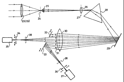
In the accompanying drawings:
Figure 1 is illustrative of the prior art; and
Figure 2 illustrates an embodiment of scanning confocal optical microscope
system
in accordance with the present invention.
In accordance with the invention there is provided a modification of a
scanning confocal
optical microscope of conventional design. The known and conventional parts
correspond
to those parts of the light path shown in Figure 1 up to the point where the
light emitted
from the specimen enters lens 19. The modification is shown in Figure 2.
CA 02343279 2001-03-07
WO 00/31577 PCT/GB99/02284
6
In Figure 2, light from the specimen is shown entering lens 23, which occupies
a position
in the confocal microscope equivalent to that of lens 19 in Figure 1. The lens
23 focusses
the light from a point in the specimen to a point situated in an image plane
24 (the primary
image plane). From plane 24 the light passes into another lens 25 of shorter
focal length
than lens 23, which relays an enlarged image of the point focus in plane 24 to
a plane 27
which is conjugate with the plane of focus within the specimen and which lies
in the
aperture of lens 26. From the lens 26 the light passes through a dispersing
means,
consisting of a prism, compound prism or multiple prisms or diffraction
grating,
symbolised in the diagram by the prism 28. Reference 29 symbolises an optional
reflector or plurality of reflectors, which might be used at any point in the
light path to
produce a folding of the beam convenient for packing the apparatus into a
small space.
The light then passes to a lens 30 and within this lens, or in the close
vicinity of the lens,
an optical spectrum, consisting of a linearly dispersed spread of images of
the beam in the
aperture of the lens 25 is produced by the focusing action of the lens 26
coupled with the
dispersing action of the dispersing means 28.
In Figure 2, multiple images are indicated by the small vertical arrows at 31
and are
shown as discrete; in reality, the light would ordinarily be composed of a
continuous
range of wavelengths, so the images would be infinite in number and any single
point in
the series at 31 would be illuminated by overlapping images. As is well known
in the an
of spectroscopy, the resolution of this projected spectrum is greater the
smaller the angular
extent of one monochromatic image in relation to the angular dispersion
according to
wavelength. Close to the plane of the images at 31 (i.e. the planes of
spectral focus) lie
adjustable beam-separating means, symbolised by an opaque baffle 32 and a
plane reflector
33. Light of wavelengths corresponding to one end of the spectrum falls upon
the baffle
32 and is absorbed. Light of the middle region of the spectrum passes between
the baffle
32 and the reflector 33 and is brought to an image-plane focus at a secondary
image plane
containing aperture 34. The plane of this aperture contains a focussed image
of the
specimen and aperture 34 therefore functions as a confocal aperture
controlling the
passage of light to a detector 35.
Light from the other end of the spectrum is reflected by the reflector 33 and
is brought
CA 02343279 2001-03-07
WO 00/31577 PCT/GB99/02284
7
to a focus at a secondary image plane containing aperture 36, which is a
second confocal
aperture controlling the passage of light to detector 37. An image of the
specimen is
formed at aperture 36 similar to that at aperture 34, except that it is
composed of different
colours, according to the wavelength range selected by altering the position
of the reflector
33 and the baffle 32. The apertures 34 and 36 are situated at foci which are
conjugate
with each other and with the primary focus at image plane 24. Alternatively or
additionally, optical filters 38 may be incorporated in the light paths
leading to the
apertures 34 and 36.
Embodiments of the invention are envisaged in which reflectors are used to
subdivide the
spectrum in other ways, for instance into more than two segments, which, by
the use of
additional baffles, may be delimited further and made non-contiguous.
Embodiments are
also envisaged which contain more than two confocal detectors, each with its
own
independently variable aperture.
In a particular construction of the embodiment of Figure 2, the beam entering
the lens 23
has a maximum diameter of 2 mm, depending on the exit pupil diameter of the
objective
lens in use. The ratio of focal lengths of the lens 23 to the lens 25 was
18:1, with the
result that the beam diameter at the aperture of lens 25 was nominally 2/18 or
0.11 mm.
The lens 26 was an achromatic doublet of focal length 100 mm used to produce
an image
of the aperture of the lens 25 at unit magnification in the plane of the
spectrum 31. This
image, if formed with monochromatic light, had a nominal size of 0.11 mm. The
dispersing means 28 was an equilateral prism of high-dispersion flint glass
which gave a
spectrum extending 25 mm from violet (450 nm) to red (650 nm). The nominal
resolution in this spectrum, neglecting diffraction effects and astigmatism,
is 0.9 nm,
which is more than sufficient for the separation of currently-used fluorescent
dyes.
When the reflector 33 and the baffle 32 were withdrawn from the system, all
the light
transmitted by the lens 30 was passed to the aperture 34. When white light
from a
tungsten-halogen source was then passed into the microscope, the image of the
characteristic region of the specimen at aperture 34 was white in colour. When
the
reflector 33 was inserted by translating it in the plane of the reflecting
surface. an image
CA 02343279 2001-03-07
WO 00/31577 PCT/GB99/02284
8
of the same region of the specimen as seen at aperture 34 appeared in the
plane of the
confocal aperture at 36, but was coloured relatively uniformly red, this being
the colour
of the portion of the spectrum reflected by the reflector 33. At the same
time, the image
at aperture 34 assumed the complementary hue (blue-green) because it received
all
wavelengths but red.
