Note: Descriptions are shown in the official language in which they were submitted.
CA 02343361 2001-03-09
WO 00/15097 PCT/US99/20776
METHODS FOR USING RESONANT ACOUSTIC AND/OR RESONANT
ACOUSTO-EM ENERGY TO DETECT AND/OR EFFECT STRUCTURES
TECHrTICAL FIELD
The present invention relates to detection of inorganic and biologic
structures and/or
disruption and/or augmentation of functions of biologic structures using
resonant acoustic
and/or resonant acousto-EM energy.
BACKGROUND OF THE INVENTION
The resonant acoustic frequency of a system is the natural free oscillation
frequency
of the system. A resonant acaustic system can be excited by a weak mechanical
or acoustic
driving force in a narrow band of frequencies, close or equal to the resonant
frequency
thereby inducing acoustic resonance in a targeted structure.
Acoustic resonance has been used to determine various properties of solid
materials.
For instance, Migliori et al in U.S. Patent Nos. 4,976,148 and 5,062,296 and
5,355,731
disclose a method for characterizing a unique resonant frequency spectroscopic
signature for
objects derived from ultrasonic excitation of objects, the use of resonant
ultrasound
spectroscopy for grading production quantities of spherical objects such as
roller balls for
bearings, and the use of resonant ultrasound spectroscopy with a rectangular
parallelpiped
sample of a high dissipation material to enable low amplitude resonance to be
detected for
use in calculating the elastic constants of the high dissipation sample.
However, the Migliori
patents are directed to solid materials and not to selectively targeting
organic or biologic
material especially when liquid systems are involved.
In addition to interacting with inanimate structures, acoustic energy also
interacts
with living, biologic organisms and structures. Acoustic energy has been used
extensively
in medicine and biology for imaging structures, by directing an acoustic wave
at a biologic
structure and analyzing the reflection pattern of the acoustic wave. Also,
acoustic energy has
been used in physical therapy medicine for delivering heat to targeted areas
of injury or pain.
However, all of the above applications depend on using acoustic energy that is
non-selective
CA 02343361 2001-03-09
WO 00/15097 PCT/US99/20776
for the specific targeted biologic structure, and as such, may affect more
than just the
targeted structure.
Vago, RE., U.S. Patent No.5,048,520 and 5,178,134 discloses ultrasonic
treatment
of animals for topical hygiene and antiviral effects. The frequencies
disclosed are in the range
of 15 kilohertz to 500 kilohertz. They also report that non-enveloped viruses
were refractive
to the inactivating effects of the ultrasound. The mechanism cited for their
antimicrobial
effects is "cavitation" on the skin surface only, and they specifically avoid
the use of resonant
frequencies in their apparatus.
Moasser, M., U. S. Patent No.4,646,725 discloses the use of an adaptor for
diagnostic
ultrasound machines for treatment of skin and mucous membrane lesions caused
by infectious
agents including herpes virus. The method of treatment was 2.0 to 3.0 minutes
at a power
output of 1.5 watts per square centimeter, with no specific frequencies being
cited. The use
of acoustic resonance is not discussed or contemplated.
Johnston, RG., U. S. Patent No.5,426,977 discloses ultrasonic measurement of
the
acoustic resonances in eggs to provide a technique for establishing the
presence of
Salmonella bacteria Johnson characterizes the eggs and determines the
difference between
the egg with and without Salmonella bacteria. As such, this method does not
detect the
actual micro-organism, but instead characterizes the vibrational modes of an
eggshell, which
are modified by the physical presence of a bacteria.
The prior art has failed to suggest a satisfactory method or system for
affecting
functions of a biologic structure without also affecting near-by tissue.
Furthermore, the prior
art does not provide for a method that allows precise detection of biologic or
inorganic
structures using acoustic resonance to produce a signature with high signal to
noise ratio,
while producing little effect in nearby structures. Still further, use of non-
resonant acoustic
energy in the prior art affects targeted and non-targeted structures equally.
SUMMARY OF THE INVENTION
For purposes of this invention, the terms and expressions below, appearing in
the
specification and claims, are intended to have the following meanings:
"Acoustic energy" as used herein is defined as energy that is produced when a
2
CA 02343361 2001-03-09
WO 00/15097 PCT/US99/20776
physical structure vibrates and the vibrational energy of motion is
transferred to the
surrounding medium which includes air, liquid, or solid.
"Detect" as used herein is defined as determining the presence or absence of a
structure, and if present identifying the structure.
"Electromagnetic (EM) properties and/or fields" as used herein includes direct
and alternating currents, electric and magnetic fields, electromagnetic
radiation, and fields
which include but are not limited to waves, current, flux, resistance,
potential, radiation or
any physical phenomena including those obtainable or derivable from the
Maxwell equations,
incorporated by reference herein.
"Electromagnetic (EM) energy pattern" as used herein represents the
electromagnetic energy produced by a structure as acoustic energy interacts
with the
structure and is manifested as electromagnetic properties and/or fields.
"Biologic structure" as used herein and used interchangeably with organic
includes
anything from the smallest organic or biochemical ion or molecule, to cells,
organs, and
entire organisms.
"Disruption" as used herein refers to deleterious effects on a biologic
structure.
"Acoustic Signature" as used herein means a unique acoustic pattern that is
produced by the structure when in acoustic resonance that may take the form of
amplitude
of signal.
"Resonant acoustic frequency" as used herein includes frequencies near or at
the
natural resonant frequency of the structure including harmonic and subharmonic
frequencies
of the natural resonant frequency to induce acoustic resonance therein.
"Acousto-EM signature" as used herein is defined as an EM energy pattern of an
object in acoustic resonance and/or an EM energy equivalent in frequency to
the resonant
acoustic frequency.
"Acousto-EM spectroscopy" as used herein is defined as detecting a unique EM
signature for a structure that is in acoustic resonance or detecting a unique
acoustic signature
that is in resonance due to the introduction of electromagnetic energy, both
of which can be
used to detect and/or identify the structure in resonance.
"Living transducer" as used herein is defined as a biologic piezoelectric or
semiconductor structure that converts electromagnetic energy or fields into
mechanical
3
CA 02343361 2001-03-09
WO 00/15097 PCT/US99/20776
energy and/or mechanical energy into electromagnetic energy or fields.
"Cavitation"as described herein is defined as the formation of vapor-filled
cavities
in liquids, i.e. bubble formation in water when brought to a boil.
"Mechanical" as described herein include mechanisms such as compression and
rarefaction which are thought to take place in the intensity/duration
threshold region between
the thermal and cavitation regions.
"Non-resonant electromagnetic signature" as used herein is defined as an EM
energy pattern produced by an object stimulated by a non-resonant acoustic
field.
"Resonant acousto-EM energy" as described herein means electromagnetic energy
or field that induces acoustic resonance in a structure.
The present invention addresses the shortcomings of the prior art by inducing
acoustic
resonance in a targeted structure with select frequencies that affect the
specific targeted
structure but have virtually no effect on nearby, non-resonating structures.
Furthermore,
acoustic energy power intensities can be reduced by introducing a source of
electromagnetic
(EM) energy that augments the acoustic energy thereby reducing the destructive
nature of
high power acoustic energy.. The interaction between EM energy and acoustic
resonance
allows for precise detection of a structure in acoustic resonance by producing
a signature
with high signal to noise ratio, while producing little effect in other
structures.
The present invention provides methods to selectively detect, identify and/or
affect
an inorganic or biologic structure by using resonant acoustic and/or acousto-
EM energy
which can transfer useful energy to targeted structures while leaving nearby
structures, which
are not in resonance, virtually unchanged.
Therefore, it is an object of the present invention to provide a method of
identifying
or detecting an inorganic or biologic structure using its resonant acoustic
and/or acousto-EM
energies.
It is an object of the present invention to provide a method using resonant
acoustic
and/or acousto-EM energies to augment and/or disrupt the growth and/or
function of
biologic structures.
It is another object of the invention to provide a method for determining
resonant
frequencies of a biologic structure.
It is also an object of the invention to provide a method using resonant
acoustic
4
CA 02343361 2001-03-09
WO 00/15097 PCT/US99/Z0776
and/or resonant acousto-EM energies to detect the presence of and/or identify
biologic
structures.
In accordance with the aforesaid objects the present invention provides for
the
detection of inorganic or biologic structures and/or disruption and/or
augmentation of growth
and/or fiu~ctions of a biologic structure using resonant acoustic and/or
resonant acousto-EM
energy.
Applying principles of acoustic resonance, the resonant acoustic frequency of
a
biologic system is the natural free oscillation frequency of the system, and
thus a biologic
system can be excited by a weak mechanical or acoustic driving force in a
narrow band of
frequencies. Also, depending on the size, shape, and composition of the
biologic structure,
there can be more than one naturally occurnng resonant acoustic frequency, as
well as
numerous subharmonic and superharmonic resonant acoustic frequencies.
When a structure, both inorganic and biologic, goes into acoustic resonance,
energy
builds up in it rapidly. The energy is either kept in the system or released
to the surrounding
environment. Energy kept in the structure can enhance the structure's
functions or cause
disruption of the structure. A small amount of the energy in a resonant system
is either
intrinsically dissipated, as electromagnetic energy, or is transmitted as
acoustic energy to the
nearby medium. The intrinsically dissipated energy is of particular interest,
because it is
dissipated through molecular and atomic vibrations, producing EM energy. This
EM energy
is referred to as acousto-EM energy because it is produced when a structure is
in acoustic
resonance and some acoustic energy interacts with the structure and is
converted into
electromagnetic energy which is intrinsically dissipated into nearby media.
The properties,
fields and/or frequencies of EM energy produced depend on the unique molecular
and atomic
components of the structure in question. Thus, the induction of acoustic
resonance in a
structwe leads to the production of a unique acousto-EM signature for that
structure, which
can be used to detect and/or identify it as disclosed in the present
invention. Conversely, if
a structure is targeted with EM energy equivalent to its acousto-EM signature,
the energy
dissipation pathway is reversed, and a state of acoustic resonance can be
induced. Reversing
the energy dissipation pathway with an acousto-EM signature can be used to
produce the
same augmentation, detection, and disruption effects that the original
resonant acoustic
energy field produces. The resonant acousto-EM signature can be used either by
itself, or in
5
CA 02343361 2001-03-09
WO 00/15097 PCTNS99/20776
combination with resonant acoustic energy. Using the resonant acousto-EM and
acoustic
energy together, allows for the use of lower power levels of both types of
energy, lessening
the potential adverse affects of electromagnetic energy and/or acoustic energy
on nearby or
adjacent nontargeted structures.
Electromagnetic energy may interact with and complement an acoustic energy
wave
in a system in at least four ways: via the piezoelectric effect, intrinsic
dissipation of
electromagnetic energy and via the acoustoelectric or magnetoacoustic effect.
In the piezoelectric effect, acoustic vibratory energy is converted
interchangeably
with EM energy by a transducer. Biologic piezoelectric structures can modulate
the same
conversion of energy, thereby acting as living transducers. When an EM field
is applied to
a biologic piezoelectric structure, an acoustic wave is produced. Likewise,
when an acoustic
wave is applied to a biologic piezoelectric structure, EM energy is produced.
The
piezoelectric effect in biologic structures has many useful applications (see
below.) This
effect becomes even more useful when principles of acoustic resonance are
applied. In the
present invention specific biologic structures can be targeted with an
acoustic wave or EM
energy at power levels that dramatically affect the target structure, but have
virtually no
effect on adjacent, nonresonant structures. Although not previously postulated
by others,
biologic structures functioning a,s living, resonant piezoelectric transducers
which modulate
the conversion of mechanical and EM energy is undoubtedly one of the major
underlying
mechanisms responsible for the interaction of EM fields with biologic
structures.
In the acoustoelectric effect, the passage of an acoustic wave through a
semiconductor induces an electric current. The passage of an acoustic wave
through the
material is postulated to cause a periodic spatial variation of the potential
energy of the
charge carriers. This results in an electric field across the ends of the
semiconductor as long
as the acoustic wave is traversing the semiconductor. Free electrons carriers
are bunched in
the potential-energy troughs, and as the acoustic wave having a specific
frequency
propagates, it drags the bunches along with it, resulting in an electric field
such as a DC field
pulsing at the specific acoustic frequency or an AC field having a frequency
equal to the
specific acoustic frequency. The effect is enhanced where there are both
positively and
negatively charged carriers, and where there are many different groups of
carriers -
conditions which are frequently found in biologic systems. The attributes of
the current
6
CA 02343361 2001-03-09
WO 00/15097 PCT/US99/20776
produced depend on the unique molecular and atomic components of the structure
in
question. This aspect alone provides a means to perform acoustoelectric
spectroscopy on
biologics many of which are senuconductors, and depending on the selected
frequency, the
acoustoelectric effect in biological structures has many potentially usefi~l
applications. Thus
understood, a targeted structure can be irradiated or exposed to acoustic
energy having non-
resonant frequency and an electromagnetic energy pattern of the
acoustoelectric effect in the
structure can be detected. This detected electromagnetic energy pattern can be
used as a
signature to detect and identify the targeted structure.
However, the acoustoelectric effect becomes even more useful when principles
of
acoustic resonance are applied. Augmentation, detection, and/or disruption of
biologics can
be targeted to specific structures at power levels that dramatically affect
the target structure,
but have virtually no effect on nearby, nonresonant structures. The current
produced by the
acoustoelectric effect in a resonant stricture will be much stronger than any
current produced
by neighboring non-resonant structures, and may be of an alternating nature.
The large signal
to noise ratio obtained from a resonant structure improves accuracy of
acoustic and EM
pattern identification and detection. Similar to reversal of the piezoelectric
effect and
acoustic resonance intrinsic energy dissipation pathway (see above),
application of the
resonant acoustoelectric EM pattern to a targeted structure will amplify the
acoustic wave
(acoustoelectric gain which peaks at the frequency for which the acoustic
wavelength is the
Debye length, where bunching is optimum). Thus, combined use of the resonant
acoustic and
acoustoelectric EM fields can allow for greater tissue penetration of high
frequency acoustic
energy that would otherwise be highly attenuated and have poor tissue
penetration. Using the
resonant acoustic frequency and acoustoelectric EM fields together also allows
for the use
of lower power levels of both types of energy, lessening the potential effects
on other
nontargeted and nonresonant structures.
The magnetoacoustic effect is the magnetio-field-dependent attenuation of an
acoustic
field in a monotonic, oscillatory, or resonant manner, depending on the
electronic properties
of the substance in question. This variability in result, depending on
structural composition,
provides a further enhancement of resonant acausto-EM spectroscopy in relation
to biologics
and other structures, via addition of a magnetic field. Also, the addition of
a magnetic field
provides the means to amplify or attenuate an acoustic field, thus improving
or modulating
7
CA 02343361 2001-03-09
WO 00/15097 PCT/U599/20776
the penetration of the acoustic field in biologic tissues.
Similarly, resonant acoustics combined with acoustic cyclotron resonance (ie.
resonant acoustic cyclotron resonance) and Doppler-shifted resonant acoustic
cyclotron
resonance presents a powerful, and precise means of selectively causing
augmentation,
detection and/or disruption of structures.
The present invention provides a method that applies the principles of
acoustic
resonance to biologic stroctures for the purpose of disruption and/or
augmentation of
functions of the specifically targeted biologic structure. The resonant
acoustic frequency of
a biologic structure may be determined by performing resonant acoustic
spectroscopy using
methods and systems well know in the art. Particularly, a resonant acoustic
frequency of a
biologic structure may by determined by the steps of
a) applying acoustic energy to the biologic structure and scanning through a
range of
acoustic energy frequencies; and
b) detecting at least one specific frequency which causes a maximum signal
output
from the biologic structure indicating the biologic structure being induced
into acoustic
resonance by the at least specific frequency.
The specific frequencies causing the maximum signals are the resonant acoustic
frequencies of the biologic structure which are defined and used herein as the
acoustic
signature of the biologic structure. Once determined, at least one resonant
acoustic
frequency may be applied to the biologic structure to affect functioning
therein and/or to
determine its acousto-EM signature.
The acoustic energy including the resonant acoustic frequencies is applied at
a power
level sufficient to affect fimctioning of the biologic structure. Depending on
the power
intensity of the acoustic energy and the type of targeted strucxure that is
induced into acoustic
resonance, the structure may have its firnctions affected, such as disruption
and/or
augmentation.
At lower power levels functions of the biologic structure can be augmented
while at
higher power levels disruption of the structure may occur. Augmentation as
used herein
encompasses beneficial effects on the biologic structure. Such augmenting of
fi~nctions or
enhancing effects include but are not limited to enhancement of growth,
reproduction,
regeneration, embryogenesis, metabolism, fermentation, and the like. The
results of such
8
CA 02343361 2001-03-09
WO 00/15097 PCTNS99/20776
enhancement include but are not limited to increase in bone mass or density,
increase in
number and maturation of eggs, increase in number and/or function of
leukocytes, increase
in fermentation products in beer, wine and cheese manufacturing, increase in
plant
germination and growth and the like.
There are some situations where the ability to selectively disrupt a structure
with
resonant acoustic energy is very useful as disclosed in the present invention.
As stated
above, disruption as used herein refers to deleterious effects on the biologic
structure. Such
deleterious effects include but are not limited to structural failure of the
biologic structure
resulting in lysis, shattering, rupture or inactivation of the biologic or of
one or more
components of the biologic stn~cture. Disruption as used herein also includes
within its ambit
inhibition of vital processes required for growth, reproduction, metabolism,
infectivity and
the like. Components which may be targeted for disruption include, but are not
limited to
DNA, RNA, proteins, carbohydrates, lipids, lipopolysaccharides, glycolipids,
glycoproteins,
proteoglycans, chloroplasts, mitochondria, endoplasmic reticulum, cells,
organs and the like.
In the case of virulent organisms, the virulence factors may be specifically
targeted for
disruption to prevent or inhibit the growth, infectivity or virulence of the
organism. Such
virulence factors include but are not limited to endotoxins, exotoxins, pili,
flagella, proteases,
ligands for host cell receptors, capsules, cell walls, spores, chitin, and the
like.
Organics, biologics or one or more targeted portions thereof which are
amenable to
disruption using the methods of the present invention include but are not
limited to viruses,
bacteria, protozoans, parasites, fungi, worms, mollusks, arthropods, tissue
masses, and the
like. The organics or biologics to be disrupted may be isolated, present in a
multicellular
organism or portion thereof, or other complex environment.
It is postulated that disruption of the targeted biologic structure without
affecting
nearby tissue or structures occurs due to acoustic resonance being induced
only in the
targeted stnrcture which until now has not been considered a mechanism to
affect a biologic
structure. This is very different from that disclosed in the prior art which
contemplates only
three mechanisms for affecting a biologic structure which include cavitation,
thermal and
mechanical.
At specific power levels, such as in lower levels, that do not cause the
actual
disruption of a structure, resonant acoustic energy can intrinsically
dissipate within the
9
CA 02343361 2001-03-09
WO 00/15097 PCT/US99/20776
structure and to the adjacent medium. This intrinsically dissipated energy can
be converted
by the structure into an electromagnetic energy having specific properties
and/or fields that
may be manifested as direct and alternating currents, electric and magnetic
fields,
electromagnetic radiation and the like. The pattern of the electromagnetic
energy represents
an acousto-EM signature of the structure.
The present invention provides a method to determine an acousto-EM signature
of
a structure which comprises irradiating the structure with acoustic energy
having a frequency
at or near a previously determined resonant acoustic frequency of the
structure to induce
resonance therein and detecting the electromagnetic energy pattern caused by
the intrinsic
dissipation of energy.
Once an acousto-EM signature is determined for a specific structure, this
structure
can be induced into acoustic resonance by applying EM energy equivalent to the
acousto-EM
signature of the structure, such as equivalent to the acousto-EM signature of
the structure.
direct and alternating current, electric and magnetic fields, and
electromagnetic radiation and
the like.
As such, the present invention applies the principles of acoustic resonance by
applying
resonant acoustic frequencies and electromagnetic energy equivalent to the
predetermined
acousto-EM signature of a targeted structure individually or in combination to
affect the
targeted structure, the method comprising the steps of
a) applying at least one resonant acoustic frequency of the targeted structure
and/or introducing electromagnetic energy equivalent to part or all of the
acousto-EM
signature of the targeted structure; and
c) applying (a) and/or (b) each at a power intensity level to induce acoustic
resonance within the targeted structure and affect functioning of the
structure.
Both the resonant acoustic frequency of the targeted structure and the acousto-
EM
signature must be predetermined, as discussed above, to provide the applicable
energy for
inducing acoustic resonance in the structure. The electromagnetic energy can
be introduced
into the targeted structure in the form of a direct or alternating current
have a specific
frequency that is equivalent to the electromagnetic energy pattern detected
when the
structure is induced into acoustic resonance. Furthermore each type of energy
can be applied
at a power level less than used individually and this allows for inducing
acoustic resonance
CA 02343361 2001-03-09
WO 00/15097 PCT/US99/20776
in the structure with the possibility of reducing damage to the structure.
The present invention provides a method for detecting and identifying
inorganic or
biologic strictures using resonant acoustic and/or acousto-EM energy. The
method includes
determining the acoustic signature of a structure by irradiating the structure
with a range of
frequencies to determine the specific frequency and/or frequencies that induce
acoustic
resonance therein to provide an acoustic signature of the structure. The
acoustic signature
can be compared with reference signatures to detect and/or identify the
structure.
Furthermore, the identification and/or detection of a structure can also be
achieved
by detecting an acousto-EM signature of a targeted structure, the method
comprising the
steps of
a) inducing acoustic resonance in the targeted structure; and
b) detecting an electromagnetic energy pattern from the targeted structure in
acoustic resonance which represents an acousto-EM signature of the structure.
The acousto-EM signature can be compared to reference signatures to detect
and/or identify
the structure.
The targeted structure can be induced into acoustic resonance by introducing
acoustic
energy including at least one resonant acoustic frequency, electromagnetic
energy equivalent
to the resonant acoustic frequency, and/or an electromagnetic energy pattern
equivalent to
the acousto-EM signature.
The electromagnetic energy pattern manifested as electromagnetic properties
and
fields may be determined by detection means well known to those skilled in the
art such as
those disclosed in Introduction to Electromagnetic Fields and Waves, by Erik
V. Bohn
Addison-Wesley Publishing Co., 1968, the contents of which are incorporated by
reference
herein.
In another embodiment of the present invention, a structure may be induced
into
acoustic resonance by applying to the structure part or all of the acousto-EM
signature of the
structure to induce the structure into acoustic resonance. If the structure is
induced into
acoustic resonance, this fact may be used to detect and/or identify the
structure. This
represents another method of the present invention that may used for
identification or
detection of a specific structure, because each structure will not only have
its own unique
acoustic signature but also will have a unique acousto-EM signature to which
it responds by
11
CA 02343361 2001-03-09
WO 00/15097 PCTNS99/Z0776
resonating acoustically. Also, depending on the power intensity of the
electromagnetic
properties and/or fields and the type of targeted structure that is induced
into acoustic
resonance, the structure may have its functions affected, such as disruption
andlor
augmentation.
In all the above embodiments the introduction of acoustic and/or
electromagnetic
energy including a resonant acoustic frequency can be applied in either
continuous and/or
periodic form depending on the desired effect.
The acoustic and/or EM fields may be applied individually or in combination.
Likewise the acoustic and/or EM fields may be detected individually or in
combination.
Many biochemical compounds and biologic structures are naturally occurring
crystals
and especially susceptible in that regard to the effects of resonant acoustic
energy. Many
biologic substances are piezoelectric materials. For instance, bone is a
piezoelectric material
and the piezoelectric properties of bone play a vital role in its biological
functions. As such,
it is further envisioned by the inventors that biologic structures having a
piezoelectric nature
may be affected by applying a sufficient amount of acoustic energy and/or
electromagnetic
energy to induce the structure into resonance thereby affecting the functions
of the biologic
structure either positively or negatively. Thus understood, biologic
structures that act as
living piezoelectric transducers may be induced into acoustic resonance by
introducing
electromagnetic energy equivalent to a resonant acoustic frequency of the
biologic structure
which is converted to mechanical energy by the living transducer thereby
inducing acoustic
resonance in the structure.
Another aspect of the invention is a system for detecting a biologic or
inorganic
structure by determining the resonant acoustic and/or acousto-EM signature of
the structure
comprising:
a) means for inducing acoustic resonance in the biologic or inorganic
structure;
b) means for detecting the acoustic signature of the biologic or inorganic
structure; and
c) means for comparing the acoustic signature of the biologic or inorganic
structure with a reference acoustic signature of the structure.
Also, the above system may also or instead comprise means for detecting a
resonant
acousto-EM energy signature of the structure in acoustic resonance which
produces an
12
CA 02343361 2001-03-09
WO 00/15097 PCT/US99/20776
electromagnetic energy pattern such as described above. The acousto-EM
signature can be
compared with a previously determined reference signature by providing means
for
comparing in a detection or identification system. The electromagnetic energy
pattern is
manifested as electromagnetic properties and/or fields that include but are
not limited to
energy in the form of direct and alternating current, electric and magnetic
fields, and
electromagnetic radiation. The targeted structure can be induced into acoustic
resonance
by introducing acoustic energy including at least one resonant acoustic
frequency,
electromagnetic energy equivalent to the resonant acoustic frequency, and/or
an
electromagnetic energy pattern equivalent to the acousto-EM signature.
In another embodiment of the present invention a system for augmenting and/or
disrupting a targeted biologic structure comprises means for applying acoustic
energy
including a previously determined resonant acoustic frequency to induce
acoustic resonance
in the biologic structure, the acoustic energy being applied at a sufficient
power input to
affect functions of the biologic structure. Alternatively, the targeted
structure may be
induced into acoustic resonance by providing electromagnetic energy equivalent
to the
resonant acoustic frequency or the acousto-EM signature that was previously
determined,
such as direct and alternating current, electric and magnetic fields, and
electromagnetic
energy.
In yet another embodiment a system is provided to introduce acoustic energies
having
acoustic frequencies at or near the resonant acoustic frequencies of the
targeted structure and
also electromagnetic energy to augment the resonant acoustic frequencies
comprising:
means for introducing a frequency at or near the resonant acoustic frequency
of the
targeted structure ; and
means for introducing electromagnetic energy equivalent to the electromagnetic
energy pattern previously determined as an acousto-EM signature of the
structure, such as
direct and alternating current, electric and magnetic fields, and/or
electromagnetic radiation
and the like.
The acoustic energy and EM energy equivalent to the acousto-EM signature may
be applied and/or detected by a single means that can apply both types of
energy.
Additional objects, advantages and novel features of the invention will be set
forth
in part in the description which follows, and in part will become apparent to
those skilled in
13
CA 02343361 2001-03-09
WO 00/15097 PCT/US99/20776
the art upon eacamination of the following or may be learned by practice of
the invention. The
objects and advantages of the invention may be realized and attained by means
of the
instrumentalities and combinations particularly pointed out in the appended
claims.
BRIEF DESCRIPTION OF THE DRAWINGS
Other objects, features and many of the advantages of the invention will be
better
understood upon a reading of the following detailed description when
considered in
connection with the accompanying drawings wherein:
Figure 1 is a block schematic of a basic Acoustic Energy Generating System.
Figure 2 is a block schematic of a basic Acoustic Energy Detection System.
Figure 3 is a block schematic of a stationary magnetic field applied to a
biologic
structure.
Figure 4 is a block schematic of an oscillating magnetic field applied to a
biologic
structure.
Figure 5 is a block schematic of a direct or alternating current applied to a
biologic
structure.
Figure 6 is a block schematic of a static charge applied to a biologic
structure.
Figure 7 is a block schematic of delivery of electromagnetic radiation to a
biologic
structure.
Figure 8 is a block schematic of detection of a stationary or oscillating
magnetic field
in a biologic structure.
Figure 9 is a block schematic of detection of a static charge in a biologic
structure.
Figure 10 is a block schematic of detection of electromagnetic radiation
emitted from
a biologic structure.
Figure 11 is a block schematic of detection of direct and alternating current
in a
biologic structure.
Figure 12 is a block schematic showing a method for determining resonant
acoustic
frequencies of viruses.
Figure 13 is a block schematic showing a method for assessing the effects of
resonant
acoustic fields on viruses.
14
CA 02343361 2001-03-09
WO 00/15097 PCTNS99/20776
Figure 14 is a block schematic showing a method for disrupting viruses extra
corporeally with resonant acoustic fields.
Figure 15 is a block schematic showing a method for disrupting viruses in vivo
intravascularly with resonant acoustic fields.
Figure 16 is a block schematic showing a method for disrupting viruses in vivo
in
multicellular organism with resonant acoustic fields.
Figure 17 is a block schematic showing a method for disrupting viruses in a
portion
of a multicellular organism with a resonant acoustic field probe.
Figure 18 is a block schematic showing a method for disrupting viruses in a
portion
of a multicellular organism with a resonant acoustic field sheet.
Figures 19 A & B are block schematics showing a method for determining
resonant
acoustic and/or acousto-EM frequencies of viruses.
Figure 20 is a block schematic showing a method for assessing effects of
resonant
acoustic and/or acousto-EM fields on viruses.
I S Figure 21 is a block schematic showing a method for disrupting viruses
extracorporeally with resonant acoustic and/or acousto-EM fields.
Figure 22 is a block schematic showing a method for disrupting viruses in vivo
intravascularly with resonant acoustic and/or acousto-EM fields.
Figure 23 is a block schematic showing a method for disrupting virus in a
portion of
a multicellular organism with resonant acoustic and/or acousto-EM field probe.
Figures 24 A & B are block schematics showing a method for determining
resonant
acoustic and/or acousto-EM frequencies of microorganisms.
Figure 25 is a block schematic showing a method for augmenting microorganisms
with resonant acoustic and/or acousto-EM fields.
Figure 26 is a block schematic showing a method for disrupting microorganisms
with
resonant acoustic and/or acousto-EM fields.
Figure 27 is a block schematic showing a method for determining resonant
acoustic
and/or acousto-EM frequencies of arthropods.
Figure 28 is a block schematic showing a method for disrupting arthropods
using
resonant acoustic and/or acousto-EM energy.
Figure 29 is a block schematic showing a method for augmenting and maintaining
CA 02343361 2001-03-09
WO 00/15097 PC'T/US99/20776
normal bone structure in individuals with osteoporosis.
Figure 30 is a block schematic showing a method for maintaining normal bone
structure in normal individuals during weightlessness.
Figure 31 is a block schematic showing a method for detecting benign or
malignant
tissue types using resonant acoustic and/or acousto-EM energy.
Figure 32 is a block schematic showing a method for stimulating and/or
disrupting
proteoglycans adhesive units between cells using resonant acoustic and/or
acousto-EM
energy.
Figure 33 is a block schematic showing a method for augmenting, identifying,
detecting, and/or disrupting structures of multicellular organisms using
resonant acoustic
and/or acousto-EM energy.
Figure 34 is a block schematic showing a method for augmenting the growth rate
of
multicellular organisms using resonant acoustic and/or acousto-EM energy.
Figures 35 A & B are block diagrams showing a method and system for
determining
acoustic and/or acousto-EM frequencies of inorganic material or structure.
DETAILED DESCRIPTION OF THE PREFERRED EMBODIMENTS
The methods of the present invention comprise delivering acoustic energy at
resonant
frequencies to an inorganic or biologic structure as shown in Figure 1. Using
methods known
to those skilled in the art, any device capable of generating and transmitting
acoustic energy
through any medium can be used to generate the resonant acoustic frequencies
utilized by the
invention. This includes, but is not limited to, devices that produce acoustic
energy using
traditional EM stimulation of piezoelectric transducers, (man-made or
naturally occurring),
purely mechanical devices (such as high frequency air whistles), and laser
devices. Individual
components for acoustic energy systems are commercially available from a wide
variety of
manufacturers, which can be configured to particular applications and
frequency ranges. (See
Thomas Directory of American Manufacturers, Photonics Buyer's Guide, 199b,
Microwave
and RF, and Electronic Engineer's Master Catalogue).
Any oscillator, also called signal generator or function generator, that
produces a
signal with predetermined characteristics such as frequency, mode, pulse
duration, shape, and
16
CA 02343361 2001-03-09
WO 00/15097 PCT/US99/20776
repetition rate may be utilized to generate the resonant acoustic frequencies
utilized by the
invention. Various oscillators or signal generators can be commercially
purchased for
frequencies ranging from Hertz to Gigahertz, such as the MicroLambda LMOS
series (500
MHz-18 GHz), the BK Precision 2005A (100 KHZ-450 MHz) (B&K Precision, Chicago,
IL), the Tektronix SME02 (5 KHZ-5 GHz), and the Tektronix 25 SME 4040 (0.5 Hz -
20
MHz) (Tektronic, Inc., Beaverton, OR), and the Matec 700 series (1-1100 MHz)
and the
like.
The frequency at which resonance occurs depends on the size, shape, and
composition of a structure. For instance, the resonant frequency of a sphere
is the frequency
at which the acoustic wavelength is equal to the sphere diameter. A more
complex structure
- a cylinder - has two resonant frequencies based on two axes of orientation,
with one of the
resonant frequency wavelengths being equal to 1.5 times the length. The more
complex the
shape of the structure, the more complex the resonant acoustic frequency
pattern, however,
the wavelength at which acoustic resonance occurs is roughly equivalent to the
size of the
structure.
The frequency which matches a particular acoustic wavelength depends on the
composition of the structure, according to the equation:
velocity = frequency x wavelength (1)
where velocity refers to the speed of the acoustic wave propagation (the speed
of sound) in
the medium composing the structure. Although the speed of sound varies among
various
biological tissues, it is roughly equivalent to the speal of sound in water
(1,500 m/s), because
most biologic organisms are composed chiefly of water. Using the speed of
sound in water
as the velocity of the acoustic wave, and using the structure size as the
rough equivalent of
the wavelength, the approximate range of resonant acoustic frequencies in
organic or biologic
structures, is given by:
Frequency = Velocity - Velocity = ]"500 m/s
Wavelength Size Size (2)
(See the chart that follows.)
Other known speeds of sound in biologic tissues vary and include:
(1) liver (1550 m/s); (2) muscle (1580 m/s); (3) fat (1459 m/s); (4) brain
(1560 m/s); (5)
17
CA 02343361 2001-03-09
WO 00/15097 PGT/US99/20776
kidney(1560 m/s); (6) spleen (1570 m/s); (7) blood (1575 m/s); (8) bone {4080
m/s); (9) lung
(650 m/s);(10) lens of eye (1620 m/s); (11) aqueous humor (1500 m/s); and (12)
vitreous
humor (1520 m/s). Resonant acoustic frequency ranges for targeted organic or
biologic
structures comprised of tissues with acoustic velocities different from the
speed of sound in
S water, are derived using the same equation (velocity/wavelength) and
correlate to the charted
ranges listed below, plus or minus, depending on the speed of sound in the
targeted tissue.
Although velocity of acoustic energy in a particular medium is for the most
part
constant, there is a slight dependence of velocity on frequency - an effect
called dispersion.
For example, over the frequency range of 1 to 20 MFIz, the acoustic velocity
changes by 1%.
Thus, in the present invention the resonant frequency(s) or at least the range
of frequencies
within which the resonant frequency can be found for a targeted structure
depend on its size,
shape, and composition, and the specific frequency range under examination.
Some
approximate acoustic resonant frequencies for biologic structures are included
in the
following Table 1.
20
30
18
CA 02343361 2001-03-09
WO 00/15097 PCT/US99/20776
TABLE 1
Anprokimate Acoustic Resonant Freauency Ranees for Bioloeic Structures
(Speed of sound =1,500 m/s)
* Hertz
m -- - Whales 150 Hz -
* KiloHertz
1 m -- - hwnans 1.5 l;Hz --
1 dm - hamster 1 S l:Hz - -
-
1 cm - beetle 150 hHz -
-
* MegaHertz
1 mm - lice 1-5 ~ - -
-
100 wn - plant cells 15 MHz --
-
10 wn - animal cells 150 MHZ - -
--
* GigaHertz
1 um -- - bacteria 1.5 gHz --
100 mn -_ - viruses 15 ~ __
10 iUn -- - proteins 150 gHz -- -
* TerraHertz
1 nm -- - small molecules 1.5 tHz --
19
CA 02343361 2001-03-09
WO 00/15097 PCT/US99/20776
To obtain the maximum transfer of acoustical energy from one medium to
another,
the characteristic acoustical impedance of each should be as nearly equal to
the other as
possible. This problem of impedance matching, as it is termed, occurs in many
branches of
physics, and is employed in acoustical techniques, as a means of matching two
media of
different acoustical impedances Rl and R2 respectively. The matching medium is
sandwiched
between the other two and should be the appropriate thickness relative to the
wavelength of
the sound transmitted, and its acoustical impedance R should be nearly equal
to ,r(R1R~. An
impedance matching device that is commercially available and which can be
utilized in this
invention includes Model 60, manufactured by Matec Instruments, Inc.
Acoustic energy can be produced by a transducer that converts received
electromagnetic energy into rapid, physical vibrations, and thus acoustic
energy. The first
acoustic transducers used the piezoelectric properties of naturally occurring
quartz to
produce acoustic energy waves.
EM energy ~ piezoelectric transducer -~ acoustic energy waves
New transducers use materials such as ferroelectric ceramics (barium titanate,
lead
titanate, or lead zirconate) and zinc oxide. Recent advances in materials
engineering have also
produced piezoelectric polymers which can be shaped into sheets and cords,
allowing a
multiplicity of applications.
Transducers are also commercially available from a wide variety of
manufacturers,
in a wide variety of designs which can be configured to particular
applications and
frequencies. Examples of acoustic transducers that may be utilized in the
present invention
and which can be commercially purchased for frequencies ranging from Hertz to
Gigahertz
include Matec broadband immersion transducers MIA series (10-196 MHz), Matec
broadband MIBO series (5-10 MHz), Matec broadband MICO (3.5 MHz), Matec
broadband
M1D0 (2.25 MHz), Matec broadband Mw0 series (50 KHZ-1 MHz), Matec GPUT series
(500 KHz-20 MHz), Matec intravascular blood flow VP-A50 series (5-30 MHz), the
Teledyne Electronic Technologies In-phase or Out-of phase broadband MHz/GHz
(up to
17.5 GHz) array transducer of zinc oxide on sapphire and optional anti-
reflective coating, and
Channel Industries Kilohertz transducers. In the ultrahigh acoustic
frequencies (upper GHz
and THz) maser and laser systems may be utilized.
The transducers can produce an acoustic wave within a range of frequencies
CA 02343361 2001-03-09
WO 00/15097 PCT/US99/Z0776
(broadband) or for one specific frequency (narrowband).
Commercially available acoustic amplifiers include but are not limited to
Matec gated
amplifier systems ( 100 KHZ-200 MHz), and EM broadband amplifier model 607L
(0.8 -
1,000 MHz.)
Complete acoustic systems including power frame, computer interface, pulse
width
generator, gated amplifier, broadband receiver, and phase detector ( 100 KHZ-
100 MHz) can
be purchased commercially from sources such as Matec.
The acoustic delivery system is variable depending on the application.
Acoustic
energy waves can be transmitted into gaseous, liquid, or solid media either by
direct contact
of the transducer with the target structure medium, or by coupling of
transmission of the
acoustic wave through other structures or mediums one of which is in direct
contact with the
target structure. In the case of biologic structures, coupling through
multiple structures or
media is a likely occurrence, as the acoustic wave travels through multiple
layers of biologic
tissue to reach its target structure. If the target structure is a liquid, a
transducer can be
placed into the liquid in direct contact with it, or the liquid can be placed
in a container whose
walls are themselves transducers, in direct contact with the liquid. Also, a
transducer can be
placed on the outside of the walls of a container in which the liquid is
placed.
If the target structure is a solid, a transducer can again be placed in direct
contact
with it. The solid can be placed in a gas or liquid which is used as a
coupling agent. A liquid
or gel-type coupling agent can also couple between a free-standing solid and a
transducer,
when the transducer is placed on a surface of the solid.
The present invention also comprises receiving and analyzing acoustic energy
derived
from an inorganic or biologic structure as shown in Figure 2. Using methods
known to those
skilled in the art, any device capable of receiving and analyzing acoustic
energy through any
medium can be used to detect the resonant acoustic and/or acousto-EM
frequencies utilized
by the invention.
Detection of acoustic energy waves is basically the reverse process of
producing
acoustic energy waves. Acoustic energy waves striking a transducer apply a
mechanical
stress, producing electric polarization proportional to the mechanical stress
via the
piezoelectric effect. The resultant EM energy is converted electronically via
oscilloscope type
devices to a readable format.
21
CA 02343361 2001-03-09
WO 00/15097 PCTNS99/20776
EM energy t- piezoelectric transducer ~- acoustic energy waves.
Thus, piezoelectric transducers may be used to both produce and detect
acoustic
energy, using the reversible piezoelectric effect.
The structure after being induced into an acoustic resonance state will emit
vibrational
waves that will cause mechanical stress in the transducer. In turn, an
alternating potential
difference having the same frequency as the acoustic wave appears as voltage
across
electrodes connected to a transducer. This voltage is converted via
oscilloscope type devices
to a readable format.
Oscilloscopes that may be utilized in the present invention include but are
not limited
to those such as the BK Precision 2160A (0-60 MHz), the Tektronix TDS 784A (0-
1 GHz),
the Tektronix TDS 820 (6-8 GHz), the Tektronix 1180 a B (0-50 GHz); and
spectrum
analyzers such as Hewlett-Packard 8577A (100 Hz-40 GHz), HP 8555A (10 MHz -40
GHz),
Tektronix 492 (50 KHZ-21 GHz), Anritsu MS62C (50 Hz-1.7GHz), and Polarad 640B
(3
MHz-40 GHz) which are all commercially available.
Complete acoustic detection and analysis systems (50 KHz-100 MHz) including
power frame, computer interface, pulse width generator, gated amplifier,
broadband receiver,
phase detector, control software, pre-amplifiers, diode expander, diplexer,
filter, and
attenuators can be purchased commercially from Matec Instruments Inc. or from
other
sources.
The acoustic energy under examination can be either reflected or transmitted.
For
example, in traditional medical ultrasound methods, an acoustic wave is
produced from a
single transducer. The acoustic wave strikes various structures. Some of the
acoustic wave
is reflected back from the structures and is detected as reflected waves by
the same single
transducer. Some of the acoustic wave may also be transmitted through the
structures.
Mamr industrial applications of acoustic energy utilize the transmitted,
rather than reflected
waves.
The present invention also comprises delivering EM energy at resonant acoustic
and/or resonant acousto-EM frequencies to a targeted structure as shown in
Figures 3- 7.
If a resonant system is embedded in a fluid environment (as is the case with
most
biologic stmctures) the dissipation of energy occurs through an intrinsic
source in the system
(i.e. via conversion to EM energy), or through loss to the nearby medium (via
coupling and
22
CA 02343361 2001-03-09
WO 00/15097 PCT/US99/20776
transmission of acoustic energy). Using methods known to those skilled in the
art, any
device capable of generating and transmitting EM energy through any medium can
be used
to generate the resonant acoustic and/or acousto-EM energy utilized by the
present invention
including, but not limited to, stationary and oscillating magnetic field
(Figure 3 and 4), direct
or alternating current (Figure 5), static charge (Figure ~, electric field,
and EM radiation
(Figure 7).
Electrodes for delivering direct and alternating current are available
commercially
from a wide variety of sources.
Magnetic field generators are commercially available and include Radio Shack
Rare
earth magnets 64-1895, GMW Model 5403AC and the like. Oscillators and signal
generators
as listed above in Figures 1 and 2 are commercially available. Likewise,
numerous EM
radiation delivery systems are commercially available including Waveline Model
99 series
Standard Gain Horns (1.7-40 GHz), and JEMA JA-1 50-MS.
Systems known to those skilled in the art for exposing biologic structures to
EM
energy include anechoic chambers, transverse electromagnetic cells (TEM),
resonant cavities,
near-field synthesizer, waveguide cell culture exposure system, and coaxial
transmission line
exposure cells.
The present invention also comprises receiving and analyzing EM energy derived
from a targeted structure as shown in Figure 8 -11. Using methods known to
those skilled
in the art, any device capable of sensing and analyzing EM energy through any
medium can
be used to detect the resonant acoustic and/or acousto-EM frequencies utilized
by the
invention. Direct and alternating current can be assessed by measuring voltage
changes
(Figure 11) with 15 voltmeters such as the BK Precision 283 lA (0-1200V, 0.1
mV
resolution, or the BK Precision 3910-1 OOOV, 10 uV resolution), detection of
static charge
(Figure 9) and by measuring stationary and oscillating magnetic field changes
(Figure 8) with
a system such as HET Micro Switch 5594A1F transducer by Honeywell, and
instrumentation
amplifier chip AD524 by Analog Devices. Monitoring electrodes which are EM
field
compatible and nonperturbing are made of carbon loaded Teflon by Technical 20
Fluorocarbons Engineering and by Polymer Corp.
Broadband survey meters are commercially available such as Aeritalia RV and
307
series (1 - 1,000 MHZ), General Microwave Raham 12 (10 MHZ- 18 GHz), Holaday
23
CA 02343361 2001-03-09
WO 00/15097 PCT/US99/20776
Industries 3000 series (5-300 MHz and 500 MHz-6 GHz), Narda Nficrowave 8608 (
10 MHz-
26 GHz), and Instruments for Industry RHM- 1(10 KHz-220 MHZ) and the like.
Electric field strength meters are commercially available through sources
including
but not limited to Rohde & Schwarz MSU (25-1000 MHz), Rohde & Schwarz MSU (0.1-
30
MHz), Saentific Atlanta 1640APZ (20 MHz-32 GHz), Electro-Metrics EMS-25 (20
KHz-1
GHz), Anritsu M, NM series (500 KHz-1 GHz) and the like.
Magnetic fields may be assessed using the Bartington Fluxgate Nanoteslameter,
Mag-
O1 and the like.
Spectrum analyzers are commercially available through sources including but
not
limited to HP 8566A (100 Hz-40GHz), HP 8555A (10 MHz-40 GHz), Tektronix 492
(50
kHz- 21 GHz), Anritsu M562C (50 Hz- 1.7 GHz), and Polarad 640B (3 MHz-40 GHz)
and
the like.
Thermocouple E-field probes are manufactured by Narda, and tissue implantable
E-
field probes include, for example, the Narda 26088, the EIT 979, and the
Holaday IME-Ol.
Field probes can be connected with the external circuitry by optical-fiber
telemetry. This
limits perturbation of the test field and eliminates RF interference, thus
improving signal to
noise detection. Optical fiber kits with transmitter and receiver are
commercially available
from Hewlett-Packard and Burr Brown.
EM transmitters, include but are not limited to the JEMA., model JA-150-MS
(139-
174MHz) and the like.
While the invention is described in relation to certain specific embodiments
and
certain system components, it will be understood that many variations are
possible, and
alternative equipment and/or arrangement of components can be used without
departing from
the invention. In some cases such variations and substitutions may require
some
experimentation, but will only involve routine testing.
The following examples and descriptions of the specific embodiments will so
firlly
reveal the general nature of the invention that others can, by applying
current knowledge,
readily modify and/or adapt for various applications such specific embodiments
without
departing from the generic concept, and therefore such adaptations and
modifications are
intended to be comprehended within the meaning and range of equivalents of the
disclosed
embodiments and system components.
24
CA 02343361 2001-03-09
WO 00/15097 PCTNS99/20776
Since the induction of resonance in a structure can lead to sudden and
irreversible
structural failure due to rupture of one or more components of that structure,
biologic
structures can be selectively disrupted using resonant acoustic energy. The
present invention
takes advantage of the rigid, crystalline structure of viruses for the
purposes of detection,
augmentation, identification and/or physical disruption of the virion
structure using acoustic
energy and/or acousto-EM at the resonant frequencies unique to each specific
virus. V'uuses
may be considered piezoelectric crystals, and therefore, can act as living
transducers.
Human illnesses caused by viruses include hepatitis, influenza, chicken pox,
mumps,
measles, small pox, acquired immune deficiency syndrome (AIDS), ebola, polio,
hemorrhagic
fever, herpes, and hairy cell leukemia.
Diseases in animals caused by viruses include but are not limited to parvo
infection
in dogs, feline leukemia, cowpox, rabies, and avian plague.
One of the most notable examples of viral diseases in plant life is the
historical potato
famine in Ireland, caused by a virus which infects potato plants.
There are two major types of virus symmetry - icosahedral and helical. The
icosahedral shape is roughly equivalent to a soccer ball, while the helical
shape looks like a
toy slinky. The majority of viruses fall into one of these groups, the
remainder being complex
or unknown. The icosahedral is roughly a spherical shape made up of 20
identical, equilateral
triangles, with 3 axes of five-fold symmetry. In the helix, the units of the
capsid spiral out
around the nucleic acid, which runs down the center of the virus, and there is
only one axis
of spiraling symmetry.
Within each symmetry group, viruses can further be separated into DNA and RNA
groups. Viruses have a central core of nucleic material, either DNA or RNA.
This nucleic
core is surrounded by a symmetrical protein shell, called a capsid or protein
coat. The capsid
is composed of individual capsomere morphological units, which are in turn
composed of
individual structural units. The structural units are also called
crystallographic units, because
they form a repeating pattern and can be demonstrated with X ray
crystallographic diffraction
techniques. Structural units are the building blocks of the virus structure
and are usually
CA 02343361 2001-03-09
WO 00/15097 PCT/US99/Z0776
identical proteins.
In some viruses, a lipoprotein membrane, or envelope, surrounds the capsid.
The
envelope is derived from host cell membranes and is modified by the virus
during its
departure from the host cell. The envelope may carry specific virus proteins
such as
hemagglutirun or neuramirudase that are important for future functions and
survival of the
virus. The envelope of some viruses is studded with projections, or peplomers,
which look
like a fringe around the edge. The fringe may also be important for function
and survival of
the virus.
Classically, the piezoelectric phenomenon is said to exist when the
application of a
mechanical stress to certain dielectric (electrically nonconducting) crystals
produces electric
polarization (electric dipole moment per cubic meter) which is proportional to
the mechanical
stress. Conversely, application of an EM field to a crystal produces
mechanical stress and
distortion, and hence acoustic energy.
A necessary condition for the piezoelectric phenomenon in a crystal is the
absence of
a center of symmetry. Twenty of the 32 classically defined crystal classes
lack a center of
symmetry and are piezoelectric. Viruses are crystalline structures and as such
are susceptible
to vibrational effects by the use of acoustic and/or acousto-EM energy at
resonant
frequencies. Icosahedral viruses have 5-fold symmetry and thus do not have a
classical center
of symmetry in their crystalline structure, the necessary condition for a
piezoelectric
substance. helical viruses likewise do not have a classical center of
symmetry, as the
spiraling capsids are offset from the 90 degree horizontal of the center axis.
In addition to the
crystalline structure of viruses being susceptible to the vibrational resonant
effects of acoustic
energy, viruses, as used in the present invention, may also function as
piezoelectric, acoustic
resonance structures.
The classical 32 groups of naturally occurring crystals defined in non-organic
chemistry, do not include a group with 5-fold or offset helical symmetry. It
is postulated by
the inventors that viruses may represent a 33rd and 34th group of naturally
occurring
crystals.
The present invention has the potential to significantly reduce the number and
severity
of viral infections suffered by the world population. The invention has the
potential to
augment production of vaccines, or viral gene transfer. Also, the present has
veterinary
26
CA 02343361 2001-03-09
WO 00/15097 PCT/US99/20776
applications, i.e. treating viral infections in livestock and poultry, as well
as agricultural
applications. Unlike prior art treatments that use non-resonant frequencies in
the ultrasound
range, the present invention uses specific frequencies that create resonance
in specific viruses,
but not in the adjacent tissues. The methods of the present invention also use
electromagnetic energy equivalent to the acousto-EM signatures produced by
viruses in a
state of acoustic resonance, and utilize the piezoelectric, intrinsic energy
dissipation,
acoustoelectric, and/or magnetoacoustic properties of viruses, either alone,
in combination
with each other or in combination with a resonant acoustic field.
The disruption of viruses is useful to treat multicellular organisms, in
particular,
animals, including mammals, birds, plants, fiuit, insects, arthropods, and the
like or portions
thereof which are susceptible to infection by viruses. Portions of a
multicellular organism
which may be treated for disruption of viruses include but are not limited to
whole body,
limbs, organs such as the kidney, spleen, liver, pancreas, heart, lung,
gastrointestinal tract,
and the like, tissue such as the cornea, bone, bone marrow, blood, cartilage
and the like.
Products derived from the multicellular organism such as blood products are
included within
the scope of the invention.
In one embodiment of the present invention used in disruption of a virus, the
body or
the portion ofthe body to be treated may be immersed in a conductive medium
and acoustic
waves applied through the medium to the body or portion thereof at a resonant
frequency to
cause resonance and disruption of the virus infecting the body or portion
thereof. The
duration of the treatment is sufficient to disrupt at least about 25% of the
virus present,
preferably at least about 50%. In one embodiment the duration of treatment is
sufiycient to
disrupt at least about 50% to about 100% of the virus and at the same time
have little or no
harmful side effects to the host multicellular organism. The power intensity
is dependent
upon the tissue or organism and may range from 1 x 10'" W/mZ to 1 x 10¹¹ Wlrt~
and
preferably from about 100 to about 10,000 W/m2.
In the case where the multicellular organism is infected with more than one
genus or
species of virus, it is desirable to treat the organism with a resonant
frequency specific to
disrupt each type of virus infecting the organism. As in the case of a human
infected with
HIV- 1, opportunistic infections may occur caused by such viruses as
cytomegalovirus,
adenovirus, Herpes Simplex virus, and the like. In such a case, the unique
resonant frequency
27
CA 02343361 2001-03-09
WO 00/15097 PCTNS99/20776
may be applied for each organism infecting the human.
The present method is beneficial in organ or tissue transplantation. Treatment
of
organs or tissues from a donor prior to transplantation prevents or inhibits
the transmission
of disease-causing viruses to the recipient. Such a method is useful in
xenotransplants,
allogenac transplants, syng~eic transplants and the like. Donor organ or
tissue to be treated
for disruption of virus include but are not limited to cornea, heart, liver,
lung, skin, bone,
bone manrow cells, blood and blood products, kidney, pancreas, and the like.
Examples of diseases caused by retroviruses which may be inhibited or treated
using
the disruption methods described herein include but are not limited to AIDS,
leukemia,
mouse mammary tumor, sarcoma and the like.
Examples of diseases caused by Hepadna viruses include but are not limited to
Hepatitis B, Hepatitis C, liver cancer, woodchuck hepatitis, ground squirrel
hepatitis, duck
hepatitis and the like.
Examples of diseases caused by Herpes viruses which may be prevented,
inhibited or
treated using the methods described herein include but are not limited to
genital and oral
herpes, chickenpox, shingles, cytomegalovirus disease (birth defects and
pneumonia),
mononucleosis, Burkitt's lymphoma, nasopharyngeal cancer, bovine maznmillitis,
pseudorabies, and the like.
Examples of diseases caused by Pox viruses which may be prevented, inhibited
or
treated using the methods described herein include but are not limited to
smallpox, cowpox,
pseudocowpox, molluscum contagiosum, contagious pustular dermatitis,
buffalopox,
camelpox, monkeypox, rabbitpox, mousepox, bovine papular otomatitis, fowlpox,
turkeypox,
sheeppox, goatpox, harepox, squirrelpox, swinepox and the like.
Examples of diseases caused by Papova viruses which may be prevented,
inhibited
or treated using the method of disrupting viruses include but are not limited
to human wart
virus, genital warts, cervical cancer, progressive multifocal
leukoencephalopathy, warts and
tumors in mice, monkeys and rabbits.
Examples of diseases caused by Adenovirus which may be prevented, inhibited or
treated using the method of disrupting viruses include but are not limited to
upper respiratory
tract infections, gastroenteritis, conjunctivitis and tumors.
Examples of diseases caused by Parvo viruses amenable to prevention,
inhibition or
28
CA 02343361 2001-03-09
WO 00/15097 PCTlUS99/20776
treatment using the methods described herein include but are not limited to
Fifth disease,
bone marrow failwe, Rheumatoid arthritis, fetal death and low birth weight,
feline leukemia
and the like.
Examples of Picorna virus related diseases which may be prevented, inhibited
or
treated using the methods described herein include but are not limited to
polio, Hepatitis A,
common cold, foot and mouth disease, encephalitis, myocarditis, enteritis,
swine vesicular
disease, contagious vesicular disease and the like.
Examples of diseases caused by Reo viruses amenable to prevention, inhibition
or
treatment using resonant acoustic energy include, but are not limited to,
upper respiratory
tract infections, Colorado tick fever, gastroenteritis and the like.
Examples of Orthomyxo virus related diseases which may be prevented, inhibited
or
treated using the methods described herein include but are not limited to
influenza of man,
pigs, horses, seals, birds and the like.
Other examples of diseases caused by viruses which may be prevented, inhibited
or
treated using resonant acoustic energy of the present invention include but
are not limited to
viral diarrhea, infantile gastroenteritis, vesicular exanthema of swine, sea
lion disease
encephalomyelitis, Dengue fever, yellow fever, rubella, equine
encephalomyelitis, hog
cholera, Bwamba fever, On'boca fever, Rift Valley fever, Congo hemorrhagic
fever, Nairobi
sheep disease, African swine fever and the like.
The present method of disrupting a virus may also be utilized in agricultural
settings.
For example, plants, fruits, vegetables, and the like, suspected of containing
disease causing
viruses may be treated using resonant acoustic and/or acousto-EM energy for
disruption of
the viruses. Portions of plants which may be treated for disruption of a virus
include but are
not limited to seeds, seedling, pulp, leaves, vegetables, fruits, and the
like.
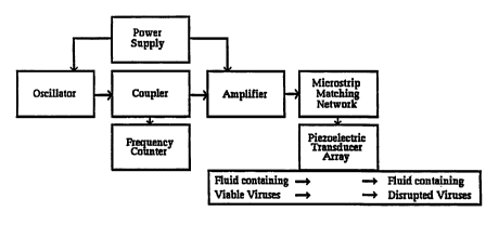
The methods of the present invention comprise delivering acoustic energy at
resonant frequencies to viruses. For example, the qualitative and quantitative
resonant
frequencies can be determined in vitro as shown by the apparatus in Figure 12.
A drop of
fluid (whole blood, serum, culture fluid, or host cells, etc.) with known
resonant acoustic
characteristics, and which also contains a known virus as determined by
standard virology
methods, is placed on a thin disc of absorptive media with known resonant
acoustic
characteristics (paper, cellulose, cotton, polymer, etc.). A thin slice of
viral-laden tissue or
29
CA 02343361 2001-03-09
WO 00/15097 PCT/US99/20776
material (embedded or sliced material such as provided commercially by
Polysciences, Inc.
JB-4 Embedding, Paraffin, Immuno-Bed Kit, LR Gold, Osteo-Bed Bone Kit,
Polyfreeze,
PEG 4000 Resin, PolyFin Para~n, etc.) can be used. The virus disc is placed
between two
broadband low GHz or high MHz transducers such as disclosed above and clamped
into
place.
The target range of frequencies to be examined for qualitative viral resonance
signatures are derived using the speed of sound in biologic tissues 1,500 m/s
divided by
desired wavelength, based on viral dimensions. If the viral dimensions are
unknown, they
may be determined by electron microscopy using techniques known in the art.
One transducer generates the acoustic signal and may sweep through a wide band
of.
target frequencies, and the other transducer detects the transmitted acoustic
signal. The
acoustic signal transmitted from the virus test disclslice is fed into the
positive lead of a signal
analyzer. The known acoustic signals from the test fluid and disc, or test
embedding material
serve as a control and are fed into the negative lead of the signal analyzer.
The control
signahues are canceled out and the remaining resonant acoustic signature
displayed is from
the virus in the sample, yielding a qualitative result.
By varying the range of frequencies analyzed and comparing the amplitudes at
each
frequency, one can identify the primary resonant frequencies, and the
associated harmonic
resonant frequencies. The primary resonant frequencies will have the highest
amplitude.
Each virus will have multiple primary frequencies depending on viral
dimensions including,
but limited to, the diameter, length (if cylindrical or helical), apical
distance, and unit distance.
See Table 2 for calculated ranges of primary resonant frequencies for
individual viruses, using
acoustic velocity as 1,500 m/s, and viral dimensions as currently determined
by standard
virology methods. Results may vary in practice depending on specific viral
factors such as
bulk modulus, dispersion, acoustic velocity in viral materials, in vivo vs. in
vitro dimensions,
etc. and thus the frequencies are in no way limited to the calculated
frequencies in Table 2.
30
CA 02343361 2001-03-09
WO 00/15097 PGT/US99/20776
TABLE 2
I. ICOSAHEDRAL SYMMETRY
A. p,~ y
VIRUS DIAMETERS APICAL LENGTH UNIT (nm) IRDQvFNCSt
(# capsomeres)(nm) 58% ave d (nm) DISTANCE (Hz)
P~'~ 21 7.143 x
10'0
(32) 23 6.522 x
10'
(Adeno-Assoc.Virus)22 6.818 x
10'
12.76 1.176 x
10'
6:63 2.26 x
10
Pol avirus 40 0
3.75 x
10
(JC Virus, 50 3.00 x
BK Virus, 10'
Simian Virus 45 3.33 x
40, 10'
Bovine, Baboon) 26.1 5.75 x
10'0
(72) 13? skewed
Papillomavirus45 3.33 x
10'
(72) 55 2.72 x
10'0
50 3.00 x
10'
29 5.17 x
10'0
? skewed
1.57 x
10
105 0
1.42 x
10
(~~ 8~~, 100 1.50 x
i0'
~~Pox, ~~, 58 2.58 x
10'0
25 6.OO x
10'
9 1.66 x
10'
Bovine herpes 95 1.57 x
virus 10'0
(162) 105 1.42 x
10'
1~ 1.50 x
10'
58 2.58 x
10'0
25 6.00 x
10'
1.66 x
10"
Herpesvirus 95 1.57 x
IV virus 10'
(162) 105 1.42 x
10'
(Epstein Barr)100 1.50 x
10'0
58 2.58 x
10
25 6.00 x
10'
1.66 x
10"
Herpesvirus 95 1.57 x
V virus 10'0
(162) 105 1.42 x
10'
(Cytomegalo) 100 1.50 x
10'
58 2.58 x
10'
25 6.00 x
10'0
9 1.66 x
10"
50 nm core 3.00 x
10'
Adaiovirus 70 2.14 x
10'
(252) 75 2.00 x
10'
72.5 2.07 x
10'
31
CA 02343361 2001-03-09
WO 00115097 PCT/US99/20776
42.05 3.57 x 10'°
8.41 1.78 x 10"
Vaccinia 200 7.5 x 10⁹
250 6.0 x 10⁹
Variola 200 7.5 x IO'
(Smallpox) 250 6.0 x 10⁹
Cowpox Virus200 7.5 x 10⁹
250 6.0 x 10⁹
Molluscum 200 7.5 x 10'
Contagiosum 250 6.0 x 10'
ORFVirus 150 1.0 x 10'
250 6.0 x 10⁹
Paravaccinia150 1.0 x 10'
250 6.0 x 10⁹
Hepatitis 40 3.75 x 10'0
B
Virus 45 (Dane Particle) 3.33 x 10'
42.5 3.53 x 10'
28 nm corc 5.36 x 10'
(Spheres and bacillary
forms noninfective)
~. B~TA ~
VIRUS DIAMETERS TRIANGLE UNIT
(# capsomers)(nm) LENGTH (nm) DISTANCE FRDQ~JII~L'Y
(nm) (Hz)
_
CaLcivnus 31 4.84 x
10'
32 35 4.28 x
10'
33 4.54 x
10'
19.14 7.84 x
10'
9.96 1.51 x
10"
Picarnavirus25 6.00 x
10'
32 30 S.OOx 10'
27.5 5.45 x
10'
15.95 9.40 x
10'
8.29 1.81 x
10"
Reovirus 70 2.14 x
10'0
(92) 75 2.00 x
10'
72.5 2.07 x
10'
42.05 3.57 x
10'
14.02 1.07 x
10'
HIV 85 1.76 x
10'
150 1.00 x
10'
100 1.76 x
10'
Surface spikes 12 1.25 x
nm 10'
18 tun 8.33 x
10'
Cone width 1/4 of
diarmter
32
CA 02343361 2001-03-09
WO 00/15097 PCT/tJS99/20776
II. HELICAL SYMMETRY
BNA Yln~
TRIANGLE UNTT
VIRUS DIAMETERS LENGTH (nm) DISTANCE FREQUENCY
Influenza 80 _ __ _ 1.88 x 10'0
HumanA,B 120 1.25 x 10'
8t C, Avian Peplomers 10 nm (A&B) 1.5 x 10'0
Peplomers 8 nm (C) 1.88 x 10"
A - 6 nm wide helix core 6.66 x 10"
C- 9nmwide helix core 1.66x 10"
Parainfluenza90 1
66 x i0'
(Mumps,Croup)300 .
5.00 x 10'
Helix 15 nra 1.00 x 10"
Helix 19 nm 7.89 x 10'0
7.5 rimby 2.00 x 10"
3~ 5.00x10"
Central canal 5 nm 3.00 x 10"
Paramyxovirvs90 1.66 x 10'0
(NewcastleDs.300 5.00 x 10'
Avian, Simian, Helix 15 nm 1.00 x 10"
Measles) Helix 19 nm 7.89 x 10'
Central canal Snm 3.00 x 10"
Respiratory 120 1.25 x 10'
Syncytial
Virus
Helix 15 nm 1.00 x 10"
Helix 19 nm 7.89 x 10'0
Central canal 5 nm 3.00 x 10"
Marburg virus80nm wide helix 1.88 x 10'0
8c Ebola Virus50 nm internal 3.00 x 10'
canal
20nm central 7.50 x 10'
canal
25 Once the qualitative viral resonant acoustic signature has been determined,
quantitative results may be determined by comparing the resonant acoustic
signature
amplitudes from samples of known concentrations of a specific virus. Samples
with higher
viral loads (concentrations) will have higher resonant acoustic signature
amplitudes. A ratio
of primary resonant frequency amplitude to viral concentration is thus
derived, allowing for
30 assessment of viral load in samples of unknown concentration.
In another embodiment, resonant acoustic signatures from the test disc/slice
may be
33
CA 02343361 2001-03-09
WO 00/15097 PCT/US99/20776
generated either by first clamping a control disclslice into the transducer
chamber and storing
the resonant acoustic signature in a microprocessor for subsequent processing
with the test
disclslice signature, or by clamping a control into a second transducer
chamber and sweeping
through the wide band of frequencies simultaneously with the test disc/slice
virus sweep.
Also, the test disc/slice may be clamped between the transducer and a
reflective surface, and
the acoustic wave generated and received by the same transducer, thus
analyzing reflected
rather than transmitted acoustic waves. Furthermore, one or more transducers
analyzing
reflected or transmitted acoustic energy may by immersed into a fluid or
medium containing
the virus.
In another embodiment one or more transducers analyzing reflected or
transmitted
acoustic energy constitute the walls of a vessel into which a fluid or medium
containing virus
is placed.
The present invention also allows the effects of the resonant frequencies to
be
determined in vitro as shown by the apparatus in Figure 13. Using standard
virology culture
methods, known to those skilled in the art, the viral culture may be placed in
a
reusable/autoclavable test cylinder. The bottom surface of the test cylinder
is the transducer,
constructed for the appropriate frequencies, such as a thin film zinc oxide on
a sapphire
substrate . The host medium thus placed in the test cylinder spreads over the
bottom of the
cylinder in a monolayer and in direct contact with the transducer. Acoustic
energy of the
desired resonant frequency is then delivered through the culture fluid and
host medium to the
viruses, and the effects on growth and function are assessed using standard
virology methods.
By varying the acoustic wave characteristics, such as amplitude, mode
(continuous vs.
pulsed), shape (sinusoidal vs. square), intensity etc., the ideal frequency
and waveform
required to obtain specific effects can be determined.
For example, in testing the augmenting and/or disrupting effects of resonant
acoustic
frequencies on HIV, uninfected T-lymphocyte host cells are first assessed in
the test cylinder
with the resonant acoustic intervention (resonant frequencies in varying
waveform patterns
for varying periods of time at varying intensities) using the trypan blue dye
exclusion test,
which excludes anomalous viral results by assessing the effects of the
acoustic intervention
on the host cells alone. Step 2 involves placing a calculated number of HIV
infected T--
lymphocytes in the test cylinder. The host cells form a monolayer on the
transducer/floor of
34
CA 02343361 2001-03-09
WO 00/15097 PCT/tJS99/20776
the test cylinder, where the acoustic intervention is delivered. The results
are then assessed
using standard in vitro methods such as the Coulter HIV- 1 p24 antigen kit,
HIV cultures,
HIV- 1 DNA by PCR, viral load measurement, quantitative measurements, time to
positivity,
and growth suppression.
The methods of the present invention also provide means to disrupt viruses in
vivo
and extracorporeally in animals as shown in Figure 14. For example, in humans
infected with
HIV, an extracorporeal blood circulation system is established using
techniques known to
those skilled in the art. The extracorporeal blood is passed over a series of
reusable/autoclavable sterilized transducers that deliver acoustic energy at
primary or
harmonic resonant frequencies. The acoustic transducer series acts in effect
as an acoustic
filter, disrupting viruses in the blood stream. Efficacy of treatment is
assessed using viral load
studies, as known to one skilled in the art, both prior to and after the
extracorporeal
treatments.
In another embodiment, the above described acoustic filter is also fitted with
a
receiving transducer mode for analysis of the blood sample. With initial
passes of blood
containing large numbers of intact virus, the resonant amplitude will be high.
After prolonged
exposure of the blood to the disrupting resonant frequencies, the resonant
amplitude will
decline as the numbers of intact viruses decline, thus giving viral load
readings and a method
to determine when cessation of the extracorporeal treatment is indicated.
In another embodiment, a sheet of piezoelectric material is fashioned into an
envelope
or mesh-type transducer, through which the extracorporeal blood is passed. In
another
embodiment, a tube of piezoelectric material is fashioned into a coil
transducer, through
which the extracorporeal blood is passed. In another embodiment, the
extracorporeal blood
is separated into red and white blood cell portions, and only the while blood
cell portion is
passed through the acoustic filter, thus reducing the time required for
treatment and reducing
mechanical damage to the red blood cell portion.
In another embodiment, banked blood is passed through an acoustic filter at
any one
of multiple points in the blood product collection and administration process
(i. e., collection
from the donor, separation into components, or administration to the
recipient).
In another embodiment, nanosystem t~hnology (seeNanosytems, by Eric Drechsler;
publications of CJ Kim, Berkley University; publications of Ralph Merck, Xerox
Co., Palo
CA 02343361 2001-03-09
WO 00/15097 PCT/US99/20776
Alto, CA) is employed to make multiple small acoustic oscillators which are
enclosed in filter
mateaial, the filter material preventing passage of the oscillators but
allowing the passage of
blood cells and blood components. The nanite virosonic filter is sterilized
and attached in line
on an extracorporeal system or in a blood products system.
In another embodiment, the resonant and/or harmonic acoustic frequencies are
generated using acoustic laser or maser systems. In similar fashion, the whole
or fractionated
blood is passed extra corporeally over or through a laser or maser acoustic
filter.
The method also provides a means to disrupt viruses in vivo and
intracorporeally in
animals as shown in Figure 15, using intravascular devices. Nanosystem
technology is
employed to make multiple small acoustic oscillators which are enclosed in
filter material, the
filter material preventing passage of the oscillators but allowing the passage
of blood cells
and blood components. The nanite virosonic filter is attached in line on a CVP
type catheter
or in a Greenfield-type filter.
In another embodiment, a central venous catheter as known to one skilled in
the art
(produced commercially by Arrow, Baxter, etc.) is engineered and fitted with a
transducer
of appropriate frequency at the tip. The catheter is inserted using standard
technique into a
large vein such as the subclavian, jugular, or femoral vein. Resonant acoustic
energy is then
delivered to the circulating blood, thereby disrupting virus in vivo.
In another embodiment, the transducer is fitted as an acoustic filter on a
larger
intravascular device such as a Greenfield filter-type device for the inferior
vena cava. The
device is fitted with a battery that is rechargeable through the skin, as
currently practiced with
rechargeable cardiac pacemakers. Once inserted, the acoustic filter reduces
viral load in the
vena caval blood flow, without the need for the patient to be restricted by
catheters.
In another embodiment, inclusion of a receiving acoustic transducer may also
detect
qualitative and quantitative resonant acoustic frequencies of the virus in the
multicellular
organism to determine efficacy and duration of treatment.
The methods of the present invention also provide a means to augment and/or
disrupt
viruses in vivo in a multicellular organism, as shown in Figure 16, using
resonant acoustic
fields. The organism is placed in a form-fitting tub filled either with water
or a coupling
medium such as castor oil (reflection coefficient 0.0043) or mineral oil, or
such other acoustic
conductive gel as is available commercially. Acoustic transducers are either
fitted into the
36
CA 02343361 2001-03-09
WO 00/15097 PCTNS99/20776
walls and floor of the tub, or are themselves the walls and floor of the tub
(i. e., piezoelectric
polymer sheets or ceramics). A predetermined acoustic field (frequencies,
harmonics,
amplitude, mode, shape, etc., at a specific intensity ) is delivered to the
organism from the
transducer tub through the coupling medium.
In another embodiment, a receiving acoustic transducer mode also detects
qualitative
and quantitative resonant acoustic frequencies of the virus in the
multicellular organism to
determine efficacy of treatment.
The present invention also provides a method to augment and/or disrupt viruses
in
viva in a portion of a multicellular organism as shown in Figure 17, using a
resonant acoustic
field probe. Acoustic transducers of desired frequency are fitted into the end
of a hand-held
probe device, as currently known to those skilled in the art of medical
ultrasonography. A
predetermined acoustic field (frequencies, harmonics, amplitude, mode, shape,
etc. at the
required intensity to effect the organism) is delivered to a predetermined
portion of the
organism, from the hand-held transducer probe. Attenuation in air is
eliminated by use of a
commercially available acoustic coupling medium such as castor oil. For
example, in a
person afflicted with hepatitis, the treatment is delivered through the skin
over the liver.
Subharmonics of the resonant acoustic frequencies can be used to minimize
acoustic
attenuation at the higher frequencies.
In another embodiment, receiving acoustic transducer mode also detects
qualitative
and quantitative resonant acoustic frequencies of the virus in the
multicellular organism to
determine eflxcacy and duration of treatment.
The present invention also provides a method to disrupt viruses in viva in a
portion
of a multicellular organism as shown in Figure 18, using a resonant acoustic
field sheet.
Piezoelectric polymer material of desired frequency is fashioned into a
flexible transducer
sheet device. A predetermined acoustic field (frequencies, harmonics,
amplitude, mode,
shape, etc.) is delivered to a predetermined portion of the organism, from the
transducer
sheet device. Attenuation in air is eliminated by use of a commercially
available acoustic
coupling medium such as castor oil. For example, in a person afflicted with
hepatitis, the
treatment is delivered by placing the sheet in contact with the skin over the
liver.
Subharmonics of the resonant acoustic frequencies can be used to minimize
acoustic
attenuation at the higher frequencies.
37
CA 02343361 2001-03-09
WO 00/15097 PCT/US99/20776
In another embodiment, receiving acoustic transducer mode also detects
qualitative
and quantitative resonant acoustic frequencies of the virus in the
multicellular organism to
determine efficacy and duration of treatment.
The present invention also provides a means to determine qualitative and
quantitative
resonant acoustic andlor acousto-EM frequencies in vitro as shown in Figure 19
A&B. A
test device, as described above and shown in Figure 12, with any and all
embodiments, is
fitted with transmitters and receivers to transmit, detect, measure, and
analyze EM energy.
When the resonant acoustic frequencies are applied to the virus test disk, a
unique
electromagnetic energy pattern is generated, according to the structure and
composition of
the virus and test disk under study, referred herein as the resonant acousto-
EM signature.
Mechanisms producing the resonant acousto-EM signature include, but are not
limited to
piezoelectricity, acoustoelectricity, magnetoacoustics, and/or intrinsic
energy dissipation. The
resonant acousto-EM signature represents one or more of several
electromagnetic properties
and/or fields including, but not limited to, direct current, alternating
current, magnetic field,
electric field, EM radiation, and/or acoustic cyclotron resonance (standard or
Doppler
shifted).
All of the above mentioned forms of EM energy are detected, measured, and
analyzed
with devices and methods known to those skilled in the art. (It should be
noted that useful
information may also be derived from application of nonresonant frequencies,
ie. current
characterization of semiconductor biologics via the acoustoelectric effect.)
This data in
combination with resonant signatures yields even greater sensitivity and
specificity to the
method. For example, Herpes simplex virus (HST I and II will have nearly
identical
resonant acoustic signatures because they are virtually identical in size and
shape. They differ
in molecular protein configuration, however, and can be distinguished by their
acousto-EM
signatures. This includes, but is not limited to, characterization at
nonresonant and resonant
frequencies of acoustoelecttic currents, acousto-EM signatures produced via
intrinsic energy
dissipation, of acoustic modulation or attenuation in the presence of a
magnetic field via the
magnetoacoustic effect, and of electric or magnetic fields induced or affected
by any of the
above processes.
In another embodiment, the test device is also fitted with any and all
combinations of
resonant acoustic and acousto-EM generating equipment. A sample of unknown
composition
38
CA 02343361 2001-03-09
WO 00/15097 PCT/US99lZ0776
is exposed to the frequency energy pattern which is included in the acousto-EM
signature for
a particular structure. Detection of the associated resonant acoustic waves
from the sample
confums the presence of the structure in the sample. Further analysis of
amplitude would
indicate the relative quantity of those particular structures in the sample.
For instance, the
combined use of resonant acoustic and acousto-EM signatures could be used to
search a
tissue slice first for the presence of HSV, and then to specify whether it is
HSV I, HSV II,
or a previously unknown and uncharacterized HSV. In addition, a quantitative
assessment
of viral load in the sample could also be performed based on relative
amplitudes. Thus, the
application of resonant acoustic and/or acousto-EM energy fields, in respect
to organic or
biologic organisms and structures, yields a form of resonant acousto-EM
spectroscopy, with
three basic stimulation and detection modes (1. acoustic, 2. EM, 3. acoustic
and EM),
producing nine basic combinations:
1. Acoustic stimulation, acoustic detection;
2. Acoustic stimulation, EM detection;
3. Acoustic stimulation, acoustic and EM detection;
4. EM stimulation, acoustic detection;
5. EM stimulation, EM detection;
6. EM stimulation, acoustic and EM detection;
7. Acoustic and EM stimulation, acoustic detection;
8. Acoustic and EM stimulation, EM detection; and
9. Acoustic and EM stimulation, acoustic and EM detection.
The more sophisticated the stimulation and detection/ analysis modes are, the
more
sensitive and specific the spectroscopy apparatus will be. It should be noted
that the use of
resonant acousto-EM spectroscopy alone or in combination with resonant
acoustic
spectroscopy in the present invention is not limited to biological materials
and can be utilized
to detect and identify inorganic materials or structures as discussed below.
The present invention also provides a method to assess the effects of resonant
acoustic and/or acousto-EM energy on viruses using any and all devices which
produce
acoustic and/or EM energy including, but not limited to, all devices and
embodiments
previously described. For instance, as shown in Figure 20, to assess the
piezoelectric effects
ofEM radiation on the crystalline structure of viruses, a test system is used
which employs
39
CA 02343361 2001-03-09
WO 00/15097 PCTNS99/20776
EM radiation of the same frequency as at least one of the resonant acoustic
frequencies of
the virus. In the case of HIV, the frequency is approximately 15 GHz. A test
box made of
EM absorptive material is fitted with a 15 GHz EM transmitter, with the EM
radiation
directed towards the floor of the box. Uninfected T-lymphocyte host cells are
first assessed
S in the test box with the 15 GHz intervention with varying exposure patterns
(resonant
frequencies in varying waveform patterns for varying periods of time and at
varying
intensities) using the trypan blue dye exclusion test, which excludes
anomalous viral results
by assessing the effects of the acousto-EM intervention on the host cells
alone. Step 2
involves placing HIV infected T-lymphocytes in the test box, where the acousto-
EM
intervention is delivered. The results are then assessed using standard in
vitro testing of anti-
HIV methods such as the Coulter HIV 1 p24 antigen kit, HIV cultures, HIV-1 DNA
by PCR,
and viral load measurement.
The present invention also provides a method to disrupt viruses
extracorporeally
and/or intravascularly in animals using resonant acoustic and/or acousto-EM
fields as shown
in Figure 21. For example, in humans infected with HIV, an extracorporeal
blood circulation
system is established using techniques known to those in the art. The
extracorporeal blood
is passed over transducers as described in Figure 14, including any and all
embodiments.
Acoustic penetration into the blood may be increased using acoustoelectric
gain by passing
a direct current into the blood parallel with the acoustic waves.
The present invention also provides a method to augment and/or disrupt viruses
in
an organ of a multicellular organism, as shown in Figure 22, using resonant
acoustic and/or
acousto-EM fields. For instance, as in Figure 16, including any and all
embodiments, a human
cadaver cornea for transplantation is placed in a form-fitting cup filled
either with water or
such other non-toxic acoustic conductive gel as is available commercially. A
predetermined
acoustic field (frequencies, harmonics, amplitude, mode, shape, etc.) is
delivered to the
cornea from a transducer tub through the coupling medium. Utilizing the
magnetoacoustic
effect, a magnetic field is placed perpendicular to the direction of the
acoustic wave
propagation, at a field strength which is a multiple of the acoustic
frequency, thereby
generating sinusoidal or peak-type resonance spikes in the acoustic power, and
improving
resonant acoustic penetration into the cornea without injuring the cornea
tissue itself.
The present invention also provides a means to disrupt viruses i» vivo in a
portion of
CA 02343361 2001-03-09
WO 00/15097 PCT/US99/20776
a multicellular organism using a resonant acoustic and/or acousto-EM field
probe. For
example, as shown in Figure 23, a hand-held probe is fitted with an EM
radiation generating
device, as currently known to those skilled in the art. A predetermined EM
radiation field
(frequencies, harmonics, amplitude, mode, shape, etc.) replicating the acousto-
EM signature
representing the intrinsic dissipation pattern of a particular virus, is
delivered to a
predetermined portion of the organism, from the hand-held probe. For example,
in a person
afflicted with an upper respiratory tract infection (a "cold"), the treatment
is delivered
through the skin over the nose, throat, and sinuses, reversing the intrinsic
energy dissipation
pathway of the rhinovirus and inducing resonant acoustic oscillations which
disrupt the
rhinovirus.
Example 2
Any micro-organism, such as bacteria, as well as structure and molecules
contained
or associated herewith, may be augmented, disrupted, detected and/or
identified in vitro or
in vivo using the methods of the present invention. Bacteria include, but are
not limited to,
those associated with animals, man, avians, reptiles, amphibians, insects,
aquatic like, plants,
fiuit, soil, water, oil, fermentation processes for food production, and the
like. In one
embodiment the bacteria include but are not limited to Streptococcus sps ,
Staphylococcus
sps., Hemophilus sps., Neisseria sps., Treponema sps., Salmonella sps,
Shigella sps,
F~scherichia colt strains, Corynebacteria spy, Bordetella sps., Chlosfridrium
sps , Rickettsia
sps., Chlamydia sps, Brucella sps , Mycobacterium sps.,Borrelia sps.,
Mycoplasma sps.,
Lactobacillus sps., strains thereof and the like. Human illnesses caused by
bacteria include
pneumonia, skin and wound infections, heart valve infections, gastroenteritis,
syphilis,
gonorrhea, the plague, urinary tract infections, lyme disease, tuberculosis,
cholera, typhoid
fever, anthrax, tetanus, and gangrene.
Fungal infections include athlete's foot, ringworm, vaginal yeast infections,
oral
thrush, histoplasmosis, and cryptococcus.
Diseases in animals caused by bacteria, fungi, protozoa and worms are similar
to
those in humans. Similarly, a wide range of micro-organisms infect plants, and
even other
micro-organisms are deemed to be beneficial (i.e. bakers yeast.).
41
CA 02343361 2001-03-09
WO 00/15097 PCT/US99/20776
Bacteria are first classified by staining characteristics as either Gram
positive, or Gram
negative. Bacterial response to staining is determined by the structure of the
cell wall. Next
bacteria are fiirther classified by shape as either cocci (spherical) or rods
(cylindrical.) Beyond
that, the classification schemes generally involve various biochemical
reactions.
Bacterial cell walls are composed of rigid peptidoglycan (mucopeptide or
murien},
a mixed polymer of hexose sugars (N-acetylglucosamine and N-acetyl muramic
acid) and
amino acids (the structural units of proteins, see below). As such, the cell
walls are
crystalline structures and are subject to vibrational effects from the use of
acoustic energy.
Thus bacteria are susceptible to augmentation, identification and detection,
or disruption by
IO resonant acoustic frequencies matched to their shape (sphere or cylinder),
size, and
composition. In addition, various organelles contained within the bacteria
structure are also
susceptible to specific resonant acoustic frequencies (i.e., pili, plasma
membrane, flagellum,
cytoplasmic inclusion bodies, basal bodies, capsule, spores, etc.). Finally,
the compounds
comprising the structure itself (crystalline proteins, etc.) also have unique
resonant
frequencies.
Fungi, protozoa, parasites, and worms are similar to bacteria in that the
organisms
are susceptible to the effects of specific resonant frequencies based on the
size and shape of
the entire organism, the size and shape of organelles making up a part of the
organism, and
the resonant characteristics of specific biochemical compounds making up the
organism.
Any fungus, including yeasts, molds and mushrooms, protozoan, parasites or
worms,
as well as structures and molecules contained or associated therewith, may be
augmented,
disrupted and/or detected in vitro or in vivo using the methods of the present
invention.
These organisms include, but are not limited to those associated with animals,
man, avians,
reptiles, amphibians, insects, aquatic life, plants, fruit, soil, water, oil,
fermentation possesses
for food production, and the like. In one embodiment, these organisms include
but are not
limited to Crypto sporidia sps., Aspergillus sps, Trichophyton sps ,
Saccharomyces sps,
Blastomyces sps , Coccidioides sys , Paracoccidioides sps., Penicillium sps ,
Rhizopus sps ,
Mucor sps , Neurospora sps., Microsporum sps, Streptomyces sps ,
Epidermophyton sps.,
Toxicara sps., Ascaris sps., Echinococcus sps., Giardia sps., Plasmodium sps.,
Tryparrosoma sps, Schistosoma sps., Brnglia sps., strains thereof and the
like.
At low acoustic and/or acousto-EM power inputs such as below 1 x 10's W/m2,
the
42
CA 02343361 2001-03-09
WO 00/15097 PCT/US99/20776
micro-organisms will be augmented in function and will emit a characteristic
acoustic and/or
acousto-EM signature which can be used to detect and diagnose the presence of
the micro-
organisms. At higher power inputs, the organisms will be disrupted and killed.
In addition to
the structures of bacteria, fungi, protozoa, and worms being susceptible to
the vibrational
resonant effects of acoustic and/or acousto-EM energy, they may also function
as
piezoelectric structures, intrinsic dissipation, acoustoelectric, and
magnetoacoustic structures.
The present invention takes advantage of the composing parts of structures, or
the
entire organism of bacteria, fungi, protozoa, and worms for the purpose of
augmentation,
identification, and/or physical disruption of the micro-organism structures
using acoustic
and/or acousto-EM energy at specific resonant frequencies, and the
piezoelectric, intrinsic
dissipation, acoustoelectric and/or magnetoacoustic properties of any and all
structures
involved, either alone or in combination with a resonant acoustic field.
Unlike treatment in the prior art using ultrasound, the present invention uses
specific
resonant frequencies, which can be used to treat a multilayer organism. The
invention also
has the potential to augment the functional activity of micro-organisms deemed
beneficial
such as baker's yeast, wine yeast, lactic acid bacteria (wine and cheese,)
petroleum yeast, and
microbes producing specific amino acids, antibiotics, enzymes, or other
chemicals. The
functional activities may include growth, metabolism, oxidation or reduction
activity and the
like.
In one embodiment, the present invention allows the resonant acoustic and/or
acousto-EM frequencies of micro-organisms to be determined in vitro as shown
by the
apparatus described in Figures 12 and 24 A & B, including any and all
embodiments, with
transducers designed for lower frequencies in the MHZ range (as provided
commercially by
Mates Instzuments). For example, in a meat packing plant concerned with the
contamination
of beef by bacteria, in particular, E. coli, a similar device can be used to
screen the meat for
bacteria, in a relatively short time span when compared to conventional
culturing methods.
First a swab of the meat surface is taken, and placed into a sterile test tube
containing sterile
saline at physiologic pH. A predetermined amount of the solution is pipetted
onto a standard
test disc, which is clamped between two transducers. Resonant or resonant
harmonic acoustic
frequencies are scanned for in the test sample, thereby screening for the
presence or absence
of potentially harmful E coli bacteria. Inspection of meat is done more
efficiently and reliably
43
CA 02343361 2001-03-09
WO 00/15097 PCT/US99/20776
than by current methods.
The present invention also allows the resonant acoustic and/or acousto-EM
fields of
micro-organisms to be used to augment these biologic organisms or their
structures. For
example, as shown in Figure 25, the bottom of a beer fermentation vat is
fitted with acoustic
transducers of appropriate frequency and power output to augment the fi~nction
of the special
strains of Saccharomyces cerevisiae yeast. This yeast is currently used to
ferment beer for
a period of 5 to 10 days, however, with resonant acoustic augmentation, the
fermentation
time is reduced. The most efficient power output level can be determined by
quantitatively
detecting concentration of yeast and conversion of starch and/or sugar
molecules to alcohol
compound.
The present invention also allows the resonant acoustic and/or acousto-EM
fields of
micro-organisms to be used to disrupt these biologic organisms or their
structures. For
example, as shown in Figure 2G, a commercial kitchen microwave is fitted with
two {2) EM
radiation horns - one for cooking and one for the resonant acoustic and/or
acousto-EM
frequencies of the common food pathogens such as E. toll and Salmonella sps.
Prior to
roasting, grilling, or such other food preparation method as may be desired,
the home chef
may decontaminate the meat or other food product of any potential pathogens by
using the
decontaminate cycle on the microwave oven.
Acoustic resonance measurements were conducted on several types of bacteria to
determine the resonant acoustic frequency of the bacteria. A Matec high
frequency 7000
pulse modulator and receiver was used in conjunction with a Matec automated
data
acquisition system and an oscilloscope. Klebsiella pneumoniae (American Type
Culture
Collection #13883) was grown on standard growth media. A Matec 90 MHz, 3/8"
diameter
transducer surface was cleaned and sterilized with alcohol. Live Klebsiella
was placed on
the s<uface of the transducer. Resonant acoustic spectroscopy was performed in
the acoustic
range of 100-200 MHz. A resonant acoustic frequency was detected for the
Klebsiella at
125-130 MHz with a centered frequency at 127.5 MHz. This was presumed to be a
resonant
subharmonic frequency.
The same measurements were performed on E. toll bacteria (American Type
Culture
Collection # 25922) using the same equipment. A resonant acoustic frequency
was detected
for the E. toll with a centered frequency of 113 MHz. This too was presumed to
be a
44
CA 02343361 2001-03-09
WO 00/15097 PCT/US99/Z0776
resonant subharmonic frequency.
Example 3
Arthropods include a diverse goup of insects that infest and feed on the blood
of
humans and animals. Examples include lice, fleas, ticks, mosquitoes, mites,
sandflies, and
tsetse flies. Aside from the general discomfort and annoyance that these
arthropods produce
when they infest a human or animal, the true danger of infestation lies in the
diseases
transmitted by the arthropods. These diseases, in general, cost the world
economy billions
of dollars a year. The overall health status of the victims is impaired and
they suffer loss of
time, quality of life, and sometimes life itself.
Mosquitoes transmit dengue fever, yellow fever, encephalitis, hemorrhagic
fever,
malaria, and lymphatic filariasis. Ticks transmit encephalitis, Lyme disease,
relapsing fever,
and Rocky Mountain spotted fever. Fleas transmit the plague (Yersinia) and
typhus. Lice
transmit typhus. Mites transmit rickettsial pox. Flies transmit African
sleeping sickness,
leishmaniasis, and Chagas disease.
The distinguishing feature of arthropods is the chitinous exoskeleton, which
covers
the body and legs. Chitin is a long, unbranched molecule consisting of
repeating units of N-
acetyl-D-glucosamine. It is found abundantly in nature and forms the hard
shell of insects,
arthropods, crustaceans, mollusks, and even the cell walls of certain fungi.
As such, chitin is
a crystalline structure and is subject to the effects of acoustic and/or
acousto-EM energy.
Thus arthropods are susceptible to detection and disruption by resonant
acoustic frequencies
matched to their shape (sphere or cylinder), and size. In addition, various
organs or
app~dages contained within the arthropod structure are also susceptible to
specific resonant
acoustic frequencies. Finally, the compounds comprising the structure itself
(chitin, crystalline
proteins, etc.) also have unique resonant frequencies.
At low acoustic power inputs, the infectious arthropods will emit a
characteristic
acoustic and/or acousto-EM signature which can be used to detect and diagnose
their
presence. At higher power inputs, the arthropods will be disrupted and killed.
The specif c
range of intensities used for detection or disruption will be determinant on
the structure and
the intensity can be determined using standard methods known to those skilled
in the art such
CA 02343361 2001-03-09
WO 00/15097 PCT/US99/20776
as discussed above. In addition to the structures of arthropods being
susceptible to the
effects of acoustic and/or acousto-EM energy, they may also function as
piezoelectric
structures.
The present invention takes advantage of composing parts of the structures or
the
entire organism of arthropods for the purpose of identification and/or
physical disruption of
the arthropod structure using acoustic and/or acousto-EM energy at specific
resonant
frequencies and patterns, and using them as piezoelectric, intrinsic
dissipation,
acoustoelectric, and or magnetoacoustic structures, either alone or in
combination with a
resonant acoustic field.
The methods of the present invention allow the resonant acoustic frequencies
of
arthropods to be determined and utilized, with devices of appropriate
frequency similar to
those previously described. For example, researchers capturing and cataloging
thousands of
insects and other arthropods in an effort to identify the source of an
infectious agent such as
Ebola, a hemorrhagic fever, or encephalitis, could use an apparatus such as
that shown in
Figure 27. The portion of the acoustic spectrum containing the resonant
frequencies of the
infectious agent in question is scanned. Known resonant frequencies of
arthropod materials
are fed into the negative lead of the spectrum analyzer and cancel out their
component
resonant frequencies in the positive lead sample scan. The remaining
frequencies are analyzed
for the resonant acoustic signature of the offending microorganism. This
provides a means
to readily identify the host reservoir of an infectious agent without the need
for expensive
and time-consuming studies.
The present invention also provides a means to kill infecting arthropods on a
large
organism, for example fleas on a dog or human, as shown in Figure 28. High kHz
to very low
MHz transducers are fitted onto a bathtub-type apparatus. The resonant
acoustic frequencies
for fleas are delivered through the water to the surface of the animal. High
power outputs for
deep tissue penetration are not required, as the infectious arthropods are
restricted to the
surface or outer-most layers of the dog or human. The same method can also be
used, for
example, to de-flea or de-louse bedding and linens in a washing machine.
46
CA 02343361 2001-03-09
WO 00/15097 PCT/US99/20776
Ezample 4
Augmentation of Bone Growth
Bone demineralization in humans is a significant health care problem.
Thousands of
elderly people sustain fractures of the hip, leg, or arm due to this bone
demineralization
(osteoporosis). These injuries cost the American health care system billions
of dollars a year,
for treatment, surgery, and rehabilitation after the injury. In addition, the
overall health status
of the victims is impaired, and they suffer loss of time and quality of life
due to these
fractures. Other conditions which contribute to bone matrix loss include
weightlessness (i.e.,
in outer space) and prolonged confinement to bed. People in certain
occupations may benefit
from an increase in the normal bone density. Examples include professional
athletes, military
personnel, and jobs requiring exposure to increased atmospheric pressures
{i.e., undersea
diving).
Living bone is organized in a calcium based crystalline structure of
hydroxyapatite,
doped with copper, and embedded in collagen fibers. The application of force
to the collagen
fibers in the bony matrix, through mechanical pressure or gravitational
fields, stimulates the
piezoelectric effect and flow of ions via fluid channels in bone. This small
electrical charge,
in turn, acts as a signal to the body's osteoblasts to deposit more
hydroxyapatite. As the
hydroxyapatite density increases, the bone becomes stronger. Thus, bones
maintain their
normal structure and density in response to pressures and forces encountered
in normal daily
activities, via a piezoelectric effect.
With aging, normal copper doping is lost, and the piezoelectric effect
diminished.
The result is that hydroxyapatite density is not maintained, and the elderly
suffer from
osteoporosis and bone fractures. The same thing occurs in the absence of
normal activity
(weightlessness and confinement to bed), with subsequent absence of the normal
piezoelectric
effect and ionic current flows.
Bone is a crystalline piezoelectric structure and as such is subject to the
vibratory
effects of acoustic energy. The operative process behind normal physiologic
bone density
maintenance is the generation of hydroxyapatite molecular movement within
collagen fibers,
compressed by macro-pressures. These occur from daily activities, and
stimulate the
piezoelectric and subsequent bone building osteoblastic effects.
47
CA 02343361 2001-03-09
WO 00/15097 PCT/US99/20776
This molecular movement and the collagen fiber compression can also be
generated
from micro-pressures within the semiconductor matrix of bone. Thus understood,
micro-
pressures can be produced by acoustic energy waves.
In addition to the piezoelectric effect, since bone is a piezoelectric and
semiconductor
structure, it will exhibit the acoustoelectric, intrinsic dissipation and
magnetoacoustic effects.
Conditions with diminished bone semiconductor fi~nction (osteoporosis) and/or
decreased
macro-pressures(weightlessness and bed confinement) can be effectively treated
through
application of acoustic micro-pressures which generate a biological
piezoelectric effect,
and/or also via acoustic resonance, intrinsic dissipation, acoustoelectric and
magnetoacoustic
effects.
Prior literature describes the use of non-resonant ultrasound to speed the
rate of
healing of bone fractures, however, the mechanism causes gross disruption of
the bone
tissues, which in turn damages the microscopic capillary bed in bone, with
leakage of serum
and cells into the bony matrix, and with subsequent bone mineralization. The
literature also
describes attempts to use ultrasound to detect resonant frequencies of the
strocture of entire
bones (femur and ulna) to diagnose a bone as normal or defective. However, the
use of
acoustics and/or acousto-EM resonant frequencies to activate the piezoelectric
effect is not
described. No consideration is given in the prior art to using bone as a
living transducer for
the piezoelectric, intrinsic dissipation, acoustoelectric, and magnetoacoustic
effects, either
alone or in combination with a resonant acoustic field.
The present invention takes advantage of the crystalline, piezoelectric
structure of
bone for the purpose of augmenting bone growth and calcification. The
invention has the
potential to significantly reduce the number and severity of bone fractures
suffered by victims
of osteoporosis. The invention has the potential to speed the healing process
of fractures.
Other conditions which contribute to bone matrix loss, such as weightlessness
(i.e., in outer
space), or prolonged confinement to bed, would also benefit from the
invention. The
invention has the potential to aid people in occupations which would benefit
from an increase
in their bone density (athletes, military personnel, and jobs requiring
exposure to increased
atmospheric pressures such as undersea diving.) The invention also has
potential veterinary
applications. Unlike prior treatment using ultrasound, the present invention
uses resonant
acoustic and/or acousto-EM frequencies of bone to stimulate at least the
piezoelectric effect
48
CA 02343361 2001-03-09
WO 00/15097 PCT/US99/20776
for augmentation of bone growth without affecting neighboring tissue.
The methods of the present invention provide a means to augment the growth and
maintenance of bone using resonant acoustic and/or resonant acousto-EM energy.
For
example, as shown in Figure 29, a sheet of piezoelectric material is fitted
into a shower mat
device. When an elderiy person, prone to osteoporosis, showers the mat is
activated. Water
in the shower acts as a conductive medium and primary or harmonic resonant
frequencies are
delivered through the soles of the feet, along the lines of force, up into the
legs and hips. The
piezoelectric effect in bone is activated and bone density is increased.
The present invention provides a method to augment the growth and maintenance
of
bone using resonant acoustic and/or acousto-EM energy, for example, as also
shown in
Figure 30. The sleeping/tether bags used by astronauts during conditions of
weightlessness
are fitted with EM radiation transmitters in the foot of the bags. The bags
are made of EM
absorptive materials. The tethers that anchor the sleeping bags to the space
vessel include the
cables to connect the antennas to signal generators in the space craft. While
sleeping, the
bone maintenance devices in the sleeping bag are activated, delivering EM
radiation to the
astronauts at a resonant frequency that activates the piezoelectric effect in
bone, and thus,
maintains their normal body density. Extraneous EM radiation which might
interfere with
other equipment on board is blocked by the EM absorptive materials in the
sleeping bags.
Example 5
There are a wide variety of tissue masses, both benign and malignant, which
afflict
humans and animals. Many tissue masses are encapsulated or are contained
within a
restricted area in the body. Nearly all benign tumors grow and expand slowly,
developing a
fibrous capsule, and producing a discrete, readily palpable and easily movable
mass.
Examples of benign tumors include fibroma, lipoma, chondroma, osteoma,
hemangioma,
lymphangioma, meningioma, leiomyoma, adenoma, papilloma, polyps, condyloma,
fibroadenoma, and rhabdomyoma. Most malignant tumors are invasive and
metastasize,
however, notable exceptions are gliomas and basal cell carcinomas. Other
tissue masses
causing disease include emboli, thrombi, abscesses, stones, and foreign
bodies.
49
CA 02343361 2001-03-09
WO 00/15097 PCT/US99/20776
By virtue of having a defined, discrete structure, many tissue masses are
susceptible
to the disnapting effects of acoustic energy at resonant frequencies matched
to their size and
shape. Prior art contains many applications for the use of acoustics at non-
resonant
frequencies to detect and even disrupt tissue masses, but to date detection of
tissue masses
via resonant acoustic energy and disruption of tissue masses via acoustic
energy at resonant
frequencies has not been disclosed.
In addition to tissue masses being susceptible to detection and disruption by
resonant
acoustic frequencies matched to their shape and size, the components
comprising the tissue
mass itself (cell types, crystalline proteins, etc.) also have unique resonant
frequencies
susceptible to detection and disruption. At lower power inputs, certain
tissues or masses can
be augmented in growth or metabolism, providing a supplemental technique for
tissue
culturing, regeneration, and growth.
Depending on their structure, certain tissue masses or types may also exhibit
resonant
acousto-EM effects as well as functioning as piezoelectric, intrinsic
dissipation,
acoustoelectric, and/or magnetoacoustic structures.
The present invention takes advantage of the discrete shape, size, and
composition
of numerous benign and malignant tissues and masses to cause the
identification,
augmentation, detection, and/or disruption of those structures using acoustic
and/or
electromagnetic energy at specific resonant frequencies. Unlike prior
treatments using
ultrasound, the present invention uses specific resonant acoustic and/or
electromagnetic
frequencies, which can be used to treat a multilayer organism by targeting a
specific structure
therein. It combines the known tumor/mass detection abilities of acoustic
energy (diagnostic
ultrasound) with the disruptive characteristics of acoustic and/or
electromagnetic energy at
resonant frequencies. The invention also has the potential to augment the
growth and
function of various tissues and masses, where desirable.
The present invention provides a means to detect and disrupt benign or
malignant
tissues and/or tissue masses using resonant acoustic and/or acousto-EM energy.
For
example, as shown in Figure 31, an acoustic transducer designed with standard
echo-
reflective capabilities is used to determine the size and dimensions of a
tissue mass. Based on
the calculated resonant frequencies, a range is scanned to determine the
precise resonant
frequencies. Then one or more of those frequencies are delivered to the mass,
disrupting its
SO
CA 02343361 2001-03-09
WO 00/15097 PCT/US99120776
structure and allowing subsequent resorption of the mass by the body.
Also, the present invention provides a means to detect benign or malignant
tissue
types using resonant acoustic and/or acousto-EM energy, using the apparatus
described in
Figures 12 and 19 A &B, including any and all embodiments, the cell test disc
or tissue
preparation is placed between two transducers and the frequencies are scanned
looking for
resonant peaks and EM patterns. Differences in the resonant peaks and EM
patterns will
differentiate between tissue types, for example between normal epithelial
cells and cancerous
epithelial cells.
Ezample 6
Augmentation, Detection and/or Disruption of Bioyhemical Comir~ou~~c nr
Ticcupc
Biologic organisms are composed of many biochemical compounds including
nucleic
acids, carbohydrates, lipids, amino acids, and steroids. Many biochemical
compounds align
themselves in regularly repeating patterns: in other words they adopt
crystalline forms.
Examples of biochemical crystals include insulin, hexokinase, aldolase,
hemoglobin,
myoglobin, and spectrin. In addition, certain tissues or cell structures adopt
crystalline form
such as bone, muscle fibers, and connective tissue fibers for the former, and
cell membranes,
Na/K membrane pumps, and visual rod receptors for the latter.
The biochemical compounds from which biological organisms are composed have
their own unique resonant frequencies, based on their innate crystalline
structure. Many of
the biochemical compounds are also piezoelectric, intrinsic energy
dissipation,
acoustoelectric, and magnetoacoustic structures. As such, biochemical
compounds are
subject to the augmenting, disrupting, and/or detecting features of resonant
acoustic and/or
acousto-EM energy. The present invention uses specific resonant acoustic
and/or acousto-
EM frequencies, which can be used to treat a multilayer organism. The present
invention also
has the potential to utilize piezoelectric, intrinsic energy dissipation,
acoustoelectric, and/or
magnetoacoustic effects to achieve desired results, either alone or in
combination with a
resonant acoustic field.
51
CA 02343361 2001-03-09
WO 00/15097 PCTNS99/20776
Eiample 7
The present invention provides a method to stimulate and/or disrupt
proteoglycans
adhesive units between cells using resonant acoustic and/or acousto-EM energy.
Millions
of operations are performed on humans every year, using metal scalpels to make
the incision.
The use of such scalpels requires closure of the incisions with stitches, a
period of healing,
and invariably results in scar formation. In addition, millions of people
suffer traumatic cuts,
tears, or ruptures of the skin, again requiring closure of the wounds with
stitches, a period
of healing, and scar formation.
In multicellular organisms, the cells are held together by proteoglycans
units, at the
rate of approximately 1,600 per cell. These units are approximately 200 um
long, with some
variation between the species.
When an incision is made, or a traumatic break in a cell layer occurs, the
cellular
adhesions are ripped apart, some cells are ruptured, and blood vessels are
torn open. White
blood cells, platelets, and fibroblasts congregate in the extracellular space
and eventually lead
to the formation of a scar which readheres the tissues. During this healing
phase the open
tissues are much more susceptible to invasion by foreign organisms, and wound
infection is
a complication that must be constantly guarded against.
Even if the wound heals without the complication of infection, a scar still
remains.
Modern plastic surgery techniques try to either minimize or hide scars, but
the formation of
a scar is inevitable.
An energy field achieving acoustic resonance with the proteoglycans units at
high
amplitudes indicating high power levels will cause separation of the adhesive
bonds between
cells, thus producing separation of tissue layers, and in essence, a non-
traumatic incision. The
same energy field at lower amplitudes will cause readhesion of the adhesive
bonds, with
nearly instantaneous and scarless healing of the readhesed incision.
The present invention dramatically improves the surgical process by
nontraumatically
separating cell layers in the tissue, and by instantly readhering the cell
layers with minimal or
no scarring, using resonant acoustic frequencies. In so much as proteoglycans
units may
52
CA 02343361 2001-03-09
WO 00/15097 PC'f/US99/20776
exhibit piezoelectric, intrinsic energy dissipation, acoustoelectric, and/or
magnetoacoustic
effects, the present invention has the potential to produce the above results
using the
electromagnetic ~~gy pattern of the acousto-EM signature, either alone or in
combination
with a resonant acoustic field. The present invention also has veterinary and
agricultural
significance, i.e., treating wounds or performing surgery in livestock and
poultry, and grafting
of various plant tissues or branches from one plant to another.
For example, as shown in Figure 33, a transducer tipped scalpel is used to
produce
an acoustic/acousto-EM wave of appropriate frequencies to disrupt the
proteoglycans
adhesive units between cells and create a surgical incision. At the end of the
procedure the
edges of the incision are held together, and another transducer of appropriate
frequencies and
type is passed over the incision, readhering the tissues.
Ezample 8
Augmentation. Detection. and/or Disruption of Structures of Multicellular
9~gap,j~g
The augmentation, identification, detection, and/or disruption of
multicellular
organisms has many applications. The world population is plagued by a variety
of pests such
as insects, rodents, and mollusks. In other situations, the detection of
various species in
particular habitats is of importance to human activities. Finally, there are
many multicellular
organisms whose growth and augmentation are desired for harvesting of food,
medicines,
jewelry, etc. Pests can be eliminated by the use of resonant acoustic and/or
acousto-EM
frequencies matched to the size and shape of their body, parts of their
bodies, or specific
biochemical compounds contained in their bodies. For example, a resonant
acoustic and/or
acousto-EM frequency matched to the size of the head, thorax, or abdomen,
could be lethal
to bees, wasps, ant, or termites. Similarly, a resonant acoustic and/or
acousto-EM frequency
matched to the size and shape of a mouse's internal organ (brain, kidney,
gonad, aorta, etc.)
could be lethal to that animal. Mollusk pests such as the zebra shell mussel
and barnacles
could be controlled or eliminated through the use of resonant acoustic and/or
acousto-EM
frequencies matched to the size and shape of their eggs, internal organs,
chitin shell, or
cement/cement plate, etc.
Detection of various pest organisms such as termites, or desired organisms
such as
53
CA 02343361 2001-03-09
WO 00/15097 PCT/US99/20776
endangered species could be aided through the use and detection of resonant
acoustic and/or
acousto-EM frequencies specific for those organisms. The use of resonant
acoustic and/or
acousto-EM frequencies could potentially aid in the identification and
differentiation of
species and subspecies throughout the animal, plant, and microbiological
kingdoms.
Examples of multicellular organisms whose growth and augmentation are desired
for
harvesting include plants and protein sources such as fish, clams, shrimp,
chickens, and other
livestock. Medicines, drugs, and chemicals harvested from a wide variety of
plant and animal
sources include hormones, perfumes, dyes, and vitamins. Other materials
harvested from
plant and animal sources are such an intrinsic part of human activities that
they are simply too
numerous to list (i.e., pearls, clothing fibers, building materials, leather,
etc.) At lower power
inputs of the resonant acoustic and/or acousto-EM frequencies, these organisms
and their
structures can be selectively augmented.
The present invention takes advantage of the discrete shape and size of
numerous
organisms to make use of resonant acoustic and/or acousto-EM frequencies
specific to those
organisms, for purposes of augmentation, identification, detection and/or
disruption. Using
the piezoelectric, intrinsic energy dissipation, acoustoelectric, and/or
magnetoacousto effects,
the invention has the potential to produce the above results using
electromagnetic energy
pattern of the specific acousto-EM signature, either alone or in combination
with a resonant
acoustic field. The present invention has the potential to provide chemical-
free control of
numerous pests. The present invention also has the potential to provide for
the detection and
identification of numerous species of organisms. Lastly, the present invention
has the
potential to augment growth and metabolism in and of structures in various
species deemed
beneficial.
The present invention provides a means to augment, detect, and/or disrupt
structures
of multicellular organisms using resonant acoustic and/or acousto-EM energy.
For example,
as shown in Figure 32, a transducer apparatus with the resonant frequency for
the cement
plate of barnacles (by which they attach themselves to the hulls of ships) is
fitted into an
underwater "scrubber" which is operated remotely from the deck of the ship via
cables, or
from inside the vessel via RF control. As the scrubber moves along the outside
of the hull,
the acoustic wave disrupts the cement plate of the barnacles, causing them to
lose their grip
on the hull and fall off into the ocean.
54
CA 02343361 2001-03-09
WO 00/15097 PCT/US99/20776
Eiample 9
The present invention provides for augmenting and/or disrupting the growth
rate of
fish in a commercial fishery as shown in Figure 34.
Two breeding pairs of small fish were maintained in a 10 gallon fish tank at
80°F.
The breeding pairs produced eggs which hatched in approximately 3-5 days. The
three day
old small-fry hatchlings were removed from the breeding tank and measured for
acoustic
resonance frequency profiles. The small-files were placed, one at a time, in a
drop of water
on top of a 2.25 MHz Matec transducer to measure and determine resonant
frequencies of
the small-fries. All of the small-fiy tested produced similar resonant
acoustic frequencies
profiles with minor individual variations. One of the strongest initial
signals was at 2.4 MHz.
TEST A. The first test was conducted on two different groupings of small-fiy,
one
group exposed to an acoustic resonant field and the other used as a control
group. The
experimental tanks were fitted with Matec 2.25 MHz acoustic transducers
through a water
tight grommet, through and parallel to the bottom of the tanks. One half of
the small-fry
were placed in a control tank that was connected to a transducer, but not
activated. The
other half of small-fry were placed in a tank with a transducer and an
acoustic field was
applied to the tank. The acoustic field transmitted at 2.4 MHz, continuously
at 10 voltsJsec.
power. The small-fiy that were in the control tank a!1 thrived and grew while
all the small-fiy
in the acoustic field died within two weeks.
TEST B. Another testing regime was conducted on small-fiy wherein the small-
fiy
were divided into three groups.
DAY 1. One third of the group was left in the breeding tank with parents as
controls.
One group was put in another small control tank, attached to a transducer but
without
activating power to the transducer. The third group was placed in a tank
attached to a
working transducer and the small-fiy were exposed to an acoustic field of 2.4
MHz, using
the pulse mode of the power source at 10 msec repetition rate with a 20
microsecond pulse
width or duration. The voltage power was set at 300 volts/s, via the Matec TB
1000.
DAY 7. Within one week there was a noticeable difference in the sizes of the
different groups of small-fry, the small-fry exposed to the acoustic resonance
field being
CA 02343361 2001-03-09
WO 00/15097 PC'T/US99/20776
larger than the two control groups.
DAY 10. On the tenth (10) day of the experiment, all the small-fry were
remeasured
and the frequency exposing the small-fiy in the acoustic exposed tank was
reduced to 2.0
MHz but all other parameters remained the same. The acoustic exposed small-fry
thrived.
DAY 14. Five of the small-fiy in the small tank control group died.
DAY 16. Eighteen of the small-fiy in the small tank control group had died by
this
time. The breeding tank group were unaffected. All remaining small-fry in all
groups were
measured using a centimeter ruler and the binocular microscope:
Acoustic group 7 mm long
Breeding tank control group 6 mm long
Small tank control group 5 mm long
DAY 18. All but one of the small-fiy in the small tank control group had died.
The
control group in the breeding tank were still alive and functioning and the
acoustic resonance
exposed group were thriving.
DAY 19. The resonant acoustic frequencies of the growing small-fry in the
acoustic
tank was measured again. The acoustic field was changed to 1.55 MHz, with all
other
parameters remaining the same except the pulse width of each repetition was
reduced to 2
microseconds. This reduction of width of pulse had a marked influence on the
growth of the
small-fiy indicating that the 20 microseconds was at the upper end of the
power range for
augmentation at these frequencies.
DAY 21. The sole remaining small-fry in the small tank control group was moved
into
the breeding control group. This sole small-fiy was noticeably smaller than
the other control
groups but all control small-fiy were noticeably smaller than the acoustic
group.
DAY 41. In the acoustic group tank, the acoustic field was changed to 0.830
MHz,
having all other parameters remain constant.
DAY 65. The acoustic field exposing the small-fiy in the acoustic group tank
was
terminated. At approximately two months old, the acoustic resonance exposed
fish were
approximately the same size as much older 4 month old controls from an earlier
control
group and much larger than their counterparts in the breeding control group.
RESULTS: There was a significant difference in level of power input or
intensity
between TEST A and TEST B. In TEST A, the power was continuous at 10
Volts/sec. In
TEST B the power was pulsed and the acoustic field was active at the most only
0.2% of the
56
CA 02343361 2001-03-09
WO 00/15097 PCT/US99l20776
time. Therefore, even though the power was 300 volt/sec, the overall yield was
only (300
V/sec x 0.002) or 0.6 Volts/sec total power.
As the small-fiy grew the acoustic resonant frequencies that induced function
changes
also changed due to difference in structure size and shape.
After termination of the acoustic field, the small fry were allowed to grow to
maturity
and breed. The fish exposed to acoustic energy at the resonant frequency
matured and laid
eggs significantly sooner than the control fish. No second generation effects
were noted in
offspring of either the acoustic exposed or control fish.
Ezample 10
Augmentation of Plant Growth
Testing was conducted to determine the effects of resonant acoustic energy on
the
germination and growth patterns of sugar snap peas. The seeds for the sugar
snap peas were
obtained from Lake Valley Seed Co., packed for lot 1997 lot A2B, 5717,
Arapahoe, Boulder
Colorado, 80303.
Initially, the resonant acoustic frequency of pea sprouts was ascertained by
determining the frequency for the maximum amplitude shown on an A-scan. By
varying the
frequency of the audio generator, the amplitude of the pea sprout was a
maximum at the
resonant frequency. Seven sugar snap peas were covered half way with room
temperature
water in a wide-mouth glass container and left on the counter to sprout. Six
days later, the
sprouts were tested as follows:
The Matec Ultrasonic Inspection System, with Tb 1000 and A to D data
acquisition
card was used. the Tb 1000 settings were:
Gain 0-20 dB
Trigger Internal +
Voltage High
Rectify None
LP filter varied
HP filter varied
Output level 100
Rep. Rate 10.000 msec
Pulse Width 2.00 user
Frequency 0.5-20 MHz
Mode Through transmission
57
CA 02343361 2001-03-09
WO 00/15097 PCT/US99/2077b
A to D settings were:
Data On
Delay none
Range 12 user
Signal path RF
Volt. Range 1 V
Channel A/AC
Trigger External
+
Threshold 1
Sample rate 100 MHz
Vid. Filtr 1.7 usec
DAC offset 1945
Transducers used in the experiment included the Matec 1.0 MHz, 2.25 MHz, 5.0
MHz and
10.0 MHz, all being 0.5 inches in diameter. These frequencies were initially
chosen because
calculation showed that based on the speed of sound in water (1,500 m/s) and
the diameter
of the sprouts (1-2 mm or 0.001-0.002 m), the resonant frequency across the
diameter of the
sprout should be in the low MHz range:
velocity = frequency x wavelength
frequency = velocity = wavelength = 1,500 m/s = 0.001 m = 1.5 MHz
Sprout #1 was excised from the pea halves, and was placed between two 2.25 MHz
transducers, coupled with a thin coat of EKG gel. The Tb 1000 was set on scan
increments
of O.OOSMHz, and the sprout was scanned from the lowest (50 KHz) frequency
available on
the system to the highest (20 MHz). Variations in amplitude were observed
during this
frequency sweeping process, and the low MHz region was quickly identified as
the highest
amplitude region. Further frequency sweeping revealed maximum amplitude at 1.7
MHz.
The same procedure was followed for test sprout #2 and #3. Test sprout #2 was
still
attached to half of the pea, and the resonant frequency of 1.64 MHz was
detected from the
entire structure, although the gain had to be increased because of the
attenuation of the
acoustic field by the pea half. Sprout #3 was an isolated sprout such as #1
and revealed a
resonant frequency of 1.78 MHz.
The same procedure was repeated with the 1.0 MHz transducer and similar
results
were obtained. Thus, it was concluded that the acoustic resonant frequency for
4-5 day old
58
CA 02343361 2001-03-09
WO 00/15097 PCT/US99/20776
sugar snap pea sprouts was 1.7 MHz t 0.1 MHz. Having successfully identified a
resonant
frequency for a multicellular biological, the next step was to show disruption
and/or
augmentation effects from the application of an acoustic field at this
frequency.
A number of germination tests were conducted using different power levels or
voltages and length of exposure at the acoustic resonant frequency.
GERMII~TATION #1
A Matec 1.0 MHz transducer was used with the Tb 1000 system having the same
settings as that described above in determining the acoustic resonant
frequency except:
Frequency 1.7 MHz
Voltage High
Rep. Rate 10 msec
Pulse Width 2 ,usec
Through Mode
Two small plastic dishes were prepared with sterile cotton balls in a single
layer in the bottom
of the dishes with seven sugar snap pea seeds and filled with distilled water
to cover the pea
seeds halfway. The pea seeds in one dish served as a control. The 1.0 MHz
transducer was
clamped tightly in a ring stand clamp, and the face of the transducer was
lowered into the
center of the dish. The acoustic field of the transducer was lowered into the
center of the
dish. The acoustic field was initiated on day one and interrupted several
times during the
next 72 hours due to frequent storms in the area. The transducer was operating
approximately only 18 hours during the first 48 hours of the test.
The experiment was terminated on day five. All seven of the acoustic pea seeds
sprouted, while only five of the control pea seeds sprouted. Several spots of
black mold were
noted in the control dish. Comparison of the root sprouts revealed that the
acoustic sprouts
were twice as long as the control sprouts (2.9 cm vs. 1.6 cm). Interpretation
of these results
was ambiguous because of the tight clamping of the transducer, the frequent
and repeated
interruption of the acoustic field and the contaminating mold in the control
dish.
Accordingly, test trays were constructed with the transducer coming up through
the bottom
of the tray.
GERMINATION #2
The same acoustic equipment and setup was used in this germination as that
used in
germination #1. The 1.0 MHz transducer was clamped loosely in a ring stand
clamp, and the
face of the transducer was lowered into a larger plastic dish. A second 1.0
MHz transducer,
59
CA 02343361 2001-03-09
WO 00/15097 PGT/US99/20776
unconnected to the signal generator was lowered into a larger control dish.
Interruptions
were infrequent.
The study was terminated on day #7. In the control dish, 79 % had sprouted and
the
avexage root sprout length was 3.95 cm (n=81.) In the acoustic dish, only 69%
had sprouted
and the average root sprout length was 3.12 cm (r~80). It was concluded that
this frequency
at the higher power voltage output demonstrated a disruptive effect on pea
sprouting and
growth. GERMINATION #3
A new setup was implemented wherein the 1 MHz transducer was fitted into the
bottom of two dishes which were modified by drilling a hole with rubber seals
to accomodate
a .5 inch diameter transducer. The transducers were placed face up through the
bottom of
the dish. Each dish was prepared with sterile cotton batting in a single layer
in the bottom.
Fifty sugar snap pea seeds were placed in the dishes and filled halfway with
water. The
control dish was prepared exactly as the acoustic dish but unconnected to the
signal
generator. The acoustic field was initiated on day #1 with the above settings
used in
gerniination #1, except that the pulse width was increased to 19.98 usec which
was about 10
times the pulse width used in germination #1. It was also 10 times the power
output as in
experiment #2. Interruptions were infrequent.
The study was terminated on day #7. In the control dish, 82% had sprouted and
the
average root sprout length was similar to germination #2. In the acoustic
dish, only 72% had
sprouted and the average root sprout was similar to gern~ination #2. This data
confirmed that
the frequency of 1.7 MHz at a high power voltage level demonstrated a
disruptive effect on
pea sprouting and growth.
GERMINATION #4
The same setup was used as that disclosed in germination #3 except:
Voltage Low
Rep. Rate 2 ,usec
Pulse Width 0.3 ~csec (this was adjusted to produce only one sonic wavelength
per repetition)
The results of this germination showed that only 84% of the control dish had
sprouted, while in the acoustic dish, 90% had sprouted. The average root
sprout length of
the acoustic peas was 24% longer than the control peas. It was concluded that
this frequency
and a lower power acoustic field has an augmenting effect on the pea sprouting
and growth.
GERMINATION #5
CA 02343361 2001-03-09
WO 00/15097 PC'f/US99/20776
The same setup and experiment disclosed in germination #4 was repeated with
similar
results. In the control dish, 84% had sprouted, while in the acoustic dish,
96% had sprouted.
The average root sprout length of the acoustic peas was 30% longer than the
control peas
(3.26 an vs. 2.49cm). It was confirmed that the acoustic resonant frequency at
low power
had an augmenting effect on the growth of the peas.
The results of the above five germination tests, shown in Table 3, confirmed
that
acoustic resonant energy can have both an disruptive and augmenting effect
depending on
the length of exposure and power intensity of exposure. Also, it was concluded
that the tight
clamping of the transducer in gemination # 1 must have damped and attenuated
the power
output from the transducer to mimic low power effect.
TABLE 3
# Frequency Power Rep. Pulse Transducer Sprouting
Voltage Rate Width Position Results
msec r~aec /.
A C*
1 1.7 MHz ITrgh 10.00 2.0 clamped I00 75
2 1.7 MHz ITtgh 10.00 2.0 clamped 69 79
3 1.7 MHz I~gh 10.00 19.98 bottom 72 82
4 1.7 MHz Low 13.00 0.3 bottom 90 84
5 1.7 MHz Low 13.00 0.3 bottom 96 84
* A and C dc6ne the percentage rates of survival and growth of Acoustic (A)
and Control (C) peas.
GERN13NATION #6
Germination trays were prepared by placing sterile cotton in the bottom of
round
plastic bowls equipped with acoustic transducers in the bottom. Seventy-five
peas (Sugar
snap, Lake Valley lot A2B 1997) were placed in each tray and distilled water
was add~l as
needed. An acoustic field was delivered to one group of peas for three days
using a Matec
1.0 MHz transducer with a repetition rate of 10 msec having a pulse width of 2
sec. The
peas were then transferred to 6 inch diameter tapered black plastic pots,
filled with plant
growing medium, having bottom openings for water drainage. Three peas were
planted in
61
CA 02343361 2001-03-09
WO 00/15097 PCT/US99/20776
each container.
The peas were grown indoors with a 1000 watt grow-light. The peas grew to
maturity and into plants bearing pea pods which were measured and weighed.
Table 4
provides information relating to the overall growth pattern of the mature pea
plants.
TABLE 4
Number of Mature Plants 64 54
Percent Plants 119% 100%
Number of Pods from Mature Plants 307 287
( Percent Pods 107% 100%
Average Plant Length 81 inches 80 inches
Weight of Peas 3.7 oz. 3 .1 oz.
Percent Weight 119% 100%
Weight per Plant 0.058 oz. 0.057 oz.
~ Volume of Peas 160 ml 130 ml
Percent Volume 123% 100%
Conclusion - The acoustically treated peas had approximately 20% greater
weight and
volume of peas. Weight of peas per plant was identical between the two groups.
Hence,
the acoustic treatment affected crop yield indirectly, by increasing
germination. The acoustic
treatment during the first three days affected germination only, and did not
affect the
subsequent growth and crop yield after the acoustic field was discontinued.
GERMINATION #7
DAY 1 Germination trays (2) were prepared as above in germination #6 with 115
peas per tray. Neither tray was equipped with acoustic transducers. In this
experiment, peas
contained in one of the prepared trays were induced into acoustic resonance by
an acousto-
EM field which was delivered via exposure in a shielded room using a 20 foot
antenna and
an E field generator. EM energy at a frequency of 1.7 MHz was applied
continuously at a
power of 8.5 volts/m. The tray containing the control peas was kept in a
second shielded
room without exposure to an acousto-EM field.
DAY 3 - 11 of the peas exposed to the acousto-EM field sprouted while only 5
of the
62
CA 02343361 2001-03-09
WO 00/15097 PCT/US99/20776
control peas sprouted. The acousto-EM exposed peas were almost twice the
length of the
control peas.
DAY 6 - 45 of the peas exposed to the acousto-EM field had sprouted while only
35 of the control group had sprouted.
DAY 10 - 61 of the peas exposed to the acousto-EM field had sprouted while
only
45 of the control group had sprouted. The average length of the leaf sprout on
the exposed
acousto-EM field group was 3.3 cm while the average length of the control goup
was only
2.7 cm.
RESULTS: Using acousto-EM energy at the resonant acoustic frequency augmented
the germination and growth rate of the peas.
Ezample 11
The methods and systems of the present invention have a wide range of useful
applications, such as on-site identification both qualitatively and
quantitatively of various
types of inorganic matter or structures, recognition of impurities in metal
alloys, recognition
of armaments and weapons, such as plastic explosives, etc.
Detection and identification can be achieved by applying acoustic energy at a
frequency closely matching the resonant frequency of an object or structure
thereby inducing
acoustic resonance therein for detection of a unique acoustic and/or acousto-
EM signature.
Using methods known to those skilled in the art, any device capable of
generating and
transmitting acoustic energy through any medium can be used to generate the
resonant
acoustic and/or acousto-EM frequencies utilized by this invention including
the apparatus
disclosed and shown above in Figure 1.
Using methods known to those skilled in the art, any device capable of
detecting and
analyzing acoustic energy and/or EM energy through any medium can be used to
detect the
resonant acoustic and/or acousto-EM frequencies utilized by the invention such
as disclosed
and shown above in Figure 2.
The system shown in Figure 12 gives a schematic overview of the necessary
components to be utilized in determining resonant acoustic frequencies of
different inorganic
materials or structures. Predetermination of the specific frequencies and
acoustic and/or
63
CA 02343361 2001-03-09
WO 00/15097 PCT/US99/20776
acousto-EM signatures will provide a database for later comparisons.
In Figures 35 A & B block diagrams show the apparatus setup wherein resonant
acoustic energy can be combined with acousto-EM energy for a spectroscopic
method to
identify, detect and distinguish similar or dis-similar objects. This can be
accomplished by
stimulating an object to resonance by the use of acoustic energy,
electromagnetic energy or
both. When the resonant acoustic frequencies are applied to the sample,
acoustic resonance
is induced and a unique electromagnetic energy pattern is generated, that
being the resonant
acousto-EM signature. Mechanisms producing the resonant acousto-EM signature
may
include, but are not limited to piezoelectricity, acoustoelectricity,
magnetoacoustics and/or
intrinsic energy dissipation. The resonant acousto-EM signature is a
manifestation of
electromagnetic properties and/or fields including, but not limited to, direct
current,
alternating current, magnetic field, electric field, EM radiation, and/or
acoustic cyclotron
resonance.
Analysis is then performed on the resultant acoustic, electromagnetic or
combined
energy spectrum produced. The distribution of acoustic and electromagnetic
frequencies
and/or properties is then characterized to describe a unique acoustic and/or
acousto-EM
signature of the object.
The present invention may be utilized in security systems such as in airports
where
concerns regarding the transport of plastic explosives or plastic weapons into
airlines
terminals and carriers are generating increased security surveillance. Metal
detectors are not
capable of detecting polymers because in most cases the polymers will not
respond to the
magnetic fields of the device. Likewise, the other alternatives such as X-rays
devices or
trained animals are not able to distinguish one polymer from another, and
therefore, some
explosives can be difficult to detect.
A detection device can be used that will recognize the unique acoustic
signature
and/or acousto-EM signature which characterizes a particular plastic
explosive.
To determine the acoustic resonant frequency of the plastic explosive, the
natural
frequency of the plastic containing the explosive has to be determined first.
The method to
determine the resonant frequency which in turn determines the frequency needed
to induce
acoustic resonance includes the following steps. A sample of the plastic
having a known
quantity of explosive material is placed between two transducers comprising
thin slices of thin
64
CA 02343361 2001-03-09
WO 00/15097 PGT/US99/20776
film zinc oxide on a sapphire substrate available from Teledyne Electronic
Technology. The
sample is adhered to the transducers by phenyl salicylate, a coupling medium
that acts as an
adhesive and also allows the transfer of energy. One of the transducers is
connected to a
Teledyne Microstrip Matching Network, which is an impedance matching device.
The
impedance matching device is in turn connected to a Hewlett-Packard Model
6286A power
source. The other transducer is also connected to a Teledyne l~crostrip
Matching Network
which in turn is connected to a B & K Precision Model 2625 spectrum analyzer.
The
acoustic signal, of the plastic test sample, transmitted from the transducer
is fed into the
positive lead of the spectrum analyzer. The known acoustic signals from the
testing fluids,
holders, transducer material served as a control and are fed into the negative
lead of the
spectrum analyzer. Using this setup the control signatures are canceled out
and the
remaining resonant acoustic signature displayed is from the plastic explosive,
yielding a
qualitative result and a unique signature.
The power source is activated and a range of voltages is transmitted to the
transducer. The electrical signal induces a mechanical strain in the
transducer material
causing an acoustical energy wave in a specific frequency range corresponding
to the voltage
that is delivered by the power source. This acoustic wave is transmitted
through the plastic
sample and received by the second transducer. The electrical output from the
transducer is
converted into a readable format by the spectrum analyzer. The resonant
frequency and in
turn the resonant acoustic signature can be determined by this method. By
varying the
voltage from the power source, the amplitude of the transmitted acoustic wave
reacts to the
different applied voltages. When the amplitude of the signal reaches a
maximum, the plastic
sample is in acoustic resonance and the frequency that induces this state
substantially
conresponds to the resonant frequency. At this point, the resonant acoustic
and/or acousto
EM signature can be determined.
Once the resonant acoustic signature of the plastic explosive is determined
then a test
can be conducted with several different types of plastic, some that contained
the explosive
and some that do not. Again each sample is placed in the same setup as
explained above.
The previously determined frequency range to induce acoustic resonance in the
sample
containing the explosive is administered by the power source using the
corresponding
voltage. The samples are individually tested and only the samples containing
explosives reach
CA 02343361 2001-03-09
WO 00/15097 PCT/US99/20776
maximum amplitude at the predetermined acoustic resonant frequency. Using this
method
a unique signature for a plastic that contains a certain type of explosive can
be determined.
Once the qualitative resonant acoustic signature has been determined it can be
stored
in a microprocessor or other memory storage device for subsequent comparative
analysis in
a recognition mode. Also once the qualitative resonant acoustic and/or acousto-
EM energy
signature has been determined, quantitative results may be determined by
comparing the
resonant acoustic signature amplitudes from samples of known concentration of
the plastic
explosives. Samples with higher concentrations of plastic explosives will have
a higher
resonant acoustic signature amplitudes. In turn, a ratio can be derived
allowing for
assessment of load in the sample of unknown concentration.
Suitcases, packages and people can be scanned at an airport terminal to
determine if
a plastic explosive is being transported into the terminal or on a carrier. A
suitcase can be
placed between two transducers, one transducer generates the acoustic signal
and sweeps
through a wide band of target frequencies, and the other transducer detects
the transmitted
acoustic signal. The acoustic signal transmitted from the suitcase is fed into
the positive lead
of a signal analyzer. The known acoustic resonant signatures for leather,
paper, fabric,
plastics, and other materials that would normally be included in passenger's
luggage or carry-
on packages are fed into the negative lead of the signal analyzer. Thus the
control signatures
cancel out their component resonant frequencies in the positive lead sample.
The remaining
frequencies are analyzed for the acoustic resonant signature of the plastic
explosive.
In another embodiment, the electromagnetic energy pattern of the acousto-EM
signature of a plastic explosive is transmitted to the suitcase. If an
acoustic transducer
detects an acoustic signal from within the suitcase which is indicating the
material has been
induced into acoustic resonance then detection is a~nned. The amplitude of the
acoustic
signal may provide additional information on the relative size or amount of
explosive in the
suitcase.
In yet another embodiment the acousto-EM signature of a plastic explosive is
transmitted to the suitcase. Both acoustic energy and acousto-EM properties of
the contents
within the suitcase are measured to detect and identify the plastic explosive.
66