Note: Descriptions are shown in the official language in which they were submitted.
CA 02344049 2008-11-06
70850-119
Reversal of Viral-Induced Systeniic Shock and Respiratory Distress by Blockade
of The Lymphotoxin Beta Pathway
Field of the Invention
This invention relates generally to methods of inducing an antiviral response
in
an individual. In particular, this invention provides methods for treating
viral-induced
systemic shock and respiratory distress in an individual. The methods involves
administration of certain "lymphotoxin-beta blocking agents".
BackQround of the Invention
Several viruses including Sin Nombre (SNV), Ebola, Marburg, Lassa, and
_ Dengue all cause acute diseases with many of the following symptoms: rapid
onset,
fever, systemic shock, and pulmonary distress (Lacy et al. (1997) Adv. Ped.
Inf. Dis.
12:2 1). Another conunonality among these infections is the systemic
distribution of
viral infection, targeting endothelial cells and macrophages (Lacy et al.
(1997) Adv.
Ped. Inf. Dis. 12:21). Most of these emerging viruses, with the exception of
SNV, were
initially identified decades ago. In the years since their discovery these
pathogens have
re-emerged in outbreaks worldwide. As of June 1998 there have been 183
confirmed
cases of SNV, the causative agent of Hantavirus Pulmonary Shock Syndrome, in
the
southwestem United States due to an increase in deer.mouse populations. Only
55% of
these cases have survived infection (Centers for Disease Control and
Prevention.
MMWR. 47, 449 (1998)). Little is currently known about the pathogenesis of
these
viruses nor how to effectively treat the thousands of patients infected
globally each year
suffering from viral-induced systemic shock and respiratory distress.
- 1 -
CA 02344049 2008-11-06
70850-119
Thus, there exists a need to identify novel
methods for treating viral-induced systemic shock and
respiratory distress in an individual.
Summary of the Invention
The present invention solves the problem referred
to above by providing pharmaceutical compositions and
methods for treating viral-induced systemic shock and
respiratory distress in an individual.
The methods and compositions of this invention
capitalize in part on the discovery that certain agents,
defined herein as lymphotoxin-beta (LT-B) blocking agents
may be used in treating viral-induced systemic shock and
respiratory distress in an individual. In a one embodiment,
the LT-B blocking agents is a lymphotoxin-beta receptor
(LT-B-R) blocking agent. In a preferred embodiment, the
LT-B-R is an antibody against a lymphotoxin-B receptor or a
soluble lymphotoxin B receptor. In a most preferred
embodiment, the LT-B-R blocking agent is a recombinant
LT-B-R fusion protein that has an LT-B-R extracellular
ligand binding domain fused to an immunoglobulin constant
heavy chain domain.
One aspect of the invention relates to use of a
lymphotoxin-P (LT-R) blocking agent or a lymphotoxin-P
receptor (LT-R-R) blocking agent, and a pharmaceutically
acceptable carrier in the preparation of a pharmaceutical
composition for inducing an antiviral response in an
individual suffering from viral-induced systemic shock and/or
pulmonary distress, wherein said pharmaceutical composition
is to be administered to an individual.
Another aspect of the invention relates to use of
a lymphotoxin-P (LT-R) blocking agent or a lymphotoxin-P
- 2 -
CA 02344049 2008-11-06
70850-119
receptor (LT-P-R) blocking agent, and a pharmaceutically
acceptable carrier in the preparation of a pharmaceutical
composition for reducing viral-induced systemic shock in an
individual, wherein said pharmaceutical composition is to be
administered to an individual.
Another aspect of the invention relates to use of
a lymphotoxin-P (LT-P) blocking agent or a lymphotoxin-p-
receptor (LT-P-R) blocking agent and a pharmaceutically
acceptable carrier in the preparation of a pharmaceutical
composition for reducing viral infection in an individual
suffering from viral-induced systemic shock and/or pulmonary
distress, wherein said pharmaceutical composition is to be
administered to an individual.
Another aspect of the invention relates to use of
a lymphotoxin-R (LT-R) blocking agent or a lymphotoxin-p-
receptor (LT-R-R) blocking agent and a pharmaceutically
acceptable carrier in the preparation of a pharmaceutical
composition for reducing viral-induced pulmonary distress in
an individual, wherein said pharmaceutical composition is to
be administered to an individual.
The foregoing and other objects, features, aspects
and advantages of the present invention, as well as the
invention itself, will be more fully understood from the
following description of preferred embodiments.
Brief Description of the Figures
Figures 1A and 1B show that infection of NZB mice
with Clone 13 LCMV results in mortality. Mortality curve of
NZB mice infected with LCMV-13 (n = 14) and viral titers in
various tissues of LCMV-13 (n = 7) infected mice six days
post-infection.
- 2a -
CA 02344049 2008-11-06
70850-119
Figure 2 shows the histological profile of
LCMV-13 infection in NZB mice. (A) Normal lung at
(100X, H+E) (B) Interstitial pneumonitis with mononuclear
cell infiltrate and alveolar wall thickening in the lung,
day 5 post-infection (100X, H+E) (C) Lymphoid depletion,
cellular necrosis and obliteration of follicular
architecture in
- 2b -
CA 02344049 2008-11-06
70850-119
the spleen (25X, H+E) (D) Higher magnification showing cellular necrosis and
karyorrhectic debris in the spleen (158, H+E) (E) LCMV-l3 positive endothelial
cells
(arrows) and macrophages (white arrows) in the lung (100X, IHC) (F) LCMV-13
positive endothelial cells endothelial cells (arrows) and mesothelial cells
(arrow heads),
and macrophages (white arrows) in the spleen (50X, IHC) (G) LCMV-13 positive
endothelial cells in the heart (IOOX, IHC) (H) LCMV-13 positive Kupffer cells
and
sinusoidal lining cells in the liver (100X, IHC).
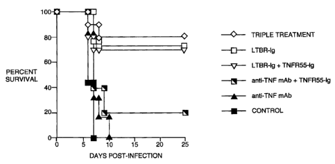
Figure 3 shows that blockage of the LT(3R signaling pathways significantly
improves survival rates among Clone 13 infected NZB mice. Mortality curves for
Clone 13 infected NZB mice treated as described are presented here. NZB mice
were
given 2.5 x 10⁶ pfu Cl 13 i.v. followed by two i.p. injections containing 250
g of TN3-
19.12 antibody in endotoxin free PBS on days 1 and day 4 post-
infection. Control mice were injected with the same volume of PBS lacking
antibody
on the same days. Mice were treated as described. For the triple treated
group, TNFR55-Ig and LTQR-Ig proteins were given on day 0 and day 3 post-
infection,
i.p., in 200 g amounts. Control mice were given human antibody used in the
synthesis
of these fusion proteins (AY1943-29) on the same days in identical amounts.
Mice
receiving LT(3R-Ig only were treated identically, except the TNFR55-Ig
injections were
omitted. Data was compiled from several experiments anti-TNF (TN3-19.12)
alone, n
16 for LTOR-Ig alone, n = 10 for the triple treatment group, (n=10 for the
triple
treatment group, n=22 for LTQR-Ig alone, n=10 for the LT(3R-Ig + TNFR55-Ig
group,
n=5 for the anti-TNF and TNFR55-Ig treated group, n=6 for anti-TNF (TN3-19.12)
alone, and n=25 for Control).
Figuu es 4A and 4B showtlhat blockage ofthe LMR pathway results in a decrease
in CD8
T cell function. Splenocytes from mice in different treatment groups were
harvested on
day 6 post-infection and stained with an Ld tetramer containing a NP 118 9mer
peptide
as previously described. Values given are adjusted for non-specific background
staining. To monitor interferon gamma production in response to the same
peptide,
cells were incubated for 5 hours at 37 C in the presence of NP118 at 0.1 g/ml
final
-3-
CA 02344049 2008-11-06
70850-119
concentration and IL-2. Values given here are adjusted for background levels
in the
absence of peptide. Spleenocytes from three niice treated with control human
Ig were
pooled as were those from two LT(3R-Ig mice (LT beta #2/3). All other results
are from
individual mice.
Figure 5 shows that depletion of CD8+ T cells, not CD4+ T cells, reverses the
lethal effects of LCMV- 13 infection in NZB mice. Mice were treated as
described for
depletion of cell populations in vivo. A mortality curve is presented for each
of the
treated groups (n = 4).
Detailed Description of the Invention
Definitions
In order to more clearly and concisely point out the subject matter of the
claimed invention, the following definitions are provided for specific terms
used in the
following written description and appended claims.
Lymphotoxin-beta (LT- beta ) is a member of the TNF family of ligands, which
also includes the ligands to the Fas, CD27, CD30, CD40, OX-40 and 4-1BB
receptors
(Smith et al., Cell, 76, pp. 959-62 (1994)). Signaling by several members of
the TNF
family-including TNF, LT- alpha, LT- beta and Fas-can induce tumor cell death
by
necrosis or apoptosis (programmed cell death). In non-tumorigenic cells, TNF
and
many of the TNF family ligand-receptor interactions influence immune system
development and responses to various immune challenges.
Lymphotoxin- beta (also called p33), has been identified on the surface of T
lymphocytes, T cell lines, B cell lines and lymphokine-activated killer (LAK)
cells. LT-
beta is the subject of applicants' co-pending international applications
PCT/US91/04588, published Jan. 9, 1992 as WO 92/00329; and PCT/US93/11669,
published Jun. 23, 1994 as WO 94/13808.
The LT- beta receptor, a member of the TNF family of receptors, specifically
binds to surface LT ligands. LT- beta -R binds LT heteromeric complexes
-4-
CA 02344049 2001-03-13
WO 00/21558 PCT/US99/23477
(predominantly LT- alpha l/ beta 2 and LT- alpha 2/ beta 1) but does not bind
TNF or
LT- alpha (Crowe et al., Science, 264, pp. 707-10 (1994)). Signaling by LT-
beta -R
may play a role in peripheral lymphoid organ development and in humoral immune
responses.
LT- beta -R mRNAs are found in human spleen, thymus and other major organs.
LT- beta -R expression patterns are similar to those reported for p55-TNF-R
except that
LT-beta -R is lacking on peripheral blood T cells and T cell lines.
The term "LT- beta - blocking agent" refers to an agent that can diminish
ligand
binding to LT- beta, cell surface LT- beta clustering or LT- beta signalling,
or that can
influence how the LT- beta signal is interpreted within the cell. Examples of
LT- beta -
blocking agents include anti-LT- beta, soluble LT- beta -R-Fc molecules, and
anti-LT-
alpha, anti-LT- alpha / beta and anti-LT- beta -R Abs. Preferably, the
antibodies do not
cross-react with the secreted form of LT-alpha.
The term "LT- beta - receptor blocking agent" refers to an agent that can
diminish ligand binding to LT- beta -R, cell surface LT- beta -R clustering or
LT- beta -
R signalling, or that can influence how the LT- beta -R signal is interpreted
within the
cell. Examples of LT- beta -R blocking agents include soluble LT- beta -R-Fc
molecules, and and anti-LT- beta -R Abs. Preferably, the antibodies do not
cross-react
with the secreted form of LT-alpha.
The term "anti-LT- beta receptor antibody" refers to any antibody that
specifically binds to at least one epitope of the LT- beta receptor.
The term "anti-LT antibody" refers to any antibody that specifically binds to
at
least one epitope of LT- alpha, LT- beta or a LT- alpha / beta complex.
The term "LT ligand" refers to a LT heteromeric complex or derivative thereof
that can specifically bind to the LT- beta receptor.
The term "LT- beta -R signaling" refers to molecular reactions associated with
the LT- beta -R pathway and subsequent molecular reactions which result
therefrom.
-5-
CA 02344049 2001-03-13
WO 00/21558 PCT/US99/23477
The term "LT- beta -R ligand binding domain" refers to the portion or portions
of the LT- beta -R that are involved in specific recognition of and
interaction with a LT
ligand.
The terms "LT- alpha / beta heteromeric complex" and "LT heteromeric
complex" refer to a stable association between at least one LT- alpha and one
or more
LT-beta subunits, including soluble, mutant, altered and chimeric forms of one
or more
of the subunits. The subunits can associate through electrostatic, van der
Waals, or
covalent interactions. Preferably, the LT- alpha /62 heteromeric complex has
at least
two adjacent LT- beta subunits and lacks adjacent LT- alpha subunits. When the
LT-
alpha / beta heteromeric complex serves as a LT- beta -R activating agent in a
cell
growth assay, the complex is preferably soluble and has the stoichiometry LT-
alpha 1/
beta 2.
Soluble LT- alpha /62 heteromeric complexes lack a transmembrane domain
and can be secreted by an appropriate host cell which has been engineered to
express
LT- alpha and/or LT- beta subunits (Crowe et al., J. Immunol. Methods, 168,
pp. 79-89
(1994)).
The terms "surface LT- alpha /62 complex" and "surface LT complex" refer to a
complex comprising LT- alpha and membrane-bound LT- beta subunits-including
mutant, altered and chimeric forms of one or more of the subunits-which is
displayed
on the cell surface. "Surface LT ligand" refers to a surface LT complex or
derivative
thereof that can specifically bind to the LT- beta receptor.
An "effective amount" is an amount sufficient to effect beneficial or desired
clinical results. An effective amount can be administered in one or more
administrations. For purposes of this invention, an effective amount of an
agent which
blocks the binding of lymphotoxin-B to its receptor is an amount of the agent
that is
sufficient to ameliorate, stabilize, or delay the development of a viral
response. In
particular, an agent that is sufficient to ameliorate, stabilize, or delay the
development
of viral-induced systemic shock and respiratory distress. Detection and
measurement of
these indicators of efficacy are known to those of skill in the art.
-6-
CA 02344049 2001-03-13
WO 00/21558 PCT/US99/23477
An "individual" refers to vertebrates, particularly members of a mammalian
species, and includes but is not limited to domestic animals, sports animals,
and
primates, including humans.
"functional equivalent" of an amino acid residue is (i) an amino acid having
similar reactive properties as the amino acid residue that was replaced by the
functional
equivalent; (ii) an amino acid of an antagonist of the invention, the amino
acid having
similar properties as the amino acid residue that was replaced by the
functional
equivalent; (iii) a non-amino acid molecule having similar properties as the
amino acid
residue that was replaced by the functional equivalent.
A first polynucleotide encoding a proteinaceous antagonist of the invention is
"functionally equivalent" compared with a second polynucleotide encoding the
antagonist protein if it satisfies at least one of the following conditions:
(a): the "functional equivalent" is a first polynucleotide that hybridizes to
the
second polynucleotide under standard hybridization conditions and/or is
degenerate to
the first polynucleotide sequence. Most preferably, it encodes a mutant
protein having
the activity of an integrin antagonist protein;
(b) the "functional equivalent" is a first polynucleotide that codes on
expression
for an amino acid sequence encoded by the second polynucleotide.
"functional equivalent" of an amino acid residue is (i) an amino acid having
similar
reactive properties as the amino acid residue that was replaced by the
functional
equivalent; (ii) an amino acid of an antagonist of the invention, the amino
acid having
similar properties as the amino acid residue that was replaced by the
functional
equivalent; (iii) a non-amino acid molecule having similar properties as the
amino acid
residue that was replaced by the functional equivalent.
A first polynucleotide encoding a proteinaceous antagonist of the invention is
"functionally equivalent" compared with a second polynucleotide encoding the
antagonist protein if it satisfies at least one of the following conditions:
(a): the "functional equivalent" is a first polynucleotide that hybridizes to
the
second polynucleotide under standard hybridization conditions and/or is
degenerate to
the first polynucleotide sequence. Most preferably, it encodes a mutant
protein having
the activity of an integrin antagonist protein;
-7-
CA 02344049 2001-03-13
WO 00/21558 PCT/US99/23477
(b) the "functional equivalent" is a first polynucleotide that codes on
expression
for an amino acid sequence encoded by the second polynucleotide.
The LT-B blocking agents used in the invention include, but are not limited
to,
the agents listed herein as well as their functional equivalents. As used
herein, the term
"functional equivalent" therefore refers to a LT-B blocking agent or a
polynucleotide
encoding the LT-B blocking agent that has the same or an improved beneficial
effect on
the recipient as the LT-B blocking agent of which it is deemed a functional
equivalent.
As will be appreciated by one of ordinary skill in the art, a functionally
equivalent
protein can be produced by recombinant techniques, e.g., by expressing a
"functionally
equivalent DNA". Accordingly, the instant invention embraces LT-B blocking
agent
encoded by naturally-occurring DNAs, as well as by non-naturally-occurring
DNAs
which encode the same protein as encoded by the naturally-occurring DNA. Due
to the
degeneracy of the nucleotide coding sequences, other polynucleotides may be
used to
encode LT-B blocking agents. These include all, or portions of the above
sequences
which are altered by the substitution of different codons that encode the same
amino
acid residue within the sequence, thus producing a silent change. Such altered
sequences are regarded as equivalents of these sequences. For example, Phe (F)
is
coded for by two codons, TTC or TTT, Tyr (Y) is coded for by TAC or TAT and
His
(H) is coded for by CAC or CAT. On the other hand, Trp (W) is coded for by a
single
codon, TGG. Accordingly, it will be appreciated that for a given DNA sequence
encoding a particular integrin there will be many DNA degenerate sequences
that will
code for it. These degenerate DNA sequences are considered within the scope of
this
invention.
The term "fusion" or "fusion protein" refers to a co-linear, covalent linkage
of
two or more proteins or fragments thereof via their individual peptide
backbones, most
preferably through genetic expression of a polynucleotide molecule encoding
those
proteins. It is preferred that the proteins or fragments thereof are from
different sources
so that this type of fusion protein is called a "chimeric" molecule. Thus,
preferred
fusion proteins are chimeric proteins that include a LT-B blocking agent or
fragment
covalently linked to a second moiety that is not a LT-B blocking agent.
Preferred fusion
proteins of the invention may include portions of intact antibodies that
retain antigen-
-8-
CA 02344049 2008-11-06
70850-119
binding specificity, for example, Fab fragments, Fab' fragments, F(ab' )2
fragments,
F(v) fragments, heavy chain monomers or dimers, light chain monomers or
dimers,
dimers consisting of one heavy and one light chain, and the like.
The most preferred fusion proteins are chimeric and comprise a LT-B blocking
agent moiety fused or otherwise linked to all or part of the hinge and
constant regions of
an immunoglobulin light chain, heavy chain, or both. Thus, this invention
features a
molecule which includes: (1) a LT-B blocking agent moiety, (2) a second
peptide, e.g.,
one which increases solubility or in vivo life time of the LT-B blocking agent
moiety,
e.g., a member of the immunoglobulin super family or fragment or portion
thereof, e.g.,
a portion or a fragment of IgG, e.g., the human IgGI heavy chain constant
region, e.g.,
CH2, CH3, and hinge regions. Specifically, a "LT-B or LT-B-R/Ig fusion" is a
protein
comprising a biologically active LT-B blocking of the invention (e.g. a
soluble LT-B-
R, or a biologically active fragment thereof linked to an N-terminus of an
immunoglobulin chain wherein a portion of the N-terminus of the immunoglobulin
is
replaced with the LT-B blocking agent. A species of LT-B or LT-B-R/Ig fusion
is an
"LT-B-R/Fc fusion" which is a protein comprising an LT-B-R of the invention
linked to
at least a part of the constant domain of an immunoglobulin. A preferred Fc
fusion
comprises a LT-B blocking agent of the invention linked to a fragment of an
antibody
containing the C terminal domain of the heavy immunoglobulin chains.
"standard hybridization conditions"- salt and temperature conditions
substantially equivalent to 0.5 X SSC to about 5 X SSC and 65 C for both
hybridization and wash. The term "standard hybridization conditions" as used
herein is
therefore an operational definition and encompasses a range of hybridization
conditions. Higher stringency conditions may, for example, include hybridizing
with
~
plaque screen buffer (0.2% polyvinylpyrrolidone, 0.2% Ficoll 400; 0.2% bovine
serum
albumin, 50 mM Tris-HCI (pH 7.5); 1 M NaCl; 0.1% sodium pyrophosphate; 1%
SDS); 10% dextran sulfate, and 100 g/ml denatured, sonicated salmon sperm DNA
at
65 C for 12-20 hours, and washing with 75 mM NaCI/7.5 mM sodium citrate (0.5
x
SSC)/1% SDS at 65 C. Lower stringency conditions may, for example, include
hybridizing with plaque screen buffer, 10% dextran sulfate and 110 g/ml
denatured,
*Trade-mark.
-9-
CA 02344049 2001-03-13
WO 00/21558 PCTIUS99/23477
sonicated salmon sperm DNA at 55 C for 12-20 hours, and washing with 300 mM
NaCI/30mM sodium citrate (2.0 X SSC)/1% SDS at 55 C. See also Current
Protocols in Molecular Biology, John Wiley & Sons, Inc. New York, Sections
6.3.1-
6.3.6, (1989).
A "therapeutic composition" as used herein is defined as comprising the
proteins
of the invention and other biologically compatible ingredients. The
therapeutic
composition may contain excipients such as water, minerals and carriers such
as
protein.
II. Description of the Preferred Embodiments
The present invention depends in part upon the discovery that LT-B blocking
agents can induce an antiviral response in an individual. It was found that
treating an
individual infected with a virus can greatly increase the survival rate of the
individual.
Specifically, it was shown that treating LCMV-13 infected NZB mice with a LT-B
blocking agent, such as LT(3R-Ig fusion protein increased their survival rate
73%.
Currently treatment for Ebola, Dengue, SNV and other viruses mentioned herein
is preventative via education on transmission of disease. Vaccines do not
exist for
these highly pathogenic viruses. Ribavirin, a guanidine analog, has been
employed as a
generic antiviral drug to several of these infections with reproducible
success only
documented in treatment of Lassa Fever when used early on in the illness (M.D.
Lacy
and R.A. Smego, Adv. Ped. Inf. Dis., 12, 21 (1997). Our data indicate that
some of the
pathology associated with these viruses may be immune mediated. Blockade of
the LT
system could greatly increase the chance for survival by transiently reducing
virus
specific CD8 T cells numbers and their functionality. Clinical trials that
employ several
means of blocking the TNFa pathway are already underway for the treatment of
several
ailments (H.I. Pass, D. Mew, H.A. Pass, et al., Chest Surg. Clin.1V. Amer. 5,
73
(1995). We believe the LT(3R-Ig treatment should be considered for further
testing in
animal models for eventual use in human trials involving patients with acute,
rapidly
progressive viral infections involving shock and/or pulmonary distress.
-10-
CA 02344049 2004-10-08
70850-119
LT-B Blocking Ago_ts
In one embodiment of this invention, the LT- beta blocking agent comprises an
antibody (Ab) directed against LT- beta that inhibits LT- beta signaling.
Preferably,
the anti-LT- beta Ab is a monoclonal antibody (mAb). Inhibitory anti-LT- beta
Abs and
other LT- beta blocking agents can be identified using screening methods that
detect
the ability of one or more agents to bind to a LT ligand, or to inhibit the
effects of LT-
beta signalling on cells.
In another embodiment of this invention, the LT- beta blocking agent comprises
an LT-beta receptor (LT-B-R) blocking agent. In a preferred embodiment, the LT-
B-R
io blocking agent is an antibody (Ab) directed against LT- beta -R that
inhibits LT- beta -
R signaling. Preferably, the anti-LT- beta -R Ab is a monoclonal antibody
(mAb). One
such inhibitory anti-LT- beta-R mAb is BDA8 mAb. Inhibitory anti-LT- beta -R
Abs
and other LT- beta -R blocking agents can be identified using screening
methods that
detect the ability of one or more agents either to bind to the LT- beta -R or
LT ligand, or
to inhibit the effects of LT-beta -R signalling on cells.
One screening method makes use of the cytotoxic effects of LT- beta -R
signalling on tumor cells bearing the LT- beta -R. Tumor cells are exposed to
one or
more LT- beta -R activating agents to induce LT- beta -R signalling. LT- beta -
R
activating agents include LT- alpha /62 heteromeric complexes (preferably
soluble LT-
alpha 1/ beta 2) in the presence of IFN- gamma, or an activating anti-LT- beta
-R Ab
(see below; also described in PCT publication
WO 96/22788 Al).
Antibodies and other agents that can block LT- beta -R signalling are selected
based on their ability to inhibit the cytotoxic effect of LT- beta -R
signalling on tumor
cells in the following assay:
1) Tumor cells such as HT29 cells are cultured for three to four days in a
series of tissue
culture wells containing media and at least one LT- beta -R activating agent
in the
presence or absence of serial dilutions of the agent being tested;
-11-
CA 02344049 2001-03-13
WO 00/21558 PCT/US99/23477
2) A vital dye stain which measures mitochondrial function such as MTT is
added to
the tumor cell mixture and reacted for several hours;
3) The optical density of the mixture in each well is quantitated at 550 nm
wavelength
light (OD 550). The OD 550 is proportional to the number of tumor cells
remaining in
the presence of the LT- beta -R activating agent and the test LT- beta -R
blocking agent
in each well. An agent or combination of agents that can reduce LT- beta -R-
activated
tumor cell cytotoxicity by at least 20% in this assay is a LT- beta -R
blocking agent
within the scope of this invention.
Any agent or combination of agents that activate LT- beta -R signalling can be
used in the above assay to identify LT- beta -R blocking agents. LT- beta -R
activating
agents that induce LT- beta -R signalling (such as activating anti-LT- beta -R
mAbs)
can be selected based on their ability-alone or in combination with other
agents-to
potentiate tumor cell cytotoxicity using the tumor cell assay described above.
Another method for selecting an LT- beta -R blocking agent is to monitor the
ability of the putative agent to directly interfere with LT ligand-receptor
binding. An
agent or combination of agents that can block ligand-receptor binding by at
least 20% is
an LT- beta -R blocking agent within the scope of this invention.
Any of a number of assays that measure the strength of ligand-receptor binding
can be used to perform competition assays with putative LT- beta -R blocking
agents.
The strength of the binding between a receptor and ligand can be measured
using an
enzyme-linked immunoadsorption assay (ELISA) or a radio-immunoassay (RIA).
Specific binding may also be measured by fluorescently labelling antibody-
antigen
complexes and performing fluorescence-activated cell sorting (FACS) analysis,
or by
performing other such immunodetection methods, all of which are techniques
well
known in the art.
The ligand-receptor binding interaction may also be measured with the BlAcore
TM instrument (Pharmacia Biosensor) which exploits plasmon resonance detection
(Zhou et al., Biochemistry, 32, pp. 8193-98 (1993); Faegerstram and
O'Shannessy,
-12-
CA 02344049 2008-11-06
70850-119
"Surface plasmon resonance detection in affinity technologies", in Handbook of
Affinity Chromatography, pp. 229-52, Marcel Dekker,lnc., New York (1993)).
The BlAcore TM technology allows one to bind receptor to a gold surface and
to flow ligand over it. Plasmon resonance detection gives direct quantitation
of the
amount of mass bound to the surface in real time. This technique yields both
on and off
rate constants and thus a ligand-receptor dissociation constant and affinity
constant can
be directly determined in the presence and absence of the putative LT- beta -R
blocking
agent.
With any of these or other techniques for measuring receptor-ligand
interactions, one can evaluate the ability of a LT- beta -R blocking agent,
alone or in
combination with other agents, to inhibit binding of surface or soluble LT
ligands to
surface or soluble LT- beta -R molecules. Such assays may also be used to test
LT- beta
-R blocking agents or derivatives of such agents (e.g. fusions, chimeras,
mutants, and
chemically altered forms)-alone or in combination-to optimize the ability of
that altered
agent to block LT- beta -R activation.
The LT- beta -R blocking agents in one embodiment of this invention comprise
soluble LT- beta receptor molecules. The sequence of the extracellular portion
of the
human LT- beta -R, which encodes the ligand binding domain is shown in Figure
1 of
U.S. patent no. 5,925,351. Using the sequence
information in Figure 1 of U.S. patent no. 5,925,351 and recombinant DNA
techniques
well known in the art, functional fragments encoding the LT- beta -R ligand
binding
domain can be cloned into a vector and expressed in an appropriate host to
produce a
soluble LT- beta -R molecule. Soluble LT- beta -R molecules that can compete
with
native LT- beta receptors for LT ligand binding according to the assays
described herein
are selected as LT- beta -R blocking agents.
A soluble LT- beta receptor comprising amino acid sequences selected from
those shown in Figure 1 of U.S. patent no. 5,925,351 may be attached to one or
more
heterologous protein domains ("fusion domain") to increase the in vivo
stability of the
receptor fusion protein, or to modulate its biological activity or
localization. Preferably,
-13-
CA 02344049 2001-03-13
WO 00/21558 PCTIUS99/23477
stable plasma proteins-which typically have a half-life greater than 20 hours
in the
circulation-are used to construct the receptor fusion proteins. Such plasma
proteins
include but are not limited to: immunoglobulins, serum albumin, lipoproteins,
apolipoproteins and transferrin. Sequences that can target the soluble LT-
beta -R
molecule to a particular cell or tissue type may also be attached to the LT-
beta -R
ligand binding domain to create a specifically-localized soluble LT- beta -R
fusion
protein. All or a functional portion of the LT- beta -R extracellular region
(FIG. 1 of
US Pat. no 5,925,351) comprising the LT- beta -R ligand binding domain may be
fused
to an immunoglobulin constant region like the Fc domain of a human IgG 1 heavy
chain
(Browning et al., J. Immunol., 154, pp. 33-46 (1995)). Soluble-receptor-IgG
fusion
proteins are common immunological reagents and methods for their construction
are
known in the art (see e.g., U.S. Pat. No. 5,225,538). A functional LT- beta -R
ligand
binding domain may be fused to an immunoglobulin (Ig) Fc domain derived from
an
immunoglobulin class or subclass other than IgGI. The Fc domains of antibodies
belonging to different Ig classes or subclasses can activate diverse secondary
effector
functions. Activation occurs when the Fc domain is bound by a cognate Fc
receptor.
Secondary effector functions include the ability to activate the complement
system, to
cross the placenta, and to bind various microbial proteins. The properties of
the
different classes and subclasses of immunoglobulins are described in Roitt et
al.,
Immunology, p. 4.8 (Mosby-Year Book Europe Ltd., 3d ed. 1993). The complement
enzyme cascade can be activated by the Fc domains of antigen-bound IgG 1, IgG3
and
IgM antibodies. The Fc domain of IgG2 appears to be less effective, and the Fc
domains
of IgG4, IgA, IgD and IgE are ineffective at activating complement. Thus one
can select
a Fc domain based on whether its associated secondary effector functions are
desirable
for the particular immune response or disease being treated with the LT- beta -
R-Fc
fusion protein. If it would be advantageous to harm or kill the LT ligand-
bearing target
cell, one could select an especially active Fc domain (IgGl ) to make the LT-
beta -R-
Fc fusion protein. Alternatively, if it would be desirable to target theLT-
beta -R-Fc
fusion to a cell without triggering the complement system, an inactive IgG4 Fc
domain
could be selected.
- 14-
CA 02344049 2008-11-06
70850-119
Mutations in Fc domains that reduce or eliminate binding to Fc receptors and
complement activation have been described (S. Morrison, Annu. Rev. Immunol.,
10,
pp. 239-65 (1992)). These or other mutations can be used, alone or in
combination, to
optimize the activity of the Fc domain used to construct the LT-beta -R-Fc
fusion
protein.
The production of a soluble human LT- beta -R fusion protein comprising
ligand binding sequences fused to a human immunoglobulin Fc domain (hLT- beta -
R-
Fc) is described in Example I of U.S. patent no. 5,925,351.
One CHO line made according to Example 1 that secretes hLT- beta -R-Fc is
called "hLT beta ;R-hGl CHO#14". A sample of this line was deposited on Jul.
21,
1995 with the American Type Culture Collection (ATCC) (Rockville, Md.)
according
to the provisions of the Budapest Treaty and was assigned the ATCC accession
number
CRL 11965.
The production of a soluble murine LT- beta -R fusion molecule (mLT- beta -R-
Fc) is described in Example 2 of U.S. patent no. 5,925,35 ;.
A CHO line made according to Example 2 of U.S. patent no. 5,925,351 that
secretes mLT- beta -R-Fc is called "mLT beta ;R-hGl CHO#1.3.BB". A sample of
this
line was deposited on Jul. 21, 1995 with the American Type Culture Collection
(ATCC) (Rockville, Md.) according to the provisions of the Budapest Treaty and
was
assigned the ATCC accession number CRL 11964.
Different amino acid residues forming the junction point of the receptor-Ig
fusion protein may alter the structure, stability and ultimate biological
activity of the
soluble LT- beta receptor fusion protein. One or more amino acids may be added
to the
C-terminus of the selected LT- beta -R fragment to modify the junction point
with the
selected fusion domain.
The N-terminus of the LT- beta -R fusion protein may also be varied by
changing the position at which the selected LT- beta -R DNA fragment is
cleaved at its
5' end for insertion into the recombinant expression vector. The stability and
activity of
each LT- beta -R fusion protein may be tested and optimized using routine
-15-
CA 02344049 2004-10-08
70850-119
experimentation and the assays for selecting LT- beta -R blocking agents
described
herein.
Using the LT- beta -R ligand binding domain sequences within the extracellular
domain shown in FIG. 1 of US 5,925,351, amino acid
sequence variants may also be constructed to
modify the affinity of the soluble LT- beta receptor or fusion protein for LT
ligand. The
soluble LT- beta -R molecules of this invention can compete for surface LT
ligand
binding with endogenous cell surface LT- beta receptors. It is envisioned that
any
soluble molecule comprising a LT- beta -R ligand binding domain that can
compete
with cell surface LT- beta receptors for LT ligand binding is a LT- beta -R
blocking
agent that falls within the scope of the present invention.
In another embodiment of this invention, antibodies directed against the human
LT- beta receptor (anti-LT- beta -R Abs) function as LT- beta -R blocking
agents for
use in treating conditions that place individuals, including human, in, or at
risk of, viral-
induced systemic shock and respiratory distress. The anti-LT- beta -R Abs of
this
invention can be polyclonal or monoclonal (mAbs) and can be modified to
optimize
their ability to block LT- beta -R signalling, their in vivo bioavailability,
stability, or
other desired traits.
Polyclonal antibody sera directed against the human LT- beta receptor
areprepared using conventional techniques by injecting animals such as goats,
rabbits,
rats, hamsters or mice subcutaneously with a human LT- beta receptor-Fc fusion
protein
(Example 1 of US pat. no. 5,925,351) in complete Freund's adjuvant, followed
by
booster intraperitoneal or subcutaneous injection in incomplete Freund's.
Polyclonal
antisera containing the desired antibodies directed against the LT- beta
receptor are
screened by conventional immunological procedures.
Mouse monoclonal antibodies (mAbs) directed against a human LT- beta
receptor-Fc fusion protein are prepared as described in U.S. patent number
5,925,351,
Example 5. A hybridoma cell line (BD.A8.AB9) which produces the mouse anti-
human
LT- beta -R mAb BDA8 was deposited on Jan. 12, 1995 with the American Type
Culture Collection (ATCC) (10801 University Boulevard, Manassas, Va. 20110-
2209)
-16-
CA 02344049 2001-03-13
WO 00/21558 PCT/US99/23477
according to the provisions of the Budapest Treaty, and was assigned the ATCC
accession number HB 11798.
Various forms of anti-LT- beta -R antibodies can also be made using standard
recombinant DNA techniques (Winter and Milstein, Nature, 349, pp. 293-99
(1991)).
For example, "chimeric" antibodies can be constructed in which the antigen
binding
domain from an animal antibody is linked to a human constant domain (e.g.
Cabilly et
al., U.S. Pat. No. 4,816,567; Morrison et al., Proc. Natl. Acad. Sci. U.S.A.,
81, pp.
6851-55 (1984)). Chimeric antibodies reduce the observed immunogenic responses
elicited by animal antibodies when used in human clinical treatments. In
addition,
recombinant "humanized antibodies" which recognize the LT- beta -R can be
synthesized. Humanized antibodies are chimeras comprising mostly human IgG
sequences into which the regions responsible for specific antigen-binding have
been
inserted (e.g. WO 94/04679). Animals are immunized with the desired antigen,
the
corresponding antibodies are isolated, and the portion of the variable region
sequences
responsible for specific antigen binding are removed. The animal-derived
antigen
binding regions are then cloned into the appropriate position of human
antibody genes
in which the antigen binding regions have been deleted. Humanized antibodies
minimize the use of heterologous (inter-species) sequences in human
antibodies, and
are less likely to elicit immune responses in the treated subject.
Construction of different classes of recombinant anti-LT- beta -R antibodies
can
also be accomplished by making chimeric or humanized antibodies comprising the
anti-
LT- beta -R variable domains and human constant domains (CH1, CH2, CH3)
isolated
from different classes of immunoglobulins. For example, anti-LT- beta -R IgM
antibodies with increased antigen binding site valencies can be recombinantly
produced
by cloning the antigen binding site into vectors carrying the human mu chain
constant
regions (Arulanandam et al., J. Exp. Med., 177, pp. 1439-50 (1993); Lane et
al., Eur. J.
Immunol., 22, pp. 2573-78 (1993); Traunecker et al., Nature, 339, pp. 68-70
(1989)). In
addition, standard recombinant DNA techniques can be used to alter the binding
affinities of recombinant antibodies with their antigens by altering amino
acid residues
in the vicinity of the antigen binding sites. The antigen binding affinity of
a humanized
-17-
CA 02344049 2001-03-13
WO 00/21558 PCT/US99/23477
antibody can be increased by mutagenesis based on molecular modeling (Queen et
al.,
Proc. Natl. Acad. Sci. U.S.A., 86, pp. 10029-33 (1989); WO 94/04679).
It may be desirable to increase or to decrease the affinity of anti-LT- beta -
R
Abs for the LT- beta -R depending on the targeted tissue type or the
particular treatment
schedule envisioned. For example, it may be advantageous to treat a patient
with
constant levels of anti-LT- beta -R Abs with reduced ability to signal through
the LT-
beta pathway for semi-prophylactic treatments. Likewise, inhibitory anti-LT-
beta -R
Abs with increased affinity for the LT- beta -R may be advantageous for short-
term
treatments.
By testing other antibodies directed against the human LT- beta receptor, it
is
expected that additional anti-LT- beta -R antibodies that function as LT- beta
-R
blocking agents in humans can be identified for treating conditions that place
individuals, including human, in, or at risk of, viral-induced systemic shock
and
respiratory distress using routine experimentation and the assays described
herein.
Another preferred embodiment of this invention involves compositions and
methods which comprise antibodies directed against LT' ligand that function as
LT- beta
-R blocking agents. As described above for the anti-LT- beta -R Abs, anti-LT
ligand
antibodies that function as LT- beta -R blocking agents can be polyclonal or
monoclonal, and can be modified according to routine procedures to modulate
their
antigen binding properties and their immunogenicity. The anti-LT antibodies of
this
invention can be raised against either one of the two LT subunits
individually, including
soluble, mutant, altered and chimeric forms of the LT subunit. If LT subunits
are used
as the antigen, preferably they are LT- beta subunits. If LT- alpha subunits
are used, it is
preferred that the resulting anti-LT- alpha antibodies bind to surface LT
ligand and do
not cross-react with secreted LT- alpha or modulate TNF-R activity (according
to the
assays described in Example 3 of US patent no. 5, 925,351).
Alternatively, antibodies directed against a homomeric (LT- beta ) or a
heteromeric (LT- alpha /62 ) complex comprising one or more LT subunits can be
raised and screened for activity as LT- beta -R blocking agents. Preferably,
LT-alpha 1/
-18-
CA 02344049 2001-03-13
WO 00/21558 PCT/US99/23477
beta 2 complexes are used as the antigen. As discussed above, it is preferred
that the
resulting anti-LT- alpha 1/ beta 2 antibodies bind to surfaceLT ligand without
binding
to secreted LT- alpha and without affecting TNF-R activity.
The production of polyclonal anti-human LT- alpha antibodies is described in
applicants' co-pending application (WO 94/13808). Monoclonal anti-LT- alpha
and
anti-LT- beta antibodies have also been described (Browning et al., J.
Immunol., 54, pp.
33-46 (1995)). Mouse anti-human LT- beta mAbs were prepared as described in
Example 6 of U.S patent number 5,925,351. Hybridoma cell line (B9.C9. 1) which
produces the mouse anti-human LT- beta -R mAb B9 was deposited on Jul. 21,
1995
with the American Type Culture Collection (ATCC) (10801 University Boulevard,
Manassas, Va. 20110-2209) according to the provisions of the Budapest Treaty,
and
was assigned the ATCC accession number 11962.
Monoclonal hamster anti-mouse LT- alpha /62 antibodies were prepared as
described in Example 7 of U.S patent number 5,925,351. A hybridoma cell line
(BB.F6. 1) which produces the hamster anti-mouse LT- alpha /62 mAb BB.F6 was
deposited on Jul. 21, 1995 with the American Type Culture Collection (ATCC)
(10801
University Boulevard, Manassas, Va. 20110-2209) according to the provisions of
the
Budapest Treaty, and was assigned the ATCC accession number MB 11963.
A fluorescence-activated cell sorting (FACS) assay was developed to screen for
antibodies directed against LT subunits and LT complexes that can act as LT-
beta -R
blocking agents as described in Examples 6 and 7 of U.S. patent number
5,925,351. In
this assay, soluble human LT- beta -R-Fc fusion protein is added to PMA-
activated II-
23 cells-which express surface LT complexes (Browning et al., J. Immunol.,
154, pp.
33-46 (1995))-in the presence of increasing amounts of the test antibody. An
antibody
that can inhibit LT- beta receptor-ligand interaction by at least 20% is
selected as a LT-
beta -R blocking agent.
Using a LT- alpha / beta complex rather than a LT subunit as an antigen to
immunize an animal may lead to more efficient immunization, or may result in
antibodies having higher affinities for surface LT ligand. It is conceivable
that by
-19-
CA 02344049 2001-03-13
WO 00/21558 PCT/US99/23477
immunizing with the LT- alpha /62 complex, antibodies which recognize amino
acid
residues on both the LT- alpha and the LT- beta subunits (e.g., residues that
form an
LT- alpha /62 cleft) can be isolated. By testing antibodies directed against
human LT-
alpha /62 heteromeric complexes, it is expected that additional anti-LT
antibodies that
function as LT- beta -R blocking agents in humans can be identified using
routine
experimentation and the assays described herein.
Administration
The compositions described herein will be administered at an effective dose in
methods for treating viral-induced systemic shock and respiratory distress in
an
individual. Determination of a preferred pharmaceutical formulation and a
therapeutically efficient dose regiment for a given application is well within
the skill of
the art taking intoconsideration, for example, the condition and weight of the
patient,
the extent of desired treatment and the tolerance of the patient for the
treatment. Doses
of about 1 mg/kg of a soluble LT- beta -R are expected to be suitable starting
points for
optimizing treatment doses.
Determination of a therapeutically effective dose can also be assessed by
performing in vitro experiments that measure the concentration of the LT- beta
-R
blocking agent required to coat target cells (LT- beta -R or LT ligand-
positive cells
depending on the blocking agent) for 1 to 14 days. The receptor-ligand binding
assays
described herein can be used to monitor the cell coating reaction. LT- beta -R
or LT
ligand-positive cells can be separated from activated lymphocyte populations
using
FACS. Based on the results of these in vitro binding assays, a range of
suitable LT- beta
-R blocking agent concentrations can be selected to test in animals according
to the
assays described herein.
Administration of the soluble LT- beta -R molecules, anti-LT ligand and anti-
LT- beta -R Abs of this invention, alone or in combination, including isolated
and
purified forms of the antibodies or complexes, their salts or pharmaceutically
acceptable derivatives thereof, may be accomplished using any of the
conventionally
accepted modes of administration of agents which exhibit immunosuppressive
activity.
-20-
CA 02344049 2001-03-13
WO 00/21558 PCT/tlS99/23477
The pharmaceutical compositions used in these therapies may also be in a
variety of forms. These include, for example, solid, semi-solid and liquid
dosage forms
such as tablets, pills, powders, liquid solutions or suspensions,
suppositories, and
injectable and infusible solutions. The preferred form depends on the intended
mode of
administration and therapeutic application.
Modes of administration may include oral, parenteral, subcutaneous,
intravenous,intralesional or topical administration. The soluble LT- beta -R
molecules,
anti-LT ligand and anti-LT- beta -R Abs of this invention may, for example, be
placed
into sterile, isotonic formulations with or without cofactors which stimulate
uptake or
stability. The formulation is preferably liquid, or may be lyophilized powder.
For
example, the soluble LT-beta -R molecules, anti-LT ligand and anti-LT- beta -R
Abs of
this invention may be diluted with a formulation buffer comprising 5.0 mg/ml
citric
acid monohydrate, 2.7 mg/ml trisodium citrate, 41 mg/ml mannitol, 1 mg/ml
glycine
and 1 mg/ml polysorbate 20. This solution can be lyophilized, stored under
refrigeration
and reconstituted prior to administration with sterile Water-For-Injection
(USP).
The compositions also will preferably include conventional pharmaceutically
acceptable carriers well known in the art (see for example Remington's
Phannaceutical
Sciences, 16th Edition, 1980, Mac Publishing Company). Such pharmaceutically
acceptable carriers may include other medicinal agents, carriers, genetic
carriers,
adjuvants, excipients, etc., such as human serum albumin or plasma
preparations. The
compositions are preferably in the form of a unit dose and will usually be
administered
one or more times a day.
The pharmaceutical compositions of this invention may also be administered
using microspheres, liposomes, other microparticulate delivery systems or
sustained
release formulations placed in, near, or otherwise in communication with
affected
tissues or the bloodstream. Suitable examples of sustained releasecarriers
include
semipermeable polymer matrices in the form of shaped articles such as
suppositories or
microcapsules. Implantable or microcapsular sustained release matrices include
polylactides (U.S. Pat. No 3,773,319; EP 58,481), copolymers of L-glutamic
acid and
ethyl-L-glutamate (Sidman et al., Biopolymers, 22, pp. 547-56 (1985)); poly(2-
-21-
CA 02344049 2001-03-13
WO 00/21558 PCT/US99/23477
hydroxyethyl-methacrylate) or ethylene vinyl acetate (Langer et al., J.
Biomed. Mater.
Res., 15, pp. 167-277 (1981); Langer, Chem. Tech., 12, pp. 98-105 (1982)).
Liposomes containing soluble LT- beta -R molecules, anti-LT ligand and anti-
LT- beta -R Abs of this invention, alone or in combination, can be prepared by
well-
known methods (See, e.g. DE 3,218,121; Epstein et al., Proc. Natl. Acad. Sci.
U.S.A.,
82, pp. 3688-92 (1985); Hwang et al., Proc. Natl. Acad. Sci. U.S.A., 77, pp.
4030-34
(1980); U.S. Pat. Nos. 4,485,045 and 4,544,545). Ordinarily the liposomes are
of the
small (about 200-800 Angstroms) unilamellar type in which the lipid content is
greater
than about 30 mol. % cholesterol. The proportion of cholesterol is selected to
control
the optimal rate of soluble LT- beta -R molecule, anti-LT ligand and anti-LT-
beta -R
Ab release.
The soluble LT- beta -R molecules, anti-LT ligand and anti-LT- beta -R Abs
ofthis invention may also be attached to liposomes containing other LT- beta -
R
blocking agents, immunosuppressive agents or cytokines to modulate the LT-
beta -R
blocking activity. Attachment of LT- beta -R molecules, anti-LT ligand and
anti-LT-
beta -R Abs to liposomes may be accomplished by any known cross-linking agent
such
as heterobifunctional cross-linking agents that have been widely used to
couple toxins
or chemotherapeutic agents to antibodies for targeted delivery.conjugation to
liposomes
can also be accomplished using the carbohydrate-directed cross-linking reagent
4-(4-
maleimidophenyl) butyric acid hydrazide (MPBH) (Duzgunes et al., J. Cell.
Biochem.
Abst. Suppl. 16E 77 (1992)).
The LT- beta -R blocking agents of the compositions and methods of this
invention can be modified to obtain a desirable level of LT- beta -R
signalling
depending on the condition, disorder or disease being treated. It is
envisioned that the
absolute level of LT- beta -R signalling can be fine-tuned by manipulating the
concentration and the affinities of the LT- beta -R blocking agents for their
respective
molecular targets. For example, in one embodiment of this invention,
compositions
comprising soluble LT- beta -R molecules are administered to a subject. The
soluble
LT- beta receptor can effectively compete with cell surface LT- beta receptors
for
binding surface LT ligands. The ability to compete with surface LT ligands
depends on
-22-
CA 02344049 2001-03-13
WO 00/21558 PCT/US99/23477
the relative concentrations of the soluble and the cell surface LT- beta -R
molecules,
and on their relative affinities for ligand binding.
Soluble LT- beta -R molecules harboring mutations that increase or decrease
the
binding affinity of that mutant soluble LT- beta -R with surface LT ligand can
be made
using standard recombinant DNA techniques well known to those of skill in the
art.
Large numbers of molecules with site-directed or random mutations can be
tested for
their ability to act as LT- beta -R blocking agents using routine
experimentation and the
techniques described herein. Similarly, in another embodiment of this
invention,
antibodies directed against either the LT- beta receptor or one or more of the
LT ligand
subunits function as LT- beta -R blocking agents. The ability for these
antibodies to
block LT- beta receptor signalling can be modified by mutation, chemical
modification
or by other methods that can vary the effective concentration or activity of
the antibody
delivered to the subject.
Uses
As a general matter, the methods of the present invention may be utilized for
inducing an antiviral response in an individual comprising administering to
the
individual an effective amount of a LT-B blocking agent and a pharmaceutically
acceptable carrier. The viral response to be treated may be caused by any
number of
known viruses, including but not limited to Sin Nombre (SNV), Ebola, Marburg,
Lassa,
and Dengue.
Epuivalents
The invention may be embodied in other specific forms without departing from
the spirit or essential characteristics thereof. The foregoing embodiments are
therefore
to be considered in all respects illustrative of, rather than limiting on, the
invention
disclosed herein. Scope of the invention thus is indicated by the appended
claims rather
than by the foregoing description, and all changes which come within the
meaning and
range of equivalency of the claims are intended to be embraced therein.
-23-
CA 02344049 2001-03-13
WO 00/21558 PCTIUS99/23477
Examale
Tumor necrosis factor (TNF cc) plays a key role in facilitating acute shock
responses to
viral infections and other immunogens (K.C.F. Sheehan, N.H. Ruddle, and R.D.
Schreiber., J. Immunol., 142, 3884 (1989); G.W.H. Wong and D.V. Goeddel Nature
323, 819 (1986); B. Beutler, I.W. Milsark, A. Cerami, Science 229, 869 (1985);
F.
Mackay, P.R. Bourdon, D.A. Griffiths, et al. J. Immunol. 159, 3299 (1997);
P.D.
Crowe, T.L. VanArsdale, B.N. Walter, et al. Science 264, 707 (1994)). During
episodes of Dengue Fever involving shock, levels of TNFa in sera from patients
are
elevated as are levels of soluble TNFR-75 (D. Hober, et al., J. Trop. Med.
Hyg., 48, 324 (1993);
D.B. Bethell, K. Flobbe, C.X.T. Phuong, et al., J. Infect. Dis., 177, 778
(1998)). We measured
TNFa levels in the sera of mice infected with a variant of lymphocytic
choriomeningitis
virus, LCMV, Clone 13 (LCMV- 13) (HH, II). TNFa levels in the sera of mice
infected
with LCMV-13 were found to be just above the level of detection for the assay
until day
4 post infection (Serum TNFa levels were measured by ELISA assay (Genzyme
Corporation, catalog
number 80-2802-00)). On days 5 and 6, when the disease is at its peak, soluble
TNFa
levels in the serum increased 3-6 fold above normal(data not shown). We
therefore
chose to block TNFa function by using a monoclonal antibody, TN3-19.12, which
is
known to bind both secreted TNFa, thus causing its depletion from the mouse as
verified by ELISA (K.C.F. Sheehan, N.H. Ruddle, and R.D. Schreiber., J.
Immunol.,
142, 3884 (1989) G.W.H. Wong and D.V. Goeddel Nature 323, 819 (1986); B.
Beutler, I.W. Milsark, A. Cerami, Science 229, 869 (1985); F. Mackay, P.R.
Bourdon,
D.A. Griffiths, et al. J. Immunol. 159, 3299 (1997); P.D. Crowe, T.L.
VanArsdale,
B.N. Walter, et al. Science 264, 707 (1994); D. Hober, et al., J. Trop. Med.
Hyg., 48,
324 (1993); D.B. Bethell, K. Flobbe, C.X.T. Phuong, et al., J. Infect. Dis.,
177, 778
(1998)). Serum TNFa levels were measured by ELISA assay (Genzyme Corporation,
catalog number 80-2802-00). NZB mice were given 2.5 x 10⁶ pfu Cl 13 i.v.
followed
by two i.p. injections containing 250 g of TN3-19.12 antibody in endotoxin
free PBS
(see reference S) on days 1 and day 4 post-infection. Control mice were
injected with
the same volume of PBS lacking antibody on the same days. This treatment (anti-
TNF)
had little effect on the survival rate of these mice (Fig. 3). Lymphotoxin
alpha (LTa),
-24-
CA 02344049 2008-11-06
70850-119
also known as TNFP, though it shares identical receptors and many of its
biological
effects with TNFa, is not recognized by this antibody (F. Mackay, P.R.
Bourdon, D.A.
Griffiths, et al. J. Immunol. 159, 3299 (1997). It is possible that targeting
both TNFa
and LTa are required to increase survival rates. To test this hypothesis, we
used the
above TN3-19.12 mAb and a receptor fusion protein that fused the extracellular
domain
of the TNF p55 receptor to CH2 and CH3 domains of human IgG 1(TNFR55-Ig)( W.R.
Force, B.N. Walter, C. Hession, et. al., J. Immunol.,155, 5280 (1995); G.T.
Miller,
P.S. Hochman, W. Meier, et. al., JEM., 178, 211 (1993); J.L. Browning, I.
Dougas, A.
Ngam-ek, et al., J. Immunol., 154:33 (1995). Mice were treated as described.
For the triple treated group, TNFR55-Ig and LT(iR-Ig proteins were given
on day 0 and day 3 post-infection, i.p., in 20%tg amounts. Control mice were
given
human antibody usecl in the synthesis of these fusion proteins (AY1943-29) on
the same
days in identical amounts. Mice receiving LTOR-Ig only were treated
identically,
except the TNFR55-Ig injections were omitted). This treatment also did not
significantly alter survival rates in LCMV- 13 infected NZB mice (See anti-TNF
and
TNFR55-Ig group). The membrane form of lymphotoxin, a heteromer of LTa and
LT(i, does not recognize TNFR-75 or TNFR-55 but rather binds to a third
receptor
called LT(3R (15). We elected to use a fusion protein containing the LT(3R
extracellular
domain also attached to CH2 and CH3 domains of human IgG 1(LT(3R-Ig).
Treatment
of the mice with anti-TNFa mAb, TNFR55-Ig and LTPR-Ig (triple treatment or
TNFR55-IG and LT(3R-Ig) resulted in a dramatic increase in survival, to 80%
and 70%
respectively. In contrast, only 20% of mice treated with anti-TNFa mAb and
TNFR55-
Ig survived infection. Recently a second ligand for LTOR, LIGHT, was
identified (D.N.
Mauri, R. Ebner, R.I. Montgomery, et al. Immunity 8, 21 (1998); R.I.
Montgomery,
M.S. Warner, B. Lum, et al. Cell 87, 427 (1996)). LIGHT has also been shown to
bind the herpesvirus entry mediator (HVEM), a type I transmembrane protein
with
significant homology to members of the TNFR family that is expressed on
activated
CD4 and CD8 T cells (D.N. Mauri, R. Ebner, R.I. Montgomery, et al. Immunity 8,
21
(1998); R.I. Montgomery, M.S. Warner, B. Lum, et al. Cell 87, 427 (1996)).
Based
on results presented here, prevention of LT(3R signaling and potentially HVEM
-25-
CA 02344049 2001-03-13
WO 00/21558 PCTIUS99/23477
signaling by the binding of LT02a, and LIGHT by LTOR-Ig was likely responsible
for
most of the effect seen in the triple treatment group. We affirmed this
hypothesis by
treating LCMV-13 infected NZB mice with just the LT(3R-Ig fusion protein. The
survival rate of mice in this group (73%) was almost as high as the triple
treated group
(Fig. 3). Taken together, these data represent the first demonstration that
the LT(3R
and/or HVEM signaling pathway is involved in the orchestration of an acute
lethal
disease involving systemic shock and respiratory distress.
In an effort to determine the mechanism of survival behind LT(3 blockage
treatment, both CD8 /tetramer co-staining for NP 118 specific T cells, the
dominant
1o CD8 epitope in the NZB LD system, and intracellular staining for interferon
gamma
production by spleenocytes stimulated with NPl 18 peptide were performed on
samples
from LCMV- 13 infected NZB mice who were treated with control antibody, LT(3R-
Ig
alone, or triple treated. Figure 4 demonstrates a reduction in the number of
NP 118
specific CD8 T cells with the greatest effect seen in the triple treatment
mice. In mice
treated with control antibody, only 10% of tetramer positive cells actively
produced
INFy. The emergence of anergic T cells during LCMV- 13 infection has been
previously documented and is likely due to high levels of viral antigen in the
mouse
(Fig. 1). Not only has the number of NP118 specific cells declined in the
LT(3R-Ig
treated mice, but the percentage of those cells producing INFy was also
reduced. This
effect was even more pronounced in the triple treatment group. Thus it is
possible that
the CD8 compartment may be the source of this lethal NZB response to LCMV- 13
infection. The fact that activated CD8s are known to display LT(32a1 is
consistent with
this hypothesis (Y. Abe, A. Horiuchi, Y. Osuka, et al., Lymph. Ctyok. Res.,
11, 115 (1992); C.F.
Ware, P.D. Crowe, M.H. Grayson, et al., J. Immunol., 149, 3881 (1992); J.L.
Browning, A. Ngam-ek, P.
Lawton, et al., Cell, 72, 847 (1993)). To support this assertion, we depleted
infected NZB
mice of their CD8 or CD4 positive T cells in vivo (Male NZB mice were given
2.5 x 10⁶ pfu
LCMV-13 i.v. followed by two 500 1 i.p. injections of anti T cell antibody.
The mAb Lyt2.43 was used
to deplete CD8+ T cells while the GK1.5 (M1) antibody was used for CD4" T cell
depletion. Both
antibodies were prepared by an ammonium sulfate precipitation from hybridoma
supernatants followed
by dialysis against PBS. FACS analysis was used to verify the depletion in
several of the mice.).
Depletion of CD4 T cells did not increase survival. In contrast, depletion of
CD8 T
-26-
CA 02344049 2008-11-06
70850-119
cells resulted in 100% survival in the absence of disease symptoms unlike the
LTOR-Ig
treated mice (Fig. 5). Because viral titers in several tissues of CD8 depleted
mice were
higher than those not treated, it is likely that death resulted from a toxic
immune
response mediated by CD8 T cells rather than from destruction of tissues by
viral
infection.
We have reported here that NZB mice when infected with a high dose of
LCMV-13 intravenously develop an acute, rapidly progressive disease that
shares
several common traits with Ebola, Marburg, Lassa, Dengue, and Sin Nombre
infections. Lethality of this illness was dependent on the presence of CD8+ T
cells
which are known to express TNFa, LTOt, and LTO when activated. Though this is
an
encouraging finding, treatment of viral infection by depletion of CD8+ T cells
would
not be advisable. Such treatment could leave patients vulnerable to other
opportunistic
infections. Furthermore, since viral clearance is unlikely in the absence of
CTLs the
risk of the patient tolerizing to the virus upon re-establishment of the CD8+
compartment is very real. We have shown that blockage of the LT(3R/HVEM
pathways by administration of LTPR-Ig represents a powerful treatment that is
transient
innature, with rapid recovery to homeostasis once treatment is stopped.
Surviving mice treated in this manner eventually cleared
virus from tissues tested (data not shown) and no longer show signs of
disease.
These data represent the fust demonstration that LT(3R signaling plays an
important role in antiviral responses and CD8 T cell function. The lymphotoxin
system is intimately linked to organization of lymphoid architecture most
likely via
control of the expression of several chemokines that direct T and B cell
organization
(. Chaplin et al. Curr. Opin. Immunol. 10, 289 (1998), J. Cyster, in press).
The mature functional
status of follicular dendritic cells is maintained by constant B cell
signaling and these
cells disappear within one day upon cessation of the LTOR signaling. These
cells are
critical for the presentation of antigen to the B and T cell compartments. A
reasonable
speculation is that some aspect of antigen presentation to CD8 cells or the
proper
positioning of these cells in a chemokine gradient during maturation is
prevented by
disruption of LT(3R signaling. Previous studies of LT function have focused
primarily
-27-
CA 02344049 2001-03-13
WO 00/21558 PCT/US99/23477
on B cell biology and the involvement in a T cell function was unforeseen.
Either LT
has additional functions or these data reflect a role for the novel ligand
LIGHT. What
role HVEM and LIGHT may play in the progression of the disease documented here
is
unclear at present.
-28-