Note: Descriptions are shown in the official language in which they were submitted.
CA 02344987 2001-04-24
t ,. ~w
SEPTAL DEFECT OCCLUDER
TECHNICAL FIELD
The present invention generally relates to devices for
occluding septal defects or shunts in the heart or the
vascular system, and particularly provides a low profile
septal defect conforming device reversibly deliverable via
catheter to a septal defect site.
BACKGROUND OF INVENTION
The term "septal defect" generally refers to a
perforation or other type hole (i.e., a defect) which passes
through a thin wall of muscle or other tissue (i.e., a septum)
which divides or separates "areas" within the body. Such
defects can occur, either congenitally or by acquisition,
between chambers of the heart (i.e., atrium or ventricle) or
the great vessels (interatrial and interventricular septal
defects or patent ductus arteriosus and aortico-pulminatry
window respectively), causing shunting of blood through the
opening.
In the case of the atrium, the presence of a
significantly large septal defect can cause blood to shunt
across the defect from the right atrium to the left atrium and
-1-
CA 02344987 2001-04-24
r
'i , ,
hence on to the left ventricle, aorta and brain. If the
defect is not closed, the risk of stroke is increased.
Shunting of blood from the left to the right side can
also have negative conseq-uences. _ Thl,s_can. ~~asl._t~deatYl-~~7P __
to cardiac failure or hemoptysis.
In patients with significant sized ventricular septal
defects or patent ductus arteriosus, there is shunting of
blood from the high pressure left ventricle or aorta, into the
right side chambers and pulmonary arteries which normally have
much lower pressures. The torrential increase in flora at a
high pressure can lead to cardiac failure and death, apart
from the serious long-term complication of high pulmonary
pressures which can cause a reversal of the direction of the
shunt.
Atrial septal defects were initially corrected by open
heart surgery which required the surgeon to open the chest of
a patient and bypass the heart temporarily (e.g., by means of
a mechanical heart or a "heart-lung machine"). The surgeon
would then physically cut into the heart and suture small
defects closed. In the case of larger defects, a patch of a
biologically compatible material would be sewn onto the septum
to cover (i.e., "patch") the defect.
In order to avoid the morbidity, mortality and long
-2-
CA 02344987 2001-04-24
recovery times associated with open heart surgery, a variety
of transcatheter closure techniques have been attempted. In
such techniques, an occluding device is delivered through a
catheter to the septal defect site. Once the closure device is
positioned adjacent the defect, it must be attached to the
rest of the septum in a manner which permits it to effectively
block the passage of blood through the defect.
One such closure device, as illustrated in U.S. Pat. No.
3,874,388 (King et al.), includes a pair of complex mechanical
umbrellas, each having a plurality of arms extending radially
from a central hub. The hubs of the two umbrellas are
mechanically connected to one another and each umbrella
includes a fabric covering over the arms, much like a common
umbrella. The ends of each arm are provided with barbs which
are anchored into the septum to hold the occluder in place.
The complex umbrellas prove rather difficult to unfold after
passage through a catheter, requiring an array of cables to
deploy the arms. This makes proper placement of the device
difficult, and the barbs on the arms prevent retraction or
repositioning of the device once it is in place. Use of this
device has been limited to adult patients because the device
requires a large catheter, such as about 23 French (7.3 mm),
for delivery.
-3-
CA 02344987 2001-04-24
t
' r'
Rashkind proposed a single-umbrella closure device
capable of delivery through a 5 mm system which permitted use
in children weighing at least about 20 kg. Similar to the King
device, this umbrella utilizes barbed hooks on the ends of
umbrella arms to ensure attachment to the septum, with the
single umbrella being placed on the left side of the atrial
septal defect. The barbs prevent disengagement of the device,
and poorly centered or seated devices requiring open heart
surgery for correction are common.
Due to the low success rate of previous devices, a
"modified double-urr~brella Rashkind occlude," in which the arms
of the device are hinged to permit them to fold back against
themselves was developed. A more compact collapsed condition
and a less intrusive delivery as by an 11 French (3.7 mm)
catheter were thereby facilitated. Furthermore, such a
"clamshell" occlude, did not include barbs at the end of the
radial arms of the umbrella, allowing it to be readjusted and
retrieved. Typically, this could be accomplished only once,
and without subsequent redeployment due to damage or
destruction of the device. Although arguably an improvement
over heretofore known devices, such a device generally
requires a complex loading jig for deployment and remains
susceptible to moderately high shunting.
_Q_
CA 02344987 2001-04-24
Sideris, in U.S. Pat. No. 4,917,089, proposed an
occlusion device which combines a single umbrella with a
separate anchoring device. Like the previous defect occlusion
devices, Sideris' invention utilizes an umbrella with a
plurality of radially extending arms. A string connects the
arms of this umbrella to a generally rhomboidally shaped
anchor which includes an internal wire skeleton and a central,
rhomboidally shaped piece of rubber. The string attached to
the struts of the umbrella is affixed to the central rubber
element of the anchor. The anchor is placed on the opposite
side of the septum from the umbrella, and the length of the
string limits movement of the occlusion device with respect to
the septum. This style of occluder is difficult to deploy,
and its overall bulkiness in the heart causes potential clot
emboli due to protrusion into the atrial cavities.
Kotula et al., U.S. Pat. No. 5,725,552, provides a
collapsible device comprising a heat-set woven metal fabric
configured as a bell, hourglass, etc. for occluding an
abnormal opening in a body organ. The device of Kotula et al.
does not adequately "fill" the defect nor fit flat against, or
readily conform to, the structures within the heart, thereby
increasing the embolization potential with the use of such
device.
-s-
CA 02344987 2001-04-24
r
Das, U.S. Pat. No. 5,334,217, teaches an occluder having
paired disks, each of which comprises a membrane, and an
elastically deformable frame carried about the periphery of
each membrane. The disks are joined only at central portions
of each membrane, thereby defining a conjoint disk. The Das
device is intended to be self-centering within the defect.
Since the ability to achieve defect conformity is limited due
to the defined conjoint disk structure, residual shunting can
occur. Furthermore, with such a device, the conjoint disk
cannot uniformly apply and distribute a force to the "second"
disk (e.g. , as when the second disk follows the first disk
into the catheter for purposes of retrieval. As a result, the
occluder is caused to contort, resulting in non-symmetrical
collapse, and the problems associated therewith.
All of the prior art devices described above suffer
shortcomings. First, most of these systems (i.e., the occluder
and delivery means) are mechanically complex and require a
great deal of remote manipulation for deployment or retrieval,
if the device is retrievable. This extensive remote
manipulation, such as by applying tension to one or more
cables in order to deploy the arms of an umbrella or to anchor
the device in place, not only increases the difficulty of the
procedure, but tends to increase the likelihood that the
-6-
CA 02344987 2001-04-24
device will be improperly deployed. This can necessitate
either retrieval, or repositioning so as to effectively
occlude the defect and minimize the risk of embolization.
Second, all of these devices, except for Kotula and Das,
essentially teach two separate members joined to each other at
a single interface. With such device, when the left atrial
member is opened, the central point tends to ride to the lower
margin of the defect. Proper centering of the device is quite
difficult, and when a self centering device as disclosed by
Das is employed, it is at the cost of defect conformity.
It is desirable, therefore, to provide a simple,
collapsible compact closure device which may be delivered
through a small catheter. It is also highly advantageous to
have such a device which can be readily reversibly deployed
and retrieved with a minimum of remote manipulation and
applied force. Further, a device which is self-centering and
self-occluding, particularly one that possesses a defect
conforming variable geometry to fill slit-like defects and
patent foramen ovate, and one that can be released while still
being tethered to the delivery mechanism to assure proper
placement and function prior to release, would be superior to
heretofore known devices. This is particularly true in view
of the need to test for shunting of blood around the occluder
CA 02344987 2001-04-24
device prior to release.
SUMMARY OF THE INVENTION
The present invention is a septal defect occluder which
has first and second occluder panels. Each occluder panel
includes a fabric support structure and fabric suspended from
a perimeter thereof. The occluder panels are conjoined at a
plurality of points which are located within an area bounded
by the perimeter of each fabric support structure, as well as
on the fabric, to thereby form a defect conforming region for
the occluder.
The present invention is thus an improved device over
structures known in the prior art. More specific features and
advantages obtained in view of those features will become
apparent with reference to the drawing figures and DETAILED
DESCRIPTION OF THE INVENTION.
BRIEF DESCRIPTION OF THE DRAWINGS
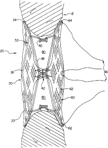
FIG. 1 is a side elevational view of the septal defect
occluder of the present invention deployed in a septal defect;
FIG. 2 is an end view of an occluder panel of the septal
defect occluder of the present invention, particularly
illustrating a center fabric attachment point;
_g_
CA 02344987 2001-04-24
r
FIG. 3 is an end view of the frame members of the device
of the subject invention having two pairs of opposing inward
eyelets;
FIG. 4 depicts a fabric sheet suitable for use with the
invention;
FIG. 5 is an end view of two frame members of FIG. 3
arranged to form a fabric support structure having both
internal and perimeter eyelets;
FIG. 6 depicts a first occluder half, corresponding to
the fabric support structure of FIG. 5, showing the
relationship between the fabric sheet of FIG. 4 and the
underlying support structure;
FIG. 7 depicts a second occluder half, corresponding to
the second fabric support structure of FIG. S, snowing the
relationship between the fabric sheet of FIG. 4 and the
underlying support structure;
FIG. 8 is a side view of the present invention being
delivered within a catheter to a septal defect site;
FIG. 9 is a side view of the present invention being
initially deployed within the septal defect, the second
occluder half having expanded to conform to a portion of the
defect;
FIG. 10 is a side view of the present invention deployed
_g_
CA 02344987 2001-04-24
within the septal defect while under the control of tension
imparting means;
FIG. 11 is a side view of the present invention being
initially retrieved into the catheter from the septal defect
site, the first occluder half being collapsed for catheter
entry; and,
FIG. 12 is a side view of the present invention on its
way to complete retrieval into the catheter from the septal
defect site.
DETAILED DESCRIPTION OF THE INVENTION
As shown generally in FIG. 1, a septal defect closure
device 20 of the invention may be attached to the septum S
(e. g., an atrial septum) to effectively conform to and block
the defect, without protruding into atrial cavities and the
like. As described in detail below, once the closure device 20
is in place, it becomes anchored to the septum and prevents
the flow of blood through the atrial septum to the adjoining
chambers of the heart. This will permit the heart to operate
normally.
Referring now to FIGS. 1 and 2, the extremely low profile
closure device includes first and second occluder panels 30,
60. Each panel 30, 60 is generally round (e. g., circular,
-10-
CA 02344987 2001-04-24
oval, elliptical etc.) so as to facilitate positioning, and
minimizes chances of erosion and puncture. Each panel 30, 60
generally comprises a fabric support structure 32, 62 and
fabric 33 suspended from a perimeter 34, 64 of the fabric
support structures 32, 62. The occluder panels 30, 60 are
conjoined at a plurality of discrete points, located or
positioned within the bounds of each of the fabric support
structures 32, 62 (i.e., within an area bounded by each
perimeter 34, 64 of the support structures 32, 62), as well as
on the fabric 33 (which will be further explained with
reference to FIGS. 2, 7 and 8) . A defect conforming region 80
for the occluder 20 is thereby formed. The nature (i.e.,
structure, relationships therebetween and function) of the
defect conforming region will be detailed hereinbelow,
particularly with reference to FIGS. 1, 2 and 10-14. At this
point it may be said that the region 80 expandingly conforms
to substantially completely and thoroughly satisfy the
perimeter of the defect geometry. This stabilizes panels 30,
60 so that complete coverage of the defect from either
direction is achieved. The defect :is thereby occluded without
even minimal shunting or distortion of the defect.
The fabric support structures 32, 62 of the occluder are
generally flexible and elastically deformable, and include
-11-
CA 02344987 2001-04-24
perimeter and traversing segments 36, 38. Resilient fabric 33
(FIG. 4) is suspended or otherwise affixed to the perimeter
segments 36 of the fabric support structures 32, 62. As
particularly shown in FIG. 2, the perimeter segments 36 of the
fabric support structures 32, 62 extend substantially around
the periphery 35 of the fabric 33. The fabric 33 may be formed
of a thin, flexible material which can be folded and pulled
taut without being damaged. Elastic polymeric materials such
as, for example, polyester knit, nylon, polypropylene,
polytetrafluoroethylene (e. g., Teflon°), and expanded
polytetrafluoroethylene (e. g., GoreTex°), as well as natural
fabrics such as silk, are acceptable.
To accommodate the need of the fabric support structure
to distort when retrieving the occluder 20 into the catheter,
excess fabric can be provided. On an area basis relative to
the support structure, an excess of fabric in the range,
typically, of about 30-35 percent, and up to 50 percent, is
sufficient. This range is required because the low stretch
characteristics of the fabric prevent the support structure
from collapsing in a manner suitable to get into the catheter.
However, the 20 denier polyester knit is advantageous in that
it possesses a low stretch character, is approximately 500
less bulky than known jersey style knit patterns which
-12-
CA 02344987 2001-04-24
facilitates the use of smaller delivery catheters, and allows
for the occluders to be retrieved into such catheters at
forces that are not detrimental to either the catheter or the
occluder (e.g., a 40 mm occluder may be pulled into a 12
French catheter using a reasonable peak force of about four
pounds). A further advantage is that two complete fabric
"patches" may be incorporated into the closure device (i.e.,
no need to remove material to reduce bulk), which thereby
creates a device having a high reliability of successful
closure.
The fabric 33 may be attached to their respective support
structures 32, 62 by any suitable means. For instance, the
fabric 33 may be directly attached to the support structures
32, 62 by means of an adhesive or the like, or the periphery
35 of the fabric 33 may be wrapped about each of the support
structures 32, 62 and the peripheral edge attached to the rest
of the fabric so as to essentially define a sleeve about each
of the support structures 32, 62. In the latter instance, the
sleeve may fit the support structure relatively loosely so
that the structure may move within the sleeve with respect to
the fabric. The peripheral edge of the fabric may be affixed
to the rest of the fabric sheet 33 in any suitable fashion
such as by sewing. Preferably, though, the periphery of the
-13-
CA 02344987 2001-04-24
fabric can be sewn to at least some portion of the perimeter
segments 36 of the support structures 32, 62 using polyester,
non-adsorbable suture.
Referring to FIG. l, the fabric support members 32, 62 of
the occluder panels 30, 60 are shown as being spaced from one
another for purposes of the present explanation, but this is
not the normal configuration (i.e., static condition) of the
panels. In a static, non deployed condition, the fabric
support structures of the device take a generally planar form,
with the two fabric support structures 32, 62 generally
abutting against, or closely proximate, one another.
Again referri ng to FIGS . 1 and 2 , the occluder panels 30 ,
60 are conjoined at a plurality of discrete points, the points
being selected to effectively link each ef the fabric support
structures 32, 62 together, as well as associate each sheet of
fabric 33 carried thereby, so as to form the variably
configurable defect conforming region 80. With such
arrangement, the resilient fabric 33 is not only inherently or
indirectly positionable in response to the defect geometry,
but also directly responsive vis-a-vis the conjoined suppcrt
structures 32, 62.
The conjoined points within the fabric support structures
32, 62, which responsively link the opposing structures,
-14-
CA 02344987 2001-04-24
comprise loops formed in the traversing segments 38 thereof,
these loops defining internal eyelets 40 for the structures
32, 62. The internal eyelets 40 of each of the structures 32,
62 are shown as being joined by suture (e. g., polyester, non-
absorbable or other suitable material), and to some extent
delimit the defect conforming region 80, and serve to center
the occluder 20 within the defect. The remaining point of
conjointment comprises the union, at a single point, of the
fabric of each of the fabric support structure so as to define
a generally central fabric attachment point 42. It is
important that fabric 33 of each support structure 32, 62 be
limitingly controlled via the union, however it is equally
important that the fabric 33 remain substantially susper_ded
for expansion during deployment, preferably exclusively about
or by its periphery 35.
As best seen in FIG. 2, the center attachment point 42 of
the occluder 20 is preferably but not exclusively configured
as a sutured cross stitch positioned in the center of the
fabric 33. Other attachment configurations or geometries are
contemplated, to the extent that tl-.e center attachment point
42 maintains its functionality, namely that of control of the
peripherally supported fabric, and generally contributing to
a centering function far the occluder. Preferably the internal
-15-
CA 02344987 2001-04-24
eyelets 40 are symmetrically oriented about the center fabric
attachment point 42.
In addition to internal eyelets 40 which are formed in
the traversing segments 38 of each of the fabric support
S structures 32, 62, the perimeter segments 36 of at least one
(i.e., structure 62) of the fabric support structures 32, 62
include loops formed therein, thereby defining perimeter
eyelets 44 for that particular support structure 62. As best
seen in FIGS. 1 and 8-12, the perimeter eyelets 44 cooperate
with urging means 46 carried by and or through a catheter 47
so as to aid in the symmetrical collapse of each of the fabric
support structures 32, 62, and the occluder panels 30, 60
thereby, during reversible retrieval of the device 20 into the
catheter 47. The perimeter eyelets 44 associated with the
"catheter side" occluder panel 60 transmit and distribute
deployn-~ent and retrieval forces imparted thereupon through the
defect conforming region 80 and to the other occluder panel
30. As will subsequently be discussed, the unique
configuration of the fabric support structure components, and
the relationships therebetween, provide numerous advantages
(for example: symmetrical collapse of the occluder, less peak
force for retrieval into a catheter for deployment, and
heretofore unsurpassed sealing of narrow slit defects without
-16-
CA 02344987 2001-04-24
the distorting defects typically associated with fixed
geometry conjoint areas such as circumferential conjoint
disks) .
Referring now to FIGS. 3-7, the fabric support structures
comprise cooperating frames 50, each of which preferably
resembles a "bowtie," as best seen in FIG. 3. A more
technical description for the frame geometry might be to
characterize it as an octagon (i.e., a frame of eight legs or
segments), particularly an octagon having a concave, rather
than convex, "top" and "bottom" (i.e., ceiling and floor). Put
yet another way, the frames resemble elongated hexagons whose
long sides are "pinched" towards each other. The frames 50 may
be generally characterized as having maximum and minimum
dimensions and corresponding axes of maximum and minimum
dimension 52, 54. The above frame description is intended to
be illustrative, not limiting, with alternating frame
geometries satisfying the general characterization being
possible.
The internal eyelets 40 of the fabric support structures
32, 62 are formed in each of the frames 50 along the axis of
minimum dimension 54 (FIG. 3), as illustrated. The resilient
internal eyelets 40 are generally disposed between adjacent
ends of two legs or frame segments 51, one end of the eyelet
-17-
CA 02344987 2001-04-24
being attached to each leg 51. The internal eyelets 40 are
shown as lying generally in the same plane as the legs 51 and
may extend generally outwardly of the periphery of each of the
support structures 32, 62, or may preferably extend inwardly
S of the periphery of the structures as shown in the figures.
The eyelets are desirably formed to function as spring hinges.
This will serve to ensure that the occluder panels 30, 60,
particularly the catheter side panel 60, elastically return
substantially to a plane-defining configuration even after
they have been collapsed and delivered through a catheter.
Frames 50 of the device have internal eyelets 40, as
previously explained. The internal eyelets 40 of one fabric
support structure (i.e., 32) mate (i.e., align or register)
with those of the cther support structure (i.e., 62} so as to
thereby conjoin the occluder panels 30, 60 (FIG. 2). The
perimeter eyelets 44 of fabric support structure 62 on the
other hand are formed in its frames 50A along the axis of
maximum dimension 54 (FIG. 3). The perimeter eyelets 44
cooperatively engage urging means 46 so as to enable remote
manipulation of the occluder 20 during retrieval.
Each fabric support structure 32, 62 comprises
perpendicularly overlying frames, the axis of maximum
dimension 52 of one frame 50 or 50A substantially aligning
-18-
CA 02344987 2001-04-24
with the axis of minimum dimension 54 of the other frame (FIG.
5). The somewhat oversized fabric 33 is shown in FIG. 6
underlaying the cooperating frames, the periphery thereof
being sewn or otherwise affixed to those portions of the
frames, which when configured as shown in FIG. 5, form a
perimeter 34, 64 for each of the fabric support structures 32,
62. It is again noted that the preferred fabric 33 contributes
to an occluder 20 that has complete opposing fabric patches
suspended by the fabric support structures 32, 62, which in
turn include a frame geometry and arrangement that generally
reduce deployment and retrieval forces. In return, fabric and
stitch wear and tear and frame "break through" (i.e.,
separation of the perimeter segments 36 from the fabric 33
upon expansion of the occluders 30, 60) are minimized.
Each frame 50 is preferably formed of a single elongate
strand of wire W. As best seen in FIG. 3, each of the legs 51
may simply comprise a length of the wire, and the wire may be
bent through greater than 360 degrees to define adjacent legs
51 and to form the loops or eyelets 40, 44. The ends of the
wire may be attached to each other in any secure fashion, such
as by means of a weldment or a suitable biocompatible
cementitious material.
The frames 50 should be formed of a flexible, elastically
-19-
CA 02344987 2001-04-24
deformable material such as a metal, and the wire comprising
the frame is formed of a superelastic material. One such
material currently known in the art is a near-stoichiometric
nickel/titanium alloy, commonly referred to as Nitinol or
NiTi. Such superelastic materials may be elastically deformed
to a much greater extent than most other materials, yet
substantially fully recover their original shape when
released. This permits the frame to be deformed sufficiently
for insertion into, and passage through, a small-diameter
catheter yet automatically elastically return to its initial
shape upon exiting the catheter.
The frames are preferably manufactured with nitinol wire
that can be wound around the pins of a forming die and
subjected to heat treatment. Each device consists of four
frames, two frames for each support structure. More
particularly, each support structure 32, 62 comprises
matchingly paired frame styles (i.e., as shown in FIGS. 6 and
7, occluder panel 30 has a pair of frames 50 whereas occluder
panel 60 has a pair of frames 50A). All eyelets 40, 44 can be
made having generally a 0.030 inch inside diameter, and, as
previously noted, be inward facing (i.e., directed toward the
center fabric attachment point 42). The wire ends of each
frame can be connected with a titanium hypo tube using a
..
-20-
CA 02344987 2001-04-24
i a
compression crimp. The titanium is more ductile than the
nitinol, providing a reliable grip with excellent corrosion
resistance. Alternately, the preferred shape of the frame may
be cut out from a sheet of such superelastic material as a
single block, by chemical etching, punching with a suitable
punch and die, or any other appropriate forming method.
In order to er~hance radiopacity so that the frame can be
viewed remotely during deployment, the frame may be provided
with a radiopaque coating, such as gold or platinum. For
instance, the wire W may be plated with a thin layer of gold
or platinum. In one particularly useful embodiment, a
helically wound length of a thin radiopaque wire (not shown)
is placed over the wire W; such core/coil structures are well
known in the art. Alternatively, radiopaque marking bands
(not shown), which are commercially available, may be
employed. By placing one such band on each leg of the frame,
a physician can remotely visualize the frame as a plurality of
small bands; when the bands are appropriately spaced from one
another on a monitor, the physician knows that the frame is
properly deployed.
Referring now to FIGS. 8-10, the closure device 20 of the
invention is shown being deployed to occlude a defect in a
septum S. The first panel 60 (i.e., catheter side occluder
-21-
CA 02344987 2001-04-24
panel) of the device 20 is positioned on one side of the
defect while the second panel 30 is generally disposed on the
other side. The frames 50 or 50A of the fabric support
structures 32, 62 are elastically biased toward the position
S shown in FIG. 2. The defect conforming region 80 is positioned
within, and expands so as to occlude the defect. Because the
support structures 32, 62, vis-a-vis their frames 50 or 50A,
are elastically biased toward their deployed configuration,
they are biased generally toward one another and engage
opposing sides of the septum about the defect. Since there are
no compressive forces acting on the frames which might cause
them to collapse, this serves to effectively hold the device
in place and occlude the defect. The device is further shown
in FIGS. 11 and 12 being retrieved from a septal defect site,
as might be required in the event of inadvertent initial
placement, size mismatch, or otherwise.
The fabric sheets 33 are formed of a relatively porous
material (FIG. 4). While this may seem to contradict the
purpose of the device, blood will tend to coagulate on the
latticework provided by the porous material. Blood flow
across the defect is usually substantially blocked after
minimal time passage. If so desired, the conjoint portion of
the device (or the entire device) may be treated with a
-22-
CA 02344987 2001-04-24
thrombogenic agent to speed this natural process or may be
impregnated with a biocompatible polymeric compound or the
like to make it relatively impervious to fluids.
The primary purpose of using a porous fabric is to
accelerate the process of permanently anchoring the device in
place. The support structures hold the fabric tautly and in
intimate contact with the surface of the septum S. This
intimate contact between the septum and perimeter of the
occluder permits ingrowth of collagen and fibrous tissue from
the septum into the fabric. Over time, the membrane resting
against the septum will become securely anchored to the septal
wall and be covered by a layer of endothelial cells.
The design of this device is in stark contrast to the
septal defect closure devices known in the art. As explained
in detail above, prior art devices employ a mechanical
umbrella of one design or another. The radially extending arms
of the umbrella contact the septum and serve to space all but
the peripheral edge of the umbrella away from the septum.
Endothelial cells, collagen and fibrous tissue are therefore
permitted to grow into only the very periphery of the
umbrella. Thus, while a closure device of the invention
essentially becomes an integral part of the septum, the
complex mechanical structure of prior art devices does not
-23-
CA 02344987 2001-04-24
enable as complete integration as the present invention.
The mechanical complexity of prior art devices also tends
to markedly affect their durability. In the case of atrial or
ventricular septal defects, for example, the heart obviously
continues to beat after tre device is in place. Since beating
of the heart is accomplished by flexure of the heart muscles,
the septum will flex to some degree with every beat of the
heart. The radial arms must therefore flex with the septum
with each and every time the heart beat . The number of cycles
of this stress-inducing movement produces repeated stresses on
the arms, which can eventually lead to mechanical failure and
fracture of the arms.
(r7hen a closure device of the invention is deployed, the
tension of the frame of the support structure opens the panel
to occlude the defect. Since there are no radial arms to prop
open the device, the occurrence of repeated flexion does not
occur due to the beating of the heart or pressure differences
between the cardiac chamber during the phase of contraction of
the heart. To the contrary, any pressure difference would urge
a frame and panel against the septum, more firmly occluding
the defect. In addition, the superelastic material of the
frame tolerates flexural stresses much better than the rigid
steel arms of the prior art devices. The present device
-24-
CA 02344987 2001-04-24
therefore will continue to flex with the septum without any
significant effect on its structural integrity.
Although the foregoing has focused on application of the
present invention to occlude atrial septal defects, the
invention is not limited to occluding such defects. For
instance, the instant closure device can be used to treat
ventricular septal defects, patent ductus arteriosus or any
other congenital or acquired orificial or tubular
communications between vascular chambers or vessels.
While a preferred embodiment of the present invention has
beer described, it should be understood that various changes,
adaptations and modifications may be made therein without
departing from the spirit of the invention. Changes may be
made in details, particularly in matters of shape, size,
material, and arrangement of parts without exceeding the scope
of the invention. Accordingly, the scope of the invention is
as defined in the language of the appended claims.
-25-