Note: Descriptions are shown in the official language in which they were submitted.
CA 02345646 2001-03-26
WO 00/18320 PCT/US99/22769
DEVICE AND METHOD FOR
RESTRUCTURING HEART CHAMBER GEOMETRY
REFERENCE TO COPENDING APPLICATION
This is a continuation in part application of United States patent application
serial no.
09/316,611, filed May 21,1999 (incorporated herein by reference), entitled
"Device and Method
for Restructuring Heart Chamber Geometry", which is a continuation in part
application of
United States patent application serial no. 09/165,887, filed September 30,
1998 (incorporated
herein by reference), entitled "Device and Method for Restructuring Heart
Chamber Geometry".
which is a continuation in part application of United States patent
application serial no.
08/581,914, filed December 23, 1997 (incorporated herein by reference),
entitled "Activation
Device for the Natural Heart and Method of Doing the Same," which is a
continued prosection
application of United States Patent application serial no. 08/581,914 filed on
January 2, 1996
(incorporated herein by reference}.
TECHNICAL FIELD OF THE INVENTION
w The present invention relates to devices and methods for treating
cardiomyopathies
andlor enlarged hearts and, more specifically, devices and methods for
decreasing a heart
chamber's wall tension.
CA 02345646 2001-03-26
WO 00/1$320 PCT/US99/22769
BACKGROUND OF THE INVENTION
The natural heart, and specifically, the cardiac muscle tissue of the natural
heart (e.g.,
myocardium) can fail for various reasons to a point where the natural heart
cannot provide
sui~cient circulation of blood for a body so that life can be maintained. More
specifically, the
heart and its chambers can become enlarged for a variety of causes and/or
reasons, including t
viral disease, idiopathic disease, valvular disease (mitral, aortic and/or
both), ischemic disease,
Chagas' disease and so forth. As the heart and its chambers enlarge, tension
of the wails of the
heart's chambers increase and thus, the heart must develop more wall tensile
stress to generate
the needed pressure for pumping blood through the circulatory system. The
process of
ventricular dilation is generally the result of chronic volume overload or
specific damage to the
myocardium. In a normal heart that is exposed to long-term increased cardiac
output
requirements, for example, that for an athlete, there is an adaptive process
of slight ventricular
dilation and muscle myocyte hypertrophy. In this way, the heart may fully
compensate for the
increase cardiac output requirements of the body. With damage to myocardium or
chronic
volume overload, however, there are increased requirements put on the
contracting myocardium
to such a level that this compensated state is never achieved and the heart
continues to dilate.
A problem with an untreated dilated ventricle is that there is a significant
increase in wall
tension and/or stress, both during the diastolic filling, and during the
systolic contraction. In a
normal heart, the adaption of muscle hypertrophy (e.g. thickening) in the
ventricular dilation
maintain a fairly constant wall tension for systolic constriction. However, in
a failing heart, the
ongoing dilation is greater than the hypertrophy, and as a result, rising wall
tension is required
for systolic contraction. This is believed to result in further muscle damage.
The increase in wall stress is also true for diastolic filling. Additionally,
because of the
lack of cardiac output, ventricular filling pressure tends to rise due to
several physiologic
2
CA 02345646 2001-03-26
WO 00/18320 PCT/US99/Z27b9
mechanisms. Moreover, in diastole, both the diameter and wall pressure
increase over normal
levels, thus contributing to higher wall stress levels. As a solution for the
enlarged natural heart,
attempts have been made in the past to provide a treatment to maintain
circulation. Prior
treatment for heart failure generally fall into three categories, namely
surgical treatments;
mechanical support systems; or pharmacological.
One such approach has been to replace the existing natural heart in a patient
with an
artificial heart or a ventricular assist device. In using artificial hearts
and/or assist devices, a
particular problem stems from the fact that the materials used for the
interior lining of the
chambers of an artificial heart are in direct contact with the circulating
blood, which can enhance
undesirable clotting of the blood, build up of calcium, or otherwise inhibit
the blood's normal
function. Hence, thromboembolism and hemolysis could occur with greater ease.
Additionally,
the lining of an artificial heart or a ventricular assist device can crack,
which inhibits
performance, even if the crack is at a microscopic level. Moreover, these
devices must be
powered by a source which can be cumbersome and/or external to the body.
Drawbacks have
1 S limited use of these devices to applications having too brief a time
period to provide a real lasting
benefit.
An alternative procedure is to transplant a heart from another human or animal
into a
patient. The transplant procedure requires removing an existing organ (i.e.,
the natural heart) for
substitution with another organ (i.e., another natural heart) from another
human, or potentially,
from an animal. Before replacing an existing organ with another, the
substitute organ must be
"matched" to the recipient, which can be, at best, difficult and time
consuming to accomplish.
Furthermore, even if the transplanted organ matches the recipient, a risk
exists that the
recipient's body will reject the transplanted organ and attack it as a foreign
object. Moreover,
the number of potential donor hearts is far less than the number of patients
in need of a
3
CA 02345646 2001-03-26
WO 00/18320 PCT/US99/22769
transplant. Although use of animal hearts would lessen the problem with fewer
donors than
recipients, there is an enhanced concern with rejection of the animal heart.
In an effort to use the existing natural heart of a patient, other attempts
have been made
to reduce wall tension of the heart by removing a portion of the heart wall,
such as a portion of
the left ventricle in a partial left ventriculectomy procedure (the Batista
procedure). A wedge
shaped portion of the ventricular muscle has been removed, which extends from
the apex to the
base of the heart. By reducing the chamber's volume, and thus its radius, the
tension of the
chamber's wall is reduced as well. There are, however, several drawbacks with
such a
procedure. First, a valve (i.e., the mitral valve) may need to be repaired or
replaced depending
on the amount of cardiac muscle tissue to be removed. Second, the procedure is
invasive and
traumatic to the patient. As such, blood loss and bleeding can be substantial
during and after the
procedure. Moreover, as can be appreciated by those skilled in the industry,
the procedure is not
reversible. Another device developed for use with an existing heart for
sustaining the
circulatory function of a living being and the pumping action of the natural
heart is an external
bypass system, such as a cardiopulmonary (heart-lung) machine. Typically,
bypass systems of
this type are complex and Large, and, as such, are limited to short term use
in an operating room
during surgery, or to maintaining the circulation of a patient while awaiting
receipt of a
transplant heart. The size and complexity effectively prohibit use of bypass
systems as a long
term solution, as they are rarely even portable devices. Furthermore, long
term use of these
systems can damage the blood cells and blood borne products, resulting in post
surgical
complications such as bleeding, thromboembolism function, and increased risk
of infection.
Medicines have been used to assist in treating cardiomyopathies. Some
inotropic agents
can stimulate cardiac work. For example, digoxin can increase the
contractibility of the heart,
and thereby enhances emptying of the chambers during systolic pumping.
Medicines, such as
4
CA 02345646 2001-03-26
WO 00/18320 PCTNS99/22769
diuretics or vasodilators attempt to reduce or decrease the heart's workload.
For example,
indirect vasodilators, such as angiotensin-converting enzyme inhibitors (e.g.,
enalopril), can help
reduce the tendency of the heart to dilate under the increased diastolic
pressure experienced
when the contractibility of the heart muscle decreases. Many of these
medicines have side
effects, such as excessive lowering of blood pressure, which make them
undesirable for long a
term therapy.
As can be seen, currently available treatments, procedures, medicines, and
devices for
treating end-stage cardiomyopathies have a number of shortcomings that
contribute to the
complexity of the procedure or device. The current procedures and therapies
can be extremely
invasive, only provide a benefit for a brief period of time, or have
undesirable side effects which
can hamper the heart's effectiveness. There exists a need in the industry for
a device and
procedure that can use the existing heart to provide a practical, long-term
therapy to reduce wall
tension of the heart, and thus improve its pumping efficiency.
SUMMARY OF THE PRESENT INVENTIQN
It is the object of the present invention to provide a device and method for
treating
cardiomyopathies that addresses and overcomes the above-mentioned problems and
shortcomings in the thoracic medicine art.
It is another object of the present invention to provide a device and method
for treating
cardiomyopathies that minimizes damage to the coronary circulatory and the
endocardium.
It is still a further another object of the present invention to provide a
device and method
for treating cardiomyopathies that maintains the stroke volume of the heart.
5
CA 02345646 2001-03-26
WO 00/18320 PCT/US99/22769
Another object of the present invention is to provide a device and method for
treating
cardiomyopathies that supports and maintains the competence of the heart
valves so that the
heart valves can function as intended.
Still another object of the present invention is to provide a device and
method than
increases the pumping effectiveness of the heart.
Yet another object of the present invention is to provide a device and method
for treating
cardiomyopathies on a long term basis.
It is yet still an object of the present invention to provide a device and
method for treating
cardiomyopathies that does not require removal of any portion of an existing
natural heart.
Still a further object of the present invention is to provide a device and
method for
treating dilated cardiomyopathies that directly reduce the effective radius of
a chamber of a heart
in systole as well as in diastole.
Additional objects, advantages, and other features of the present invention
will be set
forth and will become apparent to those skilled in the art upon examination of
the following, or
may be learned with practice of the invention.
To achieve the foregoing, a geometric reconf guration assembly for the natural
heart
having a collar configured for surrounding the natural heart. The collar can
include a plurality
of bands, such as thin bands of about . 2 mm in thickness, in a spaced
relationship to each other,
and a connector bar intersecting the plurality of bands and configured for
maintaining the spaced
relationship of the bands to each other. The collar may include a plurality of
bands, such as
from about 2 to about 10 bands, that are positioned parallel to each other.
The bands can each
be made of a biomedical material, such as polyacetal or a metal, such as
titanium or steel.
The connector bar of the present invention can be positioned tangential to the
plurality
ofbands, and may have a plurality of grooves configured to receive the
thickness of each of the
6
CA 02345646 2001-03-26
WO 00/18320 PCTNS99/22769
plurality of bands. The grooves also may be beveled to allow for the bands to
flex as the heart
beats. The connector bar's inner surface can have an outwardly convex curved
configuration,
and may even include a cushioned portion that can be made from a polymeric
material. A pad
may be positioned between the collar and the epicardial surface of the heart
that may comprise'
a low durometer polymer, or either a gel-filled cushion or a fluid-filled
cushion.
The assembly of the present invention may also comprise a closure device for
enclosing
at least one of the bands in the connector bar.
In use, the present invention can reduce the wall tension on one of the
chambers of the
heart. A yoke or collar is surrounds the heart so as to provide the chamber of
the heart as at least
two contiguous communicating regions, such as sections of truncated
ellipsoids, which have a
lesser minimum radii than the chamber before restructuring. As such, the
collar displaces at least
two portions of the chamber wall inwardly from the unrestricted position.
BRIEF DESCRIPTION OF THE DRAWINGS
While the specification concludes with claims particularly pointing out and
distinctly
claiming the present invention, it is believed the same will be better
understood from the
following description taken in conjunction with the accompanied drawings in
which:
Fig. 1 partial frontal anterior view of an exemplar natural heart;
Fig. 2 vertical cross sectional view of an exemplar natural heart and blood
vessels leading
to and from the natural heart;
Fig. 3 is a horizontal cross sectional view of an unrestrained left ventricle
of the natural
heart;
Fig. 4 is a horizontal cross sectional view of a heart restrained made in
accordance with
the present invention;
7
CA 02345646 2001-03-26
WO 00/18320 PCT/U899/22769
Fig. 5 is a perspective view of a device made in accordance with the present
invention;
Fig. 6 is an enlarged exploded perspective view of a portion of the assembly
made in
accordance with the present invention;
Fig. 7 is an enlarged perspective view of another portion of the assembly made
in
accordance with the present invention;
Fig. 8 is a cross sectional view of a connector of the present invention taken
along line
8-8 in Fig. 7;
Fig. 9A is a partial horizontal cross sectional view of an assembly made in
accordance
with the present invention while the heart is at rest;
Fig. 9B is a partial horizontal cross sectional view of an assembly made in
accordance
with the present invention while the heart is contracting:
Fig. 10 is a perspective view of the assembly made in accordance with the
present
invention and positioned on the left ventricle;
Fig. 11 is an alternative embodiment of the assembly made in accordance with
the present
1 S invention;
Fig. 12 is a cross sectional view of one embodiment of the collar of the
present invention
taken along line 12-12 in Fig. 11;
Fig. 13 is a perspective view of another alternative embodiment of the
assembly made
in accordance with the present invention;
Fig. 14 is a perspective view of yet another alternative embodiment of the
assembly made
in accordance with the invention;
Fig. 15 is another alternative embodiment of the assembly made in accordance
with the
present invention;
8
CA 02345646 2001-03-26
WO 00/18320 PCTNS99/22769
Fig. 16A is a perspective view of the assembly made in accordance with the
present
invention;
Fig 16B is a perspective view of an alternative embodiment of the assembly
made in
accordance with the present invention;
Fig. 17 is a perspective view of the assembly made in accordance with the
present
invention;
Fig. 18 is a perspective view of the assembly made in accordance with the
present
invention;
Fig. 19 is a perspective view of the assembly of Fig. 18, with the connector
cord and/or
portion of the collar secured to the assembly;
Fig. 20 is a vertical cross sectional view of one embodiment of an auxiliary
fastener made
in accordance with the present invention;
Fig. 21 is another vertical cross sectional view of the auxiliary fastener of
Fig. 16 inserted
into the assembly;
Fig. 22 the vertical cross sectional view of the embodiment of Fig. 16
illustrating the
auxiliary connecter;
Fig. 23 is a vertical cross sectional view of the auxiliary fastener of Fig.
16 a period of
time after being inserted into position;
Fig. 24 is a perspective view of an exemplar heart with the assembly of the
present
invention being positioned on the heart;
Fig. 25A is a perspective view of another embodiment of the present invention;
Fig. 25B is a top view of an exemplar heart with the assembly of Fig. 25A of
the present
invention having been positioned on the heart;
9
CA 02345646 2001-03-26
WO 00/18320 PCT/US99/22769
Fig. 26 is a perspective view of an exemplar heart with the assembly of the
present
invention having been positioned on the heart;
Fig. 27A is an enlarged perspective view of a connector portion of the
assembly made
in accordance with the present invention;
r:
Fig. 27B is a perspective view of the assembly made in accordance with the
present
invention including a connector portion;
Fig. 27C is a perspective view of the embodiment of Fig. 27B, wherein the ends
of the
connector portion have been attached to one another; and
Fig. 27D is another perspective view of the assembly made in accordance with
the
present invention in a generally elongated configuration.
DETAILED DESCRIPTION OF THE PREFERRED EMBODIMENT
Referring now to the figures in detail wherein like numerals indicate the same
elements
throughout the views, an exemplary natural heart, generally indicated in
Figures 1 and 2 as 10,
1 S has a lower portion comprising two chambers, namely a left ventricle 12
and a right ventricle 14,
which function primarily to supply the main force that propels blood through
the circulatory
system, namely the pulmonary circulatory system, which propels blood to and
from the lungs,
and the peripheral circulatory system, which propels blood through the
remainder of the body.
A natural heart 10 also includes an upper portion having two chambers, a left
atrium 16 and a
right atrium 18, which primarily serve as an entryway to the left and right
ventricles 12 and 14,
respectively, and assist in moving blood into the left and right ventricles 12
or 14. The
interventricular wall 40 of cardiac tissue 32 separates the left and right
ventricles 12 and 14, and
the atrioventricular wall 42 of cardiac tissue 32 separates the lower
ventricular region from the
upper atrium region.
CA 02345646 2001-03-26
WO 00/18320 PCT/US99122769
Generally, the left and right ventricles 12 and 14, respectively, each has a
cavity 13 and
15, respectively, that is in fluid communication with cavities 17 and 19,
respectively, of the atria
(e.g., 16 and 18) through an atrioventricular valve 50 (which are each
illustrated as being in the
closed position in Fig. 2). More specifically, the left ventricle cavity 13 is
in fluid
communication with the left atrium cavity 17 through the mitral valve 52,
while the right I
ventricle cavity 15 is in fluid communication with the right atrium cavity 19
through the tricuspid
valve 54.
Generally, the cavities of the ventricles (e.g., 13 and 15) are each in fluid
communication
with the circulatory system (i.e., the pulmonary and peripheral circulatory
systems) through a
semilunar valve 44 {which are each illustrated as being in the open position
in Fig. 2). More
specifically, the left ventricle cavity 13 is in fluid communication with the
aorta 26 of the
peripheral circulatory system through the aortic valve 46, while the right
ventricle cavity 15 is
in fluid communication with the pulmonary artery 28 of the pulmonary
circulatory system
through the pulmonic valve 48.
Blood is returned to the heart 10 through the atria (e.g., 16 and 18). More
specifically,
the superior vena cava 22 and inferior vena cava 24 are in fluid communication
with and deliver
blood, as it returns from the peripheral circulatory system, to the right
atrium 18 and its cavity
19. The pulmonary veins 30 are in fluid communication with and delivers blood,
as it returns
from the pulmonary circulatory system, to the left atrium 16, and its cavity
17.
The heart 10 is enclosed in the thoracic cavity within a double walled sac
commonly
referred to as the pericardium. Its inner layer is the visceral pericardium or
epicardium, and its
outer layer is the parietal pericardium. The heart 10 is generally made up of,
among other
materials, cardiac muscle or tissue 32, which has an exterior surface commonly
known as the
epicardial surface 34 and an interior surface, or endocardial surface 38, that
generally defines the
11
CA 02345646 2001-03-26
WO 00/18320 PCT/US99/22769
cavities (e.g., ventricular cavities i 3 and 15, respectively, and atrial
cavities 17 and 19,
respectively). Coronary arteries 36 on the epicardial surface 34 of the heart
10 provide blood
and nourishment (e.g., oxygen) to the heart 10 and its cardiac tissue 32.
By way of a non-limiting example, the present invention will be discussed in
terms of
embodiments that are used to primarily assist in the restructuring or
reconfiguring, and/or T .
operation of the left ventricle chamber (e.g., 12) of the natural heart 10.
However, it is noted that
the present invention can also be used to assist in the restructuring or
reconfiguring, and/or
operation of other portions of the natural heart 10, such as either atria ( 16
and/or 18), and/or the
right ventricle chamber (e.g., 14).
Taming now to Fig. 3, the chambers of the heart 10, including the left
ventricle chamber
12, is generally shaped as a hollow truncated ellipsoid having, at any
circular cross-section
perpendicular to its long axis, a center point "C," and a radius "R,"
extending from center point
C, to the endocardiai surface 38. The cardiac tissue 32 of the heart 10 has a
thickness "w,''
which is generally the distance between the epicardial surface 34 and the
endocardial surface 38.
An assembly 60 of the present invention preferably is configured and
positioned relative
to the natural heart 10 to displace at least two portions of the cardiac
tissue 32 inwardly (see, e.g.,
Fig. 4) from the unrestricted position, as exemplified in Fig. 3. By
displacing portions of the
cardiac tissue 32 inwardly, the shape of the chamber (e.g., the left ventricle
chamber 12) of the
heart 10 is generally restructured or reconfigured from a generally hollow
truncated ellipsoid
{see, e.g., Fig. 3) to a chamber generally shaped as having at least two
continuous
communicating portions of truncated ellipsoids (see, e.g., Fig. 4). In
generally reconfiguring or
restructuring the heart 10 as such, each of the truncated ellipsoids has an
adjusted radius "R2,"
which is preferably shorter than radius "R,."
12
CA 02345646 2001-03-26
WO 00/18320 PCT/US99/22769
Assembly 60 can be static or passive in that it does not actuate or pump the
heart 10, but
rather, displaces and holds portions of the cardiac tissue 32 in a generally
predetermined fixed
position as the heart 10 continues to contract (e.g., beat) and pump blood
through its chambers
and through the body's circulatory system. Nevertheless, assembly 60 can be
conf gored and'
constructed to permit torsional deformation as the natural heart 10 beats.
Assembly 60 of the present invention can include a yoke or collar 62, as
exemplified in
Figs. 5-7, to assist in restraining or restructuring a ventricle, such as the
left ventricle chamber
12. Collar 62 can be any desired shape and preferably surrounds or encircles
the heart 10, and
preferably one chamber (e.g., the left ventricle chamber 12) as exemplified in
Fig. 10, so as to
restructure or reconfigure the left ventricle chamber 12 as having a shape
approximating at least
two continuous communicating portions of truncated ellipsoids (see, e.g., Fig.
4). Preferably,
a portion or region 64 of the collar 62 can extend along the longitudinal
plane or along the longer
axis of the chamber. Suitable locations on the epicardial surface 34 for the
region 64 can include
the basal portion near the atrioventricular groove 43 (see, e.g., Fig. 1) and
apical portion 20 of
the heart 10, the anterolateral surface of the left ventricle chamber 12, or
the posteromedial
surface of the left ventricle chamber 12.
The collar 62 may include two or more bands (e.g., 76) configured for
positioning around
the heart 10. Preferably, bands 76 are circumferentially flat and may be
oriented with the surface
78 being positioned generally tangent to the epicardial surface 34 of the
heart 10, and having the
smaller dimension, as compared with surface 80. Surface 80 is generally
oriented perpendicular
to the epicardial surface 34. Band 76 should be sized so as to provide for low
deformation in the
direction perpendicular to the epicardial surface 34 of the heart 10, but only
require a low strain
energy for tortial deformation as the heart 10 beats. Band 76 can have a
thickness "th" across
surface 78 and a width "w" across surface 80, that each varies depending on
the selected material
13
CA 02345646 2001-03-26
WO 00/18320 PCTNS99/22769
and its particular deformation characteristics. When metallic material is used
with the present
invention, the band 76 can have a thickness "th" across surface 78 of about .2
mm, and can have
a width, "w" across surface 80 from about 5 mm to about 12 mm, and more
preferably, about 7
mm: It should be noted that the particular dimensions of each assembly 60, and
of its
components (e.g. collar 62 and its various portions, bands 76, etc.) will
depend, as will be ; ,
discussed later, according to particular anatomy, the desired application, and
upon the particular
size and configuration of the individual natural heart 10.
In constructing assembly 60 using bands 76, from about 2 to about 10 bands 76
may be
used, and preferably about 4 bands 76 are used in the present invention.
Nevertheless, the
number of bands 76 may be selected depending upon the properties of the
material selected for
each of the bands 76, as well as the load stress required to appropriately
restructure the heart
chamber geometry.
Bands 76 are each preferably made of a light weight, generally rigid material
that has a
low bending strain under expected levels of stress so that the material has
sufficient wear
resistance in use while the heart 10 beats, and maintains its desired shape in
use adjacent the
heart 10. Illustrative examples of suitable materials which may be employed as
bands 76 include
any biocompatible or biomedical materials, such as metals, including titanium
or stainless steel,
or a suitable polymer, including polyacetal, polypropylene, rigid polyurethane
or an ultra high
molecular weight polyethylene, or a combination of the same.
The collar 62 may preferably include a connector 82, and preferably a
plurality of
connectors 82 spaced along the collar 62, as exemplified best in Fig. 5. The
connectors 82 can
assist in maintaining the space relationship of the bands 76 relative to each
other, and of the
assembly 60 to the heart 10. Turning now to Figs. 6-8, the connector 82
preferably has a contact
or an inner surface 84, which is configured for placement adjacent or against
the epicardial
14
CA 02345646 2001-03-26
WO 00/18320 PCT/US99l22769
surface 34 of the natural heart 10. The inner surface 84 may be configured so
that the epicardial
surface 34 may slide along inner surface 84 during contraction and expansion
of the heart 10, and
to minimize damage to the epicardial surface 34, and the coronary arteries
(e.g., 36). Preferably,
the inner surface 84 is curved convex outwardly in a longitudinal plane (e.g.,
a positive radius
3>
of curvature) (see, e.g., Figs. 4 and 8) and has a smooth surface, and/or
preferably rounded edges
87 so that collar 62 can be configured to be positioned adjacent or on the
epicardial surface 34
whereby intimate contact can be established and maintained, even during the
contraction or
beating of the heart 10. Alternatively, the inner surface 84 may have a
negative radius of
curvature, or an infinite radius of convexity (e.g., a flat surface).
Figs. 6-8 illustrate the connectors 82 as each including one or more grooves
92, which
can extend inwardly from an opening 98 in the outer wall 86, and toward the
contact or inner
surface 84. Each groove 92 is preferably sized and configured to receive a
band 76 whereby its
surface 78 would be positioned adjacent the base wall 94, and its surfaces 80
preferably would
be positioned adjacent sidewalls 96.
In a preferred embodiment, groove 92 should be configured to assist in
allowing flexion
movement of the band 76 as the heart 10 beats and moves. As best exemplified
in Figs. 6-8,
grooves 92 may be tapered inwardly as the grooves 92 proceeds or extends from
the outer
surface 86 inwardly toward the contact surface 84. In addition, grooves 92 may
also be tapered
inwardly as the groove extends from each of the lateral surfaces 88 inwardly
(e.g., upwardi~~
and/or downwardly), as best illustrated in Fig. 6.
Connectors 82 are each preferably made of a light weight, generally rigid
material that
has a low bending strain under expected levels of stress so that the material
has sufl-icient wear
resistance in use while the heart 10 beats, and maintains its desired shape in
use adjacent the
heart 10. Illustrative examples of suitable materials which may be employed as
connectors 82
CA 02345646 2001-03-26
WO 00/18320 PCT/US99/227b9
may include any biocompatible or biomedical materials, such as metals,
including titanium or
stainless steel, or a suitable polymer, including polyacetal or an ultra high
molecular weight
polyethylene, or a combination of the same.
Turning back to Fig. 6, a structure 100 can be provided so as to assist in
maintaining the
bands 76 in the groove 92, in use. Any structure 100 contemplated for use with
assembly 60~
should assist in restricting movement of the band 76 out of the groove 92
through opening 98.
In one embodiment, the structure 100 may take the form of a plate 100 that can
be secured or
otherwise attached, and preferably releasably secured, to close off or
restrict access through one
or more openings 98. In addition to a plate-like structure, sutures (not
shown) may also be
threaded through the connector 82 to assist in restricting bands 76 movement
through opening
98. Structure 100 is preferably made of a biocompatible or biomedical
material.
Turning now to Figs. 11 and 12, an alternative embodiment of the present
invention may
include a collar or yoke 162 that provides an essentially continuous surface
which contacts the
epicardium surface 34 of the heart 10. In the present embodiment, collar 162
may take the form
of a generally continuous yoke-like structure that is essentially rigid and/or
elastic. Collar 162
preferably includes a contact or an inner surface 184, which is configured for
placement
adjacent or against the epicardial surface 34 of the natural heart 10. The
inner surface 184
should be configured so that the epicardial surface 34 may slide along the
inner surface 184
during contraction and expansion of the natural heart 10, and to minimize
damage to the
epicardial surface 34 and the coronary arteries (e.g., 36). Preferably, the
inner surface I84
is curved convexly outwardly in a longitudinal plane and has a smooth surface,
and/or
preferably rounded edges 187 so that a collar 162 can be configured to be
positioned adjacent
or against the epicardial surface 34 whereby intimate contact can be
established and
maintained, even during the contraction or expansion of the natural heart 10.
Alternatively,
16
CA 02345646 2001-03-26
WO 00/18320 PCT/US99/22769
the inner surface 184 may have a negative radius of curvature, or an infinite
radius of convexity
(e.g., a flat surface).
The collar 162 preferably is selected from a generally rigid or tough
biomedical or
biocompatable material. Examples of such suitable materials which may be
employed as collar
S I62 can include a metal, such as titanium or steel, or a polymer, such as an
ultra high molecular
weight polyethylene, polyurethane, polyacetal, or a polymer composite material
such as carbon
fiber-epoxy or fiberglass-epoxy, or a combination of the same. Moreover, the
collar 162 may
be covered, either partially or entirely, with a material that promotes tissue
ingrowth into the
collar 162, such as a soft tissue polyester fabric sheeting or
polyletrafluroethyhere (PTFE).
In other alternative embodiments, exemplified in Figs.l3-14, it is
contemplated that the
collar 162 may include an attachment system 163 that allows the collar 162 to
be placed around
the heart 10, such as inbetween the pulmonary veins 30 (e.g., the left and
right pulmonary veins
30A and 30B, respectively) near the basal portion of the heart 10 so as to
reduce the possibility
of lateral or medial displacement of the assembly 60, or about the lateral
atrium or the
atrioventricular groove region.
In one embodiment exemplified in Fig. 13, the collar 162 may include an
attachment
system 163 that permits the collar 162 to be separated and then reattached at
two or more sites
or positions along the collar 162, preferably adjacent or near the region of
the collar 162
configured forplacement adjacent or on the basal portion and/or apical portion
20 of the natural
heart 10. While the attachment system 163 is illustrated as an interlocking
pin 163B and
receptacle 163A (e.g., a ball and socket-like joint), it is contemplated, and
as would be
appreciated by those skilled in the art, other devices and assemblies for
releaseably securing the
collar 162 together can be used. Example of such devices and assemblies for
attachment system
17
CA 02345646 2001-03-26
WO 00/18320 PCT/US99/22769
163 could include sutures, a screw and bore holes through overlapping portions
of the collar 162,
clamps, or a combination of these devices and assemblies.
Alternatively, as exemplified in Fig.l4, the collar 162 may include an
attachment system
163 at one site along the collar i62, preferably adjacent or at the portion of
the collar 162 -
configured for placement adjacent or on the basal portion of the heart 10.
This embodiment of I
collar 162 preferably would include a portion 167 that can either include
flexible material, a
pivotable section 168, or both to provide movement of the collar 162 so that
the attachment
assembly 163 can open, and the collar 162 can be slipped around the heart 10,
such as between
the left and right pulmonary veins 30A and 30B, respectively.
In yet another embodiment illustrated in Figure 15, the assembly 260 may
include a collar
262 having a region 264 similar to the structure of the collar 62, exemplified
above in Figs. 4-8,
and connector portions or regions 268, similar to the structure of the collar
162, discussed above,
and exemplified in Figs. 11-14.
Additional embodiments of the present invention are exemplified in Figs. 16-
19, and may
include a collar or yoke 362 that includes an internal frame portion 374 (see,
e.g., Figs. 16A and
16B) and an external shell or skin portion 400 (see e.g., Fig. 18). The
internal frame portion 374
is preferably configured to support the external shell portion 400, in use,
and to assist in
restraining or restructuring a ventricle.
Turning now to Figs. 16A and 16B, internal frame portion 374 can include
supports 376,
380, and 382, and connectors 390. Supports 376 are sized and configured to
extend generally
along a longitudinal plane of the longer axis of the heart 10, and preferably,
are generally thin
elongated panels, with a slight arc or curvature whereby they are contoured to
match the
epicardial surface 34.
18
CA 02345646 2001-03-26
WO 00/18320 PCT/US99l22769
Support 380 is preferably sized and configured to extend generally around the
apical
portion 20 of the heart 10, whereas support 382 is generally U-shaped and is
preferably
configured to extend generally around the basal portion of the heart 10. The
supports 376 and
380 are preferably made of a light weight, generally rigid material that has a
low bending strain'
under expected stress levels so that the material has sufficient wear
resistance in use while the; '
heart 10 beats, and maintains its desired shape in use adjacent the heart 10.
Support 382 is preferably more rigid as it is configured for being positioned
around the
basal portion of heart 10, whereby it can have a greater bending movement
applied to it by the
heart 10. Furthermore, it may include a metal brace encased in a polymer.
Moreover, since
some embodiments of the invention may be encased in external shell portion
400, the internal
frame portion 374 may be selected from a group of materials that are not
biocompatible, such
as other metallic alloy or other polymer.
Illustrative examples of suitable materials which may be employed as supports
376, 380
and 382 can include any biocompatible or biomedical materials, such as metals,
including
titanium, stainless steel, or a suitable polymer, including polyacetal,
polypropylene, rigid
polyurethane, an ultra high molecular weight polyethylene, a fiber-reinforced
polymer composite
or a combination of these materials.
In the embodiment of Figs. 16A, support 3 82 may be configured to be
positioned to the
left of the left pulmonary veins 30A (as shown in Figs. 24 and 26) and/or to
the right of the right
pulmonary veins 30B (simply by reversing the orientation of collar 362 from
that shown in Fig.
26). Support 382 can be provided in a generally horn shaped configuration with
end portions
384 at each end of support 382. As illustrated in Figs. 16B, 25A and 25B,
support 382 can also
be configured to be positioned between the left and right pulmonary veins 30A
and 30B,
respectively, and in substantially the same plane as the other supports of
collar 362.
19
CA 02345646 2001-03-26
WO 00/18320 PCT/US99122769
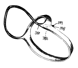
Openings 388 are preferably provided on the surface of the support number 382,
and
more preferably in the end portion 3 84, whereby a channel 3 86 extends into,
and preferably
therethrough. Openings 38$ and channels 386 are preferably sized and
configured to selectively
receive a connector cord 396, to assist in maintaining the position of the
assembly on the heart'
S 10, as will be discussed later herein. A similar horn shaped configuration
with an end portion
384 is provided on the opposite end of support 3$2 in order to receive the
other end of a
connector cord 396.
Supports 376, 380 andlor 382 are preferably connected or joined to each other
with
connectors 390, as exemplified in Figs. 16A and 16B. Connectors 390 are
generally provided
at or adjacent the end portions of the supports 376, 380 and/or 382. When
attached to the
supports 376, 380 and/or 382, connectors 390 preferably can provide for low
deformation in a
direction perpendicular to the epicardial surface 34 of the heart 10, and can
preserve freedom for
slight spontaneous systolic torsion as the heart 20 expands and contracts.
Connector 390 may
take the fornz of a ball and socket joint 392 that is made from either metal,
such as steel, a
polymer such as poiyacetal, or a combination of steel and polymer.
Turning now to Fig. 17, the area around or adjacent connectors 390 can
preferably be
provided with a packing 394 to reinforce the connector 390, and to provide a
generally smooth,
generally crevice free surface whereby the external shell portion 400 can
easily bind thereto.
Moreover, where the external shell portion 400 is not used with the present
invention, the
packing 394 can also assist is prevent tissue from becoming entangled or
embedded in the
connector 390. As such, tissue trauma may be reduced. Illustrated examples of
suitable
materials which may be employed as packing 394 may include silicon rubber or a
low durameter
polymer or a gel or an oil. Moreover, packing 394 may be reinforced with
carbon fiber, steel,
fiberglass, or another suitable reinforcing micro fiber composite materials.
When packing 394
CA 02345646 2001-03-26
WO 00/18320 PCT/US99/22769
is employed in the present invention without external shell portion 400,
packing 394 preferably
should be selected from a suitable biomedical or biocompatible material.
Turning now to Figs.16A,18, and 24, the present invention can also include one
or more
connector cords 396 to further assist in securing the collar 362 to the heart
10, and in maintaining
its position relative to the heart 10. The end of the cord 398 is preferably
joined or attached to
a portion of the support 382. As exemplified in Figs. 16A, 19, and 24,
openings 388 and through
channels 386 may be provided in support 382 and are preferably and sized and
configured to
receive at least one end 398 of connector cord 396. The connector cord 396 may
be attached
thereto by suitable devices and techniques, such as by inserting the connector
cord 396
completely through the channel 386, and providing a knot 399 at its end 398,
or otherwise
securing the connector cord 396 it so that it does not become detached or
disconnected from the
support 382.
Connector cord 396 should be sized and configured to be positioned around the
base
portion of the heart 10. In a preferred embodiment shown in Figs. 24 and 26,
connector cord 396
should be sized and configured to pass around the heart 10 through the center
along the oblique
sinus between the left and right pulmonary veins 30A and 30B, respectively.
Alternatively,
connector cord 396 may be configured to pass around heart 10 to the right of
the right pulmonary
veins 30B (see, e.g., Figs . 25A and 25B), and/or to the left of the left
pulmonary veins 30A (as
shown by the dashed lines in Figs. 25A and 25B). Also, the connector cord 396
may be sized
and configured to pass through the pericardial reflections behind either the
inferior vena cava 24
or the superior vena cava 22, and through the free space of the transverse
sinus.
Connector cord 396 is preferably made of any biocompatible flexible cord or
cord-like
material. lllustrative examples of suitable materials which may be employed as
connector cord
21
CA 02345646 2001-03-26
WO 00/18320 PCT/US99/22769
396 include a braided polyester, a flexible polyurethane, insertion tape, or a
combination of the
same.
An external shell or skin 400 is preferably provided to encase the internal
frame portion
374, and at least a portion connector cord 396 to provide an essentially
continuous surface which
contacts the epicardium surface 34 of the heart 10, in use.
Supports 376 having an external shell or skin thereon are indicated at 364,
support 380
having an external shell or skin thereon is indicated at 368, and support 382
having an external
shell or skin thereon is indicated at 370 on the drawing figures (see Fig.
18). Also, the portion
of connector cord 396 having an external shell or skin thereon is indicated at
372 in the drawing
figures.
External shell or skin 400 preferably is a one piece unit which can include a
contact or
inner surface 402, which is generally configured for placement adjacent or
against the epicardial
surface 34.
The external shell portion 400 can have a thickness of less than 80 mils,
preferably can
have as a thickness of up to 20 mils, and preferably can have a thickness from
about .5 mils to
about 4 mils.
Furthermore, the inner surface 402 should be configured so that the epicardial
surface 34
may slide along the inner surface 402 during contraction and expansion of the
heart 10, and to
minimize damage to the epicardial surface 34 and the coronary arteries (see,
e.g., 36 on Fig. 1).
Preferably, the inner surface 402 is formed to be a curved or shaped convexly
outwardly in a
longitudinal plane, and has a smooth surface and/or preferably rounded edges
so that collar 362
can be configured to be positioned adjacent or against the epicardial surface
34 of the natural
heart 10 whereby intimate contact can be established and maintained during
beating of the
natural heart 10. Alternatively, the inner surface 402 may have a negative
radius of curvature,
22
CA 02345646 2001-03-26
WO 00/18320 PCT/US99/22769
or an infinite radius of convexity (e.g., a flat surface). The inner surface
402 also may be textured
to enhance tissue integration into and/or with the inner surface 402 and the
collar 362.
External shell or skin 400 is preferably selected from a generally tough or
rigid
biocompatible or biomedical material. Illustrative examples of suitable
materials which may be
employed as external shell 400 can include a castable polyurethane solution,
such as Tecoflex~3 '
by ThemoCardio Systems of Waltham, MA or Biomer~ by Johnson & Johnson, New
Brunswick, NJ. Alternatively, external shell or skin 400 may be an elastomeric
material selected
from a group of various rubbery materials.
In the manufacture of the collar 362, it is contemplated that the internal
frame portion 374
may be assembled and one end of connector cord 396 attached thereto. The
external shell
portion 400 can be provided around or encase the external frame portion 374,
and at least a
portion 372 of the connector cord 396 by dipping it in a solution for the
external shell portion
400, or by coating the external shell portion 400 thereon. Preferably, a
stereolithography
technique or other computer-driven fabrication method may be used to form and
harden the
external shell portion 400 around the internal frame portion 374.
To assist the epicardial surface 34 in separating from any of the collars 62,
162, or 262
adjacent or at the lateral portions 85 of inner surface 84 without creating
substantial negative
pressure, pads can be positioned and/or interposed between the epicardial
surface 34 and the
inner surface of the collar. Pad 56 can be, as exemplified in Figs. 9A and 9B,
a fluid-filled or
gel-filled pad or cushion. In the embodiment of Fig. 9A, pads 56 generally
will occupy space
laterally beyond the collar 62 and the lateral portions 85 of inner surface 84
of connectors 82
while the heart 10 is in as a relaxed state. However, as the heart 10
contracts and the wall
shortens (see, e.g., Fig. 9B), generally circumferentially (reducing cavity
radius), the epicardial
surface 34 will "peel away" from the collar 62 and the lateral portions 85 of
inner surface 84 and
23
CA 02345646 2001-03-26
WO 00/18320 PCT/US99/22769
thus, fluid or gel in the pads 56 can fill this space so that the inner
surface 84 and epicardial
surface 34 remain in contact and effect focal restraint whereby the chamber 12
is restructured,
as detailed above.
In one embodiment, the pad 56 is a closed system. Alternatively, it is
contemplated that'
ad 56 can be confi
p gored such that fluid and/or gel can be added or removed to enhance
functionality of the device assembly of the present invention, as desired. For
example, one or
more lines 58 can be in fluid communication with a chamber in pad 56. Line 58
can extend from
pad 56 to an injection port 59, which can be positioned subcutaneous or
elsewhere, as desired,
for enhanced access. As will be appreciated by those skilled in the art, fluid
or gel can be
injected into the injection port 59 using a standard syringe and needle, or
other device, to
increase the size of the pad 56 and/or the pressure within the pad 56, as
desired. Alternatively,
fluid or gel can be withdrawn as desired.
Alternatively, pad 56 can be as a low durometer polymer such as a plastic or
other
material (e.g., rubber). In use, as detailed above, the material accommodates
and maintains the
contact between the collar 62, and more specifically its inner surface 84, and
epicardial surface
34 and thus, the desired reconfiguration of the heart 10 as the heart 10 beats
or deforms.
To assist each of the assembly 60 in remaining fixed in a spatial or spaced
relationship
to each other and adjacent or on the epicardial surface 34, as desired, one or
more auxiliary
connectors may be provided. These auxiliary connectors can take the form of
various
mechanical connectors used in the industry to attach and position prosthetic
devices in the body.
One type of auxiliary connector is a spike shaped object or pin 71 that is
configured to penetrate
the epicardial surface 34 into the cardiac tissue 32. Also, the auxiliary
connectors) can take the
form of a button 72 and cord 73. One end of the cord 73 can be attached or
otherwise secured
to the collar 62, and it can extend inwardly into and through the cardiac
tissue 32. A button 72
24
CA 02345646 2001-03-26
WO 00/18320 PCT/US99/22769
can be attached to or adjacent the other end of the cord 86 adjacent the
endocardial surface 38.
Button 72 can be made of any biocompatible material, and is preferably made of
a material that
enhances tissue growth around the button 72 to minimize the possibility of the
formation of
blood clots. It is further contemplated that other surgical attachment
articles and techniques can
be used in accordance with the present invention, such as screws, surgical
staples and the like,
to assist in fastening and securing the assembly 60 in position, as desired.
Furthermore, auxiliary connectors) can take the form of a peg 74, as
exemplified in Figs.
20-23, that can configured to be lockably received in a hole 67 positioned
and/or aligned on the
assembly (e.g., assembly 60,) and preferably on the connectors 82 in the case
of collar 62. Peg
74 generally comprises a generally permanent, potion 74A configured preferably
to be snugly
received in the hole 67, as discussed above. The portion 74A can be made of
any suitable
biomedical or biocompatible material. Suitable examples of materials for
portion 74A, can
include the same materials that can be used with the collar 62, as exemplified
above.
At the end of the portion 74A of the peg 74, a generally rigid absorbable
spike 74B is
provided, which preferably is a generally frustoconical shaped and tapers
inwardly as the spike
74B extends away from the portion 74A. Spike 74B is sufficiently rigid so that
it can pierce the
tissue and then be inserted into the muscle tissue (e.g., the cardiac tissue
32). The material used
for spike 74B should be a material that is absorbable by the body tissue over
a period of time.
Suitable materials can include a gelatin material, which can be partially
denatured thermally or
chemically to control solubility and the absorption rate in the tissue (e.g.,
32), a polyglycol acid,
or other materials, as will be appreciated by those skilled in the industry,
used with absorbable
surgical devices or sutures.
Within the portion 74A and spike 74B is a generally flexible extension 74C
configured,
for example, as a strip, coil, tube, or loop which preferably may include
exposed interstices
i
CA 02345646 2001-03-26
WO 00/18320 PCTNS99/22769
(mesh), holes, loops or other surface enhancements to promote tissue in
growth. Extension 74C
can be made from a material to enhance tissue integration therein. Suitable
examples of
materials for use as extension 74C can include polyester, polypropylene, and
other polymers
used in as non-dissoluble implants.
In accordance with the teachings of the present invention, the assembly of the
present .
invention should be so configured and positioned adjacent the heart 10 whereby
the wall tension
is reduced in accordance with LaPlace's theory of a chamber, which is as
follows:
(Tension of wall) = K *(chamber pressure)*(radius of chamber)(wall thickness),
wherein K is a proportionality constant.
As an illustrative example of one embodiment in accordance with the teachings
of the
present invention, calculations will be performed based on the following model
as exemplified
in Figs. 3 and 5. It is assumed that the long axis of the left ventricle 12 of
the heart 10 is 100
mm, that the equatorial or short axis of the chamber 12 is 70 mm, that the
equatorial wall
thickness "w" of the chamber is about 10 mm and the basal diameter of the
heart 10 is 60 mm.
An arbitrary slice or plane of the left ventricle I2 will be analyzed to
illustrate local dimensional
computations for the present invention.
Furthermore, this model will assume that the inner radius "R," (of the slice
or plane) of
the unrestricted heart 10 (see, e.g., Fig. 3) is about 28.982 mm and that the
heart 10 has an outer
radius of about 38.406 mm. As is known to those skilled in the industry, the
width "w" and
radius "R," can be directly obtained from high-resolution imaging, such as an
echocardiogram,
or preferably, by computation based on an assumed geometric model. The ratio
of the restraint
contract pressure of the left ventricle 12 of the device 60 to the cavity
pressure can vary from 1
to about 2. This example will further assume that the allowed ratio of the
restraint contact
pressure of the left ventricle 12 of device 60 to the cavity pressure is to be
limited to a maximum
26
CA 02345646 2001-03-26
WO 00/18320 PCTNS99/22769
of about 1.5, which is represented by symbol K in the mathematical formulas
below. Also, it is
desired to achieve an altered radius "Rz" of the left ventricle 12 to 80% of
its original radius R,,
and as such:
Rz = ,g*R~
Rz = .8*28.982 mm
Rz= 23.186 mm
In order to calculate the radius of curvature "g" of the inner surface 64 of
member 62 in
the transverse plane, the following formula can be used:
g = (w+Rz) - (k-1 )
g = (9.424 mm + 23.186 mm) = (1.5-1)
g=(32.61 rnm)-.5
g = 65.22 mm.
Now that the value of radius of curvature of the inner surface 84 "g" has been
calculated,
the angle "8" between the line g, (joining the center of curvature of the
member 62 with one
margin, in this plane, of the contact area between inner surface 84 and the
epicardial surface 34)
and line gz (joining the same center of curvature with the center of the inner
surface 84 in the
same plane) can be calculated using the following formula:
8 = (n/2) * [Rz-Ri] - (Rz+w+g)
8 = (n/2) * [28.982 mm -23.186 mm] + (28.982 mm+9.424 mm + 65.22 mm)
8 = (~/2) * [5.796 mm] = (103.636 mm)
8 = .09063 radius or 5.332 degrees
Using the formula below, the distance inwardly that the heart 10 should be
displaced can
be calculated so that the desired restructuring can be achieved. If "e" is the
distance that the
27
CA 02345646 2001-03-26
WO 00/18320 PCTNS99/22769
center of either member 62 is to be separated from the absolute center of a
remodeled ventricle
in this plane, then:
a = [(8+w+Rz) * cos9] - g
a = [(65.22 mm + 9.424 mm + 23.186 mm) * cos 5.332 degrees] - 65.22 mm
e=32.21 mm.
As such, twice a or (2*e) is 64.42 mm, and this is the preferred distance
separating the
oppositely disposed inner surfaces 64.
Based on the calculation, the wall of the heart 10 needs to be displaced or
moved
inwardly about 6.20 mm from the unrestrained position to achieve the desired
restructure or
reconfiguration whereby wall tension is adjusted, as desired. Also, using the
formula 26g to
calculate the desired contacting width of the inner surface 84, which is about
11.68 mm in this
example.
To position the assembly 60 into a body (e.g., the thoracic cavity) and around
an existing
natural heart 10, a high resolution image, such as a standard echocardiogram,
or other analysis
i 5 of the heart 10 is preferred so that certain anatomical measurements can
be electronically,
preferably digitally, recorded and calculated, as detailed above. While the
present application
only includes one set of mathematic calculations to optimize the present
invention, it is
contemplated that measurements will need to be taken along several axes,
planes, locations or
positions along the longer axis of the chamber. Pre-surgical calculations are
preferred so that
the assembly 60 can be constructed, as desired, before surgery to minimize
surgical time, and
preferably reduce or eliminate use of a heart/lung bypass machine.
Thoracic surgery may be required to implant assembly 60. Clinically sufficient
anesthesia is administered and standard cardiac monitoring is employed to the
patient and then,
28
CA 02345646 2001-03-26
WO 00/18320 PCT/US99/22769
via a sternal or lateral wall incision, the pericardial sac where the heart 10
is usually situated is
opened using standard thoracic surgical procedures, which are known to those
skilled in the art.
Once the thoracic cavity and pericardium is opened, the heart 10 must be
narrowed or
constricted so that the assembly 60 can be placed around the heart 10. In one
embodiment;
;:
inflow to the heart 10 may be occluded. This can be accomplished by placing a
tourniquet
around either the superior and/or inferior vena cava 22 and 24, respectively,
as illustrated
respectfully in Figs. 1 and 2, for a brief period of time (e.g., about 3 to 4
heartbeats) whereby the
heart 10 shrinks and empties. Thereafter, the collar 62 may be slipped around
the heart 10. The
tourniquets can be released from occlusion around the superior and/or inferior
vena cavas 22 and
I O 24, respectively, and the heart 10 re-fills with blood.
While for prolonged reduction of blood pressure by cardiac inflow occlusion,
hypothermia techniques may be employed to lower body temperature to reduce the
side effects
that can be caused by reduced blood pressure in the circulatory system.
If an open heart procedure employed in the present invention, circulation of
blood to the
natural heart IO may be bypassed so the present invention can be inserted on
and/or into the
patient. If so, refernng back now to Fig. 2, the superior vena cava 22, the
inferior vena cava 24.
and aorta 26 are cannulated. The circulatory system is connected to as a
cardiopulmonary bypass
machine so that circulation and oxidation of the blood are maintained during
the surgical
procedure. By way of example, the procedure discussed in detail will be for
insertion of the
present invention 60 to restructure or reconfigure the left ventricle chamber
12.
Turning now to Figs. 4-7 and 10, an assembly 60, which may have been
customized
according to the anatomical measurements and calculations, is preferably
positioned adjacent or
against the epicardial surface 34 in predetermined locations relative to each
other and relative
29
CA 02345646 2001-03-26
WO 00/18320 PCT/US99/22769
to the chamber (e.g., left ventricle chamber 12). Assembly 60 is positioned
around the heart 10
so that portions of the heart 10 are displaced or urged inwardly, as desired.
Turning now to Figs. 18 and 24-26, collar 362, which also may have been
customized
according to the anatomical measurements and calculations, is preferably
positioned adjacent or
against the epicardial surface 34, as discussed above. The connector cord 396
may be extended; t
around the heart 10 either to the left of the left pulmonary veins 30A (as
shown by the dashed
lines in Fig .25B), to the right of the right pulmonary veins 30B (see, e.g.,
Figs .25B), through
the center along the oblique sinus between the left and right pulmonary veins
30A and 30B,
respectively (see, e.g., Fig. 26) or any combination thereof, as desired. The
connector cord 396
can be secured to the end portion 384 of support 370. For example, an end 398
of connector cord
396 may be inserted into opening 388 and through channel 386. The end of 398
may be knotted
or otherwise configured so that the end 398 of connector cord 396 is not
permitted to become
removed or detached from the support 370.
As illustrated in Figs. 27A - 27C, a connector 406 may be provided on any
portion of the
collar 362, and preferably on support 370 whereby selective separation and
reattachment of the
first end 408 and second end 410 can be accomplished. The connector 406 can
take the form of
any suitable releasably looking mechanism that preferably includes a plurality
of various locking
positions to assist in further customizing the present invention to the heart
10, so that the degree
of geometric alterations of the present invention can be adjustable, as
desired.
The apparatus of the present invention can also be placed around the patient's
heart
10 in a minimally invasive procedure, particularly the apparatus exemplified
in Figs. 13, 14,
25A-B and 27A-C (e.g., assemblies 162 and 362). As shown in Figs. 27D,
assembly 362 can
be separated at first and second ends 408 and 410, and folded outwardly into
the configuration
shown in Fig. 27D (since connectors 390 will act as hinges). Thereafter,
assembly 362 may
CA 02345646 2001-03-26
WO 00/18320 PCTNS99/22769
be inserted into the patient through a port which provides access to
pericardial sac. The port
may comprise a simple incision which extends through the skin into the
pericardial sac.
Alternatively, the port can comprise a trocar cannula (or even the operative
port of an
endoscope) which has been inserted through the skin into the pericardial sac.
Preferably, the
port through which assembly 362 is inserted is located near the apical portion
20 of the heart' .
10, and is about 2 cm in length.
Once assembly 362 has been inserted through the port into the pericardial sac,
it is
manipulated into position using one or more surgical grasping devices in a
manner similar to
that shown in Fig. 24. In order to facilitate manipulation and proper
placement of assembly
362 about the heart 10, one or more trocars may be inserted into the patient
so as to provide
access to the pericardial sac. Preferably, these trocar(s) are inserted into
the patient at
locations which are higher on the chest wall than the port through which
assembly 362 is
inserted, and an endoscope (more particularly, a thoracoscope) is inserted
through at least one
of the trocar cannulas. The endoscope provides operative vision within the
pericardial sac
1 S (such as through a video monitor attached to the endoscope), and various
surgical grasping
instnunents and other necessary instruments may be inserted through the
operative port of the
endoscope in order to manipulate assembly 362 into position around the heart.
Of course
these surgical instruments can also be inserted into any other trocar cannulas
positioned to
provide access to the pericardial sac, including the cannula (i.e., the port)
through which
assembly 362 has been previously inserted.
Auxiliary connectors can be used to further secure the assembly 60 to the
heart 10.
Turning now to Figs. 20-23, peg 74 can be inserted in the hole 67, whereby the
spike 74B is
piercing the epicardial surface 34 and is being inserted into the tissue
(e.g., cardiac tissue 32).
Peg 74 preferably locks into position once inserted (see Fig. 17), to further
secure the assembly
31
CA 02345646 2001-03-26
WO 00/18320 PCTNS99/22769
60 in place. Over time, it is preferred that spike 74B, which has been
inserted into the tissue,
dissolve and be absorbed by the surrounding tissue. As the spike 74B is being
absorbed,
extension 74C becomes exposed to the tissue, and tissue thereby insinuates and
grows into any
exposed interstices, loops, holes, or other surface enhancements to promote
tissue ingrowth. The
__
peg 74B can thereafter be held in place by the tissue insinuation and growth
into extension 74C,~
which can assist in maintaining the position of assembly 60.
Once the assembly 60 is properly positioned and secured, termination of a
cardiopulmonary bypass, if used, is attempted and, if successful, the
thoracotomy is closed.
Alternatively, once the thoracic cavity and pericardium is open, the collar
162
exemplified in Figs. 13 or 14, can be placed around the heart 10, either
between the pulmonary
artery 28 and the superior left atrial surface or between the aorta and the
pulmonary artery 28 and
then across the posterior dorsal left atrial surface in between the left and
right pulmonary veins
30A-B, respectively. A portion of the collar 162, preferably the posterior
portion, can be placed
behind the heart 10. An opening is sharply and/or bluntly deveioped in the
leaves of the
pericardium forming the anterolateral margin of the oblique sinus. Then, a
hemostat can be used
to place a portion of the collar 162 through the opening.
Alternatively, a detachable connector cord (see, e.g., 372 and 396) with one
end attached
to the portion of the collar i 62, can be grasped and used to pull a portion
of the collar 162
through the opening. Such placement of the collar 162 across the epicardial
surface 34 of the
lateral atrium or atrioventricular junction should reduce the possibility of
adverse medial or
lateral displacement or movement of the collar 162.
An alternative method for positioning the present invention includes removing
the natural
heart 10 from the patient, positioning the components of the present invention
on or around the
32
CA 02345646 2001-03-26
WO 00/18320 PCT/US99/22769
heart 10, and auto-transplanting the natural heart 10 back into the patient
using standard
cardiectomy and cardiac transplant techniques known in the industry.
Having shown and described the preferred embodiments to the present invention,
further
adaptations of the activation device for the living heart as described herein
can be accomplished
by appropriate modifications by one of ordinary skill in the art without
departing from the scope s
of the present invention. For example, the present invention can be used with
any one or even
as a plurality of the various chambers of a living heart, and also could be
used with different
structural embodiments to restructure he chamber. Several such potential
modifications have
been discussed and others will be apparent to those skilled in the art.
Accordingly, the scope of
the present invention should be considered in terms of the following claims
and is understood
not to be limited in the details, structure and operation shown and described
in its specification
and drawings.
33